Article

Synthesis of Dacus Pheromone, 1,7-Dioxaspiro[5.5]Undecane and Its Encapsulation in PLLA Microspheres for Their Potential Use as Controlled Release Devices

by
Stavroula A. Zisopoulou
1,
Christina K. Chatzinikolaou
1,
John K. Gallos
1,
Anna Ofrydopoulou
2,
Dimitra A. Lambropoulou
2,
Eleni Psochia
3,
Dimitrios N. Bikiaris
3 and
Stavroula G. Nanaki
3,*
1
Department of Chemistry, Aristotle University of Thessaloniki, 54124 Thessaloniki, Greece
2
Laboratory of Environmental Pollution Control, Department of Chemistry, Aristotle University of Thessaloniki, 54124 Thessaloniki, Greece
3
Laboratory of Chemistry and Technology of Polymers and Dyes, Department of Chemistry, Aristotle University of Thessaloniki, 54124 Thessaloniki, Greece
*
Author to whom correspondence should be addressed.
Submission received: 1 July 2020 / Revised: 17 July 2020 / Accepted: 18 July 2020 / Published: 21 July 2020

Abstract

:
Olive fruit fly Dacus oleae is a well-known pest infecting the bark of olive fruit, leading to reduction of extracted olive oil properties. Among chemicals proposed for Dacus oleae population control, pheromone 1,7-dioxaspiro(5.5)undecane (DSU), Dacus pheromone, is considered as a promising agent, which is added in several traps. However, all proposed systems manage to sufficiently deliver DSU for only two weeks. Furthermore, an additional problem is the limited available amount of pheromone to use in such systems. To overcome this, in the present study, a novel synthetic procedure of DSU is described, including only five steps. Intermediate products were studied by High Resolution Mass Spectroscopy Electrospray Ionization (HRMS-ESI) (m/z), while the resulting DSU was further characterized by 1H and 13C-NMR. Synthesized DSU was further encapsulated in poly(L-lactic acid) (PLLA) microparticles in three different concentrations; 5, 10 and 20% w/w. Its successful incorporation was studied by FT-IR, XRD and differential scanning calorimeter (DSC) while two procedures, liquid extraction and solid phase microextraction, followed by GC-MS analysis, was used for quantification of pheromone to microparticles. It was found that microparticles loading was over 85% for all three formulations. Its release showed a prolonged profile for microparticles containing 20% w/w DSU, lasting four weeks, while the quantity of DSU released reached 100%. These microparticles could be appropriate to control Dacus oleae population.

1. Introduction

Olive oil is considered to be the “green gold” of the Mediterranean Region where Olea europaea L. is thriving. Spain, Italy and Greece are the three countries associating with the main production of olive oil and in the resent years, countries of northern Africa, such as Algeria are trying to take part in international production. Olive oil owes its benefits to its properties; i.e., high phenolic compounds leading to antioxidant and antibacterial properties. Hence, these properties are found to be reduced when olive oil fruit solves the continuity of its peel. The olive fruit fly, Bactrocera Oleae or Dacus oleae, is considered to be one of the most known insect pests leading to reduction of olive oil phenolic content [1]. Female flies lay their eggs in the unripe fruits and upon hatching the larvae feed on the fruits, thus destroying them [2]. Apart from damages to both fruit and extracted olive oil, economic losses are estimated to reach up to 15% of the olive crop. So far, controlling Bactrocera oleae is achieved by the application of chemical pesticides, which are usually a mixture of protein hydrolysate and organophosphorous insecticides [3,4]. However, their harmful consequences both in human health and the environment led to the development of novel, environment-friendly methods for Dacus population control.
In order to use alternative approaches to the classical ones, innovative methods have been proposed. Rebora et al. [5] studied the influence of epicuticular waxes in eleven different olive varieties and studied the ability of the female Bactrocera oleae to attach to the ripe olive surface of different cultivars. It was found that in all cases there was a reduction in their ability to be attached, while the differences between the varieties was mainly owing to the differences in their morphological characteristics. Another procedure in order to control Dacus population is to hang traps to olive oil trees containing either a specific food attractant, commercially available by Bioiberica under the trade name Dacus Trap®, either sex male pheromones. Pheromones are chemical substances produced by the insect itself which generate certain behavioral reactions to other members of the same species [6]. While there are different kinds of pheromones, sex pheromones seem to have the leading role in insect control because of their ability to be species-targeted, efficient in small quantities as well as due to their non-toxic effects on animals. Dacus pheromones were first identified by Baker et al. in 1980 [7] and isolated by Mazomenos and Haniotakis [8] in 1981. After isolation of pheromones, Mazomenos and Haniotakis [9] studied Dacus attraction to four synthetic components of the female sex attractant pheromone was studied under laboratory and field conditions. It was found that 1,7-dioxaspiro[5.5]undecane, one of the four components, was the pheromone that attracted the highest population of male insects. It has to be noticed that evaporation rate and ratio of components as they come out of the dispenser used appear to be critical for male response. From that time after, 1,7-dioxaspiro[5.5] undecane was studied as insecticide population control showing good results, while other factors such as various dispenser types and concentration were used [10]. Hence, due to pheromone’s evaporation, quantity was reduced day by day without use leading to reduction of its application. Sex pheromones of the rice stem borer have also been synthesized and applied in the field also showing highly effectiveness in capturing rice stem borer [11].
The effectiveness of these systems depends on their design and chemistry, while a constant release rate during the period of application is a necessity [12]. Novel system incorporating the proper pheromone was proposed by Kondilis et al. who proposed an inclusion complex of 1,7-dioxaspiro[5.5]undecane using beta-cyclodextrin as polymeric matrix and a patent was achieved [13]. It was found that this inclusion complex managed to attract male Dacus in the first two weeks while thereafter its ability decreased and lasted 4 weeks. This was attributed to the rate of 1,7-dioxaspiro[5.5]undecane released from the complex, since an amount of water was crucial in order to be able for the pheromone to be released from the complex. Kikionis et al. [14] used typical polymers; i.e., polycaprolactone (PCL), cellulose acetate (CA) and poly[(R)-3-hydroxybutyric acid] of natural origin (PHB) in order to prepare nanofibers incorporating 1,7-dioxaspiro[5.5]undecane (DSU) by electrospinning. It was found that all fibers released DSU in the first two weeks with an exception to PCL where DSU release lasted 4 weeks. It has to be mentioned that water was not needed in its release. Nonetheless, evaporation of the pheromone during preparation was the main obstacle in the mentioned study.
The aim of the present work took two different directions. First, a novel approach was used to synthesize pheromone, solving the problems of its availability in high purity and quantities. Secondly, to microencapsulate it in poly (L-lactic acid) (PLLA) microspheres producing controlled release matrices for long period, which can be used in appropriate traps to control Dacus population. PLA is one of the most well studied biodegradable polymers. It can be derived from natural and renewable resources such as potato, corn, cane molasses and beet sugar, while its bioabsorbable character and its promising mechanical properties surely make it an attractive and useful biodegradable polymer with a wide range of applications [15]. PLA is mainly used in biomedical application and drug delivery systems [16,17,18,19] in the form of micro- and nanoparticles [20,21]. The advantage of these systems is that the active compound is homogeneously encapsulated in PLA particles and the release of them can last for many days. This is also the main target of the present work. To develop of a new approach for DSU synthesis and its further encapsulation in PLLA microspheres for their potential use as suitable polymer carriers for subsequent controlled release of DSU over an extended period. As far as of our knowledge analogous study for encapsulation of Dacus pheromone has not been conducted before.

2. Materials and Methods

2.1. Materials

PLLA of molecular weight of Mn = 20,000 Da and polydispersity index (PDI) ≤ 1.3 was kindly donated from Corbion (CORB.AS) (Spain, Montmelo). Poly (vinyl alcohol) (PVA) with average Mw varying between 13,000 Da and 23,000 Da, 87–89% hydrolyzed, was purchased from Sigma Aldrich Chemical Co. (Steinheim, Germany). The pheromone’s main component 1,7-dioxaspiro [5.5] undecane (DSU), used for calibration standard only, was supplied from Alfa Aesar company. All other reagents and solvents used were of analytical grade. Solid Phase Microextraction (SPME) holder and fiber assemblies for manual sampling were provided by Supelco (Bellefonte, PA, USA). The polydimethylsiloxane (PDMS) 100 μm fiber was chosen for all determinations.

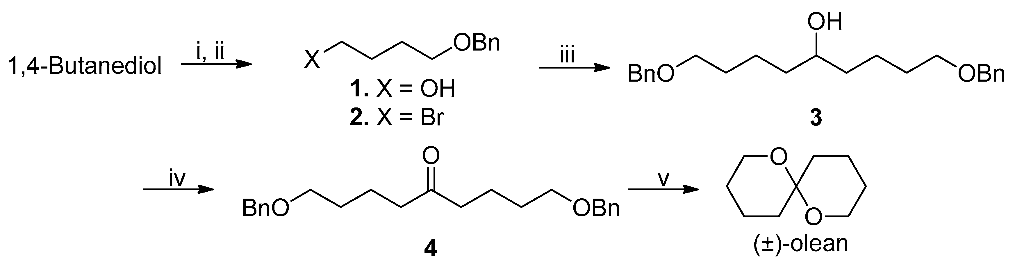
2.2. Synthesis and Characterization of Dacus Pheromone

The synthesis of pheromone was conducted by a new, simple and efficient method in five steps.
Synthesis of 4-(benzyloxy)butan-1-ol (1). Benzyl bromide (0.7 mL, 6 mmol) was added dropwise to a stirring solution of potassium hydroxide (1.5 g; 26 mmol) in 1,4-butanediol (2.3 mL; 26 mmol) at room temperature and the mixture was further stirred at the same temperature for 3 h. Then, H2O (10 mL) was added and resulting mixture was extracted thrice with diethyl ether (3 × 50 mL). The combined organic layers were dried over MgSO4, the solvent was removed in a rotavapor and the resulting residue was purified by column chromatography (hexane/ethyl acetate 4:1) to afford 4.7 g of 4-(benzyloxy)butan-1-ol (1) as an oil (99% yield based on benzyl bromide), with spectral data identical to those reported in the literature [22] 1H NMR (500 MHz, CDCl3) δ 7.41–7.23 (m, 10H), 4.52 (s, 4H), 3.64 (t, J = 5.9 Hz, 4H), 3.52 (t, J = 5.8 Hz, 4H), 1.74–1.65 (m, 8H). 13C NMR (126 MHz, CDCl3) δ 138.1, 128.4, 127.8, 127.7, 73.1, 70.3, 62.7, 30.2, 26.7.
Synthesis of ((4-bromobutoxy)methyl)benzene (2). Triphenyl phosphite (10.2 mL, 39 mmol) was added by a syringe to a solution of bromine (2 mL, 39 mmol) in CH2Cl2 (50 mL) at −30 °C, and the initial dark red solution decolorized. Pyridine (5.3 mL, 65 mmol) was then added to this mixture followed by addition of a solution of 4-(benzyloxy)butan-1-ol (1) (4.7 g, 26 mmol) in CH2Cl2 (50 mL) and the mixture allowed to warm at room temperature. The reaction mixture was quenched with 1N HCl by adjusting its pH to 7. The organic layer was washed with saturated aqueous NaCl (50 mL) and dried over MgSO4. The solvent was removed in a rotavapor and the resulting residue was purified by column chromatography (hexane/ethyl acetate 6:1) to afford ((4-bromobutoxy)methyl)benzene (2) (5.0 g, 80%) as a yellowish oil, with spectral data identical to those reported in the literature [23]. 1H NMR (500 MHz, CDCl3) δ 7.40–7.24 (m, 10H), 4.51 (s, 4H), 3.52 (t, J = 6.2 Hz, 4H), 3.45 (t, J = 6.8 Hz, 4H), 2.02–1.96 (m, 4H), 1.78 (dt, J = 12.7, 6.2 Hz, 4H). 13C NMR (126 MHz, CDCl3) δ 138.4, 128.4, 127.6, 127.6, 72.9, 69.2, 33.7, 29.7, 28.3.
Synthesis of 1,9-bis(benzyloxy)nonan-5-ol (3). A solution of ethyl formate (1.9 mL, 23.4 mmol) in dry THF (14 mL) was added dropwise to a solution of (4-(benzyloxy)butyl)magnesium bromide freshly prepared from ((4-bromobutoxy)methyl)benzene (2) (5 g, 20.7 mmol) and magnesium (0.6 g, 23.4 mmol) in refluxing dry THF (10 mL) and the reaction mixture was stirred at room temperature overnight. Then, MeOH (5 mL) was added for quench followed by addition of saturated aqueous NH4Cl (20 mL). The resulting mixture was extracted three times with ethyl acetate (3 × 50 mL) and the combined organic layers were washed with brine (50 mL) and dried over MgSO4. The solvent was then removed in a rotavapor and the residue obtained was purified by column chromatography (hexane/ethyl acetate 10:1) to afford 1,9-bis(benzyloxy)nonan-5-ol (3) (4.4 g, 60%) as a yellowish oil. 1H NMR (500 MHz, CDCl3) δ 7.37–7.29 (m, 8H), 7.29–7.25 (m, 2H), 4.50 (s, 4H), 3.59 (br s, 1H), 3.48 (t, J = 6.5 Hz, 4H), 1.62 (dd, J = 16.8, 10.3 Hz, 6H), 1.50 (dd, J = 21.6, 8.3 Hz, 4H), 1.43 (dd, J = 10.4, 4.4. 13C NMR (126 MHz, CDCl3) δ 138.6, 128.3, 127.6, 127.5, 72.9, 71.7, 70.3, 37.2, 29.7, 22.3. HRMS (ESI) (m/z): (M + H) calcd for C23H33O3, 357.2430 found 357.2390.
Synthesis of 1,9-bis(benzyloxy)nonan-5-one (4). PCC (5.3 g, 24.8 mmol) and silica gel (5.5 g) were added to a solution of 1,9-bis(benzyloxy)nonan-5-ol (3) (4,4 g, 12.4 mmol) in CH2Cl2 (50 mL) and the mixture was refluxed for 2 h. Solids were then filtered off, the solvent was evaporated in a rotavapor and the residue was chromatographed in a silica gel column (hexane/ethyl acetate 8:1) to give 1,9-bis(benzyloxy)nonan-5-one (4) (4.3 g, 97%) as a yellowish oil. 1H NMR (500 MHz, CDCl3) δ 7.35 (d, J = 1.5 Hz, 3H), 7.31–7.25 (m, 2H), 4.50 (s, 4H), 3.47 (t, J = 6.2 Hz, 4H), 2.41 (t, J = 7.2 Hz, 4H), 1.67 (dt, J = 14.7, 7.3 Hz, 4H), 1.64–1.58 (m, 4H). 13C NMR (126 MHz, CDCl3) δ 210.8, 138.5, 128.4, 127.6, 127.5, 72.9, 70.0, 42.4, 29.2, 20.6. HRMS (ESI) (m/z): [M-H] calcd for C23H31O3, 355.2273 found 355.2305.
Synthesis of (±)-1,7-dioxaspiro[5.5]undecane (5). A suspension of 1,9-bis(benzyloxy)nonan-5-one (4) (4.3 g, 12 mmol) and Pd/C 10% (w/w) (64 mg, 0.6 mmol) in MeOH (84 mL) was hydrogenated with H2 at room temperature for 16 h. Solids were then filtered off, the solvent was evaporated in a rotavapor and the residue was chromatographed in a silica gel column using methylene chloride as the eluent to afford (±)-1,7-dioxaspiro[5.5]undecane (5) (1.5 g, 80%) as a colorless liquid with spectral data identical to those reported in the literature [24]. 1H NMR (500 MHz, CDCl3) δ 3.72–3.65 (m, 2H), 3.63–3.56 (m, 2H), 1.87–1.76 (m, 2H), 1.64–1.47 (m, 8H), 1.43 (td, J = 13.3, 4.2 Hz, 2H). 13C NMR (126 MHz, CDCl3) δ 95.0, 60.3, 35.7, 25.3, 18.5.

NMR Spectroscopy (1H and 13C)

1H and 13CNMR spectra of intermediates and the final product were obtained using a Bruker AVANCE-III 300 spectrometer operating at a frequency of 500 and 126 MHz respectively. Deuterated chloroform (CDCl3) was used as solvent and the spectra were internally referenced to tetramethylsilane.

2.3. PLLA Microparticles

2.3.1. Preparation of PLLA Microparticles

Polymeric microspheres containing 1,7-dioxaspiro[5.5]undecane were prepared by oil-water (o/w) emulsification method, as was previously reported by Nanaki et al. [16,19,25]. In brief, PLLA was dissolved in 5 mL dichloromethane and 1,7-dioxaspiro[5.5]undecane was inserted in the polymeric solution in ratios 5, 10 and 20% w/w followed by probe sonicator for 1 min, cycle 1, amplitude 100% (UP50H, Hielscher Ultrasound Technology, Teltow, Germany). The dispersion was added dropwise in 20 mL of PVA solution, 1% w/v concentration, and homogenized at 30,000 rpm for 1 min (T 10 basic Ultra-Turrax, IKA, Staufen im Breisgau, Germany). After that, it was added to 20 mL H2O and left under magnetic stirring till total evaporation of dichloromethane. After centrifugation at 9000 rpm for 20 min (Heraeus™ Pico™ 17 Microcentrifuge, Thermo Fisher Scientific, Waltham, MA, USA) the resulting microparticles were washed with distilled water in order to remove the residual PVA and freeze-dried. Net microparticles of PLLA were also prepared by the same procedure for comparison reasons.

2.3.2. Characterization of Prepared Microspheres

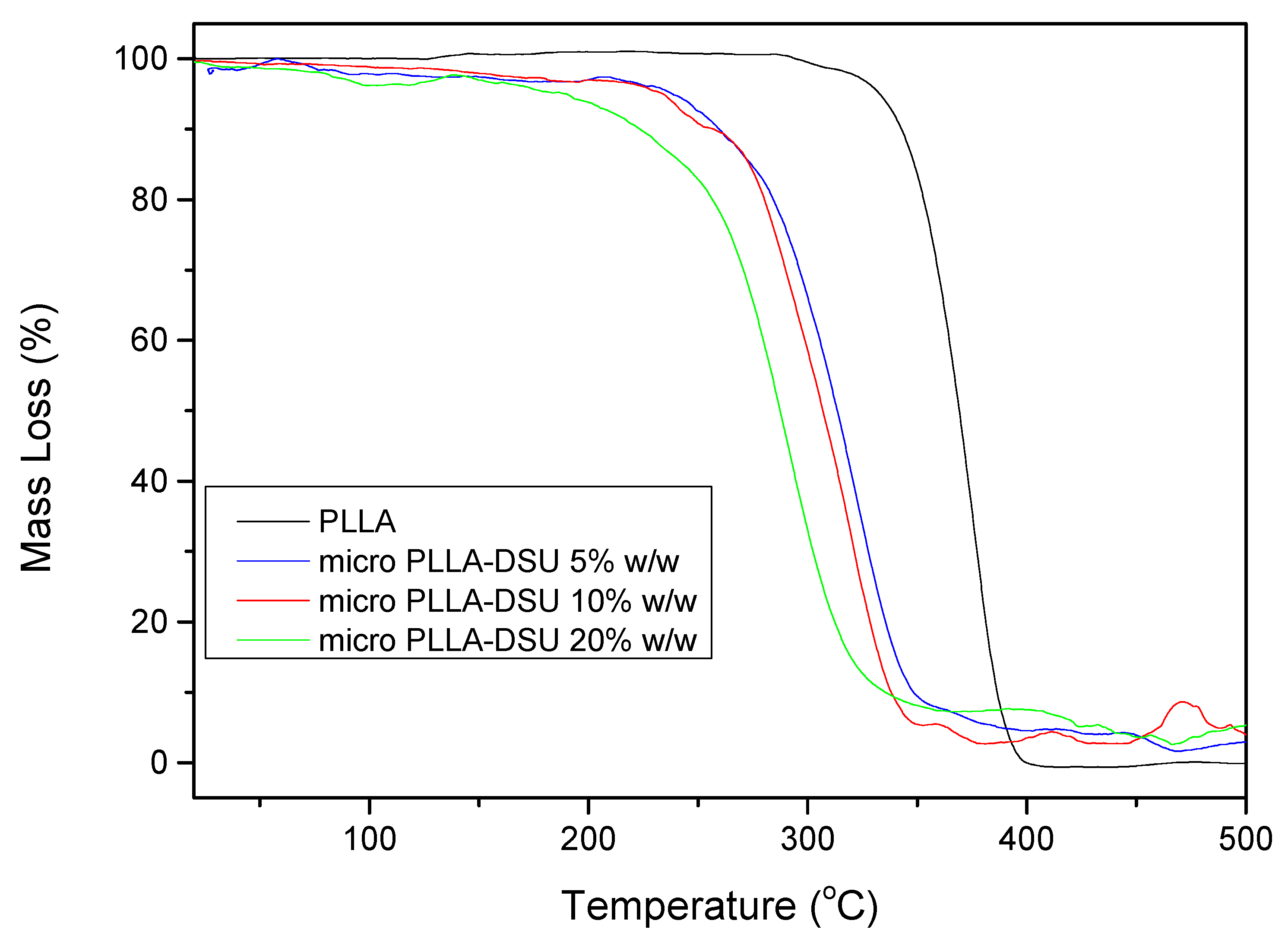
Thermogravimetric Analysis (TGA)

Thermogravimetric analysis was carried out with a SETARAM SETSYS TG-DTA 16/18. Samples (6.0 ± 0.2 mg) were placed in alumina crucibles. An empty alumina crucible was used as reference. PLLA and PLLA-DSU microparticles were heated from ambient temperature to 700 °C in a 50 mL/min flow of N2 at heating rate of 20 °C/min. Continuous recordings of sample temperature, sample weight, its first derivative and heat flow were performed.

Fourier Transform-Infrared Spectroscopy (FT-IR)

The chemical structure of the synthesized materials was determined with the use of FTIR spectroscopy. FTIR spectra of the samples were received with an FTIR spectrometer (model FTIR-2000, Perkin Elmer) using KBr discs (thickness of 500 μm). Infrared (IR) absorbance spectra were obtained between 450 and 4000 cm−1 at a resolution of 4 cm−1 using 20 co-added scans. All spectra presented were baseline corrected and normalized.

X-ray Diffraction (XRD)

X-ray powder diffraction (XRD) patterns were recorded using an XRD-diffractometer (Rigaku-Miniiflex II) with a Cu Kα radiation for crystalline phase identification (λ = 0.15405 nm for CuKα). The sample was scanned from 5 to 60 °C.

Scanning Electron Microscopy (SEM)

The morphology of the prepared microparticles was examined using a scanning electron microscope (SEM), type Jeol (JMS-840). All the studied surfaces were coated with carbon black to avoid charging under the electron beam. The instrument was operated at accelerating voltage 20 kV, probe current 45 nA and counting time 60 s.

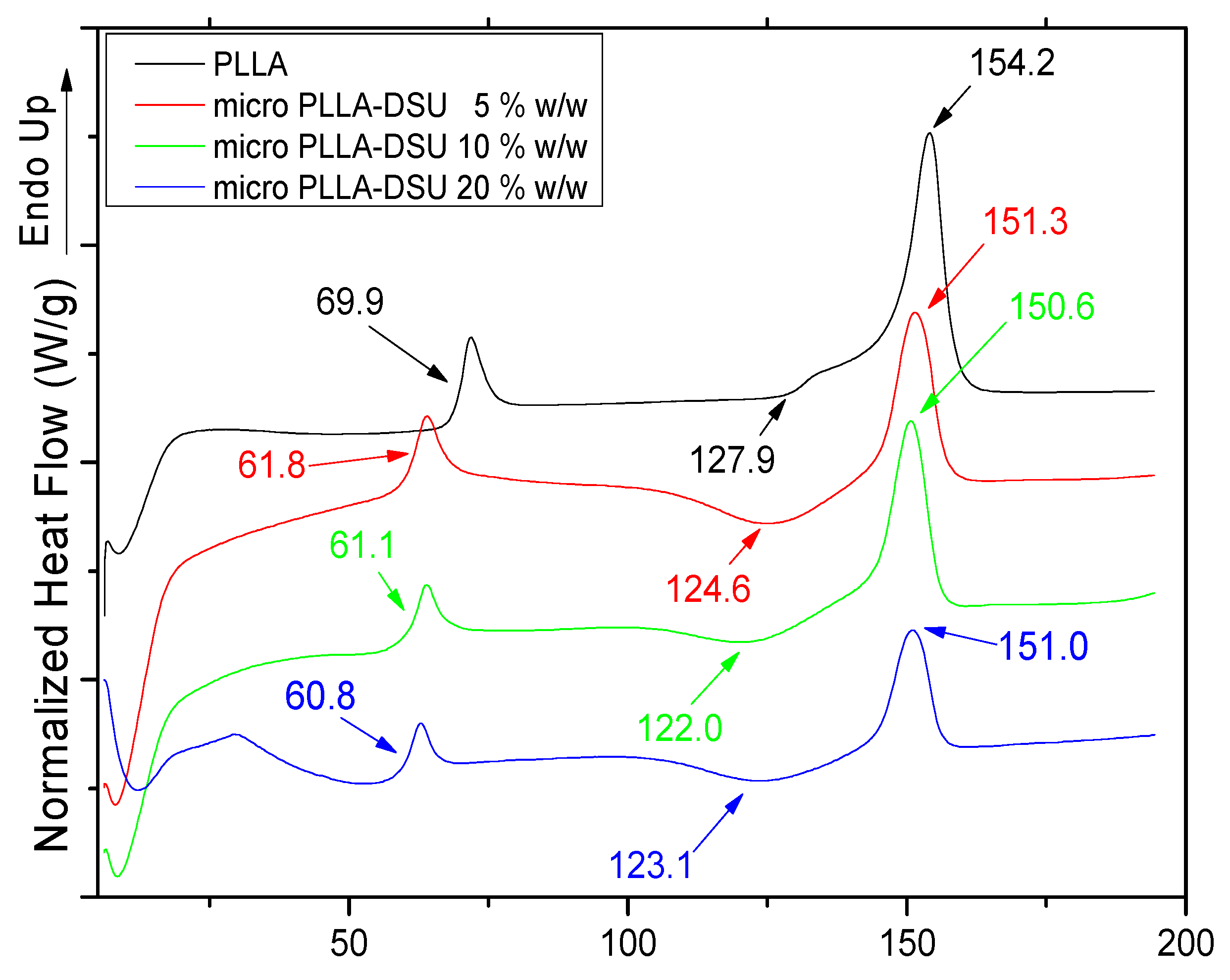
Differential Scanning Calorimetry (DSC)

A Perkin-Elmer, Pyris 1 differential scanning calorimeter (DSC), calibrated with indium and zinc standards, was employed. A sample of about 10 mg was used for each test, placed in a scaled aluminum pan and heated to 300 °C at a heating rate of 20 °C/min. The sample was held at that temperature for 5 min in order to erase any thermal history. After that it was quenched to 30 °C with liquid nitrogen and scanned immediately to 300 °C at a heating rate of 20 °C/min.

2.4. DSU Release from Microparticles

2.4.1. Determination of Amounts of Residual DSU in PLLA Microparticles

In order to determine the amounts of DSU remaining in the PLLA polymer material, microspheres were weighed (~50 mg) and placed in open glass vials at room temperature (25 ± 2 °C) for a period of 4 weeks. Samples at different time intervals such as 0, 1, 2, 3 and 4 weeks were extracted with solvent extraction. 10 mL of methanol were added in each sample and the vials were stoppered. The samples were vortexed for 30 min and then were put in shaking machine for extraction overnight. After the extraction the samples were centrifugated at 4500 rpm for 10 min in order to recover the methanol supernatant containing the released DSU. Then, 1 mL of the organic layer was filtrated using polytetrafluoroethylene (PTFE) 0.22 μm filter and transferred in an autosampler vial. Finally, an aliquot of 1 μL of each extract was subjected to GC-MS analysis. All measurements were performed in triplicate. Quantification of DSU amount on the PLLA microspheres was performed by external calibration (standard solutions at concentrations of 0.01, 0.1, 1.0, 2.5, 5.0 and 10 mg/L in MeOH). Calibration curves were calculated by linear least-squares regression using peak areas (R2 = 0.99).

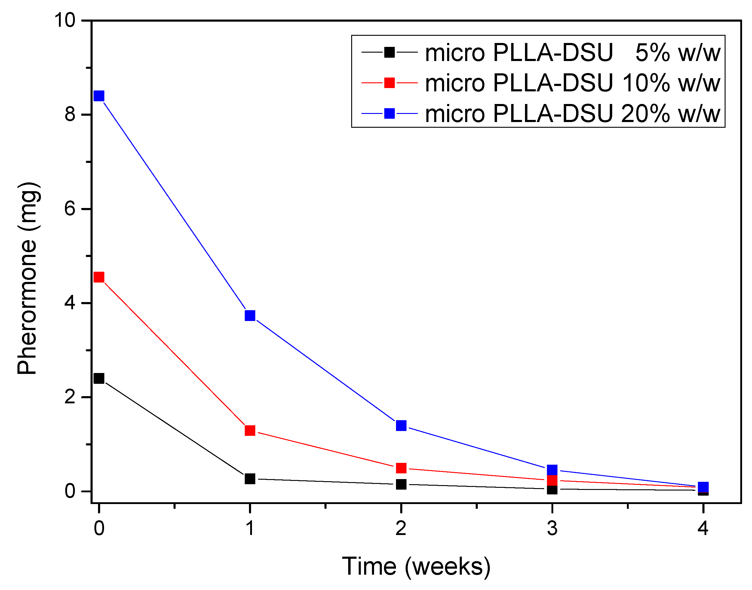
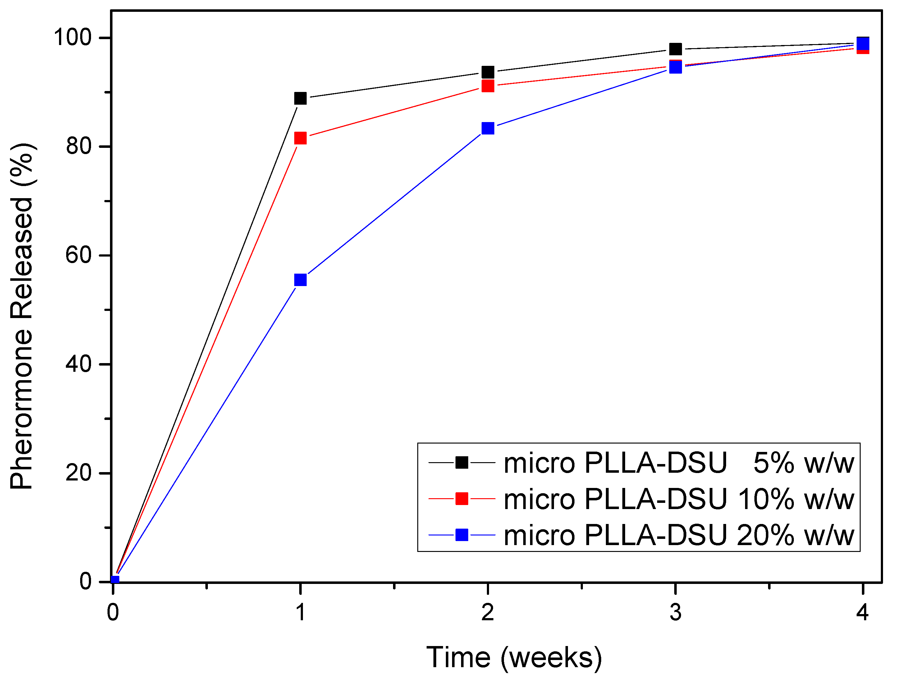
2.4.2. Determination of DSU Amount Released from PLLA Microspheres

Determination of the DSU amount released from PLLA microspheres was performed in headspace environment using the Solid Phase Microextraction (SPME) technique. Samples at different time intervals such as 0, 1, 2, 3 and 4 weeks were placed in 20 mL headspace glass vials and sealed immediately with a septum-type cap. Afterwards, the samples were placed in a water bath and left to equilibrate for 10 min. Then, a fused silica SPME fiber coated with polydimethylsiloxane (PDMS) of 100 μm diameter and 1 cm length (SUPELCO, Sigma-Aldrich, Madrid, Spain) was exposed in the headspace environment above the sample and the vials were heated for 45 min at 45 °C. Afterwards, the SPME-fiber was carefully retracted and then inserted directly into the injector of the GC-MS system for 10 min to allow for desorption of the target analyte from the fiber coating. Before measurements, the fiber was conditioned in the injector at 280 °C for 1 h. All samples were analyzed in triplicate. Quantification of DSU was performed by external calibration using standards solutions of DSU. The HS-SPME method used was considered efficient and appropriate, with high recoveries (~100%) and sensitivity (LOQ < 0.005 μg/L) for the DSU. The inter-day and intraday precision expressed, as relative standard deviation (RSD) were lower than 12%, in all cases. Compared to other extraction techniques, SPME technique proved advantageous and environmentally-friendly since it was fast, economic, does not require the use of solvents (volatile compounds are analyzed on the surface of the sample) and thus meet the requirements of “green chemistry”.
Microparticles yield (%), pheromone loading (%) and entrapment efficiency (%) were also determined immediately after their preparation according to the following equations:
Microparticles   yield   ( % ) = ( weight   of   microparticles ) ( weight   of   polymer   and   pheromone   fed   initially ) × 100
Pheromone   loading   ( % ) = ( weight   of   pheromone   in   microparticles ) ( weight   of   microparticles ) × 100
Entrapment   efficiency   ( % ) = ( weight   of   pheromone   in   microparticles ) ( weight   of   initially   used   pheromone ) × 100

2.4.3. GC-MS

Gas chromatography (Agilent 6890) equipped with MSD 5973 mass spectrometer (Palo Alto, CA, USA) was used to analyze the extracted pheromone. The capillary column was a nonpolar dimethylpolysiloxane phase (DB-5MS (30 m × 0.25 mm × 0.25 μm film thickness). The flow rate of helium was 1 mL/min in a spitless mode. The run time of the method was 14.5 min. The oven temperature was set initially at 90 °C for 1 min, then ramped to 260 °C at a rate of 20 °C/min. The temperature of the injector was set at 250 °C while the temperature of the MS Quad and MS Source was programmed at 150 °C and at 290 °C, respectively. Confirmation of the DSU was established by the retention time and the presence of the target ions by using the single-ion monitoring (SIM) mode. The characteristic ions selected for the DSU were as follows: m/z 101, m/z 100 and m/z 98.

3. Results and Discussion

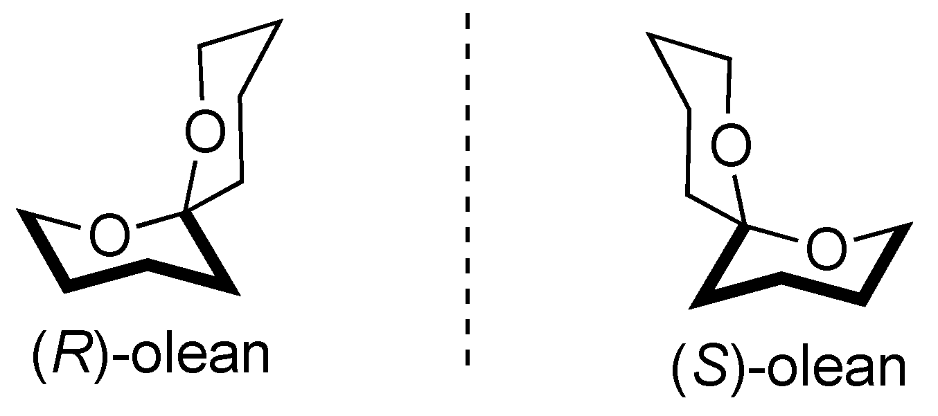
3.1. Synthesis and Characterization of Dacus Pheromone

Concerning 1,7-dioxaspiro[5.5] undecane, as was mentioned previously, was first isolated in female Dacus by Baker et al. [7] while its low quantity for commercial use lead the researchers to synthesize it in the lab. First, in 1983 it was synthesized by two independent groups [26,27]. Numerous studies have been reported in the literature thereafter concerning the synthesis of pheromone including asymmetric synthesis [28]. Kranidiotis et al. [24] proposed a novel short synthesis of racemic 1,7-dioxaspiro[5.5]undecane, named (±)-olean (Figure 1), through cross metathesis resulting in a cheap and easy to follow procedure. In the proper study, a new, simple and efficient synthesis of 1,7-dioxaspiro[5.5]undecane in five steps was developed starting from butane-1,4-diol.
Although many synthetic approaches towards olean syntheses have been reported in literature, including their asymmetric syntheses [28], we decided to developed a new simple and efficient synthesis of olean in five steps, starting from butane-1,4-diol, as outlined in Scheme 1.
Firstly, butane-1,4-diol was monobenzylated using benzyl bromide as limiting reagent in 99% yield (based on benzyl bromide). The primary hydroxyl group was then replaced by a bromine atom by a standard procedure to give bromide 2 in 80% yield and addition of the respective Grignard reagent to ethyl formate afforded good yields of alcohol 3. Subsequent oxidation of 3 with pyridinium chlorocromate (PCC) yielded quantitatively the respective ketone, which upon debenzylation with catalytic hydrogenation led to the spontaneous cyclization of the intermediately formed dihydroxyketone, to give the desired racemic olean in 80% yield. The overall yield of converting compound 1 to olean was 37%.
All compounds prepared were of high purity and characterized by their spectral and analytical data. Final (±)-olean as well as intermediate products gave accurate and clear 1H and 13C NMR spectra without the presence of any impurity.

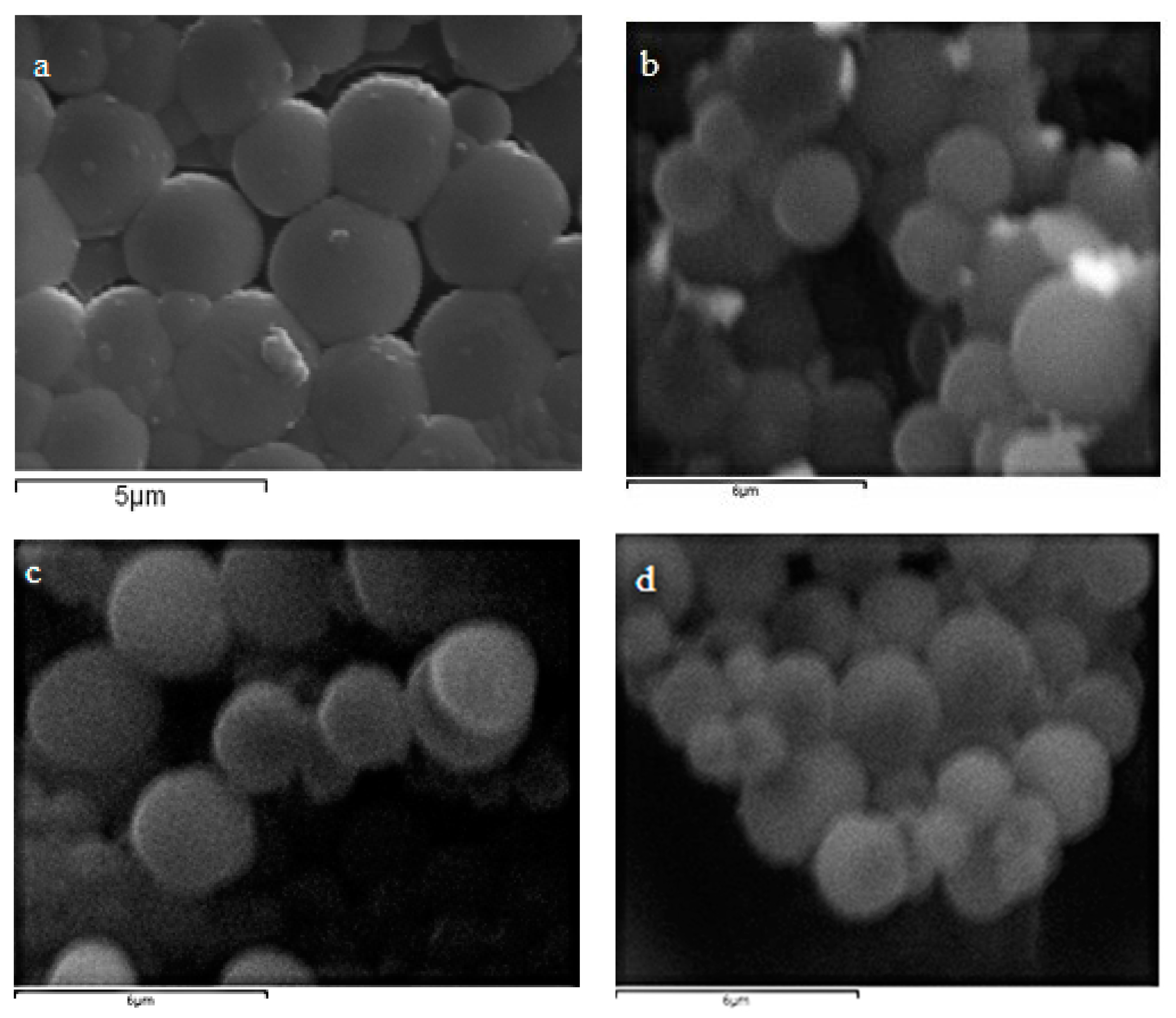
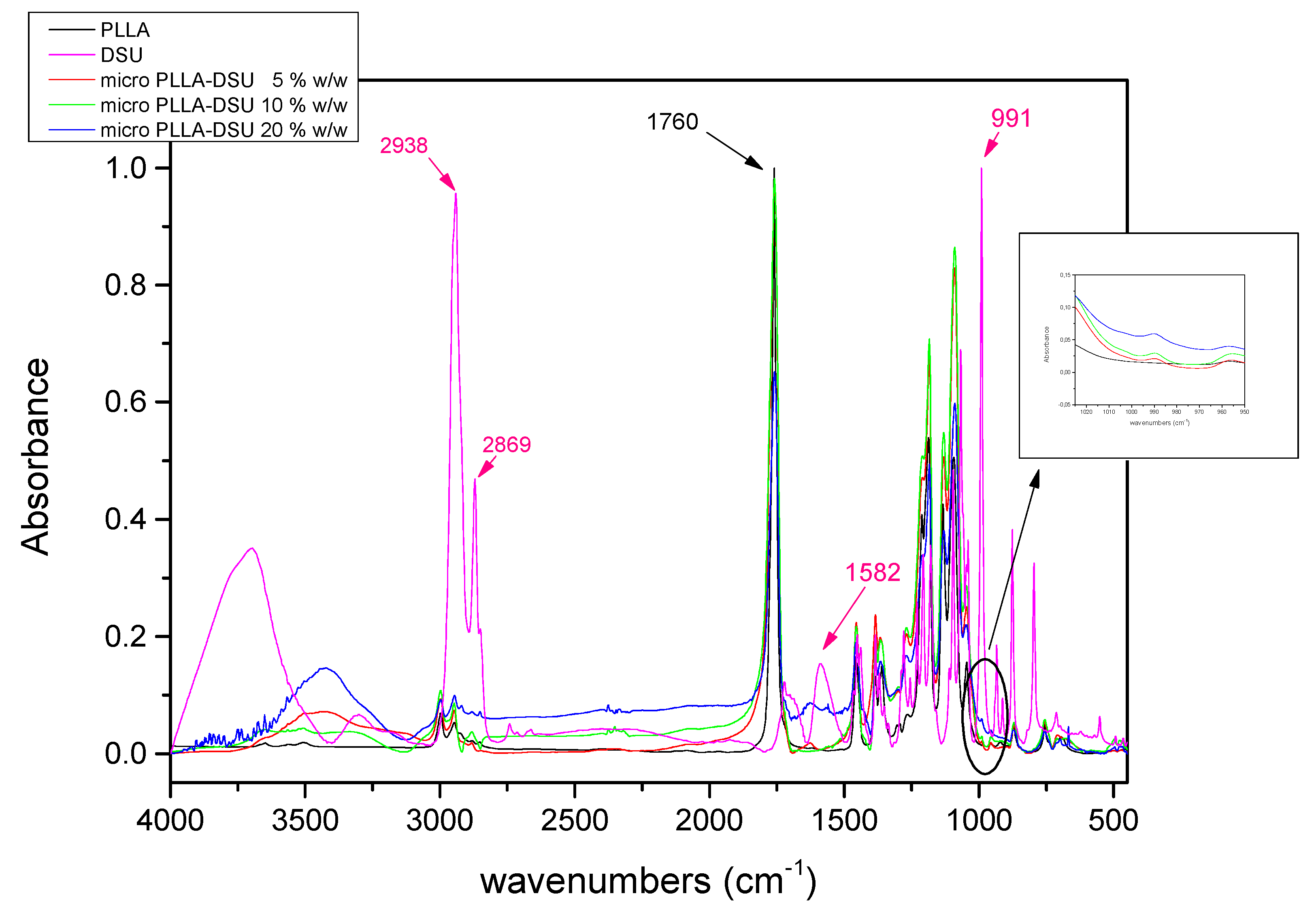
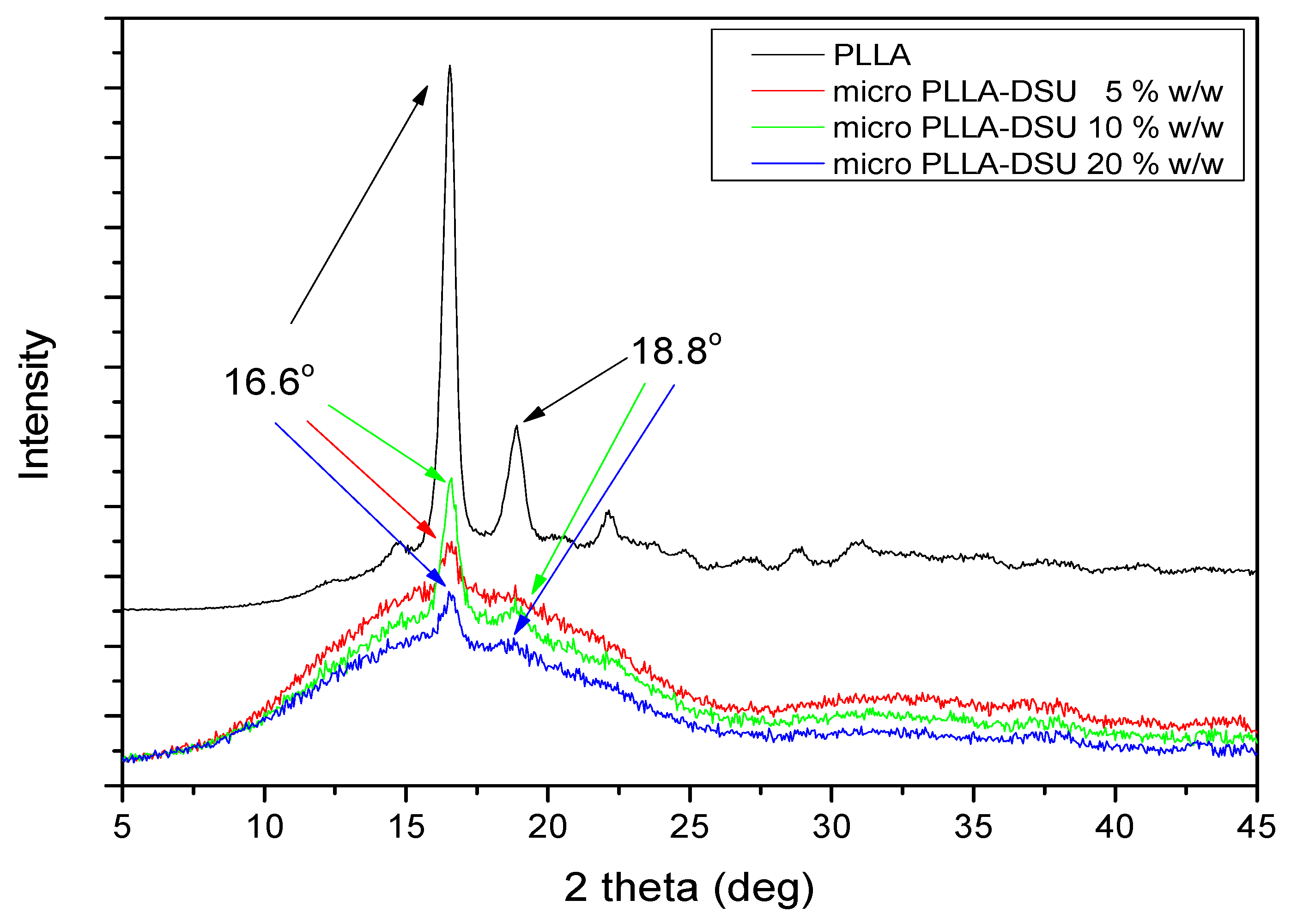
3.2. Characterization of Prepared Microparticles

Microparticles were prepared according to o/w technique as described previously [16,18,19]. SEM (Figure 2) was used in order to verify microparticles formation as well as their surface morphology. As can be seen, microparticles were successfully synthesized with sizes ranged between 1 and 2.5 μm, while no agglomeration was observed. Small differences of about 0.3 μm was observed concerning the DSU content with their sizes increased as quantity of DSU increased. Surface morphology showed to be smooth, owing to the polymer used, while no porous were observed.
FT-IR was further used to verify possible bond formation between PLLA and DSU. As shown in Figure 3, PLLA showed a single peak at 1760 cm−1 owing to carbonylic bond -C=O [18] and DSU showed a double peak at 2938 and 2869 cm−1 owing to C-H stretching vibrations and one at 991 cm−1 due to C-O-C vibrations [29]. Microparticles of 5, 10 and 20% DSU did not show significant frequency shifts, while peaks appeared at 990 cm−1 indicating the presence of pheromone in formed microparticles.
XRD was used in order to determine the crystallinity of PLLA microparticles before and after DSU’s encapsulation. PLLA is a semicrystalline polymer, as has been previously reported [19] and its recorded pattern is shown in Figure 4. Two characteristic peaks of PLLA at 16.6 and 18.8 deg were present, while after DSU encapsulation there was a reduction to intensity of the peaks. As the amount of DSU increased the reduction was much higher and this is an indication that the crystallinity of PLLA was reduced, probably because DSU could act as plasticizer to PLLA. Furthermore, higher the encapsulated amount resulted in enhancement of the proper effect. In order to confirm this, DSC was used as a further study and also to detect thermal properties of prepared microparticles.
As was already reported, PLLA is a semicrystalline polymer and exhibited a glass transition (Tg) and melting temperature (Tm) at 69.9 °C and at 154.2 °C, respectively, while cold crystallization (Tcc) was also observed at 127.9 °C (Figure 5). These are in good agreement with the corresponding values reported in literature [18,25]. Slight changes were observed in DSC thermographs of prepared microparticles after DSU incorporation. In brief, Tg values were reduced with the increase of DSU from 69.9 of neat PLLA to 61.8, 61.1 and 60.8 °C, of 5, 10 and 20% w/w, respectively. This is an indication that DSU due to its low molecular weight could act as plasticizer for PLLA polymer, which is in good agreement with XRD results. Reduction in characteristic Tcc and Tm values of PLLA was also observed in prepared microparticles. In brief, Tm values of PLLA showed reduction from 154.2 °C to 151.3, 150.6 and 151.0 °C for microparticles containing 5, 10 and 20 wt% DSU, respectively, without showing any specific trend. Concerning Tcc values reduction was observed from 127.9 °C of PLLA to 124.6, 122.0 and 123.1 °C, respectively. Reduction in Tcc value of PLLA indicates that DSU facilitates cold crystallization procedure, mainly due to looser chain formation of polymeric entanglements.
Thermal degradation of each formulation was further evaluated with thermogravimetric analysis (TGA). As shown in Figure 6, PLLA’s thermal decomposition starts at 288 °C with the first stage ending at 310 °C showing a mass loss of about 1.5%, followed by a second stage which ended at 404 °C while no further thermal decomposition was observed. Thermal decomposition of PLLA microparticles containing DSU was slightly different. PLLA microparticles containing 5% DSU started to decompose at 226 °C with a mass loss of about 4%, probably attributed to the pheromone mass loss, due to the fact that its boiling point is 194 °C. The first degradation stage lasted at 263 °C, while followed by a second stage until 351 °C. From that point after there was no significant mass loss and the mass residue was approximately 5%. For microparticles of PLLA containing 10% DSU decomposition started at 190 °C and lasted at 210 °C, showing mass loss of about 8%, and a second stage from 210 °C till 359 °C with remaining mass of about 5%. Analogous were the results for PLLA microparticles containing 20% DSU; an initial step till 203 °C, with a mass loss of 15%, and a second one till 364 °C and remaining mass 5.5%. From all these thermograms it is clear that by increasing the amount of DSU the maximum decomposition temperature of PLLA progressively shifted to lower temperatures due to the high volatility of DSU. TGA thermographs constitute an initial estimation concerning DSU concentration in microparticles. Hence, due to the fact that DSU evaporated easily and TGA did not show high sensitivity, GC-MS analysis was further used.

3.3. DSU Release from PLLA Microparticles

GC-MS analysis was conducted as referred in Section 2.4.2. Table 1 shows microparticles yield (%), pheromone loading (%) and encapsulation efficiency (%) of microparticles prepared. The proper values are referring to the zero day and the analysis conducted immediately after microparticles formation in order to avoid DSU release during storage. As can be observed, microparticles showed high yield values, over 80% for all three formulations, with that of 10% DSU reaching up to 91.5%, while no specific sequence was observed. Pheromone loading quantification was conducted according to SPME and found that DSU was present in 4.8, 9.1 and 16.8% in microparticles containing 5, 10 and 20% w/w DSU. The proper results were analogous to the ones found by TGA analysis, where DSU was found to be 4, 8 and 15%.
Figure 7 shows the remaining mass of DSU after liquid extraction procedure at 0, 1, 2, 3 and 4 weeks. As can be observed microparticles containing 5% w/w DSU showed a total mass loss in 1 week with a reduction from about 2.5 mg to 0.2 mg without any change thereafter meaning that the quantity of the pheromone remained was the total quantity of DSU was successfully released from microparticles. Concerning concentration 10% w/w DSU it was found that the main reduction in DSU content made in the first week with a loss of about 3.25 mg, while in the second week there was also observed a reduction in DSU content with an additional mass loss of 0.75 mg. Additional losses of about 0.3 and 0.1 mg was observed in the third and fourth respectively with a remaining mass of 0.08 mg. Finally, formulation of 20% w/w DSU showed to half reduce DSU content in the first week, an additional reduce of about 2.5 mg in the second week and about 1 and 0.5 mg in the third and fourth week respectively. It has to be mentioned that in all formulations the remaining mass of DSU was below 0.1 mg.
Figure 8 shows the percentage release of DSU form PLLA microparticles conducted by SPME technique. As can be observed microparticles containing 5 and 10% w/w DSU showed a release reached at 90 and 80% in the first week with an increase of about 2 and 10% in the second week, reaching 92 and 90% respectively without any significant changes thereafter. The initial burst release probably arose from the quantity of DSU remaining on the surface of the microparticles, allowing for easier release. The proper results are in accordance with the ones revealed by LE technique where, as was described previously, the total quantity of DSU was actually released in the first week for microparticles containing 5% and in the second week, with a slight increase between the two first weeks, for microparticles containing 10% DSU. Microparticles containing 20% w/w DSU showed a different profile. In brief, a sustained release profile was observed with DSU release reaching 55% in the first week, 83% in the second week, 94% in the third week and 99% in the fourth week. Concerning the proper release, it is believed that the initial release of 55% observed in the first week in probably owing to DSU release remained to the surface of microparticles, while from that time after the release could be attributed to pores formed in the surface and channels formed to the inner of microparticles renders the release easier. Finally, the recorded results are also in agreement with that obtained by LE procedure which also showed a gradual reduction in DSU mass. From these it is clear that microparticles containing 20 wt% DSU could be the ideal carrier to control Dacus oleae population for about 4 weeks.

4. Conclusions

In this study, the sexual pheromone of the insect Bactrocera oleae (DSU) was successfully synthesized following a novel, facile, five step method. The synthesized DSU was characterized by 1H and 13C NMR and with HPMS (ESI) showing its successful synthesis. DSU was further used in PLLA microparticles’ preparation at 5, 10 and 20 wt%. As was found by FT-IR and DSC measurements, DSU was successfully encapsulated to microparticles. Even though some interactions are not taking place between DSU and PLLA reactive groups, it was found that DSU due to its low molecular weight could act as plasticizer to PLLA matrix. GC-MS was used for quantification of remaining DSU in microparticles at weeks 0, 1, 2, 3 and 4 and release profile was studied. Results showed that for microparticles containing 5 and 10% DSU release practically lasted in 1 and 2 weeks respectively, while for microparticles with 20% DSU release showed a controlled profile lasted in 4 weeks. The results are promising for using the proper microparticles as innovative formulations in Dacus oleae population control.

Author Contributions

Conceptualization, J.K.G. and D.N.B.; data curation, S.A.Z., J.K.G., A.O., D.A.L., D.N.B. and S.G.N.; formal analysis, C.K.C., A.O., D.A.L. and E.P.; methodology, S.A.Z., J.K.G., A.O., D.A.L., D.N.B. and S.G.N.; supervision, J.K.G., D.A.L. and D.N.B.; validation, A.O. and D.A.L.; visualization, J.K.G. and D.N.B.; writing—original draft, S.A.Z., A.O. and S.G.N.; writing—review and editing, J.K.G., D.A.L., D.N.B. and S.G.N. All authors have read and agreed to the published version of the manuscript.

Funding

This research received no external funding.

Acknowledgments

Authors would like to acknowledge Corbion for kindly donation of PLLA and Eleni Pavlidou (Physics Department AUTH) for SEM photos captured.

Conflicts of Interest

The authors declare no conflict of interest.

References

  1. Gömez-Caravaca, A.M.; Cerretani, L.; Bendini, A.; Segura-Carretero, A.; Fernandez-Gutierrez, A.; delCarlo, M.; Compagnone, D.; Cichelli, A. Effects of fly attack (Bactrocera oleae) on the phenolic profile and selected chemical parameters of olive oil. J. Agric. Food Chem. 2008, 56, 4577–4583. [Google Scholar] [CrossRef]
  2. Bayega, A.; Djambazian, H.; Tsoumani, K.T.; Gregoriou, M.E.; Sagri, E.; Drosopoulou, E.; Mavragani-Tsipidou, P.; Giorda, K.; Tsiamis, G.; Bourtzis, K.; et al. De novo assembly of the olive fruit fly (Bactrocera oleae) genome with linked-reads and long-read technologies minimizes gaps and provides exceptional Y chromosome assembly. BMC Genom. 2020, 21, 1–21. [Google Scholar] [CrossRef] [PubMed] [Green Version]
  3. Canale, A.; Benelli, G.; Germinara, G.S.; Fusini, G.; Romano, D.; Rapalini, F.; Desneux, N.; Rotundo, G.; Raspi, A.; Carpita, A. Behavioural and electrophysiological responses to overlooked female pheromone components in the olive fruit fly, Bactrocera oleae (Diptera: Tephritidae). Chemoecology 2015, 25, 147–157. [Google Scholar] [CrossRef]
  4. Varikou, K.; Garantonakis, N.; Birouraki, A. Exposure of bombus terrestris l. To three different active ingredients and two application methods for olive pest control. Entomol. Gen. 2019, 39, 53–60. [Google Scholar] [CrossRef]
  5. Rebora, M.; Salerno, G.; Piersanti, S.; Gorb, E.; Gorb, S. Role of Fruit Epicuticular Waxes in Preventing. Insects 2020, 11, 1–17. [Google Scholar]
  6. Witzgall, P.; Kirsch, P.; Cork, A. Sex pheromones and their impact on pest management. J. Chem. Ecol. 2010, 36, 80–100. [Google Scholar] [CrossRef] [PubMed]
  7. Baker, R.; Herbert, R.; Howse, P.E.; Jones, O.T.; Francke, W.; Reith, W. Identification and synthesis of the major sex pheromone of the olive fly (Dacus oleae). J. Chem. Soc. Chem. Commun. 1980, 2, 52–53. [Google Scholar] [CrossRef]
  8. Mazomenos, B.E.; Haniotakis, G.E. A multicomponent female sex pheromone of Dacus oleae Gmelin: Isolation and bioassay. J. Chem. Ecol. 1981, 7, 437–444. [Google Scholar] [CrossRef]
  9. Mazomenos, B.E.; Haniotakis, G.E. Male olive fruit fly attraction to synthetic sex pheromone components in laboratory and field tests. J. Chem. Ecol. 1985, 11, 397–405. [Google Scholar] [CrossRef]
  10. Mazomenos, B.E.; Haniotakis, G.E.; Ioannou, A.; Spanakis, I.; Kozirakis, A. Field evaluation of the olive fruit fly pheromone traps with various dispensers and concentrations. In Fruit Flies of Economic Importance, Proceedings of the CEC/IOBC International Symposium, Athens, Greece, 16–19 November 1982; pp. 506–512.
  11. Liu, B.; Syu, K.J.; Zhang, Y.X.; Gupta, S.; Shen, Y.J.; Chien, W.J.; Tseng, J.C.; Lou, D.W. Practical Synthesis and Field Application of the Synthetic Sex Pheromone of Rice Stem Borer, Chilo suppressalis (Lepidoptera: Pyralidae). J. Chem. 2020. [Google Scholar] [CrossRef] [Green Version]
  12. Chamberlain, D.J.; Brown, N.J.; Jones, O.T.; Casagrande, E. Field evaluation of a slow release pheromone formulation to control the American bollworm, Helicoverpa armigera (Lepidoptera: Noctuidae) in Pakistan. Bull. Entomol. Res. 2000, 90, 183–190. [Google Scholar] [CrossRef] [PubMed]
  13. Mazomenos, B.; Moustakali-Mavridis, I. Inclusion Complexes of Cyclodextrin and their Use in Slow Release Formulations for Attracting Insects. PCT/EP93/01536, 17 June 1993. [Google Scholar]
  14. Kikionis, S.; Ioannou, E.; Konstantopoulou, M.; Roussis, V. Electrospun Micro/Nanofibers as Controlled Release Systems for Pheromones of Bactrocera oleae and Prays oleae. J. Chem. Ecol. 2017, 43, 254–262. [Google Scholar] [CrossRef] [PubMed]
  15. Beslikas, T.; Gigis, I.; Goulios, V.; Christoforides, J.; Papageorgiou, G.Z.; Bikiaris, D.N. Crystallization study and comparative in vitro-in vivo hydrolysis of PLA reinforcement ligament. Int. J. Mol. Sci. 2011, 12, 6597–6618. [Google Scholar] [CrossRef] [PubMed] [Green Version]
  16. Nanaki, S.; Barmpalexis, P.; Iatrou, A.; Christodoulou, E.; Kostoglou, M.; Bikiaris, D.N. Risperidone controlled release microspheres based on poly(lactic acid)-poly(propylene adipate) novel polymer blends appropriate for long acting injectable formulations. Pharmaceutics 2018, 10, 130. [Google Scholar] [CrossRef] [Green Version]
  17. Nanaki, S.; Barmpalexis, P.; Papakonstantinou, Z.; Christodoulou, E.; Kostoglou, M.; Bikiaris, D.N. Preparation of new risperidone depot microspheres based on novel biocompatible poly(alkylene adipate) polyesters as long acting formulations. J. Pharm. Sci. 2018, 107, 2891. [Google Scholar] [CrossRef]
  18. Nanaki, S.; Tseklima, M.; Terzopoulou, Z.; Nerantzaki, M.; Giliopoulos, D.; Triantafyllidis, K.; Kostoglou, M.; Bikiaris, D.N. Use of mesoporous cellular foam (mcf) in preparation of polymeric microspheres for long acting injectable release formulations of paliperidone antipsychotic drug. Eur. J. Pharm. Biopharm. 2017, 117, 77–90. [Google Scholar] [CrossRef]
  19. Nanaki, S.; Viziridou, A.; Zamboulis, A.; Kostoglou, M.; Papageorgiou, G.Z.; Bikiaris, D.N. New Biodegradable Poly (l -lactide) -Block- for Long-Acting Injectables of Naltrexone Drug. Polymers 2020, 12, 852. [Google Scholar] [CrossRef] [Green Version]
  20. Bragagni, M.; Beneitez, C.; Martín, C.; De La Ossa, D.H.P.; Mura, P.A.; Gil-Alegre, M.E. Selection of PLA polymers for the development of injectable prilocaine controlled release microparticles: Usefulness of thermal analysis. Int. J. Pharm. 2013, 441, 468–475. [Google Scholar] [CrossRef]
  21. Farah, S.; Anderson, D.G.; Langer, R. Physical and mechanical properties of PLA, and their functions in widespread applications—A comprehensive review. Adv. Drug Deliv. Rev. 2016, 107, 367–392. [Google Scholar] [CrossRef] [Green Version]
  22. Gupta, S.; Bera, S.; Mondal, D. Nascent-HBr-Catalyzed Removal of Orthogonal Protecting Groups in Aqueous Surfactants. J. Org. Chem. 2020, 85, 2635–2645. [Google Scholar] [CrossRef]
  23. Bernat, V.; Brox, R.; Heinrich, M.R.; Yves, P.; Auberson, Y.P.; Tschammer, N. Ligand-Biased and Probe-Dependent Modulation of Chemokine Receptor CXCR3 Signaling by Negative Allosteric Modulators. Chem. Med. Chem. 2015, 10, 566–574. [Google Scholar] [CrossRef] [PubMed]
  24. Kranidiotis, N.S.; Grammatoglou, C.E.; Gallos, J.K. A new short synthesis of (±)-olean through cross metathesis. Heterocycles 2018, 96, 1441–1444. [Google Scholar]
  25. Nanaki, S.; Tseklima, M.; Christodoulou, E.; Triantafyllidis, K.; Kostoglou, M.; Bikiaris, D.N. Thiolated chitosan masked microparticles with incorporated mesocellular silica foam (MCF) for intranasal delivery of paliperidone. Polymers 2017, 9, 617. [Google Scholar] [CrossRef] [PubMed] [Green Version]
  26. Koclenskl, P. Yeates, A new synthesis of 1,7-dioxasdipo[5,5] undecanes application to a rectal gland secretion of the olive fruit fly (Dacus Oleae). Tetrehedron Lett. 1983, 24, 3905–3906. [Google Scholar]
  27. Williams, B.A.; Barner, D.R. Synthetic Studies of 1,7-dioxaspir0[5.5]undecan-4-ones. Tetrahedron Lett. 1983, 24, 427–430. [Google Scholar] [CrossRef]
  28. Cala, L.; Fañanás, F.J.; Rodríguez, F. Enantioselective synthesis of spiroacetals: The conquest of a long-sought goal in asymmetric catalysis. Org. Biomol. Chem. 2014, 12, 5324–5330. [Google Scholar] [CrossRef] [Green Version]
  29. Řezanka, T.; Dvořáková, R.; Hanuš, L.O.; Dembitsky, V.M. Enteridinines A and B from slime mold Enteridium lycoperdon. Phytochemistry 2004, 65, 455–462. [Google Scholar] [CrossRef]
Figure 1. Structures of (R)- and (S)-1,7-dioxaspiro[5.5]undecane (olean).
Agronomy 10 01053 g001
Scheme 1. Reagents and conditions: (i) PhCH2Br, KOH, 20 °C, 3 h; (ii) (PhO)3P, Br2, CH2Cl2, −30 °C to 20 °C, 80%; (iii) Mg, THF, reflux, then addition of HCO2Et in THF, overnight, 60%; (iv) PCC, silica gel, CH2Cl2, reflux, 2 h, 97%; (v) Pd/C 10% (w/w), H2, MeOH, 16 h, 80%.
Agronomy 10 01053 sch001
Figure 2. SEM pictures of (a) net PLLA microparticles, (b) PLLA-DSU 5% w/w microparticles, (c) PLLA-DSU 10% w/w microparticles, (d) PLLA-DSU 20% w/w microparticles.
Agronomy 10 01053 g002
Figure 3. FT-IR spectra of PLLA, DSU and PLLA-DSU microparticles.
Agronomy 10 01053 g003
Figure 4. XRD patterns of PLLA and PLLA-DSU microparticles.
Agronomy 10 01053 g004
Figure 5. DSC thermographs of PLLA and PLLA-DSU microparticles.
Agronomy 10 01053 g005
Figure 6. TGA thermographs of PLLA and PLLA-DSU microparticles.
Agronomy 10 01053 g006
Figure 7. Amount (mg) of the remaining pheromone in PLLA microspheres after LE.
Agronomy 10 01053 g007
Figure 8. Release (%) of pheromone from PLLA microspheres after HS-SPME.
Agronomy 10 01053 g008
Table 1. Encapsulation yield, loading and efficiency percentages of PLLA on different amounts (%) of encapsulated DSU after SPME.
Sample NameMicroparticles Yield (%)Pheromone Loading (%)Encapsulation Efficiency (%)
PLLA-DSU5% w/w85.3 ± 0.24.8 ± 0.396.1 ± 0.2
PLLA-DSU10% w/w91.5 ± 0.59.1 ± 0.291.8 ± 0.4
PLLA-DSU20% w/w81.4 ± 0.316.8 ± 0.684.6 ± 0.5

