Article

Response of Rapeseed (Brassica napus L.) to Silver and Gold Nanoparticles as a Function of Concentration and Length of Exposure

by
Magdalena Tomaszewska-Sowa
1,*,
Karol Lisiecki
2 and
Dariusz Pańka
2
1
Department of Agricultural Biotechnology, Faculty of Agriculture and Biotechnology, Bydgoszcz University of Science and Technology, 6 Bernardyńska St., 85-029 Bydgoszcz, Poland
2
Department of Biology and Plant Protection, Faculty of Agriculture and Biotechnology, Bydgoszcz University of Science and Technology, 7 Kaliskiego Av., 85-796 Bydgoszcz, Poland
*
Author to whom correspondence should be addressed.
Agronomy 2022, 12(11), 2885; https://doi.org/10.3390/agronomy12112885
Submission received: 12 October 2022 / Revised: 11 November 2022 / Accepted: 14 November 2022 / Published: 18 November 2022
(This article belongs to the Special Issue Enhanced Product Quality of Plant Material from Field Crops)

Abstract

:
There is a growing demand for high quality and sustainable food in the world and this need falls within the context of the European Green Deal’s strategy “From Farm to Fork”. In order to achieve these outcomes, the use of modern and innovative technologies of plant production and protection is required. The use of nanoparticles (NPs) in agriculture and horticulture is an example of such technology. However, research on the effect of length of exposure to metal nanoparticles on seeds germination and seedlings development are limited in the literature. In our study, the effect of silver (AgNPs) and gold nanoparticles (AuNPs) on the seedling growth and biochemical response of rapeseed after 7, 14 and 21 days was analyzed. In the experiments, 0, 50 and 100 ppm concentrations of NPs were used in vitro. The level of photosynthetic pigments, anthocyanins as well as other stress parameters, such as free phenolic compounds, free sugars or H2O2, decreased due to the application of both AgNPs and AuNPs at the initial culture period; however, the differences were observed in the successive weeks of exposure. The parameters were increasing, irrespective of the kind of nanoparticles; however, as for the content of free sugars and free radicals, higher values were recorded due to the effect of AuNPs. Our results showed that length of plants exposure to NPs is very important factor modifying growth and final response of seedlings. Better understanding of its influence could speed up use of NPs in agriculture and horticulture for production of high-quality plant material (e.g., to seed priming, stimulation of seedlings’ growth and their protection), not contaminated with pesticides, fertilizers and mycotoxins.

1. Introduction

There is a growing demand for high quality and sustainable food in the world and this need falls within in the context of European Green Deal’s strategy “From Farm to Fork”. In order to achieve these outcomes, the use of modern and innovative technologies of plant production and protection is required. These technologies should ensure not only high yield but also its high quality, as well as safety for consumers and to the environment.
The use of nanoparticles (NPs) in agriculture and horticulture is an example of such technology. Due to its possible beneficial effect on the development of seeds and plants, this technology has recently gained increased attention. Thanks to multifunctional properties of nanoparticles, new areas of their application, such as nano-fertilizers, nano-pesticides and nano-priming agents, are emerging. However, there is also available literature indicating the detrimental influence of NPs on the development and physiology of plants. Thus, extensive research is underway to precisely define the parameters that affect the final result of NPs on plants.
Nanotechnology deals with designing and developing materials in which at least one of the dimensions falls within the range from 1 to 100 nm. The terms ‘nanotechnology’ and ‘nanostructures’ are relatively new concepts; however, NPs are not new for the environment and they occur, e.g., in the form of minerals. Due to high potential of nanostructures the synthetic NPs are recently created, making them suitable to various applications [1,2]. The materials made following that technology can demonstrate specific physical, chemical or biological properties, which makes them very different from the properties of single atoms or compounds with a micrometric structure [3]. The reason for that transformation is increasing the specific surface area of the volume [4,5], which can result in the NPs showing different physicochemical properties, which can change reactivity, and thus, their biological activity [2,6]. The properties of NPs, including their reactivity, are affected by a number of factors, including their size, the composition of the core, shape, the properties of the surface, purity, stability and their production method [1,7]. Next to the kind of nanoparticles and their application method, the plant genotype, age, development stage, the kind of organ or cultivation conditions also determine the response of the plant to the presence of metal NPs in the environment [8,9,10]. Most literature reports concern the commonly used nanoparticles of silver and gold. However, one can also find information on the nanoparticles of carbon, copper, aluminum, zinc, iron, silicon, titanium and others [11,12,13]. Due to its strong biocidal and fungicidal properties, of all the metal NPs, silver shows definitely the biggest number of applications [14]. Additionally, more and more products use or are composed of nanosilver (AgNPs). The materials can be successfully applied almost in all the branches of industry and technology, starting from medicine, food production, all the way to the construction industry [15,16,17]. They can be, therefore, used as components of household appliances, food packaging, textiles, medical products, antiseptics, as well as in electronic equipment due to a good electrical conductivity and photochemical properties [8], as elements of drug delivery systems or tissue imaging [18,19,20,21]. Still numerous application possibilities concern gold nanoparticles (AuNPs). They are used in drug delivery, bio-sensors and antioxidants, and they show also anticancer, antibacterial, antifungal and catalytic properties. AuNPs are applied in medicine, pharmacy, cosmetology and agriculture [22].
Controlling and sterile plant growth conditions in vitro facilitate an observation of the plant response to the presence of NPs in the environment. Studies are carried out to determine the effect of nanoparticles on seed germination, plant growth, genetic plant modifications and production of secondary metabolites [7,23,24,25]. AgNPs applied as a component of the Murashige and Skoog Basal Medium (MS) [26] are noted to stimulate shoot regeneration and growth [24,27], callus induction and proliferation [27] and embryogenesis [28], whereas AuNPs enhance the number of leaves, leaf area, plant height, chlorophyll content and the sugar content, thus leading to the better crop yield [22,29,30]. AuNPs increase the seed germination and seedling growth, vegetative growth, free radical-scavenging activity [31], embryogenesis and callus proliferation [28].
Next to the positive effect of silver and gold nanoparticles, as for the processes related to growth and development, a negative effect or indeed an inhibiting effect of development processes was also noted, especially of AgNPs. These nanoparticles lowered the seed germinability [32,33], inhibited shoot growth [34] and reduced the biomass [35].
The metal NPs, including silver and gold, can affect the development processes and biochemical and metabolic pathways. The phytotoxicity of NPs to plants in terms of physiology is predicted by reduction of chlorophyll, carotenoids or anthocyanins. AgNPs can disrupt the synthesis of chlorophyll in leaves and, thus, affect the photosynthetic system of the plants, decreasing the chlorophyll content, leading to the inhibition of plant growth [36,37,38,39].
The key mechanism responsible for AgNPs phytotoxicity is a production of excessive reactive forms of oxygen (ROS) induced by AgNPs, causing oxidative stress in plant cells. Numerous reports show that a production of ROS is considerably elevated in the plants after exposure to AgNPs [8]. Silver or gold nanoparticles phytotoxicity is related to reactive forms of oxygen accumulated in tissues, which can lead to numerous changes in plant cells. The oxidative stress markers, e.g., catalase (CAT), superoxide dismutase (SOD) and guaiacol peroxidase (GPOX), are enzymatic antioxidants, catalyzing the ROS decomposition; changes in their activity inform of a plant response to oxidative stress [8,40]. Under stress, e.g., the application of metal NPs, in plant cells, the level of certain compounds changes, such as polyphenols and flavonoids, which act as the chelators of metals and take part in ROS sweeping up by peroxidases [41,42,43]. Some evidence indicates that AuNPs induce toxicity in plants by inhibiting the aquaporin function, a group of proteins that help in the transportation of a wide range of molecules including water [44]. AuNPs enter the cells as a result of endocytosis, and within 24 h, they are enclosed in lysosomes and arranged around the perinuclear region of the cell and able to induce oxidative DNA damage as a result of oxidative stress and breaks in DNA chains, depending on the NPs’ size and cell type [16,45].
Despite numerous reports on the effect of metal NPs on the morphological parameters and physiological processes, there are still no definite data on the effect of AgNPs and AuNPs on plants. Depending on the experimental conditions and key factors, the final effect on the analyzed elements might be both positive and negative. Therefore, intense research on the role of all crucial elements influencing the metal NPs/plant interaction is highly required for a better understanding of the final outcomes. One such elements is the length of plant exposure to NPs. The available literature data on this subject are still extremely limited. Thus, the main goal of our research is to analyze the effect of rapeseed exposure length to AgNPs and AuNPs (the two most frequently investigated and described metal NPs) on the final response of seedlings, especially the morphological parameters; the length and weight of shoots and roots as well as the content of photosynthetic pigments, flavonoids, polyphenols, sugars and H2O2. Analyses were performed for 3 weeks in one-week intervals, on day 7, 14 and 21, in vitro.

2. Materials and Methods

2.1. Plant Material and In Vitro Culture Medium

Seeds of spring rapeseed ‘Feliks’ from Strzelce Plant Breeding Ltd. were used for the experiment. Both spring and winter varieties of this species are successfully used in in vitro cultures. They are characterized by an even and rapid germination period. After appropriate seed disinfection, sterile seedlings are obtained within seven days of culture. The micropropagation process has been successfully developed and described [46]. Moreover, rapeseed is a versatile industrial crop among cultivated plants. The oil extracted from rapeseed, thanks to its unique composition, is one of the food oils with the healthiest composition of fatty acids on the market. It adds to a healthy diet in humans and offers a high-protein alternative to soya in animal nutrition. In addition, rapeseed is used in various industries, due to its special fatty acid composition.
The seeds were surface disinfected. In the first step, they were rinsed in running tap water, then sterilized for 1 min in 70% ethanol. The next step was proper disinfection with 1.5% (v/v) NaClO solution (Chemistry, Bydgoszcz, Poland) for 12 min. After this step, the seeds were rinsed three times in sterilized distilled water. Sterile seeds were inoculated on the MS medium (Sigma Aldrich, Saint Louis, MO, USA), with 10 seeds per jar.
MS Basal Medium was used at 4.4 g/dm3, supplemented with 30 g/dm3 sucrose (Chempur, Piekary Śląskie, Poland). The medium was solidified with 0.7% agar (Vitro LAB-AGAR, Biocorp, Warsaw, Poland). In the final stage of preparation, the pH of the medium was set at 5.8. Then, 50 mL each of the medium was poured into 350 mL jars, closed with plastic lids, and was autoclaved at 121 °C and 0.5 atm for 20 min.
Seed germination occurred in a growth room at temperature of 24 ± 1 °C, with a 16-h photoperiod.,under photon flux density of 40 µmol m−2 s−1 light intensity. OSRAM L36W/77 Fluora lamps (OSRAM, Munich, Germany) were used as the light source.

2.2. Characteristics of NPs

AgNPs and AuNPs obtained from Nanoparticles Innovation NPIN s.c. (Lodz, Poland) were used in the study. According to the information provided by the manufacturer the nanocolloids were obtained by seeded-mediated growth method in accordance with the procedures provided by Domeradzka–Gajda et al. [47] and Pudlarz et al. [48]. The final metal concentration after the performed synthesis reached the value of 100 ppm. Measurements carried out by means of scanning transmission electron microscopy (Nova Nano SEM 450, FEI, accelerating voltage 30 kV) showed the size and size distribution of nanoparticles and 20 ± 3 nm for AgNPs and 20 ± 2 nm for AuNPs were obtained. Measurements were also performed by Dynamic Light Scattering (Nano ZS Zetasizer system, Malvern Instruments, Worcestershire, United Kingdom) and hydrodynamic sizes were 23 ± 4 for AgNPs and 24 ± 5 for AuNPs.
Nanoparticles at concentrations of 50 and 100 ppm were applied at 1 mL using a sterile pipette tip to the surface of the medium.

2.3. Biometric Analyses

Analyses of morphological parameters and biochemical analyses were carried out for each variant of the experiment after 7, 14 and 21 days after inoculation on the medium. For this purpose, rapeseed seedlings were taken out of the jars, the residual medium was removed and the shoot and root lengths (cm) were measured. Other biometric parameters were also determined, such as fresh shoot weight (mg) shoot dry weight (mg), fresh root weight (mg) and root dry weight (mg). In addition, the seedlings were visually assessed; characteristics such as leaf color and shape and any other developmental anomalies were determined.

2.4. Spectrophotometric Analyses of Plant Pigments

Determination of chlorophyll a and b and total chlorophyll was performed according to the Wettstein method [49]. Chlorophylls were extracted in the presence of 80% acetone (Chemistry, Bydgoszcz, Poland) from 100 mg of plant material 7, 14 and 21 days after inoculation onto the medium. Absorbance was measured at the wavelength characteristic of chlorophyll a and b, 645 and 663 nm, respectively. The contents of the pigments tested are presented in mg/g fresh weight (FW).
Determination of carotenoid content was performed according to the Wettstein method. Carotenoids were extracted from 100 mg of plant material in the presence of 80% acetone (Chemistry, Bydgoszcz, Poland) from plant material 7, 14 and 21 days after inoculation onto the medium. Absorbance was measured at 440 nm, a wavelength characteristic of carotenoids [49]. Carotenoid content is presented in mg/g FW.
The anthocyanin content was analyzed using the Harborne method [50]. Plant material 7, 14 and 21 days after inoculation was extracted from 200 mg of plant material in the presence of 1% hydrochloric acid in methanol (Chemistry, Bydgoszcz, Poland). Absorbance was measured at 530 nm, a wavelength characteristic of glucoside-3-cyanidin [50]. Carotenoid content is presented in mg/g FW.
The spectrophotometric analysis of the extracts was carried out using a UV-VIS BioPhotometer (Eppendorf, Hamburg, Germany).

2.5. Biochemical Analyses

Free phenolic content has been measured with the method by Maksimović et al. [51]. A total of 250 mg of fresh tissue was ground in a mortar in 2 mL of 50% EtOH. Afterward the homogenate was centrifuged in 10,000× g, for 15 min, the supernatant was collected as a source of free phenolics. In order to determine the content, 50 μL of extract was placed in microplate well. Then, 175 μL of 1 N Folin-Ciocâlteu reagent was added, followed by the addition of 5% Na2CO3. After 10 min of incubation, the absorbance was measured (724 nm). The content of free phenolics was determined on the basis of a calibration curve for gallic acid. The content of free phenolics was converted onto [g] of fresh weight.
To determine the concentration of hydrogen peroxide (H2O2), as per Zahir et al. [52], 250 mg of fresh tissue was ground in a pre-chilled mortar with 2 mL of pre-cold TCA (0.1%, w/v) in Eppendorf tube (Sigma Aldrich, Saint Louis, MO, USA). Homogenate was centrifuged for 15 min at 12,000× g. To each well containing the reaction mixture, we added 50 μL supernatant, 50 μL of 10 mM potassium phosphate buffer (pH 7.0), and 100 μL of 1 M KI (Pol-Aura, Różnowo, Poland). The absorbance was read at 390 nm. The H2O2 concentration was calculated using a standard curve with known concentrations of H2O2.
The free sugar content was determined according to the method of DuBois et al. [53], as modified by Bacete et al. [54]. Extracts (100 µL) were mixed with 100 µL of 5% phenol solution and vortexed (Vortex V-1 Plus, Thermo-Fisher Scientific, Waltham, MA, USA), then 500 µL of 96% sulfuric acid was added and vortexed again. After cooling, 250 µL of the reaction mixture was applied on a microliter plate and read at a wavelength of 490 nm. The content of free glucose equivalents was determined on the basis of a calibration curve. The content of free sugars was converted into g of fresh weight.

2.6. Statistical Analysis

The data obtained from the conducted experiments were subjected to statistical analysis performed in MS Excel and R Core Team (version 4.0.4) with the R Studio overlay. In the absence of a normal distribution of data, normalization was carried out until the moment of obtaining a normal distribution and passing the Shapiro–Wilk test. The normalization procedure was dependable on the skewness of data; therefore, square root or log10 was used. Two-way analysis of variance and determination of homogeneous groups were performed using the Tukey post-hoc test, for p ≤ 0.05 using libraries agricolae, rstatix, tidyverse, moments. In order to determine the occurrence of a correlation, an analysis of the linear r-Pearson correlation was performed (p ≤ 0.05) and presented in the form of a correlation matrix between the examined features.

3. Results

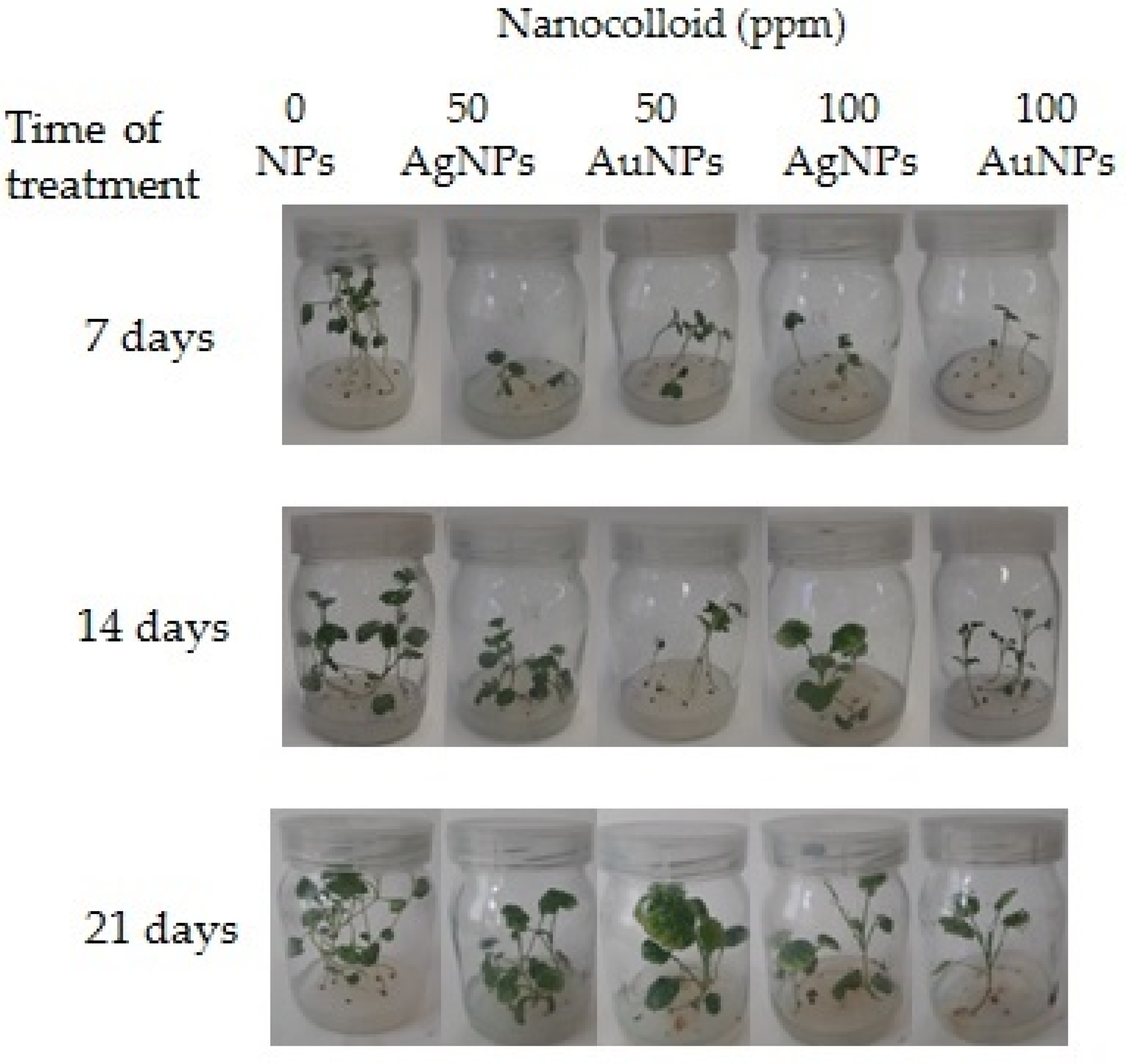
3.1. Effect of NPs on the Growth of Seedlings

3.1.1. Shoots

As demonstrated in our studies, the presence of silver and gold nanoparticles on the surface of the medium resulted in a reduction in the shoot length and weight, as compared to the control combination, irrespective of the treatment period (Table 1, Figure 1).
The application of AgNPs at the concentration of 100 ppm resulted in a decrease in the length of shoots over the first 7 days of culture, which also decreased the fresh weight and the dry weight. The longest shoots over the first days of culture were observed in the control plants. The length of these shoots was 6.33 cm, and their fresh and dry weight were 93.98 mg and 4.70 mg, respectively. After the application of AgNPs at the concentration of 100 ppm the shoots length was observed to be over 60% reduced, whereas a decrease in the fresh and dry weight accounted for 44%. The slightest changes in the morphological parameters of shoots were noted after the application of AuNPs. The shoots, when exposed to 100 ppm AuNPs, were only 34% shorter than in the control. The fresh and dry weight of shoots was similar to the same parameters of control plants in the combination AuNPs 50 ppm. In the successive weeks of culture, the biggest decreases in the shoot length and weight were noted for AuNPs 50 ppm. Fourteen days after the application, the length and the weight were, respectively, 47.3% and 50% lower than in the control. The lowest deviations in the shoot length were noted between the control combination and AgNPs 50 ppm. In the latter combination, the shoots were only about 15% shorter, while the fresh weight and the dry weight were even 23% higher compare to control. After the successive 7 days of observations, the lowest values of the parameters of shoot length and weight were noted when AuNPs at a concentration of 100 ppm were found in the environment. There was observed a decrease of the length of the shoots by 60% and the fresh weight and the dry weight by 62%. The lowest inhibiting effect was noted for AuNPs 50 ppm for the shoot length; the decrease accounted for 18%. The fresh weight and the dry weight of the shoots was higher than in the treatment without the NPs application for the variant with AgNPs 100 ppm, where, despite a reduction in the shoot length, the shoot weight was 30% higher. The application of AgNPs clearly reduced the shoot length and weight, over the first days after the application, as compared with AuNPs. A longer such impact, however, deteriorated the morphological parameters of the shoots when exposed to AuNPs.

3.1.2. Roots

The prevailing values of biometrical parameters of roots, their length and weight, decreased due to the application of AgNPs and AuNPs (Table 2). Only in the first 7 days of culture, the application of AgNPs at the concentration of 50 ppm increased the root length by over 9%. However, their fresh and dry weight was lowest, as compared with the other variants tested; about 36% lower than the control. The lowest decrease in the root weight, as compared with the control, was noted for AuNPs 100 ppm, where the root weight deviated from the values of the control treatments by 20% for the fresh weight and 25% for the dry weight of the roots.
In the successive weeks, together with an increase in the concentration of the nanoparticles applied, the maximum root length decreased both for AgNPs and for AuNPs. The shortest roots with the lowest fresh and dry weight were reported for AuNPs 100 ppm. The lowest root length and root weight losses were recorded for AgNPs 50 ppm.
After three weeks of culture, the parameters of root length and weight were highest for the combinations without the application of NPs. The greatest changes in those parameters occurred for AuNPs 100 ppm; the length as well as the fresh and dry weight of roots decreased by 68% and 61%, respectively. The smallest inhibiting effect was noted for AgNPs 100 ppm, in which the root length was reduced by 22% and the fresh and dry weight by 28%.

3.2. Effect of NPs on the Content of Chlorophyll, Carotenoids and Anthocyanins

The total chlorophyll content in rapeseed seedlings was significantly reduced under different concentrations of AgNPs and AuNPs, in comparison to the control for the first 7 days of culture (Table 3). The lowest content of total chlorophyll was recorded at the 50 ppm of AgNPs and it was 1.12 (mg/g FW), while the control was 2.15 (mg/g FW). Additionally, the treatment of the plant analyzed with AgNPs decreased the carotenoids content, especially at 50 ppm from 0.89 (mg/g FW) in the control to 0.49 (mg/g FW) in the sample studied. However, under those conditions the content of anthocyanins was 0.31 (mg/g FW) and it was higher than the control, which was 0.25 (mg/g FW). In the successive week of culture, the total content of chlorophyll as well as of carotenoids and anthocyanins was highest for the samples after the application of AgNPs 100 ppm and it was 2.09, 0.91 and 0.39 (mg/g FW), respectively. The content of photosynthetic pigments and flavonoids was also higher in the plants treated with AgNPs on the 21st day of culture. The highest content of the total chlorophyll of 2.23 (mg/g FW) was noted for AgNPs 50 ppm, the highest content of carotenoids was 0.99 (mg/g FW) and of anthocyanins—0.49 (mg/g FW); the values were significantly higher than the control (Table 3).

3.3. Effect of NPs on the Phenolics Content

The analysis of the content of free phenolic compounds (two-way ANOVA, Tukey HSD Test, p ≤ 0.05) in the plant tissues demonstrated some variation in terms of the content of the analyte (Table 4).
In the first week of the plant exposure to nanoparticles there was observed a greater variation than it is the case later. The significantly highest content of free phenols was recorded in the control plant tissues (1.32 mg/g FW), whereas the lowest significant content was recorded in the tissues of plants exposed to the effect of 100 ppm AuNPs (0.62 mg/g FW). After 14 days of exposure there was observed no greater variation in the content of free phenolic compounds across the variants; the content of phenols was very similar across the variants. Nevertheless, the lowest content was observed in the control plant tissues (0.82 mg/g FW), followed by AuNPs 100 ppm (0.93 mg/g FW) and AuNPs 50 ppm (0.97 mg/g FW). Most free phenolic compounds were recorded in the tissues of the plants treated with AgNPs (1.08 mg/g FW for AgNPs 50 ppm and 1.11 mg/g FW for AgNPs 100 ppm). Additionally, after 21 days, no greater variation was observed in the content of phenolic compounds across the combinations (Table 4). The analysis of variance showed no significance of the factor of the week of exposure to nanoparticles. However, a high significance was observed for the factor of combination (the concentration and the form of the nanoparticle) at p = 0.000482. We noted a significant interaction between the factors at p = 0.000179.

3.4. Effect of NPs on the Free Sugars Content

The analysis of the content of free sugars (two-way ANOVA, Tukey HSD Test, p = 0.05) showed some variation in terms of the content of those compounds in the plant tissues (Table 5).
After 7 days of exposure, the highest concentration of free sugars was noted in the tissues of the plants treated with AuNPs (16.88 mg/g FW for AuNPs 50 ppm, 8.62 mg/g FW for AuNPs 100 ppm). As for the control plants and the plants treated with AgNPs, we recorded no bigger differences; however, the content of sugars was non-significantly lower (7.42 mg/g FW for the control plants, 5.79 mg/g FW for AgNPs 50 ppm and 6.52 mg/g FW for AgNPs 100 ppm). After 14 days, we recorded a decrease in the content of free sugars. The lowest content was found in the plants treated with AuNPs 100 ppm (2.89 mg/g FW). As for the other variants, no significant differences were found, despite a downtrend. The highest content was reported for the plants treated with AgNPs and AuNPs at the concentration of 50 ppm; 5.88 mg/g FW (AgNPs 50 ppm) and 5.60 mg/g FW (AuNPs 50 ppm). After 21 days, the trend continued and there was observed a lower content of free sugars; 2.90 mg/g FW for the control plants, 4.02 mg/g FW for AgNPs 50 ppm, 3.98 mg/g FW for AgNPs 100 ppm, 3.44 mg/g FW for AuNPs 100 ppm, whereas in the plants treated with AuNPs (AuNPs 50 ppm), a sudden increase was observed up to 10.53 mg/g FW (Table 5). The analysis of variance showed a significance of the factor of the day for the feature at p = 0.0000285 as well as the factor of combination p = 0.00645. The interaction between the factors was significant at p = 0.09892.

3.5. Effect of NPs on the ROS Content

A number of studies demonstrated that ROS production is significantly elevated in plants after exposure to AgNPs. The exposure of plant tissues to the effect of nanoparticles, as an abiotic stress factor, is often related to an elevated level of oxidative stress markers, such as H2O2, in tissues (Table 6).
Investigating the response of plants to the presence of nanoparticles in the environment in the first week of the plant exposure, it was observed that the highest ROS concentration in form of H2O2 was found in the tissues of the plants exposed to AuNPs 100 ppm (319.99 nM/g FW). As for the other variants, the content was relatively similar. In the second and third week of exposure, there was noticed a low variation across the study variants; however, the H2O2 values in the tissues following the exposure to gold nanoparticles were higher than in the control combination. The highest concentration was noted after 14 days for the plants exposed to AuNPs 50 ppm, which was 287.22 nM/g FW and the highest concentration after 21 days was noted for plants exposed to AuNPs 100 ppm, which was 293.53 nM/g FW. The analysis of variance showed a significance of the factor of the day on feature p = 0.00000159 as well as the factor of combination p = 0.00131. The interaction between the factors was significant at p = 0.08815.

4. Discussion

The presence of AgNPs or AuNPs in the environment can result in the occurrence of some anomalies of physiological and biochemical processes and in a change in the growth rate or organ morphology. Nanoparticles can inhibit the seed germination, shoot and root elongation or reduce the leaf area [8]. Recent studies have confirmed that some NPs can get through the seed coat and affect the development processes of embryos, e.g., through a stimulation of the enzymes engaged in the metabolic processes. In the germinating seeds at the radicle appearance stage, the tissues of the root apex contact the NPs, which can get into the rhizodermis through the apoplast, with endocytosis; within the root, they flow towards the plant secretory tissue using symplastic pathways and are translocated to other plant organs [55]. Our results are consistent with such reports. The application of silver and gold nanoparticles reduced the shoot length and weight compared to the control combination. This tendency was observed in all the combinations, irrespective of the length of seed/seedlings exposure to nanoparticles. Additionally, Jiang et al. [34] noticed a drastic decrease in the fresh and dry weight of common duckweed (Spirodela polyrhiza) plants together with an increase in the concentration of AgNPs over the first few days after the application. The highest used concentration of AgNPs (10 mg/L) decreased both the fresh and dry weight of plants by almost 50%, compared to the control. Sharma et al. [56] also observed a decrease in the fresh weight of shoots in Brassica juncea but in lower concentrations of AgNPs, higher than 50 ppm. However, higher concentrations of AgNPs, exceeding 200 ppm, were needed to inhibit the shoot elongation. AgNPs concentrations on a level of 15.4 mg/L also reduced the shoot length in Physalis peruviana [12].
Our research proved that the biometrical parameters of roots decreased when exposed to AgNPs and AuNPs. The root length and weight, during 21 days of growth, were mostly inhibited by the presence of AuNPs applied in concentration of 100 ppm. Roots in this combination were the shortest and demonstrated the lowest fresh and dry weight. The reason for such an effect could be the fact that the root tissues are the first structures which come into direct contact with nanoparticles applied to the medium surface. For this reason, the effect of the nanoparticles is clearly visible in roots. Literature data indicate that the decrease in the length of the roots treated with higher concentrations of nanoparticles is a rate-related phenomenon, as lower AuNPs rates (10 ppm) in Arabidopsis thaliana did not inhibit the shoot elongation, the number of leaves and the total fresh weight [31]. Similarly, in maize plants (Zea mays L.), low AuNPs concentrations (5, 10, 15 ppm) did not inhibit growth. The seedlings under those conditions were longer than the control plants. Additionally, Sabo-Attwood et al. [57] have found that AuNPs at high concentrations (100 mg/L) deteriorated plants, whereas lower concentrations of AuNPs triggered larger-than-5 nm growth-promoting effects, supporting our findings that AuNPs enhance the plant growth.
The results of the studies and observations have suggested a conclusion that AgNPs at a high concentration result in a perforation of the cell wall penetrating the protoplast, damage the root cell vacuoles, which triggers a toxic effect [58]. Mirzajani et al. [59] observed that AgNP could not penetrate to the root cells at a low concentration (up to 30 μg/mL), whereas at higher concentrations, AgNPs showed a destructive effect on the cell structure and triggered a toxic effect by affecting various aspects of plant morphology, including a decrease in the values of growth parameters.
It was also observed that nanoparticles demonstrate a varied capacity for penetration at the highest concentration applied, depending on the parameters. A varied size of nanoparticles in various studies shows a correlation between the size and the phytotoxic effect. Usually, nanoparticles smaller in size were more destructive and inhibiting during the plant growth than those bigger in size [13,60,61]. As reported by Qian et al. [37] with an increase in the AgNPs concentration in Arabidopsis thaliana plants, a decrease was recorded in the shoot and root length. High concentrations (15.4 mg/L−1) of AgNPs also reduced the length of shoots and roots in Physalis peruviana [12]. In Brassica rapa, the concentration of 1.0 mg/L AgNPs stimulated an increase in the root length and a 10% increase in the fresh weight, while higher concentrations already showed an inhibiting effect on those parameters and the roots were clearly deformed with discolored tips [41]. Geisler-Lee et al. [62] also noted that the seedlings treated with silver nanoparticles showed shorter roots, with clearly changed brown-colored tips. The changes in the root properties and look could have been due to the fact that nanoparticles are transported through an apoplastic route in cell walls and get accumulated in structures such as plasmodesmata. The level of accumulation differed depending on the structure and accumulated progressively in the following sequence: border cells, root cap, columella and columella initials; an additional reason can be that the radicle was not adapted to the treatments [60].
Detrimental effect of AgNPs on chlorophyll content corresponds to the results of research of the impact of AgNPs in Spirodela polyrhiza [34] and in Physalis peruviana [12]. The authors observed a decreased content of chlorophyll in the initial period of plant exposure to nanoparticles. The total chlorophyll content decreased significantly when exposed to AgNPs at higher concentrations in Oryza sativa [38] and in turnip (Brassica rapa ssp. rapa L.) [41]. Sharma et al. [56] also reported on an increased content of chlorophyll after the application of 100 ppm AgNPs as well as on maximum photosystem II quantum efficiency, as measured through Fv/Fm ratio, recorded at 50 ppm AgNPs treatment, which points to a low degree of damage of the photosynthetic apparatus and to a lower frequency of photooxidative damage in plants. A corrected quantum yield positively correlates with a higher content of chlorophyll in leaves.
Anthocyanins are flavonoids found in vacuoles and they are associated with the stress response in plants [63,64]. As observed, they protect plants from damage induced by ROS [65]; they can act as non-enzymatic antioxidants to protect the cells from oxidative stress, sweeping an excess of reactive forms of oxygen as well as a chelator of metals [66] and so they are treated as antioxidant molecules [67]. The exposure of turnip seedlings to increasing AgNPs rates increased the number of anthocyanins in tissues, as compared with the control. Similar results were found for the contents of anthocyanins in A. thaliana exposed to AgNPs [37]. For that reason, an increased content of anthocyanins produced as a result of exposure to AgNPs, observed in that study, can be due to an elevated oxidative stress caused by NPs and a defense reaction against that factor in the plant.
The total polyphenol content in R. communis with the values lower than in the control samples was also found by Yasur and Rani [42], even though some data indicated that stimulation of AgNPs results in the accumulation of phenolic compounds [68]. However, Yasur and Rani [42], after the application of AgNPs, observed a greater total content of phenol in the respective parts of the plants (root and shoot). Silver in a nanoform also increased the total content of phenol in the experiments. Seed treatment with AgNP decreased the content of free phenolic compounds more than in the plants treated with AgNO3. Additionally, Spinoso-Castillo et al. [24] demonstrated an increased production of phenolic compounds in vanilla (Vanilla planifolia) in the culture with 25 and 50 mg/L AgNPs added. It is common knowledge that the effects which can be triggered in plants by AgNPs are related to their capacity to destroy cell membranes, to disrupt the DNA replication, triggering an increased production of reactive forms of oxygen and inhabiting the production of ethylene. By triggering the defense systems, the plant tries to reverse that effect, e.g., by increasing a production of phenolic compounds with a high antioxidative activity.
Presence of silver nanoparticles decreasedthe content of sugars in plants throughout the first week of culture in our study. Similar effect in the first days of culture in the plants of Lupinus termis L. was also observed by Al-Huqail et al. [69]. The exposure to 0.5 mg L−1 AgNPs decreased the content of sugar; an increase in the accumulation of proline in leaves was also noted, considered an indicator of a stressful effect of AgNPs on plants. Similar results were reported by Nair and Chung [38] in Oryza sativa L. plants. The plants exposed to 0.5 and 1 mg L−1 of AgNPs also resulted in, next to other morphological and physiological changes, a decrease in the content of sugars. However, there are reports on an increased content of soluble sugars in maize leaves exposed to AuNPs [70], which correlates with our studies with an increase in the content of free sugars as a result of the application of 50 ppm AuNPs. Additionally, it is common knowledge that the accumulation of organic substances, such as sugars or amino acids in cytoplasm, plays an important role in an osmotic regulation in plants, which affects, e.g., morphological parameters of plant growth [71,72]. In the studies of the response of the plants to stress factors, an important aspect seems to be, therefore, an identification of specific features related to the resistance to abiotic stress factors, such as the presence and concentration of compatible osmolytes (e.g., proline), sugars or proteins as potential biological markers applicable for genetic identification and modification to increase the resistance of plants and plant cells, e.g., to salinity [73].
Higher values of H2O2 were noted especially for gold nanoparticles in our study. Similarly, rice seedlings treated hydroponically with nTiO2 (1000 mg/L), nAg (0.5 and 1 mg/L), nCuO (5 mg/L) or nZnO (250 mg/L) were identified with significantly higher levels of H2O2 in the tissues [38,74,75]. Chen et al. [74] demonstrated that rice seeds exposed to nZnO at 50–1000 mg/L showed increased H2O2 levels. Similarly, Thiruvengadam et al. [41] reported the impact of AgNPs exposure in turnip seedlings and found that a higher concentration of AgNPs resulted in an excessive generation of superoxide radicals and increased lipid peroxidation; H2O2 formation also significantly increased after exposure to 5 and 10 mg/L AgNPs. Only Speranza et al. [76] reported that AgNPs treatment at a concentration of 2 mg/L delayed H2O2 production until 30 min, with a dramatic increase after the application of a higher concentration, i.e., 20 mg/L. An oxidative stress is also considered to be the one of the main mechanisms of the toxic effect of nanoparticles to plants [77].

5. Conclusions

Our results showed a variation in the effect of nanoparticles on the plants. We observed an effect of the length of exposure in relation to the kind of nanoparticles. The level of photosynthetic pigments, anthocyanins as well as other stress parameters, such as free phenolic compounds, free sugars or H2O2, decreased due to the application of both AgNPs and AuNPs at the initial culture period; however, the differences were observed in the successive weeks of exposure. The parameters were increasing irrespective of the kind of nanoparticles; however, as for the content of free sugars and free radicals, higher values were recorded due to the effect of AuNPs. Not surprisingly, the concentration has proven to play a critical role in the response of plants to nanoparticles. However, what is noteworthy is the effect of the length of exposure to nanoparticles and changes in the response of the plant in comparison to the length of its effect. Thus, our results showed that length of plants exposure to NPs is a very important factor modifying the growth and final response of seedlings. A better understanding of its influence could speed up use of NPs in agriculture and horticulture for the production of high-quality plant material (e.g., to seed priming, stimulation of seedlings’ growth and their protection against agrophages), not contaminated with pesticides, fertilizers and mycotoxins. It should be emphasized that this approach is also in line with the farm-to-fork strategy of the European Green Deal, as our findings proved the response of seedlings to AgNPs and AuNPs may vary depending on the length of exposure. Further research will be conducted to set the most efficient values of the key elements of plant/NPs interaction to ensure the priming effect of NPs on seed germination, stimulation of the seedlings’ development and protection from various biotic and abiotic factors, especially pathogens. Concluding, the potential of AgNPs and Au NPs seems to be meaningful and may lead to the application of these NPs in agriculture and horticulture as, e.g., elements of seed coatings.

Author Contributions

Conceptualization, M.T.-S.; methodology, M.T.-S. and K.L.; software, K.L. and D.P.; validation, M.T.-S. and D.P.; formal analysis, M.T.-S. and K.L.; investigation, M.T.-S.; resources M.T.-S., K.L. and D.P.; data curation, M.T.-S., K.L. and D.P.; writing—original draft preparation, M.T.-S., K.L. and D.P.; writing—review and editing M.T.-S. and D.P.; visualization, M.T.-S. and K.L.; supervision, M.T.-S.; project administration, M.T.-S.; funding acquisition, M.T.-S. and D.P. All authors have read and agreed to the published version of the manuscript.

Funding

This research received no external funding.

Data Availability Statement

Not applicable.

Conflicts of Interest

The authors declare no conflict of interest.

References

  1. Rastogi, A.; Zivcak, M.; Sytar, O.; Kalaji, H.M.; He, X.; Mbarki, S.; Brestic, M. Impact of Metal and Metal Oxide Nanoparticles on Plant: A Critical Review. Front. Chem. 2017, 5, 78. [Google Scholar] [CrossRef] [PubMed] [Green Version]
  2. Maurer-Jones, M.A.; Gunsolus, I.L.; Murphy, C.J.; Haynes, C.L. Toxicity of engineered nanoparticles in the environment. Anal. Chem. 2013, 85, 3036–3049. [Google Scholar] [CrossRef] [Green Version]
  3. Auffan, M.; Rose, J.; Bottero, J.Y.; Lowry, G.V.; Jolivet, J.P.; Wiesner, M.R. Towards a definition of inorganic nanoparticles from an environmental, health and safety perspective. Nat. Nanotechnol. 2009, 4, 634–641. [Google Scholar] [CrossRef] [PubMed]
  4. Marzec, A.; Pulit, J.; Kwaśny, J.; Banach, M. Nanometale—wybrane technologie wytwarzania. Czas. Tech.-Chem. 2012, 109, 95–107. (In Polish) [Google Scholar]
  5. Dębińska, E.; Rzepka, M.; Kremieniewski, M. Nanocząsteczki—Nowa droga w kształtowaniu parametrów świeżych i stwardniałych zaczynów cementowych. Naft. Gaz 2016, 12, 1084–1091. (In Polish) [Google Scholar] [CrossRef]
  6. Radad, K.; Al-Shraim, M.; Moldzio, R.; Rausch, W.D. Recent advances in benefits and hazards of engineered nanoparticles. Environ. Toxicol. Pharmacol. 2012, 34, 661–672. [Google Scholar] [CrossRef]
  7. Wang, P.; Lombi, E.; Zjao, F.J.; Kopittke, P.M. Nanotechnology: A new opportunity in plant sciences. Trends Plant Sci. 2016, 21, 699–712. [Google Scholar] [CrossRef]
  8. Yan, A.; Chen, Z. Impacts of Silver Nanoparticles on Plants: A Focus on the Phytotoxicity and Underlying Mechanism. Int. J. Mol. Sci. 2019, 20, 1003. [Google Scholar] [CrossRef]
  9. Barbasz, A.; Kreczmer, B.; Oćwieja, M. Effects of exposure of callus cells of two wheat varieties to silver nanoparticles and silver salt (AgNO3). Acta Physiol. Plant. 2016, 38, 76. [Google Scholar] [CrossRef] [Green Version]
  10. Barrena, R.; Casals, E.; Colón, J.; Font, X.; Sánchez, A.; Puntes, V. Evaluation of the ecotoxicity of model nanoparticles. Chemosphere 2009, 75, 850–857. [Google Scholar] [CrossRef] [Green Version]
  11. Alvarez Perez, S.; Tapia, M.A.M.; González Vega, M.E.; Ardisana, E.F.; Chávez Medina, J.A.; Flores Zamora, G.L.; Bustamante, D.V. Nanotechnology and Plant Tissue Culture. In Plant Nanobionics; Nanotechnology in the Life Sciences; Prasad, R., Ed.; Springer International Publishing: Cham, Switzerland, 2019; pp. 333–370. [Google Scholar]
  12. Timoteo de Oliveira, C.; Paiva, R.; Reis, M.V.; Claro, P.I.C.; Ferraz, L.M.; Marconcini, J.M.; de Oliveira, J.E. In vitro growth of Physalis peruviana L. affected by silver nanoparticles. 3 Biotech 2019, 9, 145. [Google Scholar] [CrossRef]
  13. Cvjetko, P.; Milošic, A.; Domijan, A.M.; Vinkovic Vrcek, I.; Tolic, S.; Peharec-Štefanic, P.; Letofsky-Papst, I.; Tkalec, M.; Balen, B. Toxicity of silver ions and differently coated silver nanoparticles in Allium cepa roots. Ecotoxicol. Environ. Saf. 2017, 137, 18–28. [Google Scholar] [CrossRef]
  14. Marambio-Jones, C.; Hoek, E.M. A review of the antibacterial effects of silver nanomaterials and potential implications for human health and the environment. J. Nanopart. Res. 2010, 12, 1531–1551. [Google Scholar] [CrossRef]
  15. Kokura, S.; Handa, O.; Takagi, T.; Ishikawa, T.; Naito, Y.; Yoshikawa, T. Silver nanoparticles as a safe preservative for use in cosmetics. Nanomed. Nanotechnol. Biol. Med. 2010, 6, 570–574. [Google Scholar] [CrossRef]
  16. Singh, N.; Manshian, B.; Jenkins, G.J.; Griffiths, S.M.; Williams, P.M.; Maffeis, T.G.; Wright, C.J.; Doak, S.H. Nano genotoxicology: The DNA damaging potential of engineered nanomaterials. Biomaterials 2009, 30, 3891–3914. [Google Scholar] [CrossRef]
  17. Tiede, K.; Boxall, A.B.; Tear, S.P.; Lewis, J.; David, H.; Hassellöv, M. Detection and characterization of engineered nanoparticles in food and the environment. Food Addit. Contam. 2008, 25, 795–821. [Google Scholar] [CrossRef]
  18. Godin, B.; Sakamoto, J.H.; Serda, R.E.; Grattoni, A.; Bouamrani, A. Emerging applications of nanomedicine for the diagnosis and treatment of cardiovascular diseases. Trends Pharmacol. Sci. 2010, 31, 199–205. [Google Scholar] [CrossRef] [Green Version]
  19. Kohl, Y.; Kaiser, C.; Bost, W.; Stracke, F.; Fournelle, M.; Wischke, C.; Thielecke, H.; Lendlein, A.; Kratz, K.; Lemor, R. Preparation and biological evaluation of multifunctional PLGA-nanoparticles designed for photoacoustic imaging. Nanomed. Nanotechnol. Biol. Med. 2011, 7, 228–237. [Google Scholar] [CrossRef]
  20. Kreuter, J.; Gelperina, S. Use of nanoparticles for cerebral cancer. Tumori 2008, 94, 271–277. [Google Scholar] [CrossRef]
  21. Meng, H.; Liong, M.; Xia, T.; Li, Z.; Ji, Z.; Zink, J.I.; Nell, A.E. Engineered design of mesoporous silica nanoparticles to deliver doxorubicin and P-glycoprotein siRNA to overcome drug resistance in a cancer cell line. ACS Nano 2010, 4, 4539–4550. [Google Scholar] [CrossRef]
  22. Gopinath, K.; Gowri, S.; Karthika, V.; Arumugam, A. Green synthesis of gold nanoparticles from fruit extract of Terminalia arjuna, for the enhanced seed germination activity of Gloriosa superba. J. Nanostruct. Chem. 2014, 4, 1–11. [Google Scholar] [CrossRef]
  23. Ruttkay-Nedecky, B.; Krystofova, O.; Nejdl, L.; Adam, V. Nanoparticles based on essential metals and their phytotoxicity. J. Nanobiotechnol. 2017, 15, 33. [Google Scholar] [CrossRef] [PubMed]
  24. Spinoso-Castillo, J.L.; Chavez-Santoscoy, R.A.; Bogdanchikova, N.; Pérez-Sato, J.A.; Morales-Ramos, V.; Bello-Bello, J.J. Antimicrobial and hormetic effects of silver nanoparticles on in vitro regeneration of vanilla (Vanilla planifolia Jacks. ex Andrews) using a temporary immersion system. Plant Cell Tissue Organ Cult. 2017, 129, 195–207. [Google Scholar] [CrossRef]
  25. Wang, Y.; Dimkpa, C.; Deng, C.; Elmer, W.H.; Gardea-Torresdey, J.; White, J.C. Impact of engineered nanomaterials on rice (Oryza sativa L.): A critical review of current knowledge. Environ. Pollut. 2022, 297, 118738. [Google Scholar] [CrossRef]
  26. Murashige, T.; Skoog, F. A revised medium for rapid growth and bioassays with tobacco tissue cultures. Physiol. Plant. 1962, 15, 473–499. [Google Scholar] [CrossRef]
  27. Aghdaei, M.; Salehi, H.; Sarmast, M.K. Effects of silver nanoparticles on Tecomella undulata (Roxb.) seem, micropropagation. Adv. Hortic. Sci. 2012, 26, 21–24. [Google Scholar]
  28. Kokina, I.; Gerbreders, V.; Sledevskis, E.; Bulanovs, A. Penetration of nanoparticles in flax (Linum usitatissimum L.) calli and regenerants. J. Biotechnol. 2013, 165, 127–132. [Google Scholar] [CrossRef]
  29. Arora, S.; Sharma, P.; Kumar, S.; Nayan, R.; Khanna, P.K.; Zaidi, M.G.H. Gold-nanoparticle induced enhancement in growth and seed yield of Brassica juncea. Plant Growth Regul. 2012, 66, 303–310. [Google Scholar] [CrossRef]
  30. Lahuta, L.B.; Szablińska-Piernik, J.; Głowacka, K.; Stałanowska, K.; Railean-Plugaru, V.; Horbowicz, M.; Pomastowski, P.; Buszewski, B. The Effect of Bio-Synthesized Silver Nanoparticles on Germination, Early Seedling Development, and Metabolome of Wheat (Triticum aestivum L.). Molecules 2022, 27, 2303. [Google Scholar] [CrossRef]
  31. Kumar, V.; Guleria, P.; Kumar, V.; Yadav, S.K. Gold nanoparticle exposure induces growth and yield enhancement in Arabidopsis thaliana. Sci. Total Environ. 2013, 461, 462–468. [Google Scholar] [CrossRef]
  32. Razavizadeh, R.; Rostami, F. Risks and Benefits Assessments of Silver Nanoparticles in Tomato Plants under in vitro Culture. Eng. Res. J. 2015, 3, 51–57. [Google Scholar]
  33. El-Temsah, Y.E.; Joner, E.J. Impact of Fe and Ag nanoparticles on seed germination and differences in bioavailability during exposure in aqueous suspension and soil. Environ. Toxicol. 2010, 27, 42–49. [Google Scholar] [CrossRef] [PubMed]
  34. Jiang, H.S.; Li, M.; Chang, F.Y.; Li, W.; Yin, L.Y. Physiological analysis of silver nanoparticles and AgNO3 toxicity to Spirodela polyrhiza. Environ. Toxicol. Chem. 2012, 31, 1880–1886. [Google Scholar] [CrossRef] [PubMed]
  35. Kaveh, R.; Li, Y.S.; Ranjbar, S.; Tehrani, R.; Brueck, C.L.; Van Aken, B. Changes in Arabidopsis thaliana gene expression in response to silver nanoparticles and silver ions. Environ. Sci. Technol. 2013, 47, 10637–10644. [Google Scholar] [CrossRef]
  36. Tripathi, D.K.; Tripathi, A.; Singh, S.; Singh, Y.; Vishwakarma, K.; Yadav, G.; Sharma, S.; Singh, V.K.; Mishra, R.K.; Upadhyay, R.G.; et al. Uptake, accumulation and toxicity of silver nanoparticle in autotrophic plants, and heterotrophic microbes: A concentric review. Front. Microbiol. 2017, 8, 7. [Google Scholar] [CrossRef] [Green Version]
  37. Qian, H.; Peng, X.; Han, X.; Ren, J.; Sun, L.; Fu, Z. Comparison of the toxicity of silver nanoparticles and silver ions on the growth of terrestrial plant model Arabidopsis thaliana. J. Environ. Sci. 2013, 25, 1947–1955. [Google Scholar] [CrossRef]
  38. Nair, P.M.G.; Chung, I.M. Physiological and molecular level effects of silver nanoparticles exposure in rice (Oryza sativa L.) seedlings. Chemosphere 2014, 112, 105–113. [Google Scholar] [CrossRef]
  39. Shaikhaldein, H.O.; Al-Qurainy, F.; Nadeem, M.; Khan, S.; Tarroum, M.; Salih, A.M. Biosynthesis and characterization of silver nanoparticles using Ochradenus arabicus and their physiological effect on Maerua oblongifolia raised in vitro. Sci. Rep. 2020, 10, 17569. [Google Scholar] [CrossRef]
  40. Homaee, M.B.; Ehsanpour, A.A. Silver nanoparticles and silver ions: Oxidative stress responses and toxicity in potato (Solanum tuberosum L.) grown in vitro. Hortic. Environ. Biotechnol. 2016, 57, 544–553. [Google Scholar] [CrossRef]
  41. Thiruvengadam, M.; Gurunathan, S.; Chung, I.M. Physiological, metabolic, and transcriptional effects of biologically-synthesized silver nanoparticles in turnip (Brassica rapa ssp. rapa L.). Protoplasma 2015, 252, 1031–1046. [Google Scholar] [CrossRef]
  42. Yasur, J.; Rani, P.U. Environmental effects of nanosilver: Impact on castor seed germination, seedling growth, and plant physiology. Environ. Sci. Pollut. Res. 2013, 20, 8636–8648. [Google Scholar] [CrossRef] [PubMed]
  43. Szollosi, R.; Molnár, A.; Kondak, S.; Kolbert, Z. Dual Effect of Nanomaterials on Germination and Seedling Growth: Stimulation vs. Phytotoxicity. Plants 2020, 9, 1745. [Google Scholar] [CrossRef] [PubMed]
  44. Shah, V.; Belozerova, I. Influence of metal nanoparticles on the soil microbial community and germination of lettuce seeds. Water Air Soil Pollut. 2009, 197, 143–148. [Google Scholar] [CrossRef]
  45. Xie, H.; Mason, M.M.; Wise, J.P., Sr. Genotoxicity of metal nanoparticles. Rev. Environ. Health 2011, 26, 251–268. [Google Scholar] [CrossRef] [PubMed]
  46. Tomaszewska-Sowa, M.; Siwik-Ziomek, A.; Figas, A.M.; Bocian, K. Assessment of metal nanoparticle-induced morphological and physiological changes in in vitro cultures of rapeseed (Brassica napus L.). EJPAU 2018, 21, 10. [Google Scholar] [CrossRef]
  47. Domeradzka-Gajda, Y.-K.; Nocuń, M.; Roszak, J.; Janasik, B.; Quarles, C.D., Jr.; Wąsowicz, W.; Grobelny, J.; Tomaszewska, E.; Celichowski, G.; Ranoszek-Soliwoda, K.; et al. A study on the in vitro percutaneous absorption of silver nanoparticles in combination with aluminum chloride, methyl paraben or di-n-butyl phthalate. Toxicol. Lett. 2017, 272, 38–48. [Google Scholar] [CrossRef] [PubMed]
  48. Pudlarz, A.; Czechowska, E.; Ranoszek-Soliwoda, K.; Tomaszewska, E.; Celichowski, G.; Grobelny, J.; Szemraj, J. Immobilization of Recombinant Human Catalase on Gold and Silver Nanoparticles. Appl. Biochem. Biotechnol. 2018, 185, 717–735. [Google Scholar] [CrossRef]
  49. Wettstein, D. Chlorophyll–letale und der submikroskopische Formwechsel der Plastiden. Exp. Cell Res. 1957, 12, 427–487. [Google Scholar] [CrossRef]
  50. Harborne, J.B. Comparative Biochemistry of the Flavonoids; Academic Press: London, UK, 1967. [Google Scholar]
  51. Maksimović, J.J.D.; Živanović, B.D. Quantification of the antioxidant activity in salt-stressed tissues. In Plant Salt Tolerance; Humana Press: Totowa, NY, USA, 2012; pp. 237–250. [Google Scholar]
  52. Zahir, S.; Zhang, F.; Chen, J.; Zhu, S. Determination of Oxidative Stress and Antioxidant Enzyme Activity for Physiological Phenotyping During Heavy Metal Exposure. In Environmental Toxicology and Toxicogenomics; Humana: New York, NY, USA, 2021; pp. 241–249. [Google Scholar]
  53. DuBois, M.; Gilles, K.A.; Hamilton, J.K.; Rebers, P.A.; Smith, F. Colorimetric method for the detemination of sugars and related substances. Anal. Chem. 1956, 28, 350–356. [Google Scholar] [CrossRef]
  54. Bacete, L.; Melida, H.; Pattathil, S.; Hahn, M.G.; Molina, A.; Miedes, E. Characterization of plant cell wall damage-associated molecular patterns regulating immune responses. In Plant Pattern Recognition Receptors: Methods and Protocols. Methods in Molecular Biology; Shan, L., He, P., Eds.; Springer Science + Buisness Media, Humana Press: New York, NY, USA, 2017; pp. 13–24. [Google Scholar]
  55. Rizwan, M.; Ali, S.; Qayyum, M.F.; Ok, Y.S.; Adrees, M.; Ibrahim, M.; Zia-ur-Rehman, M.; Farid, M.; Abbas, F. Effect of metal and metal oxide nanoparticles on growth and physiology of globally important food crops: A critical review. J. Hazard. Mater. 2017, 322, 2–16. [Google Scholar] [CrossRef]
  56. Sharma, P.; Bhatt, D.; Zaidi, M.G.H.; Saradhi, P.P.; Khanna, P.K.; Arora, S. Silver Nanoparticle-Mediated Enhancement in Growth and Antioxidant Status of Brassica juncea. Appl. Biochem. Biotechnol. 2012, 167, 2225–2233. [Google Scholar] [CrossRef] [PubMed]
  57. Sabo-Attwood, T.; Unrine, J.M.; Stone, J.W.; Murphy, C.J.; Ghoshroy, S.; Blom, D.; Bertsch, P.M.; Newman, L.A. Uptake, distribution and toxicity of gold nanoparticles in tobacco (Nicotiana xanthi) seedlings. Nanotoxicology 2012, 6, 353–360. [Google Scholar] [CrossRef] [PubMed]
  58. Mazumdar, H.; Ahmed, G.U. Phytotoxicity effect of silver nanoparticles on Oryza sativa. Int. J. Chem. Tech. Res. 2011, 3, 1494–1500. [Google Scholar]
  59. Mirzajani, F.; Askari, H.; Hamzelou, S.; Farzaneh, M.; Ghassempour, A. Effect of silver nanoparticles on Oryza sativa L. and its rhizosphere bacteria. Ecotoxicol. Environ. Saf. 2013, 88, 48–54. [Google Scholar] [CrossRef] [PubMed]
  60. Yin, L.; Colman, B.P.; McGill, B.M.; Wright, J.P.; Bernhardt, E.S. Effects of silver nanoparticle exposure on germination and early growth of eleven wetland plants. PLoS ONE 2012, 7, e47674. [Google Scholar] [CrossRef] [PubMed] [Green Version]
  61. Jiang, H.S.; Qiu, X.N.; Li, G.B.; Li, W.; Yin, L.Y. Silver nanoparticles induced accumulation of reactive oxygen species and alteration of antioxidant systems in the aquatic plant Spirodela polyrhiza. Environ. Toxicol. Chem. 2014, 33, 1398–1405. [Google Scholar] [CrossRef] [PubMed]
  62. Geisler-Lee, J.; Wang, Q.; Yao, Y.; Zhang, W.; Geisler, M.; Li, K.; Huang, Y.; Chen, Y.; Kolmakov, A.; Ma, X. Phytotoxicity, accumulation and transport of silver nanoparticles by Arabidopsis thaliana. Nanotoxicology 2013, 7, 323–337. [Google Scholar] [CrossRef]
  63. Solfanelli, C.; Poggi, A.; Loreti, E.; Alpi, A.; Perata, P. Sucrose-specific induction of the anthocyanin biosynthetic pathway in Arabidopsis. Plant Physiol. 2006, 140, 637–646. [Google Scholar] [CrossRef] [Green Version]
  64. Guo, N.; Cheng, F.; Wu, J.; Liu, B.; Zheng, S.; Liang, J.; Wang, X. Anthocyanin biosynthetic genes in Brassica rapa. BMC Genom. 2014, 15, 426. [Google Scholar] [CrossRef] [Green Version]
  65. Nagata, T.; Todoriki, S.; Masumizu, T.; Suda, I.; Furuta, S.; Du, Z.; Kikuchi, S. Levels of active oxygen species are controlled by ascorbic acid and anthocyanin in Arabidopsis. J. Agric. Food Chem. 2003, 51, 2992–2999. [Google Scholar] [CrossRef]
  66. Carocho, M.; Ferreira, I.C. A review on antioxidants, pro-oxidants and related controversy: Natural and synthetic compounds, screening and analysismethodologies and future perspectives. Food Chem. Toxicol. 2013, 51, 15–25. [Google Scholar] [CrossRef]
  67. Gould, K.S.; McKelvie, J.; Markham, K.R. Do anthocyanins function as antioxidants in leaves? Imaging of H2O2 in red and green leaves after mechanical injury. Plant Cell Environ. 2002, 25, 1261–1269. [Google Scholar] [CrossRef]
  68. Kim, M.J.; Kwak, H.S.; Kim, S.S. Effects of germination on protein, -aminobutyric acid, phenolic acids, and antioxidant capacity in wheat. Molecules 2018, 23, 2244. [Google Scholar] [CrossRef] [Green Version]
  69. Al-Huqail, A.A.; Hatata, M.M.; Al-Huqail, A.A.; Ibrahim, M.M. Preparation, characterization of silver phytonanoparticles and their impact on growth potential of Lupinus termis L. seedlings. Saudi J. Biol. Sci. 2018, 25, 313–319. [Google Scholar] [CrossRef]
  70. Mahakham, W.; Theerakulpisut, P.; Maensiri, S.; Phumying, S.; Sarmah, A.K. Environmentally benign synthesis of phytochemicals-capped gold nanoparticles as nanopriming agent for promoting maize seed germination. Sci. Total Environ. 2016, 573, 1089–1102. [Google Scholar] [CrossRef]
  71. Ashraf, M.; Harris, P.J.C. Potential biochemical indicators of salinity tolerance in plants. Plant Sci. 2004, 166, 3–16. [Google Scholar] [CrossRef]
  72. Watanabe, S.; Kojima, K.; Ide, Y.; Sasaki, S. Effects of saline and osmotic stress on proline and sugar accumulation in Populus euphratica in vitro. Plant Cell Tissue Organ Cult. 2000, 63, 199–206. [Google Scholar] [CrossRef]
  73. Shonjani, S. Salt Sensitivity of Rice, Maize, Sugar Beet, and Cotton during Germination and Early Vegetative Growth. Ph.D. Thesis, Institute of Plant Nutrition, Justus Liebig University, Giessen, The Netherlands, 2002. [Google Scholar]
  74. Chen, J.; Liu, X.; Wang, C.; Yin, S.S.; Li, X.L.; Hu, W.J.; Simon, M.; Shen, Z.J.; Xiao, Q.; Chu, C.C.; et al. Nitric oxide ameliorates zinc oxide nanoparticles-induced phytotoxicity in rice seedlings. J. Hazard. Mater. 2015, 297, 173–182. [Google Scholar] [CrossRef]
  75. Wang, S.; Lui, H.; Zhang, Y.; Xin, H. The effect of CuO NPs on reactive oxygen species and cell cycle gene expression in roots of rice. Environ. Toxicol. Chem. 2015, 34, 554–561. [Google Scholar] [CrossRef]
  76. Speranza, A.; Crinelli, R.; Scoccianti, V.; Taddei, A.R.; Iacobucci, M.; Bhattacharya, P.; Ke, P.C. In vitro toxicity of silver nanoparticles to kiwifruit pollen exhibits peculiar traits beyond the cause of silver ion release. Environ. Pollut. 2013, 179, 258–267. [Google Scholar] [CrossRef]
  77. Brunner, T.J.; Wick, P.; Manser, P.; Spohn, P.; Grass, R.N.; Limbach, L.K.; Bruinink, A.; Stark, W.J. In Vitro Cytotoxicity of Oxide Nanoparticles: Comparison to Asbestos, Silica, and the Effect of Particle Solubility. Environ. Sci. Technol. 2006, 40, 4374–4381. [Google Scholar] [CrossRef] [PubMed]
Figure 1. Comparison of the effect of the nanoparticles’ (NPs) type and concentration on the morphological parameters of seedlings. Abbreviations: AgNPs—silver nanoparticles, AuNPs—gold nanoparticles.
Agronomy 12 02885 g001
Table 1. Comparison of the influence of nanoparticles’ type and concentration on the shoot length and fresh and dry weight of rapeseed seedlings.
Treatment Time (Days)Concentration of NanoparticlesShoot Length (cm)Shoot Fresh Weight (mg)Shoot Dry Weight (mg)
7Control6.33 ± 2.06 a–e *93.98 ± 19.14 c–e4.70 ± 0.95 c–e
AgNPs 50 ppm3.67 ± 1.03 ef82.17 ± 19.55 de4.11 ± 0.97 de
AgNPs 100 ppm2.50 ± 1.22 f52.67 ± 8.64 e2.63 ± 0.43 e
AuNPs 50 ppm3.33 ± 1.36 f93.00 ± 42.87 de4.65 ± 2.14 de
AuNPs 100 ppm4.17 ± 0.98 c–f75.67 ± 19.45 e3.78 ± 0.97 e
14Control9.17 ± 1.83 a155.50 ± 34.61 b–d7.78 ± 1.72 b–d
AgNPs 50 ppm7.83 ± 1.50 ab191.33 ± 32.61 ab9.57 ± 1.63 ab
AgNPs 100 ppm7.25 ± 1.63 a–c150.50 ± 25.09 b–d7.53 ± 1.25 b–d
AuNPs 50 ppm4.83 ± 1.57 b–f76.50 ± 30.22 e3.83 ± 1.51 e
AuNPs 100 ppm5.08 ± 2.63 b–f97.33 ± 47.93 de4.87 ± 2.39 de
21Control9.83 ± 1.72 a237.00 ± 40.92 ab11.85 ± 2.04 ab
AgNPs 50 ppm6.17 ± 1.16 a–e171.50 ± 72.31 a–d8.58 ± 3.61 a–d
AgNPs 100 ppm6.67 ± 1.03 a–d307.00 ± 56.49 a15.35 ± 2.82 a
AuNPs 50 ppm8.00 ± 1.78 ab197.67 ± 95.77 a–c9.88 ± 3.64 a–c
AuNPs 100 ppm4.00 ± 1.09 d–f90.67 ± 40.10 de4.53 ± 2.00 de
* Values are presented as means ± standard deviation; the same lowercase letters indicate non-statistically significant differences according to Tukey’s test (p ≤ 0.05). Abbreviations: AgNPs—silver nanoparticles, AuNPs—gold nanoparticles.
Table 2. Comparison of the influence of nanoparticles’ type and concentration on the root length and fresh and dry weight of rapeseed seedlings.
Treatment Time (Days)Concentration of NanoparticlesMax Root Length (cm)Root Fresh Weight (mg)Root Dry Weight (mg)
7Control5.33 ± 1.94 ab *81.67 ± 28.57 b4.08 ± 1.42 b
AgNPs 50 ppm5.83 ± 1.16 a51.67 ± 11.69 b–d2.58 ± 0.58 b–d
AgNPs 100 ppm4.00 ± 0.89 a–d60.00 ± 8.36 bc3.00 ± 0.42 bc
AuNPs 50 ppm5.00 ± 1.78 ab54.33 ± 7.65 b–d2.72 ± 0.38 b–d
AuNPs 100 ppm4.33 ± 0.81 a–d61.17 ± 14.27 bc3.06 ± 0.71 bc
14Control4.75 ± 1.08 ab132.50 ± 29.78 a6.63 ± 1.48 a
AgNPs 50 ppm3.58 ± 0.64 a–e60.33 ± 17.03 b–d3.02 ± 0.85 bc
AgNPs 100 ppm3.42 ± 0.37 a–e51.67 ± 8.16 b–d2.58 ± 0.40 b–d
AuNPs 50 ppm2.33 ± 0.40 c–e47.50 ± 18.09 bc2.38 ± 0.90 b–d
AuNPs 100 ppm2.25 ± 0.61 de26.83 ± 6.17 d1.34 ± 0.30 d
21Control6.00 ± 1.54 a80.83 ± 16.36 b4.04 ± 0.81 b
AgNPs 50 ppm3.92 ± 1.02 a–d49.83 ± 11.92 b–d2.49 ± 0.59 b–d
AgNPs 100 ppm4.67 ± 1.72 a–c57.67 ± 25.42 b–d2.88 ± 1.27 b–d
AuNPs 50 ppm3.00 ± 1.54 b–e33.17 ± 27.83 cd1.66 ± 1.39 cd
AuNPs 100 ppm1.92 ± 1.02 e31.00 ± 17.49 cd1.55 ± 0.87 cd
* Values are presented as means ± standard deviation; the same lowercase letters indicate non-statistically significant differences according to Tukey’s test (p ≤ 0.05).
Table 3. Comparison of the influence of nanoparticles’ type and concentration on the photosynthetic pigments—chlorophyll (Chl.), carotenoids and anthocyanins content in rapeseed seedlings.
Treatment Time (Days)Concentration of
Nanoparticles
Chl. a
(mg/g FW)
Chl. b
(mg/g FW)
Chl. Total
(mg/g FW)
Carotenoids
(mg/g FW)
Anthocyanins
(mg/g FW)
7Control1.44 a–d *0.72 a2.15 ± 0.32 ab0.89 ± 0.12 a–c0.25 ± 0.08 ab
AgNPs 50 ppm0.75 e0.37 b–e1.12 ± 0.12 f0.49 ± 0.03 d0.31 ± 0.08 ab
AgNPs 100 ppm0.86 de0.33 de1.18 ± 0.21 ef0.55 ± 0.08 cd0.25 ± 0.03 ab
AuNPs 50 ppm1.16 a–e0.38 b–e1.55 ± 0.24 a–f0.73± 0.11 a–d0.23 ± 0.02 ab
AuNPs 100 ppm0.91 c–e0.31 e1.22 ± 0. 13 d–f0.58 ± 0.06 b–d0.24 ± 0.01 ab
14Control1.00 b–e0.35 c–e1.35 ± 0.36 b–f0.67 ± 0.17 a–d0.31 ± 0.07 ab
AgNPs 50 ppm1.16 a–e0.42 b–e1.58 ± 0.43 a–f0.73 ± 0.21 a–d0.18 ± 0.01 b
AgNPs 100 ppm1.51 a–c0.57 ab2.09 ± 0.47 ab0.91 ± 0.19 ab0.39 ± 0.36 ab
AuNPs 50 ppm1.35 a–d0.44 b–e1.78 ± 0.37 a–f0.84 ± 0.17 a–c0.24 ± 0.06 ab
AuNPs 100 ppm1.40 a–d0.44 b–e1.84 ± 0.62 a–f0.86 ± 0.27 a–c0.25 ± 0.05 ab
21Control1.54 a–c0.54 a–d2.08 ± 0.86 a–d0.93 ± 0.35 ab0.33 ± 0.10 ab
AgNPs 50 ppm1.69 a0.54 a–c2.23 ± 0.37 a0.99 ± 0.16 a0.49 ± 0.18 a
AgNPs 100 ppm1.46 a–d0.44 b–e1.90 ± 0.5 a–e0.86 ± 0.20 a–c0.32 ± 0.12 ab
AuNPs 50 ppm1.00 b–e0.32 e1.31 ± 0.49 c–f0.64 ± 0.21 a–d0.20 ± 0.03 b
AuNPs 100 ppm1.55 ab0.49 a–e2.04 ± 0.33 a–c0.94 ± 0.12 ab0.29 ± 0.07 ab
* Values are presented as means ± standard deviation; the same lowercase letters indicate non-statistically significant differences according to Tukey’s test (p ≤ 0.05).
Table 4. Comparison of the influence of nanoparticles’ type and concentration on the free phenolics content in rapeseed seedlings.
Concentration of
Nanoparticles
Phenolics mg/g Fresh Weight (FW)
7 Treatment Time (Days)14 Treatment Time (Days)21 Treatment Time (Days)
0NPs1.32 ± 0.04 a *0.82 ± 0.11 bc1.06 ± 0.04 ab
AgNPs 50 ppm1.01 ± 0.09 ab1.08 ± 0.12 ab0.97 ± 0.08 a–c
AgNPs 100 ppm0.85 ± 0.03 bc1.11 ± 0.12 ab0.98 ± 0.02 a–c
AuNPs 50 ppm1.00 ± 0.03 ab0.97 ± 0.13 a–c0.97 ± 0.00 a–c
AuNPs 100 ppm0.62 ± 0.02 c0.93 ± 0.05 bc0.82 ± 0.03 bc
* Values are presented as means ± standard deviation; the same lowercase letters indicate non-statistically significant differences according to Tukey’s test (p ≤ 0.05).
Table 5. Comparison of the influence of nanoparticles’ type and concentration on the free sugars content in rapeseed seedlings.
Concentration of
Nanoparticles
Sugars mg/g FW
7 Treatment Time (Days)14 Treatment Time (Days)21 Treatment Time (Days)
0NPs7.42 ± 1.79 a–c *4.83 ± 1.16 a–c2.90 ± 0.37 bc
AgNPs 50 ppm5.79 ± 0.35 a–c5.88 ± 1.14 a–c4.02 ± 0.65 a–c
AgNPs 100 ppm6.52 ± 0.49 a–c3.96 ± 0.65 a–c3.98 ± 0.96 a–c
AuNPs 50 ppm16.88 ± 0.84 a5.60 ± 2.39 a–c10.53 ± 0.03 a
AuNPs 100 ppm8.62 ± 1.60 ab2.89 ± 0.53 c3.44 ± 0.96 bc
* Values are presented as means ± standard deviation; the same lowercase letters indicate non-statistically significant differences according to Tukey’s test (p ≤ 0.05).
Table 6. Comparison of the influence of nanoparticles’ type and concentration on the H2O2 content in rapeseed seedlings.
Concentration of
Nanoparticles
H2O2 nM/g FW
7 Treatment Time (Days)14 Treatment Time (Days)21 Treatment Time (Days)
0NPs351.15 ± 26.18 a *251.54± 8.80 cd290.2 ± 9.73 a–d
AgNPs 50 ppm267.51 ± 9.53 b–d243.90 ± 13.59 d272.76 ± 14.64 b–d
AgNPs 100 ppm280.21 ± 1.59 b–d253.23 ± 5.66 cd268.60 ± 4.04 b–d
AuNPs 50 ppm308.66 ± 16.78 a–c287.22 ± 16.90 a–d285.84 ± 0.51 a–d
AuNPs 100 ppm319.99 ± 7.50 ab258.87 ± 13.87 b–d293.53 ± 11.24 a–d
* Values are presented as means ± standard deviation; the same lowercase letters indicate non-statistically significant differences according to Tukey’s test (p ≤ 0.05).
Publisher’s Note: MDPI stays neutral with regard to jurisdictional claims in published maps and institutional affiliations.

