Article

Cultivar Susceptibility to Olive Knot Disease and Association with Endophytic Microbiota Community

1
Council for Agricultural Research and Economics, Research Centre for Olive, Fruit and Citrus Crops, Corso Savoia 190, 95024 Acireale, Italy
2
Department of Physics and Astronomy, University of Catania, Via Santa Sofia 68, 95123 Catania, Italy
3
Department of Agriculture, Food and Environment, University of Catania, Via Santa Sofia, 100, 95123 Catania, Italy
*
Author to whom correspondence should be addressed.
Submission received: 28 December 2022 / Revised: 25 January 2023 / Accepted: 1 February 2023 / Published: 4 February 2023

Abstract

:
Olive knot disease (OKD) induced by the bacterium Pseudomonas savastanoi pv. savastanoi seriously affects olive production in the Mediterranean basin. Nowadays, the only strategies to control the disease are pruning and the application of cupric products. An essential strategy to enhance protection is represented by the identification of resistant cultivars, which represents a crucial opportunity for future investments and breeding. We undertook a three-year-long survey at the International Olive Germplasm Collection of “Villa Zagaria” (Sicily, Italy) on thirty-six Sicilian cultivars that were monitored for symptom development. Cultivars with different levels of susceptibility were divided into five clusters. Moreover, in order to investigate possible interactions with endophytic microbial communities, two cultivars with contrasting susceptibilities, Zaituna (highly resistant) and Giarraffa (highly susceptible), were selected for an amplicon-based metagenomic analysis. Distinct endophytic communities colonized the two cultivars, suggesting an interaction between the resident bacterial community and the pathogen. Significantly higher bacterial richness was detected in the shoots of the susceptible cv. Giarraffa, although it had lower diversity. The opposite trend was observed for fungal communities. Among the microbes resulted to be enriched in cv. Giarraffa, it is important to underline the presence of Pseudomonas among the bacterial genera, and Alternaria, Neofusicoccum, Epicoccum, Ascochyta, and Elsinoe among the fungal genera, which include many species often described as plant pathogens and biocontrol agents. Starting from this basic information, new strategies of control, which include breeding for resistance and integrated disease management, can be envisaged.

1. Introduction

The importance of olive cultivation in Italy, among other Mediterranean countries, is well-known. According to ISTAT data, in 2020, more than 1 million hectares of occupied land area in Italy were dedicated to olive cultivation, yielding a total of more than l26 million quintals of olives produced (http://dati.istat.it/, accessed on 22 November 2022). In Sicily, olive production in 2022 covered more than 161,000 hectares of surface, with 3 million quintals produced.
Serious losses in terms of production and olive oil quality can be caused by olive knot disease (OKD), one of the most important olive tree (Olea europaea L.) diseases worldwide [1]. Its main symptom is the formation of overgrowth tissues (knots) in the aerial parts of olive trees, mainly on twigs and branches [2]. Pseudomonas savastanoi pv. savastanoi (Psv) is the causal agent of OKD and is considered one of the major threats to olive tree production, especially in the Mediterranean region, where climatic conditions often favor the spread of the disease [3,4]. The disease rarely induces the death of a tree, but olive production can be severely affected [1,5].
Psv lives epiphytically on the surface of the aerial parts of olive trees [5,6], and under favorable weather conditions, it penetrates into olive tissues, leading to the formation of tumorous overgrowths after a few weeks or several months, depending on climate conditions and the timing of inoculum occurrence [5,7]. Moreover, wounds caused by falling leaves or pruning release signals that activate the response of Psv, inducing tumor formation [2]. Environmental stresses, such as mechanical wounding due to hail events or intense frosts, are considered the main causes of bacterium entrance and symptom development, especially in the northern regions of Italy [8].
Seasonal variation in Psv populations has also been shown to be higher in warm and rainy months (fall and spring) and lower in hot and dry months (summer and winter) in both the leaf and stem surfaces [9,10]. Psv cells growing on any part of a plant may serve as inoculum reservoirs for infection.
Nonpathogenic bacteria belonging to the species Erwinia toletana, Pantoea agglomerans, and Erwinia oleae are frequently associated with Psv within olive knots, communicating with each other by quorum sensing signals and cooperating with the pathogen to increase disease severity [11,12].
Currently, the only strategies to control the disease are pruning and the application of cupric products. However, considering the EU limitations for the use of copper [13], an essential strategy to enhance protection is represented by the identification of less susceptible cultivars [14]. Previous observations (very limited and not complete) stated that there were no cultivars resistant to OKD [15]. However, very few studies have been undertaken to explore cultivar susceptibility to OKD, especially under field conditions [5,16,17,18,19]. Not enough data are available on the pathogen susceptibility of Italian cultivars and, in particular, of Sicilian cultivars that have been gathered by field investigation or artificial inoculation [16,20,21]. A correlation between cultivar susceptibility and late winter frost was demonstrated among some Italian cultivars [22]. However, susceptibility to frost is not homogeneous among cultivars [23,24] and is somehow related to late frost tolerance [25].
A recent comprehensive survey on cultivar susceptibility was carried out after the screening of a worldwide germplasm olive collection maintained in California, USA (National Clonal Germplasm Repository in Winters), containing 104 genotypes; among them, 15 came from Italy, with only cv. Ogliarola Messinese from Sicily [21].
The traditional idea that disease symptoms are caused by a single pathogen is changing toward a new concept that involves complex microbial interactions. The protection of a plant, as well the decrease in health status, are dependent on the set of bacteria or fungi living within the plant in a relationship of either cooperation or antagonism. The microbial consortium that leads to the progression of a disease was recently termed the pathobiome [26]. Microorganisms of different kingdoms can interact with each other, resulting in enhanced or decreased pathogenicity. For this reason, the pathobiome plays a crucial role in the different manifestations of symptoms visible in a host, which can show different rates of susceptibility. In this sense, the role of endophytic fungi has been recognized as crucial in the protection of host plants against biotic adversities through cross-talking signals that allow the onset of compatible interactions [27,28]. In perennial crops, such as olive, where the factor of time has a great impact on cultivation, a strong equilibrium between the endophytic microbiota and the host plant was clearly established [29]. Moreover, through the production of bioactive compounds and stimulation of the defense reaction, endophytic fungi were able to procure beneficial effects for their host plants [28].
A recent study on OKD used as a model demonstrated that the composition and abundance of epiphytic and endophytic fungal communities were affected not only by the presence or absence of the disease, but also by the host plant genotype [30]. Similarly, OKD is the main driver of bacterial communities in olive plants showing different levels of susceptibility, with higher effects on susceptible cultivars and endophytic communities. This confirms that host plants and plant habitats have crucial roles in shaping pathobiome composition, and disease is the result of intricate relationships among all these factors [31]. With reference to the outbreak of the quick decline syndrome incited by Xylella fastidiosa in southern Italy, research on the potential association with microbial endophytes was carried out for two cultivars of high and low susceptibilities, revealing no effects on the mechanism of resistance [32].
The main objectives of this research are: (1) to investigate OKD susceptibility on a representative collection of Sicilian olive cultivars, located in Sicily in the International Olive Germplasm Collection (IOGC) of “Villa Zagaria”, and (2) to describe the structure of the endophytic microbial community of cultivars showing different levels of susceptibility.

2. Materials and Methods

2.1. Orchard, Plant Material, and Field Monitoring

We evaluated 36 Sicilian cultivars (Table 1) of cultivated olive trees conserved as a live collection in the International Olive Germplasm Collection (IOGC) of “Villa Zagaria” (lat. 37°30′52″ N; long. 14°17′46″ E) that hosts more than 400 cultivars distributed in 4 plots: plot A (local cultivars from Enna province), plot B (Sicilian cvs), plot C (Italian cvs), and plot D (international cvs). The olive trees were planted in 2004 and are occasionally treated with copper-based bactericides.
Symptoms of olive knot disease were visually monitored over 3 years (2018–2020). In December of each year, we recorded the disease intensity for each variety (4 replicates) according to a 0–4 scale [33], as follows: 0—no knots found on at least 10 branches, 1—a few single knots, 2—many knots, usually well-separated from each other, 3—knots frequently coalescing, and 4—knots covering branches, with separate knots no longer recognizable (Figure 1).

2.2. Pseudomonas savastanoi pv. savastanoi Isolation and Molecular Confirmation

Bacterial colonies were isolated from young knots collected from olive stems and branches during December 2018. After removal of the external layers of knot surfaces with the help of a sterile scalpel, small fragments (5–10 mm) were aseptically cut and placed in a sterile Petri dish. Each fragment was reduced to smaller pieces in 100 µL of sterile water, allowing bacterial spill (exit). About 20 µL of contaminated water was spread on plates containing King’s medium B (KB) [34] and cycloheximide (100 μg/mL) and incubated at 26 °C for 3 days. Single fluorescent representative colonies of predominant morphological types of bacterial isolates were re-streaked onto new KB plates to obtain pure single colonies. Bacterial lysates were prepared by dissolving 1 colony in 200 µL of sterile water in a 1.5 mL Eppendorf tube, boiling in water for 10 min, and cooling on ice. An amount of 2 µL of bacterial lysate was used as a target template for PCR identification of Psv using the following primers: IAALF (5′-GGCACCAGCGGCAACATCAA-3′) and IAALR (5′-CGCCCTCGGAACTGCCATAC-3′) [35]. Briefly, PCR was performed in a Veriti 96-well Thermal Cycler (Applied Biosystems, Foster City, CA, USA) at a total volume of 25 μL. PCR mixtures contained 1 μL of bacterial lysate, 0.6 μM of each primer, and 1x PCR DreamTaq Green PCR Master Mix (Thermo Scientific Inc., Waltham, MA, USA). Amplification conditions constituted an initial denaturation step of 5 min at 94 °C, followed by 40 cycles of 30 s at 94 °C, 30 s at 62 °C, and 30 s at 72 °C, with a final elongation step of 5 min at 72 °C. Amplified products (5 μL) were separated by electrophoresis (100 V) on 1.5% agarose gel stained with GelRed (Invitrogen Co., Carlsbad, CA, USA). Amplicons of 454 bp were obtained for a total of 39 Psv colonies, which were stored in 20% glycerol at −80 °C.

2.3. Sample Collection and Processing for Microbiome Analysis

Endophytic microbiomes were analyzed from six field-grown olive trees of the susceptible Giarraffa and resistant Zaituna cultivars (three each) exposed to natural Psv infection at “Villa Zagaria”. Trees were sampled in Spring 2021 from the cv. Giarraffa, showing high numbers of confluent knots around branches (disease severity score of 4), and cv. Zaituna, which were symptomless with no relevant knots (disease severity score of 0). Ten twigs of about 0.5 cm in diameter were collected from each tree in the middle part of the canopy from the four cardinal points, avoiding tissues in an advanced stage of desiccation. Samples were immediately stored in sealed plastic bags and kept refrigerated at 4 °C to avoid dehydration until processing in the laboratory within the following 24 h. For microbiome DNA extraction, twigs from each tree sample were cut into small pieces and washed with running tap water before surface sterilization by sequential dipping in 2% sodium hypochlorite for 2 min, 70% ethanol for 2 min, and three rinses in sterile, distilled water. Sterilized tissues were kept at −80 °C for around 1 week and then were shaken with TissueLyser (Qiagen, Milan, Italy) at 4 °C for 5 min at 30 Hz. One hundred milligrams were used for DNA extraction with a DNA Plant mini kit (Qiagen, Milan, Italy) following the manufacturer’s instructions. The presence of Psv in the DNA extracts was assessed by quantitative polymerase chain reaction (qPCR) according to Bella et al. [36].

2.4. 16S and ITS Metagenomic Sequencing Library Preparation, Sequencing, and Analysis

Next-generation sequencing experiments composed of quality control and a primary bioinformatics analysis were performed with Genomix4life S.R.L. (Baronissi, Salerno, Italy). DNA quality control was performed using a NanoDropOne spectrophotometer (Thermo Scientific Inc., Waltham, MA, USA) and a Qubit Fluorometer 4.0 (Invitrogen Co., Carlsbad, CA, USA). The 16S amplifications were performed with the following primers: forward (5′-CCTACGGGNGGCWGCAG-3′) and reverse (5′-GACTACHVGGGTATCTAATCC-3′) [37]. These targeted the hypervariable V3 and V4 regions of the 16S rRNA gene. ITS amplification was performed with the ITS3–ITS4 primers of forward (5′-GCATCGATGAAGAACGCAGC-3′) and reverse (5′-TCCTCCGCTTATTGATATGC-3′). Each PCR reaction was assembled according to Metagenomic Sequencing Library Preparation (Illumina, San Diego, CA, USA). For the PCR, we used two PNA oligos (PCR Blockers, PNA Bio, Newbury Park, CA, USA) against chloroplast and mitochondrial 16S sequences from diverse plant species to correct amplification bias and sequencing errors. A negative control without a DNA template was included in the workflow, and it consisted of all the reagents used during sample processing (16S amplification and library preparation) but did not contain a sample to ensure no contamination. Libraries were quantified with a Qubit fluorometer (Invitrogen Co., Carlsbad, CA, USA) and pooled to an equimolar amount of each index-tagged sample to a final concentration of 4 nM, including the Phix Control Library. Pooled samples were subject to cluster generation and sequenced on MiSeq (Illumina, San Diego, CA, USA) in a 2 × 300 paired-end format. Data analysis was performed with GAIA pipeline [38], which uses BWA to map the reads and pairs from any platform against custom-made databases created using NCBI as the source. Then, reads and pairs were classified into the most specific taxonomic level using an in-house lowest common ancestor (LCA) algorithm. Identity thresholds were applied to classify reads into species, genus, family, phylum, and domain levels. Alpha and beta diversities were calculated using R software (v. 4.0.3) [39] and the phyloseq package [40]. The statistical significance was assessed using the Kruskal–Wallis and permutational multivariate analysis of variance (PERMANOVA) with 999 permutations for the alpha and beta diversities, respectively. Differential abundance analysis was performed using DESeq2 [41]. Graphical representation was carried out with the R package ggplot2 [42].

2.5. Statistical Analysis

Data were analyzed using StatSoft 6.0 [43] for analysis of variance (ANOVA) and mean separation by Tukey’s honest significant difference (HSD) test. To investigate the level of similarity between the genotypes, a K-mean cluster analysis was performed for the severity index using the Factoextra package [44] implemented in R software (v. 4.0.3) [39]. The plots were constructed using the R package ggplot2 [42].
Table 1. Susceptibility to olive knot disease of 36 Sicilian cultivars located at the IOGC “Villa Zagaria” (Sicily, Enna province), revealed by visual assessment and according to disease intensity and cluster analysis.
CultivarDisease Intensity *Cluster **
Giarraffa3.92 AHighly susceptible
Nocellara del Belice, Gioconda, Randazzese3.58 ABHighly susceptible
Riondello3.42 ABCHighly susceptible
Nerba Catanese3.33 ABCDHighly susceptible
Buscionetto3.29 ABCDHighly susceptible
Nasitana3.21 ABCDSusceptible
Cerasuola3.17 ABCDHighly susceptible
Luminario, Tortella, San Benedetto3.00 ABCDHighly susceptible
Biancolilla nana (992)2.79 BCDSusceptible
Bottone di Gallo, Aitana2.46 CDESusceptible
Castriciana22.38 DEFSusceptible
Marmorina, Tonda Iblea1.75 EFGLow resistant
Leucocarpa1.54 FGHLow resistant
Minuta1.50 EFGHILow resistant
Virdisa1.46 FGHILow resistant
Santagatese1.38 GHILow resistant
Vaddarica1.33 GHILow resistant
Verdella1.21 GHIResistant
Biancolilla nana (1064), Biancolilla1.08 GHILow resistant
Nocellara messinese1.04 GHIResistant
Castriciana1, Calatina1.00 GHIResistant
Passalunara0.96 GHIJResistant
Rizza0.79 GHIJResistant
Calamignara0.71 HIJResistant
Montonica, Zaituna0.54 IJHighly resistant
Verdello grosso, Turdunazza0.00 JHighly resistant
* Disease intensity was calculated by the mean disease intensity (4 plants over 3 years of pooled data) using a 0–4 scale, as described by Pyrowolakis and Weltzien [33]. Values indicated by different letters are significantly different (p < 0.001) after ANOVA using Tukey’s HSD test. ** Cluster analysis was performed using the Factoextra package [44] implemented in R (v. 4.0.3) software [39].

3. Results

3.1. Evaluation of Olive Knot Disease Susceptibility in Open Field

The evaluation of cultivar susceptibility to OKD of 36 Sicilian cultivars at the IOGC “Villa Zagaria” was conducted on adult plants over 3 years (2018–2020) according to visual assessment (number, shape, and location of knots). First symptoms started to appear on the most sensitive cultivars in 2008–2010. In 2015, exceptional climatic events along with particularly favorable climatic conditions favored an awful spread of the disease.
Examination of the olive trees revealed that olive knot symptoms were present in 95% of the plants. Twelve out of thirty-six cultivars showed very severe symptoms (disease severity scores > 3), with high numbers of knots, often with large sizes (more than 4 cm in diameter), located on all the branches and the trunk, frequently coalescing. By applying cluster analysis to disease intensity, calculated as the mean of the 3-year assessment, the 36 cultivars were clustered in 5 groups: “highly resistant” (HR), “resistant” (R), “low resistant” (LR), “susceptible” (S), and “highly susceptible” (HS) (Table 1, Figure 2).
The Montonica, Zaituna, Verdello grosso, and Turdunazza cultivars were classified as “highly resistant”, with disease intensity values ranging from 0 to 0.54 (Table 1, Figure 2). In particular, the Verdello grosso and Turdunazza cultivars did not show any knots on branches (DI: 0) and were statistically different (p ≤ 0.001) compared to all the cultivars, except cvs. Rizza, Zaituna, Montonica, Calamignara, and Passalunara, which showed few knots of small size. Six cultivars have been classified as “resistant”, showing intensity values ranging from 0.71 to 1.21. Cvs. Marmorina, Tonda Iblea, Leucocarpa, Minuta, Virdisa, Santagatese, Vaddarica, Biancolilla nana (1064), and Biancolilla were classified as “low resistance”, with disease intensities of 1.08–1.75 (Table 1, Figure 2).
Additionally, cvs. Nasitana, Biancolilla nana (992), Bottone di Gallo, Aitana, and Castriciana2 showed many knots well-separated or coalescing and were clustered as susceptible. Highly susceptible cultivars included Giarraffa, Nocellara del Belice, Gioconda, Randazzese, Riondello, Nerba Catanese, Buscionetto, Cerasuola, Luminario, Tortella, and San Benedetto, with disease intensity ranging from 3.00 to 3.92. Cv. Giarraffa showed the highest disease index (3.92), statistically different (p ≤ 0.001) from the cultivars belonging to S (except ‘Nasitana’), LR, R, and HR clusters (Table 1).
Due to the high variability within the replicates, the disease intensity values of the cvs. Nasitana (3.21) and Verdella (1.21) over the three years of monitoring were outside the corresponding cluster ranges (“susceptible” and “resistant”, respectively) (Table 1).
Statistically significant increases in disease severity in both 2019 and 2020 were detected in comparison to 2018 (p ≤ 0.05) (Figure 3).
Psv colonies were isolated from young knots of different sizes and ages on KB and PVF-1 agar plates. Single colonies tested positive for PCRs according to Penyalver et al. [19].

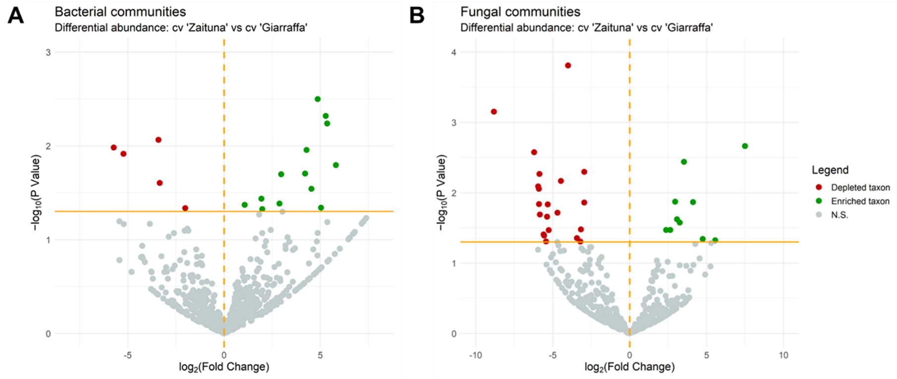
3.2. Endophytic Microbial Community in Susceptible and Resistant Cultivars

In order to unveil the possible role of the microbial endophytic community in the disease process, we analyzed and compared the microbiomes of two cultivars, Giarraffa and Zaituna, which were “highly susceptible” and” resistant” to OKD, respectively.
Next-generation sequencing was carried out on the 16S rRNA gene, resulting in 1,925,942 raw reads, nearly 72% of which (1,385,762) passed quality filtering. To investigate the taxonomic compositions of the microbial communities, the bacterial and fungal OTUs of known and identified taxa were assigned to five taxonomic levels (Table 2).
At the phylum level, considering the average relative bacterial abundance of both cvs. Zaituna and Giarraffa, Proteobacteria (82.6%) was the dominant phylum, followed by unidentified bacteria (12.4%), Actinobacteria (2.4%), and Firmicutes (1.5%) (Figure 4A). In the fungal community, according to the average relative abundance in the cultivars, the dominant phylum was Ascomycota (82%), followed by unidentified fungi (18%) (Figure 4B). The most relatively abundant orders were Rhizobiales (10.9%), Micrococcales (5.3%), and Rhodospirillales (2.6%) in cv. Zaituna, whereas Pseudomonadales (18.5%), Clostridiales (5.7%), and Bacterioidales (3.0%) were more abundant in cv. Giarraffa (Figure 4C). Regarding the taxonomic fungal orders, Sordariales (11%), Glomerallales, Ostropales, and Dothidedales (3%) were more relatively abundant in cv. Zaituna samples, whereas Pleosporales (35.6%) was more abundant in cv. Giarraffa samples (Figure 4D).
Alpha diversity was estimated by Chao1 richness and Shannon diversity indices. In particular, the comparison between the cvs. Zaituna and Giarraffa bacterial communities highlighted significantly increased richness (Figure 5A) in the shoots of the susceptible cv. Giarraffa, although it showed lower diversity (Figure 5B) compared to the cv. Zaituna. The opposite trend was observed for fungal components (Figure 5C,D).
In order to assess the beta diversity in the two cultivars, the principal coordinates analysis (PCoA) based on the Bray–Curtis dissimilarity index was not able to significantly distinguish bacterial and fungal populations in two distinct clusters according to the two cultivars (Supplementary Figure S1B).
The PCoA plot based on the Bray–Curtis dissimilarity index showed that fungal symptomatic and asymptomatic samples formed two distinct clusters (Supplementary Figure S1B).
Differential abundance in bacterial communities, which were identified at the OTU level, was assessed between the cvs. Zaituna and Giarraffa. According to a positive or negative log fold change (logFC), significantly (p < 0.05) enriched or depleted bacteria were considered, respectively (Figure 6).
In cv. Zaituna, bacterial OTUs representing endophytes as Amnibacterium (Actinobacteria) (logFC 5.35 and logFC 4.21) and unidentified Mollicutes (Tenericutes) (logFC 1.07) were found to be enriched, as well as Methylobacterium (logFC 4.87 and 1.95) and Sphingomonas (Proteobacteria) (logFC 5.28). On the other hand, Pseudomonas (Proteobacteria) was highly enriched in cv. Giarraffa (logFC 5.73 and 5.23) (Supplementary Table S1).
The same comparison performed for the fungal community showed that Alternaria (logFC 6.20), Neofusicoccum (logFC 5.94), Ascoschyta (logFC 5.89), Elsinoe (logFC 4.69), Devriesia (logFC 3.42), and Pseudocercospora (logFC 3.21) (all Ascomycota) were enriched in cv. Giarraffa, whereas Neofabraea (Ascomycota) (logFC ₋7.50) and unidentified Tremellomycetes (Basidiomycota) (logFC ₋5.56) were depleted in the same cultivar (Supplementary Table S2). Moreover, the more susceptible cv. Giarraffa presented significant (p < 0.05) enrichment (logFC 5.86) of Epicoccum nigrum (logFC 5.86) in comparison with cv. Zaituna. In addition, cv. Zaituna was colonized by a higher quantity of Pithomyces chartarum (logFC 4.74).

4. Discussion

With this work, we attempted to investigate the susceptibility of 36 Sicilian olive cultivars to Psv and to disclose the potential role of the Psv endophytic microbial community in the development of OKD.
Currently, in the absence of efficient strategies that disregard the use of copper to control Psv dissemination, the selection of cultivars that show some ability to resist the disease represents a good starting point for future management [5]. New, efficient breeding programs for olive knot-resistant genotypes can benefit from the identification of candidate cultivars only if data on disease progress under field conditions are available [19]. With this aim, the evaluation of olive germplasm collections can be crucial, especially if located in areas with favorable climatic conditions for the development of the disease.
Most olive oils produced in Sicily are extra-virgin oils, and 8 are recognized as Protected Designation of Origin (PDO), while 28 are certified as Protected Geographical Indication (PGI). Olive cultivation in Sicily originated in ancient times and its germplasms show high values of genetic variability [45,46].
However, very few data are available on resistance evaluations of Sicilian varieties [20,21,47]. To our knowledge, this is the first evaluation of the OKD susceptibility of Sicilian olive varieties in the open field conserved at the International Germplasm Collection of “Villa Zagaria”, one of the most important collections of the Mediterranean area, in which local and indigenous cultivars showing high genetic variability and many fruit and oil traits are well-maintained [45,48].
According to our evaluation, the results showed that the Calamignara, Montonica, Zaituna, Verdello grosso, and Turdunazza cultivars were able to strongly resist OKD. To the best of our knowledge, these cultivars have never been evaluated for OKD susceptibility. They are very ancient Sicilian indigenous cultivars able to produce high-quality olive oils whose cultivation has been reduced in recent years due to certain agronomical traits but could become of strategic importance for future investigations.
The high susceptibility found for cvs. Giarraffa and Nocellara del Belice to OKD is confirmed by previous data obtained by visual assessment of olive plants and by artificial inoculation [20,47]. According to our investigation, cv. Biancolilla showed very slight symptoms (resistant), whereas no symptoms were detected by Catara et al. [20], confirming the resistant behavior of this cultivar to OKD. However, after artificial inoculation, it produced knots of medium size after 20 days, revealing susceptibility to these conditions [20]. These data confirm that all cultivars are susceptible under experimental conditions, especially if a high inoculum dose is used [19]. For this reason, more realistic data are yielded by field observations, although they can be affected by several conditions and different variables.
Moreover, a parallel investigation on Sicilian cultivars was recently undertaken at the IOGC “Villa Zagaria” to investigate anthracnose susceptibility [49]. Similar to OKD, cv. Nocellara del Belice was highly susceptible to anthrachnose, while cvs. Biancolilla, Vaddarica, and Santagatese were less sensitive after artificial inoculation with three Colletotrichum species [49].
Different OKD susceptibility levels are the results of different factors (genetical, morphological, and physiological) that contribute to the ability to better protect plants from tissue damage provoked not only by pruning, but also by the exposure of olive trees to some environmental stresses [22,50].
Previous studies have demonstrated that OKD incidence and severity are positively correlated with frost-damaged organs and, in the case of damage by late winter frost, susceptible cultivars showed symptoms after 6 months [22]. Unfortunately, to the best of our knowledge, no data are available regarding the frost tolerance of Sicilian cultivars, and we are confident that this aspect needs to be further investigated in relation to OKD susceptibility. However, it is worth noting that the IOGC is located in an area characterized by late or even intense cold in the spring, followed by increases in temperature.
The possible role of the endophytic community in OKD cannot be excluded, so to answer this hypothesis, we undertook an investigation of two cultivars, Giarraffa and Zaituna, which according to our investigation, showed contrasting susceptibilities to the disease.
An analysis of the twig endosphere resulted in microbial communities whose compositions at the phylum level were in accordance with data obtained for other cultivars [31,32]. The significantly higher richness revealed in the twigs of the highly susceptible cv. Giarraffa is in accordance with previous similar data obtained for the susceptible cv. Verdeal Transmontana, which resulted in greater bacterial abundance, diversity, and composition in comparison to the resistant cv. Cobrançosa [31], although we found a lower level of diversity. This revealed that the pathobiome community is influenced by the host cultivar, which in response to pathogen invasion, activates a defense response that involves the biosynthesis of defensive compounds [50]. In addition, differences in bacterial compositions can be preceded by the presence of Psv, which could have a role in the increase in bacterial richness in cv. Giarraffa as a result of the pathogen–bacteria interaction [31]. Distinct endophytic communities seemed to colonize the two cultivars, suggesting an interaction between the resident bacterial community and Psv. The enrichment of Pseudomonas bacterial species in the cv. Giarraffa was similarly found in Verdeal Transmontana, confirming that these bacteria represent a stable consortium that greatly contributes to OKD disease in cooperation with Psv [31].
Moreover, our study demonstrated that there is great diversity in fungal endophytes in the twigs of both cultivars. Gomes et al. [30] previously showed that the fungal communities of OKD-symptomatic and -asymptomatic twigs varied depending on the cultivar and the presence of disease, as well as that fungal abundance and diversity were higher in the most tolerant cultivar, Cobrançosa. Similarly, we found that the effect of OKD on fungal abundance and diversity was greater in cv. Zaituna than in the highly susceptible cv. Giarraffa, although with less richness. These data support the hypotheses that the host cultivar has an effect on microbial endophytic communities [32] and that specific interactions between a cultivar and Psv can promote or inhibit the colonization of specific fungal families, as previously described not only in twigs but also in knots [30].
Our comparative analysis between the two cultivars on the abundance of fungal communities showed an enrichment in the highly susceptible cv. Giarraffa of genera such as Alternaria, Neofusicoccum, Epicoccum, Ascochyta, Neocucurbitaria, and Elsinoe, which include many pathogenic species. Although considered a pathogen of minor importance for olives, the genus Alternaria includes species such as A. alternata and has been described as being responsible for fruit rot [51], leaf spot [52], and decline in cuttings [53]. A. alternata was found in knots of cv. Madural, and the Alternaria genus was found to be indicative of asymptomatic twigs in cv. Cobrançosa [30]. Moreover, Alternaria is frequently described in association with Epicoccum spp. within the endophytic fungal consortium in olive trees [29]. However, the higher concentration of E. nigrum in the cv. Giarraffa apparently contrasts with the ability of culture filtrates extracted from this species to inhibit Psv in vitro [51,54]. Moreover, the higher level found in cv. Zaituna of Pithomyces chartarum, described as a fungus with relevant enzymatic and antagonistic activity, has been similarly found on the sapwood of Leccino plants, hypothesizing a role also as a biocontrol agent of Xylella fastidiosa [55]. P. chartarum was also described for its moderate antimicrobial activity against A. alternata [56]. Thanks to their ability to produce diverse toxins and antimicrobial compounds, these fungi were hypothesized to have a possible role in olive tree defense against OKD.
As suggested by Gomes et al. [30], who found the plant pathogen of Pseudocercospora at an increased quantity among endophytic fungi on olive knots, we can similarly hypothesize that its enrichment in cv. Giarraffa could be the result of the production of specific compounds by Psv that could benefit endophytic fungi, as described for other pathogenic bacteria [57]. All these facts show that OKD is the result of more complex interactions between microorganisms.
Moreover, with the fungal genera of Neocucurbitaria and Elsinoe, Alternaria was found to be the most positively correlated to trees with high abundances of Xylella fastidiosa [32]. However, Neocucurbitaria has never been reported in the literature for an association with plants [58], and Elsinoaceae is known to include plant pathogens, although it is not well-studied [59].

5. Conclusions

The data obtained in this work described for the first time the susceptibility of Sicilian olive cultivars to OKD in the open field. These data can be useful to help growers in the process of cultivar selection for new productive and efficient olive plantations. Additionally, these results can be crucial for future programs of breeding, although as a longer-term approach. The study of microbial communities associated with olive plants with different levels of susceptibility needs to be considered an important tool for the future investigation of the complex interactions between host plants, pathogens, and resident microbes, as well as for the identification of potential biocontrol agents. All these data demonstrate that detailed investigations on cultivar susceptibility, preferably in the open field, represent a milestone to acquire important information useful for future OKD management programs.

Supplementary Materials

The following supporting information can be downloaded at: https://0-www-mdpi-com.brum.beds.ac.uk/article/10.3390/agronomy13020468/s1, Figure S1: PCoA plot of bacterial (A) and fungal communities (B). Table S1: Differential abundance of the bacterial OTUs between cvs. Giarraffa and Zaituna samples. Table S2: Differential abundance of the fungal OTUs between cvs. Giarraffa and Zaituna samples.

Author Contributions

Conceptualization, G.L. and P.C.; formal analysis, P.C. and M.P.R.; investigation, G.L., P.C., M.P.R. and S.D.S.; microbiome analysis, V.C., A.M. and G.L.; writing—review and editing, G.L., P.C. and V.C.; visualization, D.P. All authors have read and agreed to the published version of the manuscript.

Funding

This research was financially supported by “MIPAAF-D.M. n. 0033437 del 21/12/2017” Project 2018–2022 “Salvaguardia e valorizzazione del patrimonio olivicolo italiano con azioni di ricerca nel settore della difesa fitosanitaria”—SALVAOLIVI.

Data Availability Statement

The data presented in this study are available upon request from the corresponding author.

Acknowledgments

The authors thank Andrea Scoto, Antonio Aveni, and Libero Consorzio Comunale di Enna for the use of the experimental orchard of IOGC “Villa Zagaria”.

Conflicts of Interest

The authors declare no conflict of interest.

References

  1. Schroth, M.N.; Osgood, J.W.; Miller, T.D. Quantitative assessment of the effect of the olive knot disease on olive yield and quality. Phytopathology 1973, 63, 1064–1065. [Google Scholar] [CrossRef]
  2. Wilson, E.E. The Olive knot disease: Its inception, development, and control. Hilgardia 1935, 9, 234–266. [Google Scholar] [CrossRef]
  3. Gardan, L.; Bollet, C.; Ghorrah, M.A.; Grimont, F.; Grimont, P.A.D. DNA Relatedness among the pathovar strains of Pseudomonas syringae subsp. savastanoi Janse (1982) and proposal of Pseudomonas savastanoi sp. nov. Int. J. Syst. Evol. Microbiol. 1992, 42, 606–612. [Google Scholar] [CrossRef]
  4. Quesada, J.M.; Penyalver, R.; López, M.M. Epidemiology and control of plant diseases caused by phytopathogenic bacteria: The case of olive knot disease caused by Pseudomonas savastanoi pv. savastanoi. In Plant Pathology; Cumagun, C.J., Ed.; Intech: Kayl, Luxembourg, 2012; pp. 299–326. [Google Scholar]
  5. Quesada, J.M.; Penyalver, R.; Pérez-Panadés, J.; Salcedo, C.I.; Carbonell, E.A.; López, M.M. Dissemination of Pseudomonas savastanoi pv. savastanoi populations and subsequent appearance of olive knot disease. Plant Pathol. 2010, 59, 262–269. [Google Scholar] [CrossRef]
  6. Lavermicocca, P.; Surico, G. Presenza epifitica di Pseudomonas syringae pv. savastanoi e di altri batteri sull’Olivo e sull’Oleandro. Phytopathol. Mediterr. 1987, 26, 137–141. [Google Scholar]
  7. Teviotdale, B.L.; Krueger, W.H. Effects of timing of copper sprays, defoliation, rainfall, and inoculum concentration on incidence of olive knot disease. Plant Dis. 2004, 88, 131–135. [Google Scholar] [CrossRef]
  8. Larcher, W. Temperature stress and survival ability of Mediterranean sclerophyllous plants. Plant Biosyst. 2000, 134, 279–295. [Google Scholar] [CrossRef]
  9. Ercolani, G.L. Pseudomonas savastanoi and other bacteria colonizing the surface of olive leaves in the field. Microbiology 1978, 109, 245–257. [Google Scholar] [CrossRef]
  10. Quesada, J.M.; Garcia, A.; Bertolini, E.; López, M.M.; Penyalver, R. Recovery of Pseudomonas savastanoi pv. savastanoi from symptomless shoots of naturally infected olive trees. Int. Microbiol. 2007, 10, 77. [Google Scholar]
  11. Hosni, T.; Moretti, C.; Devescovi, G.; Suarez-Moreno, Z.R.; Fatmi, M.; Guarnaccia, C.; Pongor, S.; Onofri, A.; Buonaurio, R.; Venturi, V. Sharing of quorum-sensing signals and role of interspecies communities in a bacterial plant disease. ISME J. 2011, 5, 1857–1870. [Google Scholar] [CrossRef]
  12. Buonaurio, R.; Moretti, C.; da Silva, D.P.; Cortese, C.; Ramos, C.; Venturi, V. The olive knot disease as a model to study the role of interspecies bacterial communities in plant disease. Front. Plant Sci. 2015, 6, 434. [Google Scholar] [CrossRef] [PubMed]
  13. European Commission Commission. Implementing Regulation (EU) 2018/785 of 29 May 2018 amending Implementing Regulation (EU) No 540/2011 as regards the conditions of approval of the active substance thiamethoxam. Off. J. Eur. Union. 2015, 132, 40–44. [Google Scholar]
  14. Sisto, A.; Iacobellis, N.S. Olive knot disease: Pathogenic and epidemiological aspects and defence strategies. Olivo Olio 1999, 2, 32–38. [Google Scholar]
  15. Young, J.M. Olive knot and its pathogens. Australas. Plant Pathol. 2004, 33, 33–39. [Google Scholar] [CrossRef]
  16. Hassani, D.; Buonaurio, R.; Tombesi, A. Response of some olive cultivars, hybrid and open pollinated seedlings to Pseudomonas savastanoi pv. savastanoi. In Pseudomonas syringae and Related Pathogens; Springer: Berlin/Heidelberg, Germany, 2003; pp. 489–494. [Google Scholar]
  17. Marcelo, A.; Fernandes, M.; Fatima Potes, M.; Serrano, J.F. Reactions of some cultivars of Olea europaea L. to experimental inoculation with Pseudomonas syringae pv. savastanoi. In Proceedings of the III International Symposium on Olive Growing, Manila, Philippines, 6–8 October 1997; Volume 474, pp. 581–584. [Google Scholar]
  18. Varvaro, L.; Surico, G. Behaviour of different olive (Olea europaea L.) cultivars to inoculation with Pseudomonas savastanoi (EF Smith) Stevens. Phytopathol. Mediterr. 1978, 17, 174–177. [Google Scholar]
  19. Penyalver, R.; García, A.; Ferrer, A.; Bertolini, E.; Quesada, J.M.; Salcedo, C.I.; Piquer, J.; Pérez-Panadés, J.; Carbonell, E.A.; Del Rio, C. Factors affecting Pseudomonas savastanoi pv. savastanoi plant inoculations and their use for evaluation of olive cultivar susceptibility. Phytopathology 2006, 96, 313–319. [Google Scholar] [CrossRef]
  20. Catara, V.; Bella, P.; Tessitori, M.; Tirrò, A.; Colina, P. Variabilità di Pseudomonas savastanoi pv. savastanoi in un’area olivicola della Sicilia e comportamento di alcune varietà di olivo alle inoculazioni [Olea europaea L.]. Tec. Agric. 2005, 57, 41–52. [Google Scholar]
  21. Salman, M.; Greenhut, R.; Preece, J.; Ferguson, L.; Kluepfel, D. Field evaluation of olive (Olea europaea) genotypes for resistance to Pseudomonas savastanoi pv. savastanoi. J. Plant Pathol. 2020, 102, 663–670. [Google Scholar] [CrossRef]
  22. Valverde, P.; Zucchini, M.; Polverigiani, S.; Lodolini, E.M.; López-Escudero, F.J.; Neri, D. Olive knot damages in ten olive cultivars after late-winter frost in central Italy. Sci. Hortic. 2020, 266, 109274. [Google Scholar] [CrossRef]
  23. Mancuso, S. Electrical resistance changes during exposure to low temperature measure chilling and freezing tolerance in olive tree (Olea europaea L.) plants. Plant. Cell Environ. 2000, 23, 291–299. [Google Scholar] [CrossRef]
  24. Petruccelli, R.; Bartolini, G.; Ganino, T.; Zelasco, S.; Lombardo, L.; Perri, E.; Durante, M.; Bernardi, R. Cold stress, freezing adaptation, varietal susceptibility of Olea europaea L.: A Review. Plants 2022, 11, 1367. [Google Scholar] [CrossRef] [PubMed]
  25. Lodolini, E.M.; Alfei, B.; Santinelli, A.; Cioccolanti, T.; Polverigiani, S.; Neri, D. Frost tolerance of 24 olive cultivars and subsequent vegetative re-sprouting as indication of recovery ability. Sci. Hortic. 2016, 211, 152–157. [Google Scholar] [CrossRef]
  26. Bass, D.; Stentiford, G.D.; Wang, H.-C.; Koskella, B.; Tyler, C.R. The pathobiome in animal and plant diseases. Trends Ecol. Evol. 2019, 34, 996–1008. [Google Scholar] [CrossRef] [PubMed]
  27. Lata, R.; Chowdhury, S.; Gond, S.K.; White, J.F., Jr. Induction of abiotic stress tolerance in plants by endophytic microbes. Lett. Appl. Microbiol. 2018, 66, 268–276. [Google Scholar] [CrossRef] [PubMed]
  28. Yan, L.; Zhu, J.; Zhao, X.; Shi, J.; Jiang, C.; Shao, D. Beneficial effects of endophytic fungi colonization on plants. Appl. Microbiol. Biotechnol. 2019, 103, 3327–3340. [Google Scholar] [CrossRef]
  29. Nicoletti, R.; Di Vaio, C.; Cirillo, C. Endophytic fungi of olive tree. Microorganisms 2020, 8, 1321. [Google Scholar] [CrossRef]
  30. Gomes, T.; Pereira, J.A.; Lino-Neto, T.; Bennett, A.E.; Baptista, P. Bacterial disease induced changes in fungal communities of olive tree twigs depend on host genotype. Sci. Rep. 2019, 9, 5882. [Google Scholar] [CrossRef]
  31. Mina, D.; Pereira, J.A.; Lino-Neto, T.; Baptista, P. Impact of plant genotype and plant habitat in shaping bacterial pathobiome: A comparative study in olive tree. Sci. Rep. 2020, 10, 3475. [Google Scholar] [CrossRef]
  32. Giampetruzzi, A.; Baptista, P.; Morelli, M.; Cameirão, C.; Lino Neto, T.; Costa, D.; D’Attoma, G.; Abou Kubaa, R.; Altamura, G.; Saponari, M. Differences in the endophytic microbiome of olive cultivars infected by Xylella fastidiosa across seasons. Pathogens 2020, 9, 723. [Google Scholar] [CrossRef]
  33. Pyrowolakis, E.; Weltzien, H.C. Studies on the distribution of Olive knot, induced by Pseudomonas savastanoi (Sm.) Stev. in the Greek island of Crete. Phytopathol. Mediterr. 1974, 13, 118–120. [Google Scholar]
  34. King, E.O.; Ward, M.K.; Raney, D.E. Two simple media for the demonstration of pyocyanin and fluorescin. J. Lab. Clin. Med. 1954, 44, 301–307. [Google Scholar]
  35. Penyalver, R.; García, A.; Ferrer, A.; Bertolini, E.; López, M.M. Detection of Pseudomonas savastanoi pv. savastanoi in olive plants by enrichment and PCR. Appl. Environ. Microbiol. 2000, 66, 2673–2677. [Google Scholar] [CrossRef]
  36. Bella, P.; Licciardello, G.; Tessitori, M.; Catara, V. A real-time PCR quantitative detection assay for Pseudomonas savastanoi pv. nerii in Nerium oleander. Phytopathol. Mediterr. 2008, 47, 204–213. [Google Scholar]
  37. Klindworth, A.; Pruesse, E.; Schweer, T.; Peplies, J.; Quast, C.; Horn, M.; Glöckner, F.O. Evaluation of general 16S ribosomal RNA gene PCR primers for classical and next-generation sequencing-based diversity studies. Nucleic Acids Res. 2013, 41, e1. [Google Scholar] [CrossRef]
  38. Paytuví, A.; Battista, E.; Scippacercola, F.; Cigliano, R.A.; Sanseverino, W. GAIA: An integrated metagenomics suite. bioRxiv 2019, 804690. [Google Scholar]
  39. R Core Team. R Development Core Team. R A Lang. Environ. Stat. Comput. 2016, 55, 275–286. [Google Scholar]
  40. McMurdie, P.J.; Holmes, S. Phyloseq: An R Package for reproducible interactive analysis and graphics of microbiome census data. PLoS ONE 2013, 8, e61217. [Google Scholar] [CrossRef]
  41. Love, M.; Anders, S.; Huber, W. Differential analysis of count data–the DESeq2 package. Genome Biol. 2014, 15, 10–1186. [Google Scholar]
  42. Wickham, H. Data analysis. In ggplot2; Springer: Berlin/Heidelberg, Germany, 2016; pp. 189–201. [Google Scholar]
  43. Weiß, C.H. StatSoft, Inc., Tulsa, Ok.: Statistica, Version 8. AStA Adv. Stat. Anal. 2007, 91, 339–341. [Google Scholar] [CrossRef]
  44. Kassambara, A.; Mundt, F. Package ‘factoextra’. Extr. Vis. Results Multivar. Data Anal. 2017, 76. [Google Scholar]
  45. Las Casas, G.; Scollo, F.; Distefano, G.; Continella, A.; Gentile, A.; La Malfa, S. Molecular characterization of olive (Olea europaea L.) Sicilian cultivars using SSR markers. Biochem. Syst. Ecol. 2014, 57, 15–19. [Google Scholar] [CrossRef]
  46. Mantia, L.; Lain, T.; Caruso, T.; Testolin, R. SSR-based DNA fingerprints reveal the genetic diversity of Sicilian olive (Olea europaea L.) germplasm. J. Hortic. Sci. Biotechnol. 2005, 80, 628–632. [Google Scholar] [CrossRef]
  47. Bottari, V.; Spina, P. Le varietà di olivo coltivate in Sicilia. Ann. Ist. Sper. Agron. 1953, 7, 937–1004. [Google Scholar]
  48. Mousavi, S.; De la Rosa, R.; Moukhli, A.; El Riachy, M.; Mariotti, R.; Torres, M.; Pierantozzi, P.; Stanzione, V.; Mastio, V.; Zaher, H. Plasticity of fruit and oil traits in olive among different environments. Sci. Rep. 2019, 9, 16968. [Google Scholar] [CrossRef] [Green Version]
  49. Licciardello, G.; Moral, J.; Strano, M.C.; Caruso, P.; Sciara, M.; Bella, P.; Sorrentino, G.; Di Silvestro, S. Characterization of Colletotrichum strains associated with olive anthracnose in Sicily. Phytopathol. Mediterr. 2022, 61, 139–151. [Google Scholar] [CrossRef]
  50. Tugizimana, F.; Mhlongo, M.I.; Piater, L.A.; Dubery, I.A. Metabolomics in plant priming research: The way forward? Int. J. Mol. Sci. 2018, 19, 1759. [Google Scholar] [CrossRef]
  51. Moral, J.; De la Rosa, R.; León, L.; Barranco, D.; Michailides, T.J.; Trapero, A. High susceptibility of olive cultivar FS-17 to Alternaria alternata in Southern Spain. Plant Dis. 2008, 92, 1252. [Google Scholar] [CrossRef]
  52. Basım, E.; Basım, H.; Abdulai, M.; Baki, D.; Öztürk, N. Identification and characterization of Alternaria alternata causing leaf spot of olive tree (Olea europaea) in Turkey. Crop Prot. 2017, 92, 79–88. [Google Scholar] [CrossRef]
  53. Tziros, G.T.; Karpouzis, A.; Lagopodi, A.L. Alternaria alternata as the cause of decline and necrosis on olive tree cuttings in Greece. Australas. Plant Dis. Notes 2021, 16, 7. [Google Scholar] [CrossRef]
  54. Berardo, C.; Bulai, I.M.; Venturino, E.; Baptista, P.; Gomes, T. Modeling the endophytic fungus Epicoccum nigrum action to fight the “olive knot” disease caused by Pseudomonas savastanoi pv. savastanoi (Psv) bacteria in Olea europaea L. Trees. Trends Biomath. Model. Optim. Comput. Probl. 2018, 189–207. [Google Scholar]
  55. Hanani, A.; Valentini, F.; Sanzani, S.M.; Santoro, F.; Minutillo, S.A.; Gallo, M.; Cavallo, G.; Mourou, M.; El Moujabber, M.; D’Onghia, A.M. Community analysis of culturable sapwood endophytes from apulian olive varieties with different susceptibility to Xylella fastidiosa. Agronomy 2021, 12, 9. [Google Scholar] [CrossRef]
  56. Tuppad, D.S.; Shishupala, S. Evaluation of endophytic fungi from Butea monosperma for antimicrobial and enzyme activity. J. Med. Plants Stud. 2014, 2, 38–45. [Google Scholar]
  57. Frey-Klett, P.; Burlinson, P.; Deveau, A.; Barret, M.; Tarkka, M.; Sarniguet, A. Bacterial-fungal interactions: Hyphens between agricultural, clinical, environmental, and food microbiologists. Microbiol. Mol. Biol. Rev. 2011, 75, 583–609. [Google Scholar] [CrossRef]
  58. Valenzuela-Lopez, N.; Cano-Lira, J.F.; Stchigel, A.M.; Rivero-Menendez, O.; Alastruey-Izquierdo, A.; Guarro, J. Neocucurbitaria keratinophila: An emerging opportunistic fungus causing superficial mycosis in Spain. Med. Mycol. 2019, 57, 733–738. [Google Scholar] [CrossRef]
  59. Jayawardena, R.S.; Ariyawansa, H.A.; Singtripop, C.; Li, Y.M.; Yan, J.; Li, X.; Nilthong, S.; Hyde, K.D. A re-assessment of Elsinoaceae (Myriangiales, Dothideomycetes). Phytotaxa 2014, 176, 120–138. [Google Scholar] [CrossRef]
Figure 1. Representative scale of symptoms used for the evaluation of the disease intensity according to Pyrowolakis and Weltzien, 1974 [33]: level 0 (A), level 1 (B), level 2 (C), level 3 (D), and level 4 (E).
Agronomy 13 00468 g001
Figure 2. Cluster graph produced according to the disease intensity of 36 cultivars calculated as the mean of the 3-year assessment, revealing the presence of 5 clusters. HS: highly susceptible, S: susceptible, LR: low resistant, R: resistant, and HR: highly resistant.
Agronomy 13 00468 g002
Figure 3. Disease intensity trend of olive knot disease symptoms of 36 olive cultivars during a 3-year survey (2018–2020). For each cultivar, the standard deviation is shown by error bars. Different colored points and the regression lines of disease intensity in 2018, 2019, and 2020 are also shown.
Agronomy 13 00468 g003
Figure 4. Relative abundances of the bacterial (A) and fungal (B) communities at the phylum level and the bacterial (C) and fungal (D) communities at the order level. Bacterial and fungal phyla and orders less abundant than 1% are reported as “Other Taxa”.
Agronomy 13 00468 g004
Figure 5. Alpha diversity of the bacterial (A,B) and fungal communities (C,D) in cvs. Zaituna and Giarraffa using the Chao1 and Shannon indices to evaluate richness and diversity, respectively. The Kruskal–Wallis test was performed considering the p-value (*, <0.05) to evaluate the statistical significance between the two cultivars for both the Chao1 and Shannon indices.
Agronomy 13 00468 g005
Figure 6. Differentially abundant bacterial (A) and fungal (B) OTUs. Log fold change values and negative log-transformed p-values were reported on the x-axis and y-axis, respectively. The orange horizontal line represents the p-value threshold transformed into a negative logarithm (1.30). The orange dotted vertical line represents the log2 fold change cut-off (0), which separates the significantly enriched (green dots) and the depleted (red dots) bacterial and fungal OTUs from the nonsignificant ones (grey dots).
Agronomy 13 00468 g006
Table 2. Number of OTUs representing identified taxa from phylum to genus taxonomic levels.
OTUsPhylumClassOrderFamilyGenus
16S3465141286678
ITS52264144292
Disclaimer/Publisher’s Note: The statements, opinions and data contained in all publications are solely those of the individual author(s) and contributor(s) and not of MDPI and/or the editor(s). MDPI and/or the editor(s) disclaim responsibility for any injury to people or property resulting from any ideas, methods, instructions or products referred to in the content.

