Article

Genome-Wide Identification and Expression Pattern Analysis of the F5H Gene Family in Flax (Linum usitatissimum L.)

Heilongjiang Academy of Agricultural Sciences, Harbin 150000, China
*
Author to whom correspondence should be addressed.
Submission received: 15 March 2023 / Revised: 6 April 2023 / Accepted: 11 April 2023 / Published: 13 April 2023
(This article belongs to the Special Issue Genomics and Genetic Improvement of Bast Fiber Plants)

Abstract

:
Ferulate 5-hydroxylase (F5H) is a cytochrome P450-dependent monooxygenase that plays a key role in the biosynthesis of syringyl (S) lignin. In this study, mining of flax (Linum usitatissimum) genomic data enabled the identification of nine LuF5H genes. Bioinformatics analysis revealed the physicochemical properties, gene structures, conserved motifs, phylogenetic evolutionary features and promoter cis-acting elements related to these genes and their encoded proteins. Based on the gene structural and phylogenetic features, the nine LuF5Hs were assigned to two subclasses. The expression levels of the nine LuF5Hs was analyzed by the RNA-seq technique, and the RNA-seq data were verified by qRT-PCR. The results of the RNA-seq analysis showed that LuF5H genes belonging to the same subclass exhibited similar expression patterns. Notably, the transcripts of the LuF5H3 and LuF5H7 subclass 1 genes accumulated at high levels in stem tissues, thus indicating that LuF5H3 and LuF5H7 are the main LuF5H genes involved in flax lignin biosynthesis. Furthermore, the expression levels of LuF5H1, LuF5H3, LuF5H4 and LuF5H7 were upregulated 1.2–1.9-fold under drought, NaCl stress and brassinosteroid treatment conditions. This first comprehensive study of the flax F5H gene family provides valuable data for use in gene function analysis toward improving flax fiber quality and reducing flax manufacturing costs and associated environmental pollution.

1. Introduction

Flax (Linum usitatissimum L.) is an important fiber crop that has been utilized by humans for thousands of years [1,2]. Flax fiber possesses high tensile strength, softness and fineness, and, thus, it is an excellent textile raw material [3]. Flax textiles are favored by consumers due to the characteristics related to their softness, high hygroscopicity, air permeability and anti-electrostatic, antibacterial and antioxidant properties [4,5]. High lignin content is an important factor affecting flax fiber yield and quality, while high raw flax lignin content is associated with increased degumming costs and generation of environmental pollutants [6]. Therefore, strategies that reduce flax lignin content are urgently sought by the flax industry to improve flax fiber quality while reducing flax manufacturing costs and associated environmental impacts.
Lignin, a complex phenolic polymer that is mainly found in plant secondary cell walls, is second only to cellulose in natural abundance [7,8]. Lignin is essential for maintaining the structural integrity of vascular plants and occupies the space between wall polysaccharides, including cellulose and hemicellulose, to enhance plant mechanical strength, water transport capability and resistance to environmental stressors [9,10]. Lignin biosynthesis has been intensively researched to reduce costs related to lignin removal from raw materials in order to increase utilization of renewable lignocellulosic materials (e.g., chemical pulp, biofuel and raw flax) [8,11,12,13]. Lignin consists of three main units, namely p-hydroxyphenyl lignin (H-lignin), guaiacyl lignin (G-lignin) and syringyl lignin (S-lignin), which are formed by oxidative polymerization of p-coumaryl alcohol, coniferyl alcohol and sinapyl alcohol, respectively [14,15,16,17]. The susceptibility of a given lignin molecule to degradation depends on its composition; a G-lignin monomer contains a single methoxy group that can form stable C-C bonds with other monomers, while an S-lignin monomer contains two methoxy groups (one of which occupies the C-5 position) that mainly bond with other monomers via β-O-4 linkages [10,18]. Lignins containing S units in high abundance exhibit low degrees of condensation, have relatively loose structures, and are more easily removed than other monomers, thus supporting greater industrial-scale plant resource utilization [9,10,19,20]. To date, most research studies on lignin biosynthesis have focused on reducing lignin content [21,22], altering lignin structure [11,23] or on both of these goals [24].
Ferulate 5-hydroxylase (F5H), which is also known as coniferaldehyde 5-hydroxylase (CAld5H), is a cytochrome P450-dependent monooxygenase that belongs to the CYP84 family [9,25]. As a lignin synthesis pathway enzyme, F5H catalyzes the hydroxylation of three substrates, including ferulate, coniferyl alcohol and coniferaldehyde, at the phenyl ring C-5 position. Importantly, F5H is one of several key enzymes that regulate S-lignin synthesis [26,27], and, thus, this enzyme plays a key role in shaping plant lignin composition by influencing plant S-lignin/G-lignin ratios. The first F5H gene was discovered by Meyer in 1996 and later became the focus of efforts to alter lignin compositions of vascular plants [28]. Previous studies had shown that Arabidopsis with F5H-deficient fah1 mutant contains almost no S-lignin [19,29,30]. Conversely, overexpression of F5H has been reported to increase both S-lignin content and the S/G ratio [10,20,23,31].
The flax genome was assembled in 2012 [32], a feat that has led to significant advances in flax genetics, molecular biology and breeding. With the publication of the flax genome, many genes related to important agronomic traits were identified [2,3,33,34,35], thus highlighting the utility of genome-wide gene identification as a rapid, accurate and effective strategy for studying gene families. Using this strategy, Tombuloglu employed a local blastP (v2.6.0) tool to identify 175 MYB genes within the flax genome [35]. Similarly, in this study, nine flax F5H genes were identified using the BLASTP plugin of the Phytozome database as a more accurate number of LuF5H genes than was previously reported [36].
In recent years, F5H homologues have been identified and characterized for diverse plant species, such as Arabidopsis thaliana [28,29], Eucalyptus globulus [26], Populus tomentosa [31,37], Oryza sativa [9], Sorghum bicolor [10], Brassica napus [22] and Corchorus capsularis [8]. However, few studies have focused on flax F5H genes, which has prompted this study. Here, we provide the first detailed analysis of flax F5H gene structures, conserved motifs, phylogenetic evolution, promoter cis-acting elements and expression patterns and identify the main F5H genes involved in flax lignin biosynthesis. The results presented here provide a rich theoretical basis to guide future F5H cloning, functional verification and flax breeding efforts toward reducing flax lignin content and improving flax fiber quality.

2. Materials and Methods

2.1. Identification of F5H Genes in Linum usitatissimum

Using the F5H protein sequences of A. thaliana, E. globulus, P. tomentosa and O. sativa as the query sequences, homologous F5H sequences within the flax genome (L. usitatissimum v1.0) were detected based on results of BLASTP sequence alignments (using an e value cutoff of 10−10) against the Phytozome database (https://phytozome-next.jgi.doe.gov/, accessed on 1 August 2022). The presence of the F5H conserved domain was confirmed using the NCBI-CDD (http://0-www-ncbi-nlm-nih-gov.brum.beds.ac.uk/Structure/cdd/, accessed on 1 August 2022) online tool. The F5H protein sequences of A. thaliana (Q42600.1), P. tomentosa (APX43202.1), E. globulus (ACU45738.1) and O. sativa (XP_015614553.1) were downloaded from the NCBI database.

2.2. Sequence Analysis and Structural Characterization of LuF5Hs

The online program ExPAsy (https://web.expasy.org/protparam/, accessed on 6 August 2022) was used to predict the basic characteristics of LuF5H proteins from their sequences [38]. ProtComp (http://www.softberry.com/berry.phtml?topic=index&group=programs&subgroup=proloc, accessed on 6 August 2022) was used to predict subcellular localization sites of F5H proteins. The DNAMAN software was used to conduct multiple sequence alignments of encoded proteins. The exon/intron configurations of flax F5H genes were analyzed using the GSDS tool (http://gsds.gao-lab.org/, accessed on 9 August 2022) [39]. The conserved motifs of LuF5H proteins were analyzed online using the MEME website (http://meme-suite.org/tools/meme, accessed on 14 August 2022) [40,41], with the parameter defining the maximum number of identified motifs set to 12. The results were visualized using the TBtools.

2.3. Phylogenetic Analysis of the LuF5H Gene Family

After the flax F5H protein sequences were obtained, they were first aligned using ClustalW, a plugin that is provided with MEGA 7.0. Next, a phylogenetic tree was built using MEGA 7.0 with the neighbor-joining (NJ) method and with 1000 repetitions employed for the bootstrap test [42].

2.4. Analysis of cis-Acting Elements in the LuF5H Gene Promoter

Thereafter, cis-acting elements of LuF5H gene promoters were analyzed using TBtools [42]; then, the 2.0 kb upstream region of the LuF5H gene’s protein-coding sequence (CDS) was extracted from the flax genome using the TBtools and saved as a FASTA file. The PlantCARE (http://bioinformatics.psb.ugent.be/webtools/plantcare/html/, accessed on 1 December 2022) web server was used to predict cis-acting elements [43], and then the results were visualized using the TBtools.

2.5. PPI Network of F5H Proteins in Linum usitatissimum

Meanwhile, the PPI (protein–protein interaction) network of flax F5H proteins was constructed and then used to predict the PPI network of LuF5H proteins using the String database (https://cn.string-db.org/, accessed on 27 November 2022). The results were downloaded as a TSV file; then, the PPI network of LuF5H proteins was visualized using Cytoscape 3.9.1.

2.6. Analysis of Expression Pattern of LuF5Hs

The plant materials used for LuF5H expression analysis included fiber flax plants of the variety “Diana”. The plants were grown under natural conditions in the experimental field of the Institute of Industrial Crops of the Heilongjiang Academy of Agricultural Sciences (Harbin, China). The samples were collected at the seedling stage, fir-like stage, early fast-growing stage, fast-growing stage, bud stage, flowering stage, green stage and mature stage, with the growth stages defined as reported by Yuan et al. [2]. The stems of the flax samples in the fast-growing stage were divided into 20 equal parts that were designated as S1–S20. Next, the samples were quickly frozen in liquid nitrogen and stored at −80 °C until they were used for RNA extraction and transcriptome analysis. The FPKM values were converted to log10-fold change values; then, the TBtools was used for heat map generation and cluster analysis [44].

2.7. Drought, NaCl and Brassinosteroid (BR) Treatments

The flax plants were grown outdoors under natural environmental conditions in a field located in Harbin, China, at the Institute of Industrial Crops of the Heilongjiang Academy of Agricultural Sciences. The flax plants were exposed to drought stress, NaCl stress or brassinosteroid (BR) treatment. For the plants exposed to the drought conditions, healthy flax plants in the fast-growing stage with similar characteristics were selected; then, each whole plant was sampled after watering had been stopped for 10 days and the leaves began to wilt. The control plants were grown under normal conditions and watered every 2 days. For the NaCl treatment, similar flax plants in the bud stage were treated with 300 mmol·L−1 NaCl, and the middle 1/3 of each stem was sampled after 12 h; plants treated with water served as the controls. For the BR treatment, similar flax plants at the bud stage were sprayed with 0.2 mg·L−1 BR, and then the middle 1/3 of each stem was sampled after 12 h; plants sprayed with distilled water served as the controls. Three biological replicates were prepared for each treatment. The harvested samples were frozen in liquid nitrogen and stored at −80 °C until they were used for RNA preparation and transcriptome analysis.

2.8. RNA Extraction

For RNA extraction, each plant sample was ground into a powder using a mortar and pestle precooled in liquid nitrogen. The material for RNA extraction must be a fresh sample. If it could not be extracted immediately, it must be frozen in liquid nitrogen and stored at −80 °C. The material used for RNA extraction must be protected from RNase contamination. Total RNA preparations from the plant samples were extracted using the cetyltrimethylammonium bromide (CTAB) method. DNA was digested using DNase I (Promega, Madison, WI, USA) to remove contaminating genomic DNA (gDNA). RNA integrity was verified via 1% agar gel electrophoresis, and then RNA concentration and quality were measured using a Nanodrop and Agilent 2100 Bioanalyzer (Thermo Fisher Scientific, Waltham, MA, USA).

2.9. DGE Library Preparation and Sequencing

The RNA samples were purified using oligo-dT magnetic beads. Thereafter, the purified mRNAs were fragmented into shorter molecules via treatment with a fragment buffer. The fragmented mRNA preparations were next used as templates for first-strand cDNA synthesis via reverse transcription using random hexamer primers; then, second-strand cDNA synthesis was conducted. Thereafter, end repair was performed to generate library-template DNA preparations, which were then subjected to PCR enrichment to generate the final cDNA libraries. The library quality was determined using an Agilent 2100 Bioanalyzer (Thermo Fisher Scientific, MA, USA). The volume of each library was adjusted to 50 µL (while ensuring the library concentration was ≥1 ng/µL), and then transcriptome sequencing was performed. The general characteristics of the transcriptome sequence results are presented in Table S1.
SOAPnuke (v1.5.2) [45] was used to screen and filter the sequence data using the following steps: (1) remove reads containing sequencing adapter; (2) remove reads with low-quality base rates of >20%; and (3) remove reads with unknown base (N) rates of >5%. Thereafter, clean reads were mapped to the reference genome using HISAT2 (v2.0.4) [46]; then, the clean reads were compared with the reference coding gene set using Bowtie2 (v2.2.5) [47]. Finally, gene expression levels were calculated using RSEM (v1.2.12) [48].

2.10. Real-Time Quantitative PCR Analysis

The abovementioned RNA extracts were used to synthesize cDNA using a PrimeScript™ RT Reagent Kit with a gDNA Eraser kit (TaKaRa) in a 20 µL reaction volume containing 1 µg of RNA template. In this study, the relative quantitative method was used to compare gene expression levels. Real-Time PCR was performed according to the instructions provided with the TB Green® Premix Ex Taq™ II kit (Tli RNase H Plus) (TaKaRa) in a 20 µL PCR reaction volume at a Tm of 60 °C. Three technical replicates and two biological replicates were performed in all experiments. The GAPDH and EF1A genes served as the internal reference genes for use in normalizing the results across the samples. According to the Ct values, the relative expression levels were calculated using the 2−ΔΔCt method. The RT-PCR primers (Table S2) were designed using Primer 5.0.

2.11. Determination of Lignin Content and Correlation Analysis

The flax samples were taken at different developmental stages for lignin content determinations. The details regarding the experimental materials, plant cultivation environment and sampling periods are shown in Section 2.6. Lignin contents in the plant samples were determined by UV spectrophotometry using commercially available kits based on the acetyl bromide method (QIYI, Shanghai, China) [2]. Correlation analysis between lignin content and LuF5Hs gene expression was conducted using the SPSS 23.0 software.

3. Results

3.1. Identification and Basic Information of F5H Genes in Linum usitatissimum

The results of the BLASTP comparisons between the reference F5H protein sequences of A. thaliana, P. tomentosa, E. globulus and O. sativa and the sequences within the Phytozome database revealed homologous F5H protein sequences encoded by the flax genome. Then, the conserved domain of LuF5Hs was confirmed by the CDD database. Ultimately, nine LuF5Hs were identified that all contained the conserved P450 domain, thus indicating that these LuF5Hs belong to the cytochrome P450 superfamily. Based on previously reported findings and genome assembly scaffold data, the LuF5H genes were named in sequential order (LuF5H1 to LuF5H9). The ExPASy website online tools were used to analyze the physicochemical properties of predicted LuF5H proteins (Table 1). The LuF5H sequences ranged from 388 (LuF5H9) to 515 (LuF5H1) aa, with an average length of 489 aa. The predicted molecular weights (MWs) ranged from 43893.39 Da (LuF5H9) to 58072.41 Da (LuF5H8), with an average MW of 55063.06 Da. The theoretical pI values ranged from 5.12 (LuF5H9) to 6.68 (LuF5H8), with an average predicted pI of 6.14. Overall, the LuF5H family proteins had pI values that were <7, thus indicating they are acid proteins. An analysis of the nine LuF5Hs using the online ProtComp tool, which is used to predict subcellular localization of proteins, predicted that all nine LuF5Hs localize to the plasma membrane (PM).

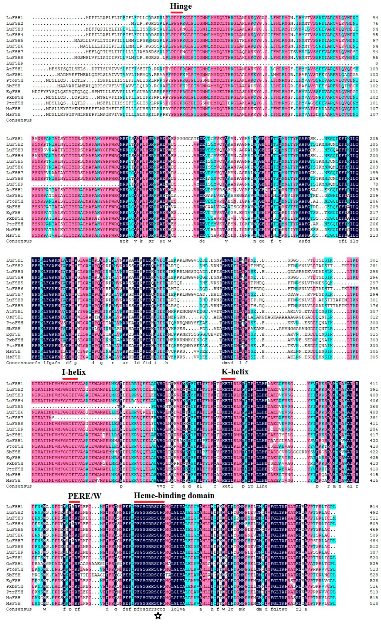
3.2. Multiple Sequence Alignment of LuF5H Proteins

The results of the multiple sequence alignment analysis conducted using the DNAMAN tool showed that the protein homology rates of the LuF5H proteins ranged from 50.58% to 94.56% (Figure 1). Figure 1 also shows that the flax F5H amino acid sequences share high degrees of identity with the F5H sequences of other plants. For example, LuF5Hs 1–9 share 68.18%, 63.89%, 69.73%, 70.04%, 56.79%, 62.90%, 65.07%, 62.57% and 47.92% sequence identity with AtF5H1 (Q42600.1), respectively, and 72.85%, 64.82%, 70.33%, 73.17%, 58.65%, 65.77%, 66.47%, 64.50% and 49.90% sequence identity with Populus trichocarpa F5H (CAB65335.1), respectively. LuF5Hs and other plant F5Hs are highly homologous and contain the heme-binding domain (PFGSGRRSCPG), wherein a conserved cysteine acts as a heme-binding ligand, thus providing further evidence that LuF5Hs belong to the cytochrome P450 family.

3.3. LuF5H Gene Structures and Conserved Motifs

In order to study LuF5H gene structures, we analyzed their DNA sequences to determine their intron and exon compositions and overall lengths (Figure 2A). The results of this analysis revealed significant differences in the lengths of the flax F5H gene family members. The shortest gene length was noted for the LuF5H9 gene (1164 bp), and the greatest gene length was noted for the LuF5H4 gene (3595 bp). The nine LuF5H genes were assigned to two subclasses according to their intron number. The four genes that contain one intron (LuF5H1, LuF5H3, LuF5H4 and LuF5H7) were assigned to subclass 1, and the five remaining LuF5Hs that contain no intron (LuF5H2, LuF5H5, LuF5H6, LuF5H8 and LuF5H9) were assigned to subclass 2. Different intron sizes were observed for subclass 1 members, with sizes of 621 bp, 1035 bp, 2066 bp and 569 bp, respectively.
To assess LuF5H protein structural diversity, 12 conserved motifs (motif 1–12) of the L. usitatissimum F5H family were identified using the MEME online program. As shown in Figure 2B, the motif compositions of most LuF5H proteins are similar overall but exhibit individual differences. LuF5H9 was found to lack Motif3, Motif5 and Motif10, whereas LuF5H5 and LuF5H7 were found to lack Motif7. The results of the multiple sequence alignment and conserved domain analyses of LuF5H amino acid sequences revealed that the predicted LuF5H proteins share motifs with typical cytochrome P450 proteins (Figure 1 and Figure 2C). These shared motifs include several conserved P450 motifs, such as the proline-rich membrane hinge, the I-helix, the K-helix and the heme-binding domain with a conserved cysteine residue [49].

3.4. Phylogenetic Analysis and Classification of the F5H Gene Family in Linum usitatissimum

To reveal the evolutionary relationships among the F5H gene family members of flax and other plants, we downloaded F5H protein sequences of 12 plant species, including L. usitatissimum, A. thaliana, P. trichocarpa, S. bicolor and O. sativa, and then constructed a NJ (neighbor-joining) phylogenetic tree using MEGA 7.0. An analysis of the phylogenetic tree revealed that the F5H protein family members cluster into two groups according to their monocot versus dicot status (Figure 3). Notably, LuF5Hs cluster with F5Hs of dicots, such as A. thaliana, of which several members of this cluster have been confirmed to participate in lignin biosynthesis. Furthermore, based on the protein sequences, the flax F5Hs could be assigned to two subclasses that are consistent with the abovementioned subclass assignments of LuF5Hs based on the gene intron characteristics; subclass 1 includes LuF5H1, LuF5H3, LuF5H4 and LuF5H7, and subclass 2 includes LuF5H2, LuF5H5, LuF5H6, LuF5H8 and LuF5H9, thus confirming the accuracy of the phylogenetic tree.

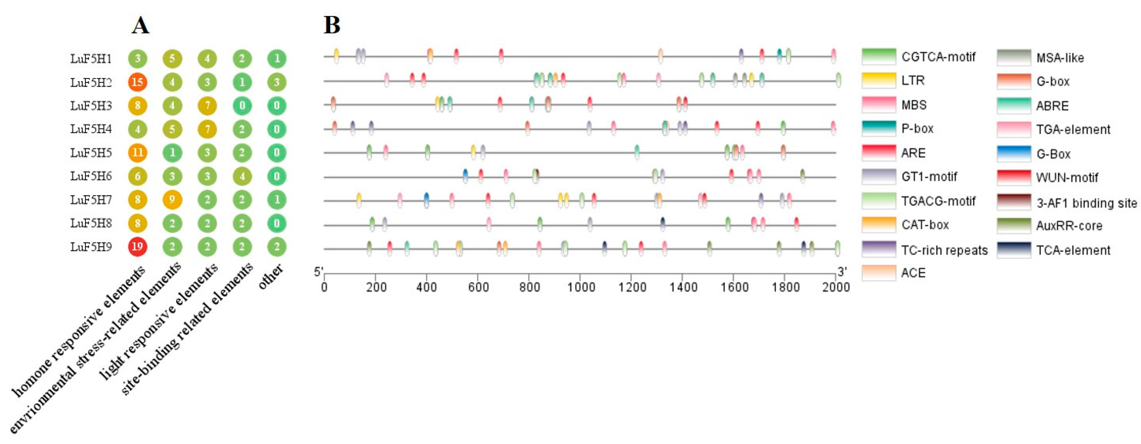
3.5. Cis-Element Analysis in the Promoters of LuF5H Genes

In order to obtain more information about F5H gene functions in flax, the online tool PlantCARE was used to analyze the promoters of the nine LuF5H genes. The results of this analysis revealed various types of cis-elements within the LuF5H promoter regions that are related to plant responses to hormone exposure, environmental stress, light, site binding and others (Figure 4A and Table S4). Abundant hormone response-related elements that can respond to methyl jasmonate (MeJA), abscisic acid (ABA), salicylic acid (SA), auxin and gibberellic acid (GA) were detected, of which the most numerous and widely distributed elements include the CGTCA motif (MeJA-response element) and the TGACG motif (MeJA-response element), followed by ABRE (ABA-responsive element) (Figure 4B). ARE, an anaerobic stress-response element, is present in eight LuF5H promoters (Figure 4B), while many promoters contain other cis-elements related to drought response (MBS), low-temperature response (LTR), wound response (WUN-motif), meristem expression (CAT-box) and light response (GT1-motif, G-box). Taken together, these results suggest that LuF5H genes may be critical players in numerous developmental processes and various stress responses.

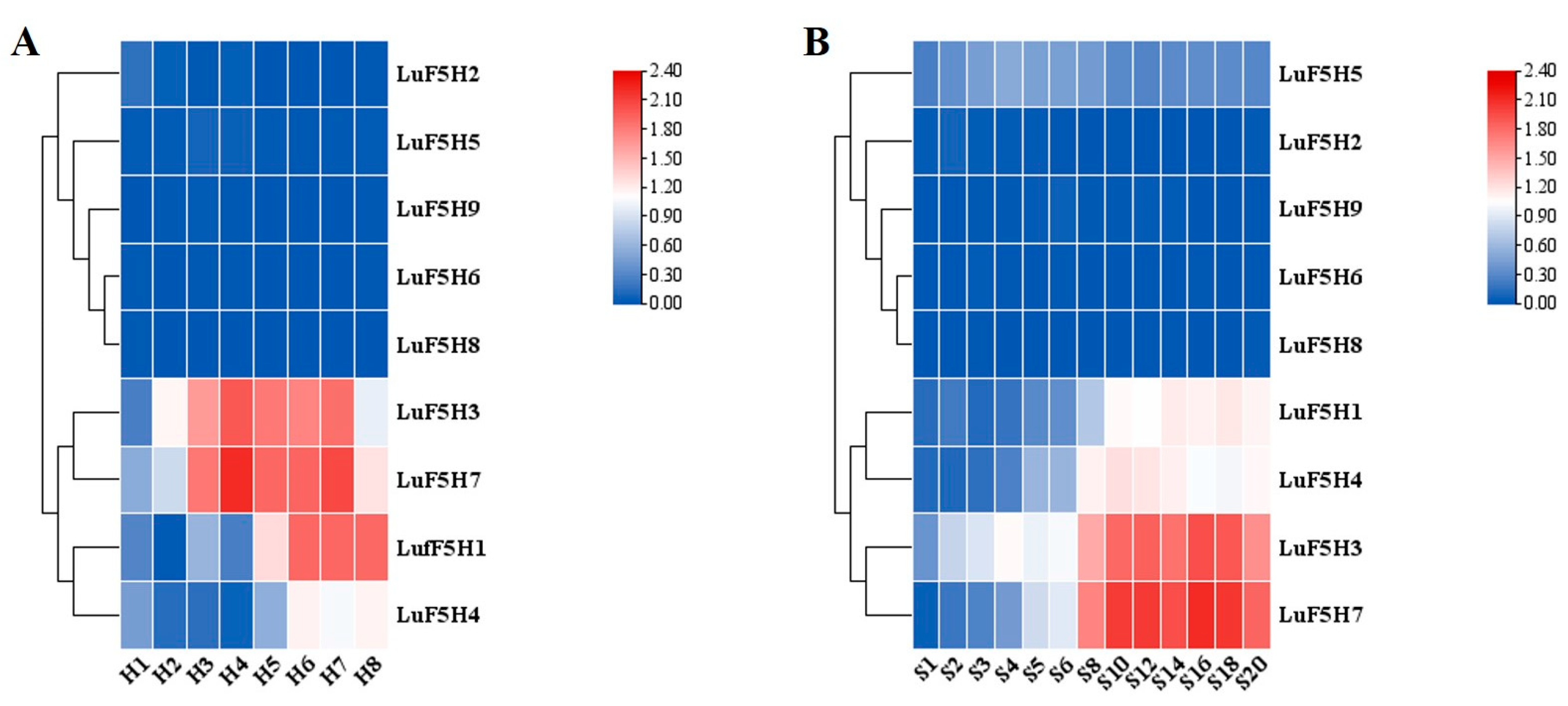
3.6. Analysis of Expression Pattern of LuF5H Genes

Transcriptome analysis was performed on the specimens collected from different flax developmental stages and different stem regions during the fast-growing stage in order to compare LuF5H gene transcript level differences among the experimental groups. Based on the expression patterns of the LuF5H genes, these genes could be assigned to two subclasses (Figure 5). The second subclass consisted of five genes (LuF5H2, LuF5H5, LuF5H6, LuF5H8 and LuF5H9), which were expressed at very low levels or not at all during different developmental stages and in different parts of the stem. The first subclass contained the remaining four genes, which were assigned to two subgroups, G1 and G2, based on their expression patterns. Members of the G1 subgroup (LuF5H3 and LuF5H7) shared the same expression pattern, whereby high-level transcript accumulation was observed in the analyzed tissues. Moreover, the levels of LuF5H3 and LuF5H7 transcripts first increased and then decreased as development progressed, and finally, they reached peak levels during the fast-growing stage. Furthermore, LuF5H3 and LuF5H7 were present at high levels in mature stems (S10–S18) and at relatively lower levels in young stems (S1–S3). The expression levels of the G2 subgroup members (LuF5H1 and LuF5H4) were relatively greater after flowering. Their expression patterns in different parts of the stem resembled the corresponding patterns observed for the G1 members, but both genes were expressed at relatively lower levels than their G1 counterparts.
The transcriptome data in different parts of flax stem were further validated via the qRT-PCR analysis of nine genes. The nine randomly selected genes included LuF5H3 (Lus10028361), LuFOMT1 (Lus10015576), LuFOMT2 (Lus10032929), Lu4CL (Lus10005390), LubZIP (Lus10040069), LuTCP15 (Lus10037190), LuGOMT (Lus10006258), Expansin (Lus10000957) and LuZF-HD (Lus10015370). It can be seen from Figure 6 that the change trend of the nine genes is consistent with the RNA-seq data, indicating that the RNA-seq data are reliable. The accuracy of transcriptome data at different developmental stages has been verified by Yuan et al. [2].

3.7. Interaction Analysis between F5H Proteins in Linum usitatissimum

In order to identify LuF5H gene functions, protein–protein interaction (PPI) networks of LuF5H proteins were built using the information provided by the String database (Figure 7A). The results of this analysis revealed that LuF5Hs did not interact with each other and that each LuF5H interacted with flavonol 3′-O-methyltransferase (Lus10015576, Lus10032929), cinnamyl-alcohol dehydrogenase (Lus10010149, Lus10017354), 4-coumarate-CoA ligase (Lus10016630, Lus10022542, Lus10005390, Lus10026143, Lus10002791) and peroxidase (Lus10011079). Due to the fact that cinnamyl-alcohol dehydrogenase, 4-coumarate-CoA ligase and peroxidase are all involved in lignin biosynthesis, these results imply that LuF5Hs participate in lignin biosynthesis. Furthermore, flavonol 3′-O-methyltransferase catalyzes the methylation of flavonol, which can enhance plant hydrophobicity to protect plant cells from microbial pathogens, thus suggesting that LuF5H functions may be related to pathogen resistance. After we analyzed the expression patterns of the interacting proteins within the PPI network (Figure 7B), we found that the expression patterns of LuF5H3 and LuF5H7 were similar to those of flavonol 3-O-methyltransferase and 4-coumarate-CoA ligase, whereby the expression levels of these genes initially increased and then decreased as plant development progressed.

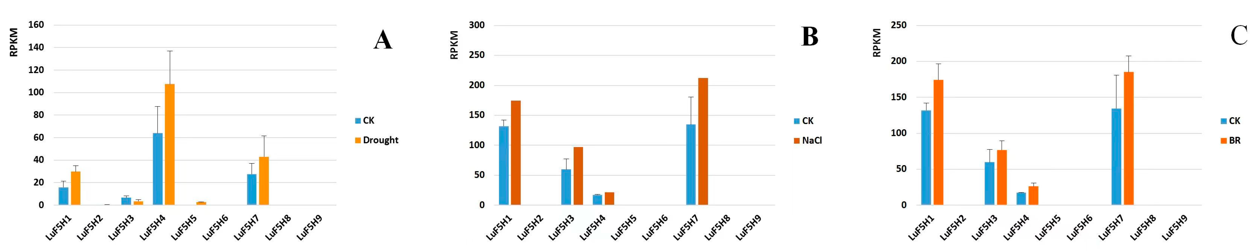
3.8. Expression Profiles of LuF5H Genes in Response to Drought, NaCl and BR Treatments

The results of previous studies show that F5H proteins participate in abiotic stress responses, which prompted us to examine the transcription levels of LuF5H genes under drought and NaCl stress conditions. After the flax plants were exposed to simulated drought conditions for 10 days, the expression levels of LuF5H1, LuF5H4 and LuF5H7 were found to be upregulated by 1.9-fold, 1.7-fold and 1.5-fold, respectively, when compared to the corresponding control group levels (Figure 8A). By contrast, after the flax plants were exposed to high NaCl stress conditions for 12 h, the expression levels of LuF5H1, LuF5H3, LuF5H4 and LuF5H7 transcripts increased 1.3-fold, 1.6-fold, 1.2-fold and 1.6-fold, respectively (Figure 8B), with similar trends observed in the expression levels of the same genes in the BR-treated plants (Figure 8C). However, the expression levels of LuF5H2, LuF5H5, LuF5H6, LuF5H8 and LuF5H9 exhibited no significant changes in response to any treatments.

3.9. Correlation Analysis between Lignin Content and LuF5H Gene Expression Levels

In order to study the relationship between the lignin content and expression patterns of LuF5H genes, we determined the lignin content of flax plants during different developmental stages (Figure 9). In general, lignin content increased slowly as development progressed and then reached a large value during the mature stage. The results of the correlation analysis revealed that lignin content was positively correlated with the expression levels of LuF5H1, LuF5H3, LuF5H4 and LuF5H7 genes (based on correlation coefficients of 0.475, 0.478, 0.453 and 0.591, respectively), although these results were not statistically significant (Table 2).

4. Discussion

Flax, an important bast fiber crop, is one of the most widely used fibrilia by the global textile industry. The chemical composition of flax fiber consists mainly of cellulose, which generally accounts for 62%–77% of flax fiber content, followed by hemicellulose and lignin [4,5]. Raw flax must be degummed before it can be used to make textiles, with lignin in flax gum being the most difficult component to remove during the degumming process. The high lignin content in raw flax not only increases the cost of degumming but also pollutes the environment [6]. At the same time, lignin is easily oxidized, which can make flax fiber hard and brittle and reduce flax fiber quality. Therefore, the flax fiber industry has urgently worked to reduce flax fiber lignin content in order to address this issue.
The relative proportions of the three main lignin monomers within plant lignins vary across species. The lignin of gymnosperms consists of G units only, while those of dicotyledonous plants are mainly G-S units, and those of non-woody monocotyledonous plants contain G-S lignin and more H lignin than is found in other plant types [17]. To date, fiber lignins of some bast fiber crops have been structurally characterized, with the molar H/G/S compositional ratio of fiber lignin in jute reported to be 2:33:65 [50], that of hemp reported to be 13:53:34 [51] and that of G-lignin-rich flax reported to be 13:72:15 [52]. Importantly, S-lignin units are easier to remove than G-lignin units due to differences in the bond types between monomers. Therefore, plant materials that are rich in S-lignin units are preferred as flax breeding stock.
F5H is a cytochrome P450-dependent monooxygenase (P450s), which can catalyze the hydroxylation of ferulate, coniferyl alcohol and coniferaldehyde phenyl ring in the phenylpropanoid pathway, and is a key enzyme in the regulation of S-lignin synthesis. Thus, since its discovery, F5Hs have been the focus of efforts to alter lignin compositions of vascular plants. Arabidopsis F5H homologues, AtF5H1 and AtF5H2, were the first identified ferulate 5-hydroxylase [7,28], of which AtF5H1 has been characterized in many plants, including Arabidopsis, tobacco and poplar [23,29]. Sibout et al. cloned PopF5H in P. trichocarpa and found that this gene was highly expressed in lignified tissues. Moreover, this gene was able to complement the A. thaliana fah1-2 gene mutation to reverse its associated lignin synthesis defect when expressed in mutant plants under the control of the CaMV 35S promoter [31]. In other work, Takeda et al. identified three F5H genes in rice, namely CYP84A5, CYP84A6 and CYP84A7, and found that the expression of CYP84A5 (OsCAld5H1) in rice vegetative tissues correlated with lignin content [9]. In addition, Takeda et al. reported that downregulation of OsCAld5H1 increased the proportion of G-lignin, such that upregulation of OsCAld5H1 led to the production of syringyl-rich lignins [9]. Meanwhile, the Sorghum genome has been found to contain three F5H genes, namely Sobic.001G196300.1, Sobic.002G029700.1 and Sobic.005G088400.1 [10]. Due to the fact that Sobic.001G196300.1 is the major F5H gene involved in Sorghum lignin biosynthesis, it is designated SbF5H [10]. Notably, when the expression of this gene was placed under the control of the CaMV 35S promoter, overexpression of SbF5H occurred, which led to increased S-lignin content and an increased lignin S/G ratio [10].
In this study, we identified nine F5H genes from the flax genome and from the analyses of the physicochemical properties, gene structures, conserved motifs, phylogenetic evolution, promoter cis-acting elements, interacting proteins and expression patterns. Our results revealed that the putative LuF5Hs, in addition to being highly similar to F5H genes of other plants, also encode proteins with cytochrome P450 features, such as the heme-binding domain (FGSGRRSCPG), the proline-rich membrane hinge, the I-helix and the K-helix, as consistent with the F5H protein structural features reported by Meyer and Zhang [28,49]. Notably, a conserved cysteine residue acts as a critically important heme-binding ligand and, thus, is present in all cytochrome P450 proteins [53]. The I-helix consensus sequence (AGxD/ET) plays a crucial role in oxygen binding [54]. Ultimately, the LuF5Hs identified here possess all of the abovementioned conserved P450 features and, thus, should be assigned to the cytochrome P450 CYP84 family based on their sequence characteristics.
In this study, the ProtComp online software tool was used to conduct subcellular localization analysis of all nine LuF5Hs, which revealed that all of these proteins are present on the PM. In contrast, the results of most other studies indicated that cytochrome P450 proteins are found within the endoplasmic reticulum, including the results reported by Bai et al. for CYP99A44 and CYP704A177 proteins [55] and the results reported by Lu et al. for CYP81A6 [56]. Due to these contradictory results, we should verify our subcellular localization results for LuF5Hs in future studies.
Importantly, our phylogenetic analysis results indicated that LuF5Hs could be assigned to two subclasses (designated 1 and 2) based on their gene structural characteristics and gene expression patterns. The genes in subclass 2 (LuF5H2, LuF5H5, LuF5H6, LuF5H8 and LuF5H9) exhibited very low expression levels or no expression during different developmental stages and in different tissue parts of stems, while high-level transcript accumulation was observed for the genes of subclass 1 (LuF5H1, LuF5H3, LuF5H4 and LuF5H7) (Figure 5). Moreover, the transcriptional expression levels of LuF5H3 and LuF5H7 increased and then decreased as development progressed, as consistent with previously reported HcF5H expression patterns in Hibiscus cannabinus stems [7]. Furthermore, peak expression levels were observed for LuF5H3 and LuF5H7 in mature flax stems, as consistent with the expression pattern reported previously for the A. thaliana F5H gene FAH1 [57], which is highly expressed in the rachis (flowering stem) of adult plants [57]. Of particular note, the results of the expression pattern analysis conducted in this study showed that LuF5H3 and LuF5H7 were the main F5H genes involved in flax lignin biosynthesis.
Intriguingly, the results of previous studies have found that F5H gene expression is induced by various environmental stresses and phytohormones [7,49]. Under drought conditions, plant cell walls become impermeable due to lignin accumulation (lignification), which reduces water transpiration, as consistent with the results obtained here showing upregulated LuF5H1, LuF5H4 and LuF5H7 expressions under drought conditions. Similarly, increased lignification or changes in cell wall lignin monomer composition may help plants survive salt stress [58], as consistent with previously reported results in kenaf [7] and the flax results obtained here showing that LuF5H1, LuF5H3, LuF5H4 and LuF5H7 were induced and upregulated by NaCl stress. Additionally, the results of this study revealed upregulated expressions of LuF5H1, LuF5H3, LuF5H4 and LuF5H7 in plants treated with the plant hormone BR. BRs play important roles in the regulation of plant growth and development [59,60], while also improving plant tolerance to abiotic stresses, such as drought, salt, high temperature and heavy metal exposure [61,62,63].

5. Conclusions

The current study is the first reported genome-wide investigation to identify and comprehensively analyze LuF5H gene family members (LuF5Hs 1-9). Our results revealed the presence of nine LuF5Hs within the flax genome that were assigned to two subclasses based on their gene structure and phylogenetic relatedness. LuF5H3 and LuF5H7 exhibited high transcript accumulation throughout development and were the main LuF5H genes involved in flax lignin biosynthesis. Meanwhile, the expression levels of LuF5H1, LuF5H3, LuF5H4 and LuF5H7 were upregulated in flax exposed to abiotic stress. Our future research efforts will focus on investigating the mechanisms employed by LuF5H3 and LuF5H7 to regulate flax lignin biosynthesis, the roles of upstream LuF5H genes in regulating LuF5H expressions and flax fiber lignin structural and compositional characteristics. The results of this study provide a rich theoretical resource to facilitate future functional studies of LuF5Hs for improving flax fiber quality.

Supplementary Materials

The following supporting information can be downloaded at https://0-www-mdpi-com.brum.beds.ac.uk/article/10.3390/agronomy13041108/s1, S1: Sequence logos for the conserved motifs of LuF5H proteins; Table S1: Transcriptome data statistics; Table S2: Primer sequences used for qRT-PCR analysis; Table S3: Sequence details used in the multiple sequence alignment; Table S4: Classification statistics of LuF5H promoter cis-elements.

Author Contributions

Performing experiments and data analysis, D.L. and H.Y.; methodology, D.L.; resources, Q.K. and X.S.; investigation and sample, L.C., L.T. and S.C.; data curation, Y.Y. and H.Y.; writing—original draft preparation, D.L.; writing—review and editing, G.W. and H.Y. All authors have read and agreed to the published version of the manuscript.

Funding

This work was supported by the National Natural Science Foundation of China (31701480, 31471546), the China Agriculture Research System of MOF and MARA (CARS-16-S3), and the Heilongjiang Provincial Scientific Research Institute Scientific Research Business Expense Project (CZKYF2022-1-B026).

Data Availability Statement

The data presented in this study are available in the Supplementary Materials.

Acknowledgments

The authors thank Hongmei Yuan (Heilongjiang Academy of Agricultural Sciences) and Guangwen Wu (Heilongjiang Academy of Agricultural Sciences) for their support in this study, as well as their guidance and suggestions on the writing of this manuscript.

Conflicts of Interest

The authors declare no conflict of interest.

References

  1. Sa, R.; Yi, L.X.; Siqin, B.; An, M.; Bao, H.Z.; Song, X.L.; Wang, S.Y.; Li, Z.W.; Zhang, Z.; Hazaisi, H.; et al. Chromosome-Level Genome Assembly and Annotation of the Fiber Flax (Linum usitatissimum) Genome. Front. Genet. 2021, 12, 735690. [Google Scholar] [CrossRef] [PubMed]
  2. Yuan, H.M.; Guo, W.D.; Zhao, L.J.; Yu, Y.; Chen, S.; Tao, L.; Cheng, L.L.; Kang, Q.H.; Song, X.X.; Wu, J.Z.; et al. Genome-wide identification and expression analysis of the WRKY transcription factor family in flax (Linum usitatissimum L.). BMC Genom. 2021, 22, 375. [Google Scholar] [CrossRef]
  3. Yuan, H.M.; Guo, W.D.; Zhao, L.J.; Yu, Y.; Chen, S.; Cheng, L.L.; Kang, Q.H.; Song, X.X.; Wu, J.Z.; Huang, W.G.; et al. Expression Characteristics Analysis of CesA/Csl genes in Flax during the Fast Growing Stage. J. Nat. Fibers 2021, 19, 7864–7877. [Google Scholar] [CrossRef]
  4. Zimniewska, M. Antioxidant Activity of Fibres Originating from Traditional Varieties of Polish Flax Plants. Fibres Text. 2015, 23, 41–47. [Google Scholar] [CrossRef]
  5. Zimniewska, M.; Rozanska, W.; Gryszczynska, A.; Romanowska, B.; Kicinska-Jakubowska, A. Antioxidant Potential of Hemp and Flax Fibers Depending on Their Chemical Composition. Molecules 2018, 23, 1993. [Google Scholar] [CrossRef] [PubMed] [Green Version]
  6. Duan, S.W.; Xu, B.R.; Cheng, L.F.; Feng, X.Y.; Yang, Q.; Zheng, K.; Gao, M.Q.; Liu, Z.Y.; Liu, C.J.; Peng, Y.D. Bacterial strain for bast fiber crops degumming and its bio-degumming technique. Bioprocess Biosyst. Eng. 2021, 44, 2503–2512. [Google Scholar] [CrossRef]
  7. Kim, J.; Choi, B.; Park, Y.H.; Cho, B.K.; Lim, H.S.; Natarajan, S.; Park, S.U.; Bae, H. Molecular Characterization of Ferulate 5-Hydroxylase Gene from Kenaf (Hibiscus cannabinus L.). Sci. World J. 2013, 2013, 421578. [Google Scholar] [CrossRef] [Green Version]
  8. Shafrin, F.; Das, S.S.; Sanan-Mishra, N.; Khan, H. Artificial miRNA-mediated down-regulation of two monolignoid biosynthetic genes (C3H and F5H) cause reduction in lignin content in jute. Plant Mol. Biol. 2015, 89, 511–527. [Google Scholar] [CrossRef]
  9. Takeda, Y.; Koshiba, T.; Tobimatsu, Y.; Suzuki, S.; Murakami, S.; Yamamura, M.; Rahman, M.M.; Takano, T.; Hattori, T.; Sakamoto, M.; et al. Regulation of CONIFERALDEHYDE 5-HYDROXYLASE expression to modulate cell wall lignin structure in rice. Planta 2017, 246, 337–349. [Google Scholar] [CrossRef]
  10. Tetreault, H.M.; Gries, T.; Palmer, N.A.; Funnell-Harris, D.L.; Satoz, S.; Ge, Z.X.; Sarath, G.; Sattler, S.E. Overexpression of ferulate 5-hydroxylase increases syringyl units in Sorghum bicolor. Plant Mol. Biol. 2020, 103, 269–285. [Google Scholar] [CrossRef]
  11. Huntley, S.K.; Ellis, D.; Gilbert, M.; Chapple, C.; Mansfield, S.D. Significant increases in pulping efficiency in C4H-F5H-transformed poplars: Improved chemical savings and reduced environmental toxins. J. Agric. Food Chem. 2003, 51, 6178–6183. [Google Scholar] [CrossRef] [PubMed]
  12. Welker, C.M.; Balasubramanian, V.K.; Petti, C.; Rai, K.M.; DeBolt, S.; Mendu, V. Engineering Plant Biomass Lignin Content and Composition for Biofuels and Bioproducts. Energies 2015, 8, 7654–7676. [Google Scholar] [CrossRef] [Green Version]
  13. Bewg, W.P.; Poovaiah, C.; Lan, W.; Ralph, J.; Coleman, H.D. RNAi downregulation of three key lignin genes in sugarcane improves glucose release without reduction in sugar production. Biotechnol. Biofuels 2016, 9, 270. [Google Scholar] [CrossRef] [PubMed] [Green Version]
  14. Humphreys, J.M.; Chapple, C. Rewriting the lignin roadmap. Curr. Opin. Plant Biol. 2002, 5, 224–229. [Google Scholar] [CrossRef] [PubMed]
  15. Ryu, J.; Kwon, S.J.; Sung, S.Y.; Kim, W.J.; Kim, D.S.; Ahn, J.W.; Kim, J.B.; Kim, S.H.; Ha, B.K.; Kang, S.Y. Molecular cloning, characterization, and expression analysis of lignin biosynthesis genes from kenaf (Hibiscus cannabinus L.). Genes Genom. 2016, 38, 59–67. [Google Scholar] [CrossRef]
  16. Tang, Y.H.; Liu, F.; Xing, H.C.; Mao, K.Q.; Chen, G.; Guo, Q.Q.; Chen, J.R. Correlation Analysis of Lignin Accumulation and Expression of Key Genes Involved in Lignin Biosynthesis of Ramie (Boehmeria nivea). Genes 2019, 10, 389. [Google Scholar] [CrossRef] [PubMed] [Green Version]
  17. Vanholme, R.; Demedts, B.; Morreel, K.; Ralph, J.; Boerjan, W. Lignin Biosynthesis and Structure. Plant Physiol. 2010, 153, 895–905. [Google Scholar] [CrossRef] [Green Version]
  18. Ralph, J.; Lundquist, K.; Brunow, G.; Lu, F.; Kim, H.; Schatz, P.F.; Marita, J.M.; Hatfield, R.D.; Ralph, S.A.; Christensen, J.H.; et al. Lignins: Natural polymers from oxidative coupling of 4-hydroxyphenyl-propanoids. Phytochem. Rev. 2004, 3, 29–60. [Google Scholar] [CrossRef]
  19. Ciesielski, P.N.; Resch, M.G.; Hewetson, B.; Killgore, J.P.; Curtin, A.; Anderson, N.; Chiaramonti, A.N.; Hurley, D.C.; Sanders, A.; Himmel, M.E.; et al. Engineering plant cell walls: Tuning lignin monomer composition for deconstructable biofuel feedstocks or resilient biomaterials. Green Chem. 2014, 16, 2627–2635. [Google Scholar] [CrossRef]
  20. Stewart, J.J.; Akiyama, T.; Chapple, C.; Ralph, J.; Mansfield, S.D. The Effects on Lignin Structure of Overexpression of Ferulate 5-Hydroxylase in Hybrid Poplar. Plant Physiol. 2009, 150, 621–635. [Google Scholar] [CrossRef] [Green Version]
  21. Bhinu, V.S.; Li, R.; Huang, J.; Kaminskyj, S.; Sharpe, A.; Hannoufa, A. Perturbation of lignin biosynthesis pathway in Brassica napus (canola) plants using RNAi. Can. J. Plant Sci. 2009, 89, 441–453. [Google Scholar] [CrossRef] [Green Version]
  22. Cao, Y.R.; Yan, X.Y.; Ran, S.Y.; Ralph, J.; Smith, R.A.; Chen, X.P.; Qu, C.M.; Li, J.N.; Liu, L.Z. Knockout of the lignin pathway gene BnF5H decreases the S/G lignin compositional ratio and improves Sclerotinia sclerotiorum resistance in Brassica napus. Plant Cell Environ. 2022, 45, 248–261. [Google Scholar] [CrossRef] [PubMed]
  23. Franke, R.; McMichael, C.M.; Meyer, K.; Shirley, A.M.; Cusumano, J.C.; Chapple, C. Modified lignin in tobacco and poplar plants over-expressing the Arabidopsis gene encoding ferulate 5-hydroxylase. Plant J. 2000, 22, 223–234. [Google Scholar] [CrossRef] [PubMed] [Green Version]
  24. Li, L.; Zhou, Y.H.; Cheng, X.F.; Sun, J.Y.; Marita, J.M.; Ralph, J.; Chiang, V.L. Combinatorial modification of multiple lignin traits in trees through multigene cotransformation. Proc. Natl. Acad. Sci. USA 2003, 100, 4939–4944. [Google Scholar] [CrossRef] [Green Version]
  25. Kim, J.H.; Yang, D.H.; Kim, J.S.; Baek, M.H.; Park, Y.M.; Wi, S.G.; Cho, J.Y.; Chung, B.Y. Cloning, characterization, and expression of two cDNA clones for a rice ferulate-5-hydroxylase gene, a cytochrorne P450-dependent monooxygenase. J. Plant Biol. 2006, 49, 200–204. [Google Scholar] [CrossRef]
  26. Garcia, J.R.; Anderson, N.; Le-Feuvre, R.; Iturra, C.; Elissetche, J.; Chapple, C.; Valenzuela, S. Rescue of syringyl lignin and sinapate ester biosynthesis in Arabidopsis thaliana by a coniferaldehyde 5-hydroxylase from Eucalyptus globulus. Plant Cell Rep. 2014, 33, 1263–1274. [Google Scholar] [CrossRef]
  27. Humphreys, J.M.; Hemm, M.R.; Chapple, C. New routes for lignin biosynthesis defined by biochemical characterization of recombinant ferulate 5-hydroxylase, a multifunctional cytochrome P450-dependent monooxygenase. Proc. Natl. Acad. Sci. USA 1999, 96, 10045–10050. [Google Scholar] [CrossRef] [Green Version]
  28. Meyer, K.; Cusumano, J.C.; Somerville, C.; Chapple, C.C.S. Ferulate-5-hydroxylase from Arabidopsis thaliana defines a new family of cytochrome P450-dependent monooxygenases. Proc. Natl. Acad. Sci. USA 1996, 93, 6869–6874. [Google Scholar] [CrossRef] [Green Version]
  29. Meyer, K.; Shirley, A.M.; Cusumano, J.C.; Bell-Lelong, D.A.; Chapple, C. Lignin monomer composition is determined by the expression of a cytochrome P450-dependent monooxygenase in Arabidopsis. Proc. Natl. Acad. Sci. USA 1998, 95, 6619–6623. [Google Scholar] [CrossRef] [Green Version]
  30. Marita, J.M.; Ralph, J.; Hatfield, R.D.; Chapple, C. NMR characterization of lignins in Arabidopsis altered in the activity of ferulate 5-hydroxylase. Proc. Natl. Acad. Sci. USA 1999, 96, 12328–12332. [Google Scholar] [CrossRef] [Green Version]
  31. Sibout, R.; Baucher, M.; Gatineau, M.; Doorsselaere, J.V.; Mila, I.; Pollet, B.; Maba, B.; Pilate, G.; Lapierre, C.; Boerjan, W.; et al. Expression of a poplar cDNA encoding a ferulate-5-hydroxylase/coniferaldehyde 5-hydroxylase increases S lignin deposition in Arabidopsis thaliana. Plant Physiol. Biochem. 2002, 40, 1087–1096. [Google Scholar] [CrossRef]
  32. Wang, Z.W.; Hobson, N.; Galindo, L.; Zhu, S.L.; Shi, D.; McDill, J.; Yang, L.F.; Hawkins, S.; Neutelings, G.; Datla, R.; et al. The genome of flax (Linum usitatissimum) assembled de novo from short shotgun sequence reads. Plant J. Cell Mol. Biol. 2012, 72, 461–473. [Google Scholar] [CrossRef] [PubMed] [Green Version]
  33. Xie, D.W.; Dai, Z.G.; Yang, Z.M.; Sun, J.; Zhao, D.B.; Yang, X.; Zhang, L.G.; Tang, Q.; Su, J.G. Genome-Wide Association Study Identifying Candidate Genes Influencing Important Agronomic Traits of Flax (Linum usitatissimum L.) Using SLAF-seq. Front. Plant Sci. 2018, 8, 22–32. [Google Scholar] [CrossRef] [PubMed] [Green Version]
  34. Guo, D.L.; Jiang, H.X.; Yan, W.L.; Yang, L.J.; Ye, J.L.; Wang, Y.; Yan, Q.C.; Chen, J.X.; Gao, Y.F.; Duan, L.P. Resequencing 200 Flax Cultivated Accessions Identifies Candidate Genes Related to Seed Size and Weight and Reveals Signatures of Artificial Selection. Front. Plant Sci. 2020, 10, 1682. [Google Scholar] [CrossRef] [PubMed] [Green Version]
  35. Tombuloglu, H. Genome-wide identification and expression analysis of R2R3, 3R- and 4R-MYB transcription factors during lignin biosynthesis in flax (Linum usitatissimum). Genomics 2020, 112, 782–795. [Google Scholar] [CrossRef]
  36. Gao, Y.; Chen, X.B.; Long, S.H.; Deng, X.; Zou, J.; Liu, A.L. Cloning and Sequence Analysis of Partial Fragments of Critical Genes Involved in Flax Lignin Synthesis. Acta Agron. Sin. 2008, 34, 337–340. [Google Scholar] [CrossRef]
  37. Jiang, W.T.; Zeng, Q.Q.; Jiang, Y.; Gai, Y.; Jiang, X.N. Molecular and functional characterization of ferulate-5-hydroxylase in Populus tomentosa. J. Plant Biochem. Biotechnol. 2021, 30, 92–98. [Google Scholar] [CrossRef]
  38. Franke, S.; Gabella, C.; Lisacek, F.; Stockinger, H.; Ioannidis, V.; Durinx, C. Expasy, the Swiss bioinformatics resource portal, as designed by its users. Nucleic Acids Res. 2021, 49, 216–227. [Google Scholar] [CrossRef]
  39. Ayaz, A.; Saqib, S.; Huang, H.D.; Zaman, W.; Lü, S.Y.; Zhao, H.Y. Genome-wide comparative analysis of long-chain acyl-CoA synthetases (LACSs) gene family: A focus on identification, evolution and expression profiling related to lipid synthesis. Plant Physiol. Biochem. 2021, 161, 1–11. [Google Scholar] [CrossRef]
  40. Bailey, T.L.; Boden, M.; Buske, F.A.; Frith, M.; Grant, C.E.; Clementi, L.; Ren, J.; Li, W.W.; Noble, W.S. MEME SUITE: Tools for motif discovery and searching. Nucleic Acids Res. 2009, 37, 202–208. [Google Scholar] [CrossRef]
  41. Li, D.H.; Zaman, W.; Lu, J.J.; Niu, Q.Q.; Zhang, X.H.; Ayaz, A.; Saqib, S.; Yang, B.M.; Zhang, J.X.; Zhao, H.Y.; et al. Natural lupeol level variation among castor accessions and the upregulation of lupeol synthesis in response to light. Ind. Crops Prod. 2023, 192, 116090. [Google Scholar] [CrossRef]
  42. Shen, S.Y.; Zhang, Q.R.; Shi, Y.; Sun, Z.M.; Zhang, Q.Q.; Hou, S.J.; Wu, R.L.; Jiang, L.B.; Zhao, X.Y.; Guo, Y.Q. Genome-Wide Analysis of the NAC Domain Transcription Factor Gene Family in Theobroma cacao. Genes 2020, 11, 35. [Google Scholar] [CrossRef] [PubMed] [Green Version]
  43. Lescot, M.; Déhais, P.; Thijs, G.; Marchal, K.; Moreau, Y.; Peer, Y.V.; Rouzé, P.; Rombauts, S. PlantCARE, a database of plant cis-acting regulatory elements and a portal to tools for in silico analysis of promoter sequences. Nucleic Acids Res. 2002, 30, 325–327. [Google Scholar] [CrossRef] [PubMed]
  44. Chen, C.J.; Chen, H.; Zhang, Y.; Thomas, H.R.; Frank, M.H.; He, Y.H.; Xia, R. TBtools: An Integrative Toolkit Developed for Interactive Analyses of Big Biological Data. Mol. Plant 2020, 13, 1194–1202. [Google Scholar] [CrossRef]
  45. Li, R.Q.; Li, Y.R.; Kristiansen, K.; Wang, J. SOAP: Short oligonucleotide alignment program. Bioinformatics 2008, 24, 713–714. [Google Scholar] [CrossRef] [Green Version]
  46. Kim, D.; Langmead, B.; Salzberg, S.L. HISAT: A fast spliced aligner with low memory requirements. Nat. Methods 2015, 12, 357–360. [Google Scholar] [CrossRef] [Green Version]
  47. Langmead, B.; Salzberg, S.L. Fast gapped-read alignment with Bowtie 2. Nat. Methods 2012, 9, 357–359. [Google Scholar] [CrossRef] [Green Version]
  48. Li, B.; Dewey, C.N. RSEM: Accurate transcript quantification from RNA-Seq data with or without a reference genome. BMC Bioinform. 2011, 12, 323. [Google Scholar] [CrossRef] [Green Version]
  49. Zhang, F.L.; Wang, F.Q.; Li, D.H. Cloning and molecular characterization of a ferulate-5-hydroxylase gene from water chestnuts (Trapa bicornis Osbeck.). J. Plant Biochem. Biotechnol. 2018, 27, 139–146. [Google Scholar] [CrossRef]
  50. del Río, J.C.; Rencoret, J.; Marques, G.; Li, J.; Gellerstedt, G.; Jiménez-Barbero, J.; Martínez, A.T.; Gutiérrez, A. Structural characterization of the lignin from jute (Corchorus capsularis) fibers. J. Agric. Food Chem. 2009, 57, 10271–10281. [Google Scholar] [CrossRef] [Green Version]
  51. Gutiérrez, A.; Rodríguez, I.M.; del Río, J.C. Chemical characterization of lignin and lipid fractions in industrial hemp bast fibers used for manufacturing high-quality paper pulps. J. Agric. Food Chem. 2006, 54, 2138–2144. [Google Scholar] [CrossRef] [PubMed] [Green Version]
  52. del Río, J.C.; Rencoret, J.; Gutiérrez, A.; Nieto, L.; Jiménez-Barbero, J.; Martínez, Á.T. Structural characterization of guaiacyl-rich lignins in flax (Linum usitatissimum) fibers and shives. J. Agric. Food Chem. 2011, 59, 11088–11099. [Google Scholar] [CrossRef] [PubMed] [Green Version]
  53. Chapple, C. Molecular-genetic analysis of plant cytochrome P450-dependent monooxygenases. Annu. Rev. Plant Physiol. Plant Mol. Biol. 1998, 49, 311–343. [Google Scholar] [CrossRef]
  54. Dueholm, B.; Krieger, C.; Drew, D.; Olry, A.; Kamo, T.; Taboureau, O.; Weitzel, C.; Bourgaud, F.; Hehn, A.; Simonsen, H.T. Evolution of substrate recognition sites (SRSs) in cytochromes P450 from Apiaceae exemplified by the CYP71AJ subfamily. BMC Evol. Biol. 2015, 15, 122. [Google Scholar] [CrossRef] [Green Version]
  55. Bai, S.; Yin, M.J.; Lyu, Q.; Jiang, B.; Li, L.X. Cytochrome P450 BsCYP99A44 and BsCYP704A177 Confer Metabolic Resistance to ALS Herbicides in Beckmannia syzigachne. Int. J. Mol. Sci. 2022, 23, 12175. [Google Scholar] [CrossRef] [PubMed]
  56. Lu, H.P.; Edwards, M.; Wang, Q.Z.; Zhao, H.J.; Fu, H.W.; Huang, J.Z.; Gatehouse, A.; Shu, Q.Y. Expression of cytochrome P450 CYP81A6 in rice: Tissue specificity, protein subcellular localization, and response to herbicide application. J. Zhejiang Univ. Sci. B 2015, 16, 113–122. [Google Scholar] [CrossRef] [PubMed] [Green Version]
  57. Ruegger, M.; Meyer, K.; Cusumano, J.C.; Chapple, C. Regulation of Ferulate-5-Hydroxylase Expression in Arabidopsis in the Context of Sinapate Ester Biosynthesis. Plant Physiol. 1999, 119, 101–110. [Google Scholar] [CrossRef] [Green Version]
  58. Neves, G.Y.S.; Marchiosi, R.; Ferrarese, M.L.L.; Siqueira-Soares, R.C.; Ferrarese-Filho, O. Root Growth Inhibition and Lignification Induced by Salt Stress in Soybean. J. Agron. Crop Sci. 2010, 196, 467–473. [Google Scholar] [CrossRef]
  59. Krishna, P. Brassinosteroid-Mediated Stress Responses. J. Plant Growth Regul. 2003, 22, 289–297. [Google Scholar] [CrossRef]
  60. Wang, L.; Xu, Y.Y.; Li, J.; Powell, R.A.; Xu, Z.H.; Chong, K. Transgenic rice plants ectopically expressing AtBAK1 are semi-dwarfed and hypersensitive to 24-epibrassinolide. J. Plant Physiol. 2007, 164, 655–664. [Google Scholar] [CrossRef]
  61. Mahesh, K.; Balaraju, P.; Ramakrishna, B.; Rao, S.S.R. Effect of brassinosteroids on germination and seedling growth of radish (Raphanus sativus L.) under PEG-6000 induced water stress. Am. J. Plant Sci. 2013, 4, 2305–2313. [Google Scholar] [CrossRef] [Green Version]
  62. Sharma, P.; Kumar, A.; Bhardwaj, R. Plant steroidal hormone epibrassinolide regulate- heavy metal stress tolerance in Oryza sativa L. by modulating antioxidant defense expression. Environ. Exp. Bot. 2016, 122, 1–9. [Google Scholar] [CrossRef]
  63. Otie, V.; Udo, I.; Shao, Y.; Itam, M.; Okamoto, H.; An, P.; Eneji, E. Salinity effects on morpho-physiological and yield traits of soybean (Glycine max L.) as mediated by foliar spray with brassinolide. Plants 2021, 10, 541. [Google Scholar] [CrossRef] [PubMed]
Figure 1. Multiple alignment of F5H amino acid sequences. The amino acids conserved in all the sequences are shaded in black. Sequence identity ≥75% is shown in pink, and sequence identity ≥50% is shown in blue. The signature motifs in P450 proteins, including the proline-rich membrane hinge, the AGxD/ET (I-helix), the KETRL (K-helix), the PERF/W and the heme-binding domain, are indicated by solid red bars. The black pentagram marks the cysteine residue important for heme binding. Details of the F5H sequences are shown in Table S3.
Agronomy 13 01108 g001
Figure 2. Gene structure and motif analysis of LuF5Hs. (A) Exon-intron structure of LuF5H genes. Exons are represented by yellow boxes, and introns are represented by black lines. (B) Motif composition of LuF5H proteins. Different colored boxes indicate different motifs. (C) Sequence logos of the conserved domains in cytochrome P450 proteins. The typical motifs of the proline-rich membrane hinge (Motif5), the I-helix (Motif7), the K-helix (Motif2) and the heme-binding domain (Motif1) are marked by solid bars, respectively. Details of each motif are provided in Supplementary Material S1.
Agronomy 13 01108 g002
Figure 3. Phylogenic tree of F5H orthologs. The phylogenetic tree was generated using the neighbor-joining method of the Mega7 program. Bootstrap support was based on 1000 replicates. The red dots represent Linum usitatissimum F5H, and the yellow squares are genetically characterized F5H. The F5H sequences of Sorghum bicolor and Linum usitatissimum were obtained from the Phytozome database, and the other F5H sequences were downloaded from the NCBI.
Agronomy 13 01108 g003
Figure 4. Cis-acting elements in LuF5H promoters. (A) Each LuF5H promoter contains the number of cis-acting elements detected, which are divided into four types. (B) Visualization of hormone-responsive elements in the LuF5H promoters by TBtools.
Agronomy 13 01108 g004
Figure 5. Expression patterns of LuF5H genes in flax. (A) Different developmental stages: H1 seedling stage, H2 fir-like stage, H3 early fast-growing stage, H4 fast-growing stage, H5 bud stage, H6 flowering stage, H7 green stage, and H8 maturity stage. (B) Different parts: flax stems of fast-growing stage were divided into 20 equal parts that were named S1–S20. S1 is closest to the shoot apex, and S20 is closest to the root. Red represents a high expression level, and blue represents a low expression level.
Agronomy 13 01108 g005
Figure 6. Validation of RNA-seq data by qRT-PCR. Flax stems of fast-growing stage were divided into 20 equal parts that were named S1–S20. S1 is closest to the shoot apex, and S20 is closest to the root. Five parts were selected for the qRT-PCR verification. Each gene was normalized with S1 as the control.
Agronomy 13 01108 g006
Figure 7. PPI networks of F5H proteins and interacting protein expression patterns in flax. (A) PPI networks of F5H proteins in flax. Different shapes represent different functions of proteins. The ellipse is the LuF5H protein to be studied, and the colors from yellow to red indicate a greater proportion of this protein in the PPI network. (B) Interacting protein expression patterns. The periods represented by H1–H8 are shown in Figure 5. Red represents a high expression level, and green represents a low expression level.
Agronomy 13 01108 g007
Figure 8. Expression patterns of LuF5H genes in response to various abiotic stresses: (A) drought treatment; (B) NaCl treatment; and (C) BR treatment. The data were obtained from the RNA-seq data (RPKM). The average expression value was calculated by repeated calculations of three independent replicates.
Agronomy 13 01108 g008
Figure 9. Lignin content in flax at different developmental stages. The periods represented by H1–H8 are shown in Figure 5. Lignin content data were obtained from Yuan et al. [2].
Agronomy 13 01108 g009
Table 1. Information of LuF5H gene family members.
Gene NameGene IDaaMV (Da)pIGRAVYAliphatic
Index
Subcellular
Localization
LuF5H1Lus1001427351557,508.515.75−0.08789.20PM
LuF5H2Lus1004151149355,865.446.37−0.21085.45PM
LuF5H3Lus1002836151257,743.956.07−0.11192.54PM
LuF5H4Lus1002597551057,051.186.16−0.07289.69PM
LuF5H5Lus1001258247053,319.636.68−0.17785.67PM
LuF5H6Lus1002230351057,656.916.43−0.09691.98PM
LuF5H7Lus1004181148554,456.205.99−0.07995.10PM
LuF5H8Lus1003430051358,072.416.68−0.05490.68PM
LuF5H9Lus1000032638843,893.395.12−0.18484.68PM
Table 2. Correlation analysis between LuF5H expression and lignin content.
LuF5H1LuF5H3LuF5H4LuF5H7Lignin Content
LuF5H110.0900.982 **0.1030.475
LuF5H30.0901−0.2040.982 **0.478
LuF5H40.982 **−0.2041−0.0030.453
LuF5H70.1030.982 **−0.00310.591
lignin content0.4750.4780.4530.5911
** represents p < 0.01.
Disclaimer/Publisher’s Note: The statements, opinions and data contained in all publications are solely those of the individual author(s) and contributor(s) and not of MDPI and/or the editor(s). MDPI and/or the editor(s) disclaim responsibility for any injury to people or property resulting from any ideas, methods, instructions or products referred to in the content.

