Article

Cadmium Exposure Impairs Development, Detoxification Mechanisms and Gene Expression of Glyphodes pyloalis Walker (Lepidoptera: Pyralidae)

1
Jiangsu Key Laboratory of Sericultural Biology and Biotechnology, School of Biotechnology, Jiangsu University of Science and Technology, Zhenjiang 212100, China
2
Key Laboratory of Silkworm and Mulberry Genetic Improvement, Ministry of Agriculture and Rural Affairs, The Sericultural Research Institute, Chinese Academy of Agricultural Sciences, Zhenjiang 212100, China
*
Authors to whom correspondence should be addressed.
Submission received: 6 February 2024 / Revised: 14 March 2024 / Accepted: 18 March 2024 / Published: 20 March 2024

Abstract

:
Cadmium (Cd) is the most serious heavy metal pollutant in the agricultural soil of China and can transfer and accumulate through the food chain and affect the growth, development and physiological processes of phytophagous insect. Glyphodes pyloalis Walker (G. pyloalis) is one of the most important pests of mulberry, and there are few studies on the adverse effects of heavy metals on insects, especially mulberry pests. To understand the toxicology of Cd exposure on G. pyloalis, we investigated the effects of three different concentrations of Cd (0, 3.89 and 51.69 mg/kg, labeled as control check (CK), low dose (LD) and high dose (HD)) on the development and detoxification mechanism of G. pyloalis and explored the molecular mechanism of Cd on G. pyloalis larvae using RNA-seq technology. Transcriptome analysis showed that compared with the CK, a total of 63 differentially expressed genes (DEGs) were identified in LD exposure, including 24 upregulated and 39 downregulated candidates. In CK versus HD groups, 395 upregulated DEGs and 436 downregulated DEGs were identified and the expression patterns of 12 genes related to detoxification and metabolism were verified using qPCR. These DEGs were relevant to multiple specific peroxisome and drug metabolism-cytochrome P450 pathways. Gene annotation and quantitative real-time PCR revealed that a high concentration of Cd significantly stimulated the expression of metabolic detoxification enzyme genes. The results revealed that Cd exposure changed the pupal weight, adult emergence rate and the activities of AKP, ACP and GST in G. pyloalis and induced notable adverse effects at the molecular level on detoxification and metabolism such as concentration and time differences. In addition, we silenced CYP12A2 by RNA interference. Bioassays showed that after silencing CYP12A2, the survival rate of G. pyloalis under Cd exposure was lower than that of the control group.

1. Introduction

The swift progress of industrialization and urbanization has resulted in the substantial release of heavy metals into the environment. Consequently, heavy metal pollution is progressively emerging as a primary global environmental concern [1]. Anthropogenic activities have led to the substantial discharge of heavy metals into the ecosystem, resulting in an increased concentration of these metals in soil, water and even the atmosphere [2]. Plants have the capacity to absorb heavy metals from the soil, leading to their accumulation in various plant parts [3]. Phytophagous insects can accumulate these heavy metals in their bodies through feeding on contaminated plants. Accumulation of heavy metals in phytophagous insects can impact their growth and reproduction [4]. The survival rate can decline, the reproductive rate can be reduced, abnormal growth can occur and their lifespan can shorten [5,6,7,8]. Furthermore, the accumulation of heavy metals can trigger the excessive generation of reactive oxygen species (ROS) in insects, thereby causing various oxidative stress reactions in insects [9,10] and can induce or inhibit gene expression and destroy the function of essential metals homeostasis [11], even causing apoptosis or morphological changes in insects [12]. Moreover, initial exposure to heavy metals can influence the sensitivity of insects to subsequent exposure sources [13]. For example, insects exposed to heavy metal exposure show reduced sensitivity to insecticides, making pest control more difficult [14]. In addition, insect nutrient accumulation is inhibited by heavy metals [15].
Detoxifying enzymes play a crucial role in the defense system of insects against external stress. Changes in the activity of detoxification enzymes can reveal the physiological state of insects under stress conditions, serving as an early warning signal for stress to various pollutants [16,17,18]. Glutathione S-transferase (GST) comprises a family of detoxification enzymes that operate through various isoenzymes. It serves as a crucial component of the intracellular detoxification mechanism in almost all animals. GST exhibits significant heavy metal binding activity, a feature that plays a protective role in shielding insects from the deleterious effects of heavy metal toxicity [19]. Phosphatases are metalloenzymes that catalyze the hydrolysis of phosphomonoesters and participate in transphosphorylation processes [20]. Acid phosphatase (ACP) and alkaline phosphatase (AKP) play pivotal roles in insect detoxification. Cd exposure induces noteworthy alterations in the activities of AKP and ACP in Lymantria dispar, thereby influencing their physiological traits [21,22]. Stubberud et al. assessed ACP enzymes as a sensitive biomarker [23]. In addition, RNA interference (RNAi) has great potential in the study of gene function. Zhang et al. revealed that employing RNAi to silence MT2B significantly diminished the survival rate of Hermetia illucens following exposure to Cd exposure [24].
Glyphodes pyloalis Walker (Lepidoptera: Pyralidae) is a major pest of mulberry trees that is widely distributed in the Asia-Pacific region and Africa and is easy to reproduce and outbreak [25], seriously affects the yield of mulberry leaves. This pest not only competes with Bombyx mori Linnaeus (Lepidoptera: Bombycidae) for food but also contaminates mulberry leaves with excreted feces. At the same time, the G. pyloalis may also carry the virus that causes silkworms disease, increasing in the incidence of silkworm disease [26]. Cd is the most serious heavy metal contaminant in agricultural soils in China and research has revealed that mulberry trees are prone to accumulating Cd [27]. However, the response mechanism of G. pyloalis to Cd exposure is still unclear.
In the present study, we determined the impact of Cd exposure on the development and growth of G. pyloalis larvae. Additionally, we evaluated the molecular responses to detoxifying enzymes (AKP, ACP and GST) and used RNA-sequencing (RNA-seq) to elucidate the molecular mechanism of the effect of Cd on the detoxification of G. pyloalis larvae at the transcriptional level. The transcriptome after Cd exposure at different levels were analyzed using RNAi to silence key detoxification genes, providing a novel insight into the mechanism underlying the Cd exposure response of G. pyloalis. The main purpose of this study is to understand the biological damage and detoxification mechanism of Cd exposure to G. pyloalis larvae from the aspects of metabolism, detoxification and growth.

2. Materials and Methods

2.1. Insect Rearing and Cd Exposure

G. pyloalis were sampled from the mulberry garden of Jiangsu University of Science and Technology, Zhen Jiang, Jiangsu Province, China. G. pyloalis were artificially raised in the laboratory for more than 60 generations. The newly hatched larvae were reared with mulberry leaves in an illuminated incubator at 26 °C, 65 ± 1% RH and 16:8 L:D photoperiod. Upon reaching the second instar, G. pyloalis larvae were partitioned into three groups, with each group comprising three replicates and each replicate consisting of 80 larvae. The G. pyloalis larvae were raised in plastic boxes (22 × 14 × 9 cm) to fifth instar. Each treatment set three biology repetitions. Three different concentrations of CdCl2 solutions (0, 3.89 and 51.69 mg/kg) were prepared with CdCl2 2.5H2O (Aladdin, Shanghai, China). Fresh mulberry leaves were soaked in CdCl2 solution for 10 s and then taken out. After natural drying, they were used to feed G. pyloalis larvae and replace mulberry leaves daily until G. pyloalis reached the fifth instar. G. pyloalis from each treatment group were collected, three per group, each treatment was replicates three times, and the larvae were stored at −80 °C for RNA extraction.
Refer to the concentration selection method of Jiang et al. [28,29]. The Cd concentrations were 0 (CK), 3.89 (LD) and 51.69 (HD) mg/kg. According to our preliminary test, this was based on 10% inhibitory concentration (IC10, the concentration of Cd exposure that was required for 10% weight inhibition of G. pyloalis larvae from the second instar to fifth instar) and 50% inhibitory concentration (IC50) (Supplementary Materials).

2.2. Effects of Cd Exposure on the Growth and Development of G. pyloalis

Three concentrations of Cd (CK, LD and HD) were used to treat the second instar G. pyloalis, and the survival and pupation rate of the larvae were evaluated. Pupae weights of G. pyloalis were evaluated 48 h after pupation, and the emergence rate of the G. pyloalis was determined. Each treatment comprised 30 larvae, with three replicates established. Mulberry leaves were replaced daily until all larvae either pupated or succumbed. Survival, pupation and emergence rates were calculated as follows: survival rate of larvae = (the number of larvae close to pupation/total number of test insect) × 100%; pupation rate = (the number of pupae/the number of larvae close to pupation) × 100%; emergence rate = (the number of adults/the number of pupae) × 100%.

2.3. Assay of Detoxification Enzymes

The treatment procedure for G. pyloalis larvae remained consistent with the aforementioned method. Subsequently, the larvae of the three treatment groups were collected, with six larvae sampled at each instar. This process was repeated three times. Following weighing, normal saline was added at a ratio of 1:9 (m/V). The mixture was homogenized in an ice bath, after which the homogenate underwent centrifugation at 4 °C and 2500 r/min for 10 min. The resulting supernatant solution was utilized as the test solution for detoxifying enzymes. The activities of detoxification enzymes AKP, ACP and GST in the third to fifth instar larvae of G. pyloalis were determined using commercial kits (Nanjing Jiancheng Bioengineering Institute, Nanjing, China). The determination method followed the instructions provided for each enzyme activity kit and the protein content of the enzyme solution was assessed using the Bradford method [30].
ACP and AKP (U/mg protein) = (measurement group absorbance value/standard group absorbance value) × the amount of phenol in standard group/(protein concentration of sample to be teste × sampling amount of homogenate supernatant)
GST activity (U/mg protein) = [(control group absorbance value − measurement group absorbance value)/(standard group absorbance value − blank group absorbance value)] × concentration of standard preparation × dilution multiple/reaction time/(sampling amount of homogenate supernatant × protein concentration of sample to be tested)

2.4. RNA Extraction, Library Construction and Illumina Sequencing

The fifth instar G. pyloalis treated with three Cd concentrations were collected, with six larvae collected for each treatment and three replicates per group. RNA was extracted from whole bodies of insect. TRIzol Reagent (Invitrogen, Waltham, MA, USA) was used to extract total RNA according to the manufacturer protocol. Transcriptome sequencing was carried out by OE biotech Co., Ltd. (Shanghai, China). The quality and integrity of the RNA were evaluated using a 2100 Bioanalyzer (Agilent Technologies, Santa Clara, CA, USA) and agarose gel electrophoresis. Subsequently, libraries were constructed using VAHTS Universal V6 RNA-seq Library Prep Kit (Illumina, San Diego, CA, USA) for Illumina according to the manufacturer’s instruction. The libraries were finally sequenced using the Illumina Novaseq 6000 platform (Illumina, San Diego, CA, USA). A total of 150 bp paired-end reads were generated. The raw reads from the images were generated using the Illumina GA Pipeline V.1.6 (Illumina, San Diego, CA, USA).
The raw data was processed by Trimmomatic to remove adapter sequences, poly-N, and lower quality sequences, thereby producing clean reads for subsequent analysis [31]. The obtained clean reads were utilized by the Trinity software (version: r20140413p1) to reassemble the transcriptome (accession number SUB13926501), resulting in the generation of contigs and unigenes [32].

2.5. De Novo Assembly and Annotation

Different databases were used to make single genes in the unigenes in the transcription group of G. pyloalis differences, including the non-redundant protein (NR) and non-redundant nucleotide (NT) sequence databases of NCBI, the Kyoto Encyclopedia of Genes and Genomes (KEGG) database, clusters of orthologous groups of proteins (KOG/COG), cluster of orthologous groups of proteins/Eukaryotic Orthologous Group (COG/KOG), protein family (Pfam) database and Swiss-Prot protein (Swiss-Prot) [33,34]. Based on these seven databases, identify candidate cDNA sequences used the Basic Local Alignment Search Tool (e-value <1 × 10⁻⁵).

2.6. Analysis of DEGs

The expression levels of the genes were evaluated by RNA-Seq by expectation maximization (RSEM) and fragments per kilobase of exon per million fragments mapped (FPKM). After annotating the unigene, the number of reads aligned to the unigene and the FPKM of the unigene for each sample species were calculated using bowtie2 (v2.4.5) and eXpress software (v3.0.1), respectively [35,36]. The fold difference was calculated using DESeq2 software (v1.10.1) [37], and unigenes meeting the criteria of p < 0.05 and fold change > 2 or fold change < 0.5 were considered to be significantly differentially expressed. GO enrichment and KEGG pathway enrichment analysis of DEGs were performed respectively using R (v 3.2.0) based on the hypergeometric distribution.

2.7. RT-qPCR Analysis

To verify the transcriptome sequencing data, a total of 12 genes related to detoxification metabolism of G. pyloalis were selected from LD vs. CK and HD vs. CK for real-time PCR (qPCR) analysis (Table S5). cDNA was synthesized through reverse transcription reaction using Hifair® Ⅲ 1st Strand cDNA Synthesis SuperMix for qPCR.
The gene-specific primer was designed by PrimerBlast (http://0-www-ncbi-nlm-nih-gov.brum.beds.ac.uk/tools/primer-blast, accessed on 22 May 2022) and synthesized by Sangon Biotech Co., Ltd. (Shanghai, China) (Table S1). G. pyloalis ribosomal protein L32 (Rpl32) was used as a stably expressed reference gene. qPCR was performed in 20 μL reaction volumes including10 μL of 2 × SYBR® Green Supermix qPCR Master Mix, 2 μL of cDNA, 0.8 μL of both forward and reverse primers and 7.2 μL of RNase-free H2O using LightCycler® 96 platform (Roche, Basel, Switzerland). The qPCR cycling conditions consisted of an initial denaturation at 95 °C for 30 s, followed by 45 cycles of annealing at 95 °C for 10 s and extension at 60 °C for 30 s. The analysis of relative gene expression levels was conducted employing the 2−△△Ct method [38].

2.8. RNAi and Bioassay

SiRNA oligos targeting specific genes were designed using BLOCK-i™ RNAi Designer (Table S6). Green fluorescent protein (GFP) was employed as a control. Subsequently, following the manufacturer instructions, siRNA was synthesized using an in vitro Transcription T7 Kit (Takara, Dalian, China) [39]. The second instar G. pyloalis larvae were exposed to Cd with CK, LD and HD concentrations. According to the expression of CYP12A2 in different instars, 1 μL (1 μg/μL) of siCYP12A2 was injected into the third instar larvae by microinjector, and the same amount of siGFP was injected as the control group. After the injection was completed, the treated larvae were placed back to the original treatment conditions to continue feeding. After 24 h of injection, the larvae of each treatment were collected and the interference efficiency was verified by qPCR. To investigate the impact of CYP12A2 knock-down on the susceptibility of G. pyloalis to Cd, toxicity bioassays were conducted simultaneously with siRNA injection. Subsequently, the injected larvae were reintroduced to their initial treatment conditions for continuous feeding until pupation, and their survival rates were recorded. Each concentration involved the injection of 30 larvae, with three replicates established.

2.9. Statistical Analysis

The data were analyzed by one-way analysis of variance (ANOVA) followed by LSD multiple comparison test using SPSS 20.0 and the differences were considered statistically significant (p < 0.05). The different letters indicate significant differences at p < 0.05. Values are given as means ± SD.

3. Result

3.1. Effect of Cd on the Growth and Development of G. pyloalis

To determine the impact of Cd exposure on the growth and development of the G. pyloalis, the larval survival rate, pupation rate, pupal weight and adult emergence rate were measured (Figure 1). Cd exposure exhibited no discernible effect on the larval survival rate and pupation rate. However, the pupal weight of LD and HD groups were significantly less than that of CK group and exhibited a positive correlation with Cd concentration (Figure 1B). The adult emergence rate of HD group was significantly lower than that of CK and LD groups (Figure 1D).

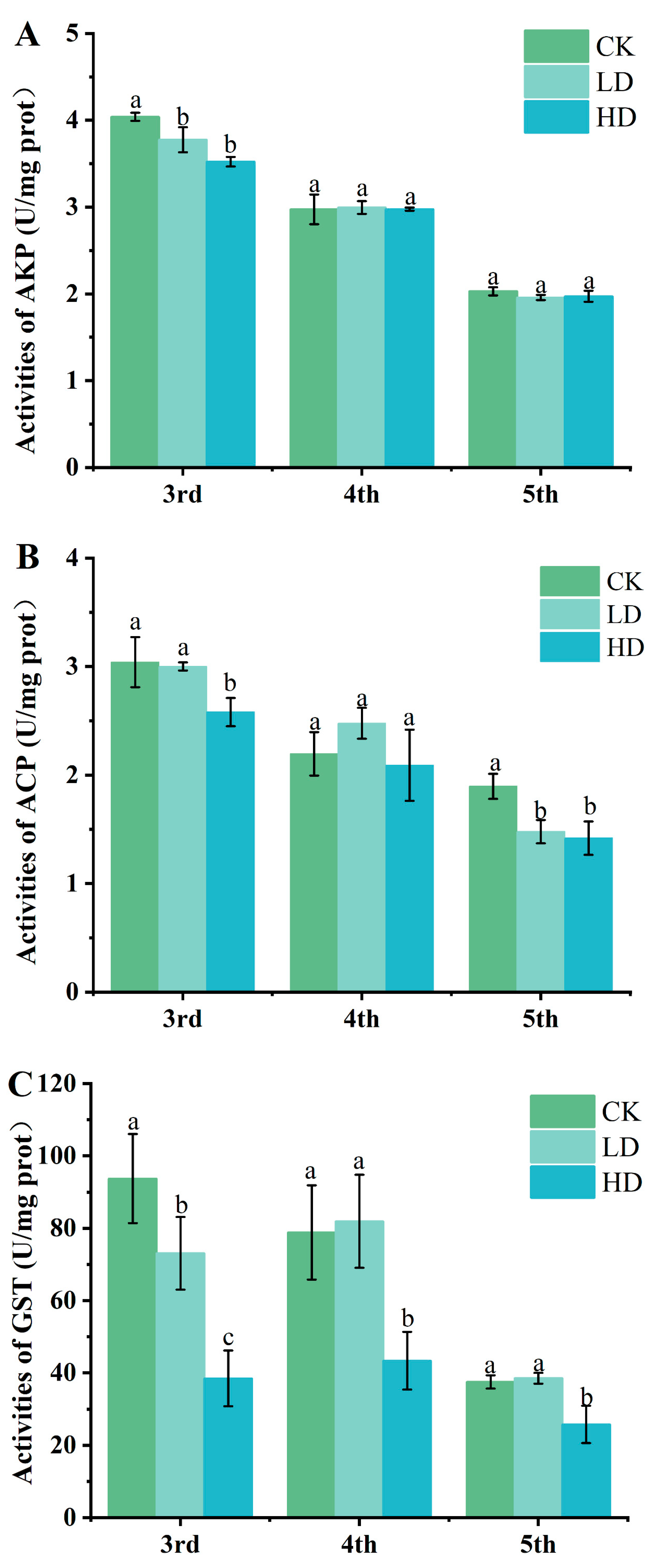
3.2. Effects of Cd Exposure on Activities of Detoxification Enzymes

The detoxification enzyme activity of the third instar larvae of G. pyloalis treated with three doses of Cd (CK, LD and HD) was measured (Figure 2). Compared with the CK group, the AKP activity of the third instar larvae in the LD and HD groups significantly reduced while the activity in the fourth and fifth instar larvae remained unaffected by the Cd concentration (Figure 2A). Compared with the CK group, the ACP activity of the LD group was significantly reduced in the fifth instar larvae however the difference between the fourth instar was not significant. Under HD treatment, the ACP activity of third and fifth instar larvae were significantly lower than that of the CK group while it was slightly lower at fourth instar than that of the CK group. However, the difference was not statistically significant (Figure 2B).
After Cd exposure, the GST activity in G. pyloalis exhibited significant changes. Compared with the CK group, the GST activity of the LD group decreased significantly at the third instar but was slightly higher than that of the CK group at the fourth and fifth instar. When G. pyloalis was exposed to high concentration of Cd, GST activity was significantly reduced in all three groups (Figure 2C). The difference in enzyme activity in the larvae of G. pyloalis may be attributed to the detoxification strategy in G. pyloalis under Cd exposure.

3.3. Sequencing of the G. pyloalis Transcriptome and De Novo Assembly

To study the transcription reaction of G. pyloalis to varying concentrations of Cd, nine cDNA libraries were built. Libraries were named correspondingly to the Cd treatment each received (CK, LD and HD), and each treatment generated three biological replicates. The nine cDNA libraries produced 66.54 G of raw data. Following the removal of low-quality reads, 64.64 G of clean data were obtained (Table S2).
Obtaining a total of 29,978 unigenes, the cumulative length reached 41,173,146 bp. The longest unigene had a length of 28,864 bp, while the average unigene length was 1373 bp, with an N50 of 2240 bp. Of these, 9159 (30.55%), 7963 (26.56%), 6509 (21.71%) and 6347 (21.17%) unigens had lengths of 301–500 bp, 501–1000 bp, 1001–2000 bp and >2000 bp, respectively (Figure S1). These results shows that the data quality was high and suitable for subsequent analysis and annotation.

3.4. Functional Annotation and Classification of Genes

A total of 29,978 unigenes were annotated based on seven databases, including NR, eggNOG, KEGG, Swiss-ProT, Pfam, GO and KOG databases. Among them, 17,368 (57.94%) unigenes were annotated in at least one database, while 3407 (11.37%) unigenes being annotated in seven databases (Table S3). In addition, 12,610 (42.06%) unigenes remained unannotated in any database, which may represent homologous genes.
According to the analysis of the annotation results, unigenes were mostly annotated by the NR database, as high as 17,292 (57.68%). According to the NR database annotation results, the most homologous genes of Ostrinia furnacalis (51.92%), subsequently Chilo suppressalis (11.35%), Galleria mellonella (4.33%) and Amyelois transitella (3.18%). The remaining 29.22% sequences and other insect sequences have good homology (Figure S2).

3.5. SSR Analysis

MISA tools were used to predict 29,978 unigenes and the SSR sites were subsequently analyzed with Prime3. This analysis resulted in the identified 4479 SSR sites (accounting for 14.94% of the total unigenes), with 3631 of the sequences containing more than one SSR (Table S4). Within all SSRs, mononucleotides had the most repeat types, accounting for 63.36%. Pentanucleotide had the least repeat types, accounting for 0.25% (Figure S3).

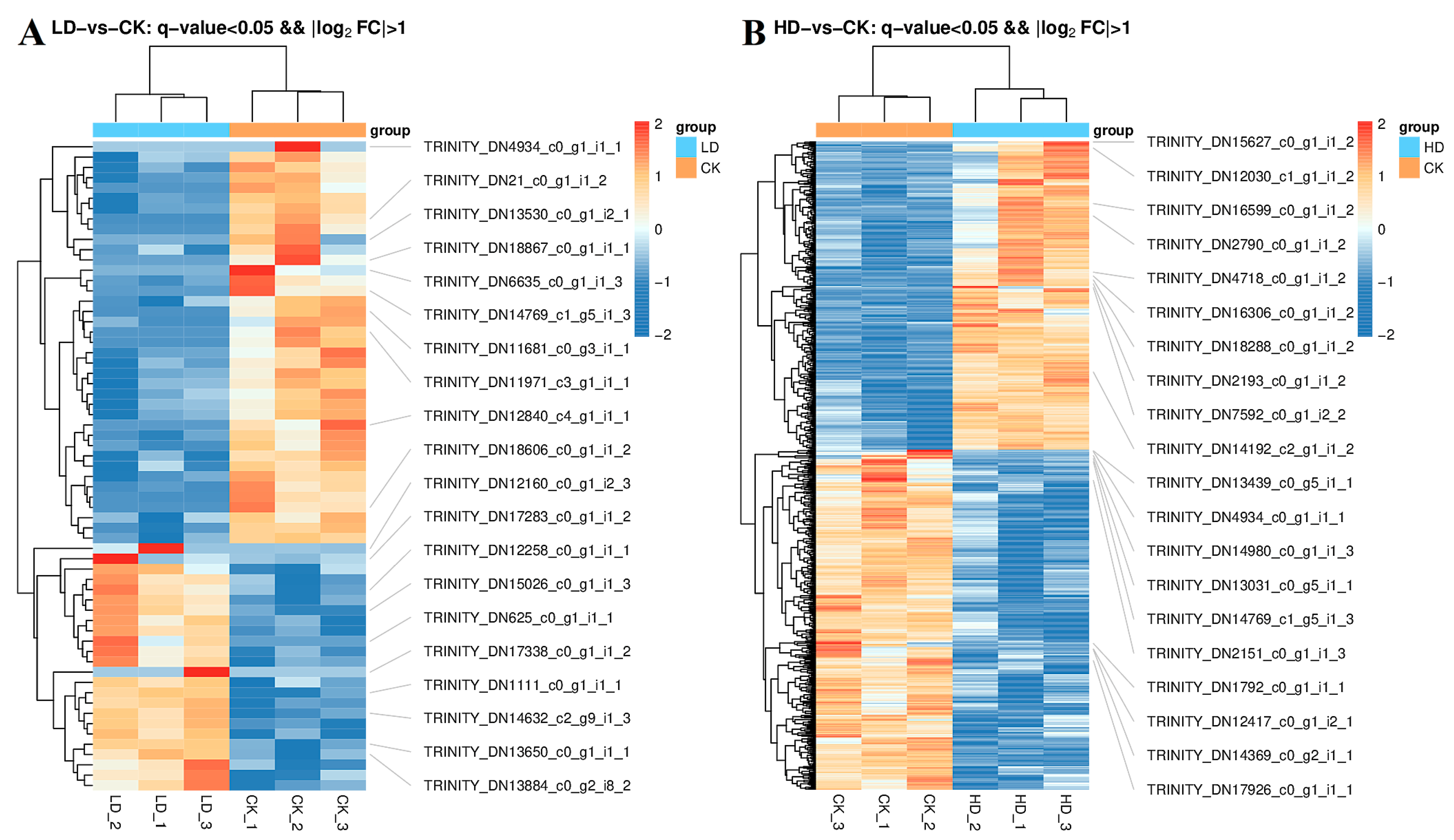
3.6. Statistical Analysis of DEG Number in Different Treatments

To explore the expression of DEGs under Cd stress and reveal the toxic mechanism of Cd stress, the DEGs of expression levels among the three groups (CK, LD and HD) were calculated and compared and the DEGs of the LD and HD groups compared with the CK group were identified. There was a total of 63 DEGs between CK and LD, of which 24 were upregulated and 39 were downregulated. There were a total of 831 DEGs between CK and HD, of which 395 were upregulated and 436 were downregulated. The quantity of DEGs between CK and HD was significantly higher than that between CK and LD. In addition, the main overview of the main expression of LD and HD on the heat map provides a general understanding of changes in gene expression (Figure 3).

3.7. GO Analysis of DEGs

Single genes were usually grouped into three different categories: biological process (BP), cell component (CC) and molecular function (MF). In order to clarify the main biological function of G. pyloalis under Cd exposure, the differences of the obtained genes were analyzed according to the three categories. Through sequence homologous analysis, 10,173 (33.93%) genes were divided into 53 functional groups (Figure S4). Cell, cell part, cellular process, binding and organelle were the top five abundant levels of GO terms. The DEGs numbers were 8199 (80.60%), 8172 (80.33%), 7734 (76.02%), 6439 (63.29%) and 6417 (63.08%), respectively.
In this study, the biological functions of the DEGs were assessed through GO enrichment analysis (Figure 4). The DEGs that were upregulated between CK and LD were significantly enriched in 18 functional categories, including “zinc ion binding”, “fatty acid elongation, monounsaturated fatty acid”, “fatty acid elongation, polyunsaturated fatty acid”, “fatty acid elongation” and other terms, while the downregulated DEGs were significantly enriched in 15 GO terms. The number of downregulated DEGs was observed to be lower than that of upregulated DEGs (Figure 4A). The DEGs upregulated between CK and HD were significantly enriched in 25 functional categories, including DNA integration, nucleic acid binding, endonuclease activity and other terms, while the downregulated DEGs were mainly enriched in the extracellular region, Z disc and nutrient reservoir activity (Figure 4B). The GO functional analysis revealed that the number of genes differentially expressed between CK and HD was higher than that between CK and LD, which may explain the higher percentage of genes involved in response to stimuli [40]. The results of the study show that exposure to high concentrations of Cd can stimulate more genes.

3.8. KEGG Analysis of DEGs

KEGG pathways were categorized into six categories: metabolism, genetic information processing, environmental information processing, cellular processes, organismal systems and human diseases. The biological significance of DEGs was further evaluated through the KEGG pathway enrichment analysis (Figure 5).
Figure 5 shows the 20 most-enriched KEGG pathways. The DEGs were enriched in peroxisome, longevity regulating pathway, AMPK signaling pathway, insulin signaling pathway, drug metabolism (cytochrome P450), metabolism of xenobiotics by cytochrome P450, adrenergic signaling in cardiomyocytes and fatty acid biosynthesis. These results suggest that exposure to Cd promotes the expression of several related metabolic and detoxification genes of G. pyloalis.

3.9. Validation by RT-qPCR

To further validate the accuracy of the DEGs library, Rpl32 was used as the reference gene, 12 DEGs related to detoxification and metabolism were selected and RT-qPCR was used to tests relative expression level. The gene expression results in the RT-qPCR analysis were accordance with the trend variation shown in the RNA-seq data (Figure 6). This is consistent with the trend of transcriptome changes (Table S5).

3.10. RNAi and Survivial Rate

To examine the role of CYP12A2 in G. pyloalis under Cd exposure, RNAi was employed to inhibit its expression in G. pyloalis larvae. Figure 7 shows the expression levels of CYP12A2 in various instars of G. pyloalis. Given the imminent pupation of the fifth instar larvae and the high expression of CYP12A2 in the third instar larvae of G. pyloalis, siRNA was injected during the third instar larvae stage. After 24 h, compared with the control group, the expression levels of CYP12A2 in CK, LD and HD decreased to 65.7%, 69.8% and 62.1%, respectively (Figure 7B). The survival rate of G. pyloalis injected with siCYP12A2 was markedly lower than that of G. pyloalis injected with siGFP. This suggests that the knockdown of CYP12A2 significantly heightened the sensitivity of the G. pyloalis to Cd (Figure 7C).

4. Discussion

As a prevalent environmental pollutant, heavy metals have been increasingly demonstrated to impact the physiology and biochemistry, life parameters and population dynamics of phytophagous insects [41,42,43]. The study showed the effects of Cd exposure on the development, detoxification mechanism and gene expression of G. pyloalis. The results revealed that Cd exposure had no significant effects on the survival and pupation rate of G. pyloalis. This finding aligns with the results of Jiang and Yan’s study, who observed that the survival and pupation of Lymantria dispar were unaffected when fed on Populus alba berolinensis exposed to heavy metal [44]. The reason for this result may be that insects have an effective detoxification system in their bodies which can reduce the impact of heavy metals exposure on insect survival rate and pupation rate. After Cd enters the body of G. pyloalis larvae, it triggers detoxification enzymes and protective enzymes to protect G. pyloalis which may weaken the toxicity of Cd to G. pyloalis larvae. Furthermore, our study showed that Cd exposure had decreased the pupal weight and adult emergence rate of the G. pyloalis larvae. Previous study has confirmed that the weight, body length, head capsule width and pupal weight of Pterostichus oblongopunctatus were inhibited under Cd exposure [45]. After the organism is subjected to external stress, a portion of the energy intake is utilized for detoxification metabolism, thereby affecting its own physiological function. [46]. However, there are also different results. For example, Stolpe and Müller found that the survival rate of Myzus persicae larvae was significantly reduced under Zn and Cd exposure [47]. Studies also have demonstrated that varying concentrations of heavy metals exert distinct effects on the growth and development of phytophagous insects. When exposed to low concentrations of Cu, the developmental duration of Spodoptera litura larvae significantly shortened while it markedly prolonged under high concentration exposure [48].
Previous research has reported that one of the main toxic effects of Cd on insects was the generation of oxidative stress, which can lead to the production of reactive ROS within insects and subsequent cellular damage [49,50]. Detoxifying enzymes serve a crucial role in averting oxidative stress and protecting organisms against ROS [51]. In the defense system of insects, AKP and ACP are important detoxification enzymes, which play an important part in resisting the harm of foreign substances [21]. In this study, the alterations in ACP activity in third and fifth instar larvae exhibited a negative correlation with the Cd concentration. Simultaneously, the AKP activity of the third instar of G. pyloalis was negatively correlated with the Cd concentration. Wu and Yi’s study showed that Pb exposure inhibited the activities of ACP and AKP in Galleria mellonella [52]. In addition, after being subjected to Pb and Cu exposure, the Sphaerodema urinator exhibits a decrease in AKP activity [53]. On one hand, the decline in ACP activity may be attributed to the direct impact of Cd on enzyme molecules, as numerous in vitro experiments have demonstrated the inhibitory effect of Cd on the enzyme [54]. On the other hand, it could result from the damage inflicted on cells by ROS induced by Cd, indirectly leading to a reduction in enzyme activity. The suppression of AKP activity may result from Cd-induced alterations in the structure of its enzyme molecules [55]. Furthermore, the modulation of AKP activity implies an enhancement of the body’s resistance to external interference, thereby indirectly reinforcing its non-specific immunity. The decrease of ACP and AKP activity suggest that Cd could interfere with the detoxification and metabolic function of G. pyloalis to a certain extent, showing its toxic effect.
GST is a commonly used biomarker for ecotoxicity detection due to its easy induction and measurement characteristics [56]. Its primary function is involvement in the detoxification and metabolism of harmful substances in insects. GST, as a class II detoxification enzyme system, plays a central role in secondary metabolism of exogenous substances and defending against environmental pollutants [57]. Additionally, a deficiency in GST can increase the risk of cellular oxidative damage [19,58]. In our study, Cd exposure inhibited the activity of GST in G. pyloalis larvae. Zhang et al. similarly reported that GST activity in O. chinensis was inhibited after Cd exposure, which increased the risk of oxidative damage to cells [59].
Genes associated with stress are frequently employed as indicators to monitor ecotoxicity, as their expression is influenced by heavy metals and other environmental pollutants [60,61]. For example, in Trachyderma hispida, the expressions of HSP70 and MT genes were also used to monitor oxidative stress parameters in the assessment of soil pollution in industrial areas [62]. In this study, the RNA-seq was used to study the molecular mechanism of the different responses of G. pyloalis larvae under Cd exposure. A total of 63 and 831 DEGs were identified under LD and HD, respectively. These results showed that after G. pyloalis were exposed to higher concentrations of Cd, in order to offset unfavorable environmental conditions, it changed the expression of multiple genes. The GO functional analysis unveiled a greater number of DEGs between CK and HD compared to CK and LD, which explained the higher proportion of genes involved in response to stimuli. These results of the study implied that high concentrations of Cd exposure activate various cellular processes, enzymes and metabolic pathways in G. pyloalis larvae, resulting in the regulating and activating of a higher number of genes. The DEGs between CK and LD are also enriched in DNA integration and nucleic acid binding, indicating that Cd seriously affected the process of DNA replication, transcription and translation in cells [63].
KEGG enrichment analysis revealed highly significant expression of DEGs between CK and LD and HD on the peroxisome pathway. Peroxisomes are organelles within cells that perform essential functions such as redox reactions and detoxification reactions and has the function of detoxification and metabolism [64]. Such enrichment underlines the innate response of the G. pyloalis to manage Cd induced exposure by initiating metabolic and detoxification responses. In addition, the DEGs between CK and HD were significantly enriched in the insulin signaling pathway, which plays a crucial role in regulating the growth, senescence and reproduction of insects [65]. The DEGs between CK and HD were also significantly enriched in the fatty acid synthesis, which usually plays an active role in resisting environmental stress [66]. Xenobiotic biodegradation and metabolism emerge as crucial strategies employed by insects to combat xenobiotic harm and thrive in challenging environments [67]. Of which, xenobiotic biodegradation and metabolism includes drug metabolism-cytochrome P450, drug metabolism-other enzymes and metabolism of xenobiotics by cytochrome P450 [68,69]. The results of this study show that metabolism of xenobiotics by cytochrome P450 was one of the most abundant pathways in CK and HD. P450s were a class of metabolic detoxification enzymes, which play an important role in the resistance of the insect to pesticides and heavy metals [13,70]. Meng et al. found that the transcription levels of CsCYP307Al, CsCYP306A1 and CsCYP314Al were induced when Chilo suppressalis was exposed to chlorantraniliprole at specific developmental stages [71]. This observation suggests that the mechanism of insect tolerance to heavy metals may share similarities with that of certain pesticides. Our findings suggest that CYP12A2 may serve as a pivotal detoxification gene in response to Cd exposure. The knockdown of CYP12A2 resulted in a significant decrease in the survival rate of G. pyloalis under Cd exposure, indicating a key role for CYP12A2 in the response of G. pyloalis to Cd exposure. In addition, the contamination of the environment with heavy metals decreased the sensitivity of insects to insecticides, thereby posing a threat to agricultural production. For example, the sensitivity of S. litura to alpha-cypermethrin and fenvalerate decreased under Pb exposure [72]. Therefore, in order to reduce the cost of pest control, the control intensity should be adjusted according to the degree of heavy metal pollution in the local area so as to obtain better control effect. In addition, whether the change in the sensitivity to insecticides is suitable for pre-exposed Cd G. pyloalis larvae will be studied in our subsequent experiments.

5. Conclusions

In conclusion, we discussed the toxic effects of Cd on G. pyloalis and focused on development, detoxification mechanisms and gene expression. The study showed that Cd exposure affects the growth of G. pyloalis, influencing the activities of AKP, ACP and GST. These variations were observed to be dependent on both time and concentration. The decrease of detoxification enzyme activity suggested that Cd could interfere with the detoxification and metabolic functions of G. pyloalis. Transcriptome revealed that different concentrations of Cd had different effects on metabolism and detoxification of G. pyloalis. G. pyloalis triggers a response strategy to Cd toxicity by changing the expression of genes that are associated with metabolism and detoxification system. This study further elucidated the toxicity of G. pyloalis exposed to different concentrations of Cd exposure at the transcriptome level and gained a comprehensive understanding of the molecular mechanism of G. pyloalis exposure to Cd toxicity. CYP12A2 was identified as a crucial gene affecting Cd exposure by RNAi, which can be further studied. Furthermore, investigating the alterations in insecticidal sensitivity following heavy metal stress in insects is a focal point of our forthcoming research.

Supplementary Materials

The following supporting information can be downloaded at: https://www.mdpi.com/article/10.3390/agronomy14030626/s1, Figure S1: Length distribution of G. pyloalis; Figure S2: Species classification based on the NR database; Figure S3:The repeat type frequency of SSR motifs in tested G. pyloalis transcriptome; Figure S4: Classification of the gene ontology for the transcriptome of G. pyloalis 10,173 (33.93%) of the total unigenes were categorized into 53 function groups; Table S1: Primers used for PCR in G. pyloalis; Table S2: Quality of G. pyloalis clean reads; Table S3: Functional annotation of the G. pyloalis transcriptome; Table S4: Summary of SSR searing in the G. pyloalis transcriptome; Table S5: List of DEGs related to detoxification metabolism of G. pyloalis larvae; Table S6: Primers used to synthesize siRNA.

Author Contributions

S.Z.: conceptualization, data curation, formal analysis, methodology, roles/writing—original draft, writing—review and editing. W.M.: data curation, formal analysis, methodology. X.P.: formal analysis, software. P.L.: formal analysis, investigation. S.S.: funding acquisition, project administration, investigation. W.Z.: funding acquisition, project administration, resources, supervision, validation, visualization, writing—review and editing. F.W.: funding acquisition, project administration, writing—review and editing. All authors have read and agreed to the published version of the manuscript.

Funding

This work was supported by the Key Research and Development Program (Modern Agriculture) of Jiangsu Province (BE2021344), the Jiangsu Agricultural Science and Technology Innovation Fund (CX (21) 3179) and the Doctoral Starting up Foundation of Jiangsu university of Science and Technology (1732932004).

Data Availability Statement

Data will be made available upon request.

Conflicts of Interest

All authors declare that they have no known competing financial interests or personal relationships that could have influenced the work reported in this paper.

References

  1. Chen, Y.G.; He, X.L.S.; Huang, J.H.; Luo, R.; Ge, H.Z.; Wołowicz, A.; Wawrzkiewicz, M.; Gładysz-Płaska, A.; Li, B.; Yu, Q.X.; et al. Impacts of Heavy Metals and Medicinal Crops on Ecological Systems, Environmental Pollution, Cultivation, and Production Processes in China. Ecotoxicol. Environ. Saf. 2021, 219, 112336. [Google Scholar] [CrossRef] [PubMed]
  2. Wang, S.L.; Xu, X.R.; Sun, Y.X.; Liu, J.L.; Li, H. Bin Heavy Metal Pollution in Coastal Areas of South China: A Review. Mar. Pollut. Bull. 2013, 76, 7–15. [Google Scholar] [CrossRef] [PubMed]
  3. Hu, Z.; Zhao, C.; Li, Q.; Feng, Y.; Zhang, X.; Lu, Y.; Ying, R.; Yin, A.; Ji, W. Heavy Metals Can Affect Plant Morphology and Limit Plant Growth and Photosynthesis Processes. Agronomy 2023, 13, 2601. [Google Scholar] [CrossRef]
  4. Shephard, A.M.; Mitchell, T.S.; Henry, S.B.; Oberhauser, K.S.; Kobiela, M.E.; Snell-Rood, E.C. Assessing Zinc Tolerance in Two Butterfly Species: Consequences for Conservation in Polluted Environments. Insect Conserv. Divers. 2020, 13, 201–210. [Google Scholar] [CrossRef]
  5. Nikolić, T.V.; Kojić, D.; Orčić, S.; Batinić, D.; Vukašinović, E.; Blagojević, D.P.; Purać, J. The Impact of Sublethal Concentrations of Cu, Pb and Cd on Honey Bee Redox Status, Superoxide Dismutase and Catalase in Laboratory Conditions. Chemosphere 2016, 164, 98–105. [Google Scholar] [CrossRef] [PubMed]
  6. Gao, H.H.; Zhao, H.Y.; Du, C.; Deng, M.M.; Du, E.X.; Hu, Z.Q.; Hu, X.S. Life Table Evaluation of Survival and Reproduction of the Aphid, Sitobion avenae, Exposed to Cadmium. J. Insect Sci. 2012, 12, 44. [Google Scholar] [CrossRef] [PubMed]
  7. Di, N.; Hladun, K.R.; Zhang, K.; Liu, T.X.; Trumble, J.T. Laboratory Bioassays on the Impact of Cadmium, Copper and Lead on the Development and Survival of Honeybee (Apis mellifera L.) Larvae and Foragers. Chemosphere 2016, 152, 108839. [Google Scholar] [CrossRef]
  8. Vlahović, M.; Matić, D.; Mutić, J.; Trifković, J.; Đurđić, S.; Perić Mataruga, V. Influence of Dietary Cadmium Exposure on Fitness Traits and Its Accumulation (with an Overview on Trace Elements) in Lymantria Dispar Larvae. Comp. Biochem. Physiol. Part—C Toxicol. Pharmacol. 2017, 200, 27–33. [Google Scholar] [CrossRef]
  9. Vlahović, M.; Ilijin, L.; Mrdaković, M.; Todorović, D.; Matić, D.; Lazarević, J.; Mataruga, V.P. Glutathione S-Transferase in the Midgut Tissue of Gypsy Moth (Lymantria dispar) Caterpillars Exposed to Dietary Cadmium. Environ. Toxicol. Pharmacol. 2016, 44, 13–17. [Google Scholar] [CrossRef]
  10. Mirčić, D.; Blagojević, D.; Perić-Mataruga, V.; Ilijin, L.; Mrdaković, M.; Vlahović, M.; Lazarević, J. Cadmium Effects on the Fitness-Related Traits and Antioxidative Defense of Lymantria dispar L. Larvae. Environ. Sci. Pollut. Res. 2013, 20, 209–218. [Google Scholar] [CrossRef]
  11. Zhao, J.; Sun, Y.; Xiao, L.; Tan, Y.; Bai, L. Molecular Characterization and Expression of Vitellogenin Gene from Spodoptera exigua Exposed to Cadmium Stress. Gene 2016, 593, 179–184. [Google Scholar] [CrossRef] [PubMed]
  12. Borowska, J.; Pyza, E. Effects of Heavy Metals on Insect Immunocompetent Cells. J. Insect Physiol. 2011, 57, 760–770. [Google Scholar] [CrossRef] [PubMed]
  13. Lu, K.; Li, W.; Cheng, Y.; Ni, H.; Chen, X.; Li, Y.; Tang, B.; Sun, X.; Li, Y.; Liu, T.; et al. Copper Exposure Enhances Spodoptera Litura Larval Tolerance to β-Cypermethrin. Pestic. Biochem. Physiol. 2019, 160, 127–135. [Google Scholar] [CrossRef] [PubMed]
  14. Poupardin, R.; Reynaud, S.; Strode, C.; Ranson, H.; Vontas, J.; David, J.P. Cross-Induction of Detoxification Genes by Environmental Xenobiotics and Insecticides in the Mosquito Aedes Aegypti: Impact on Larval Tolerance to Chemical Insecticides. Insect Biochem. Mol. Biol. 2008, 38, 540–551. [Google Scholar] [CrossRef] [PubMed]
  15. Gauthier, M.; Aras, P.; Jumarie, C.; Boily, M. Low Dietary Levels of Al, Pb and Cd May Affect the Non-Enzymatic Antioxidant Capacity in Caged Honey Bees (Apis mellifera). Chemosphere 2016, 144, 848–854. [Google Scholar] [CrossRef] [PubMed]
  16. Stone, D.; Jepson, P.; Laskowski, R. Trends in Detoxification Enzymes and Heavy Metal Accumulation in Ground beetles (Coleoptera: Carabidae) Inhabiting a Gradient of Pollution. Comp. Biochem. Physiol.—C Toxicol. Pharmacol. 2002, 132, 105–112. [Google Scholar] [CrossRef] [PubMed]
  17. Ilijin, L.; Mrdaković, M.; Todorović, D.; Vlahović, M.; Gavrilović, A.; Mrkonja, A.; Perić-Mataruga, V. Life History Traits and the Activity of Antioxidative Enzymes in Lymantria dispar L. (Lepidoptera, Lymantriidae) Larvae Exposed to Benzo[a]Pyrene. Environ. Toxicol. Chem. 2015, 34, 2618–2624. [Google Scholar] [CrossRef] [PubMed]
  18. Bauer, A.K.; Velmurugan, K.; Plöttner, S.; Siegrist, K.J.; Romo, D.; Welge, P.; Brüning, T.; Xiong, K.N.; Käfferlein, H.U. Environmentally Prevalent Polycyclic Aromatic Hydrocarbons Can Elicit Co-Carcinogenic Properties in an in Vitro Murine Lung Epithelial Cell Model. Arch. Toxicol. 2018, 92, 1311–1322. [Google Scholar] [CrossRef]
  19. Xu, Z.B.; Zou, X.P.; Zhang, N.; Feng, Q.L.; Zheng, S.C. Detoxification of Insecticides, Allechemicals and Heavy Metals by Glutathione S-Transferase SlGSTE1 in the Gut of Spodoptera litura. Insect Sci. 2015, 22, 503–511. [Google Scholar] [CrossRef]
  20. Calvo-Marzal, P.; Rosatto, S.S.; Granjeiro, P.A.; Aoyama, H.; Kubota, L.T. Electroanalytical Determination of Acid Phosphatase Activity by Monitoring P-Nitrophenol. Anal. Chim. Acta 2001, 441, 207–214. [Google Scholar] [CrossRef]
  21. Vlahović, M.; Lazarević, J.; Perić-Mataruga, V.; Ilijin, L.; Mrdaković, M. Plastic Responses of Larval Mass and Alkaline Phosphatase to Cadmium in the Gypsy Moth Larvae. Ecotoxicol. Environ. Saf. 2009, 72, 1148–1155. [Google Scholar] [CrossRef] [PubMed]
  22. Vlahović, M.; Ilijin, L.; Matić, D.; Lazarević, J.; Nenadović, V.; Perić-Mataruga, V.; Mrdaković, M. Cadmium Effects on the Ratio of Activities of Lysosomal and Total Acid Phosphatases (ACPLys/ACPTot) in Lymantria dispar Larvae. Arch. Biol. Sci. 2013, 65, 345–352. [Google Scholar] [CrossRef]
  23. Stubberud, H.E.; Hønsi, T.G.; Stenersen, J. Purification and Partial Characterisation of Tentatively Classified Acid Phosphatase from the Earthworm Eisenia Veneta. Comp. Biochem. Physiol.—B Biochem. Mol. Biol. 2000, 126, 487–494. [Google Scholar] [CrossRef] [PubMed]
  24. Zhang, J.; Shi, Z.; Gao, Z.; Wen, Y.; Wang, W.; Liu, W.; Wang, X.; Zhu, F. Identification of Three Metallothioneins in the Black Soldier Fly and Their Functions in Cd Accumulation and Detoxification. Environ. Pollut. 2021, 286, 117146. [Google Scholar] [CrossRef] [PubMed]
  25. Khosravi, R.; Sendi, J.J. Biology and Demography of Glyphodes Pyloalis Walker (Lepidoptera: Pyralidae) on Mulberry. J. Asia. Pac. Entomol. 2010, 13, 273–276. [Google Scholar] [CrossRef]
  26. Shao, Z.M.; Li, Y.J.; Zhang, X.R.; Chu, J.; Ma, J.H.; Liu, Z.X.; Wang, J.; Sheng, S.; Wu, F.A. Identification and Functional Study of Chitin Metabolism and Detoxification-related Genes in Glyphodes Pyloalis Walker (Lepidoptera: Pyralidae) Based on Transcriptome Analysis. Int. J. Mol. Sci. 2020, 21, 1904. [Google Scholar] [CrossRef] [PubMed]
  27. Feng, R.; Zhu, Q.; Xu, Y.; Li, W.; Ding, Y.; Han, L.; Rensing, C.; Wang, R. Safe Utilization of Polluted Soil by Arsenic, Cadmium and Lead through an Integrated Sericultural Measure. Sci. Total Environ. 2019, 659, 1234–1241. [Google Scholar] [CrossRef]
  28. Jiang, D.; Tan, M.; Zheng, L.; Wu, H.; Li, Y.; Yan, S. Cd Exposure-Triggered Metabolic Disruption Increases the Susceptibility of Lymantria dispar (Lepidoptera: Erebidae) Larvae to Mamestra Brassicae Nuclear Polyhedrosis Virus: A Multi-Omics Study. Ecotoxicol. Environ. Saf. 2022, 232, 113280. [Google Scholar] [CrossRef]
  29. Jiang, D.; Zhou, Y.; Tan, M.; Zhang, J.; Guo, Q.; Yan, S. Cd Exposure-Induced Growth Retardation Involves in Energy Metabolism Disorder of Midgut Tissues in the Gypsy Moth Larvae. Environ. Pollut. 2020, 266, 115173. [Google Scholar] [CrossRef]
  30. Bradford, M.M. A Rapid and Sensitive Method for the Quantitation of Microgram Quantities of Protein Utilizing the Principle of Protein-Dye Binding. Anal. Biochem. 1976, 72, 248–254. [Google Scholar] [CrossRef]
  31. Bolger, A.M.; Lohse, M.; Usadel, B. Trimmomatic: A Flexible Trimmer for Illumina Sequence Data. Bioinformatics 2014, 30, 2114–2120. [Google Scholar] [CrossRef] [PubMed]
  32. Grabherr, M.G.; Haas, B.J.; Yassour, M.; Levin, J.Z.; Thompson, D.A.; Amit, I.; Adiconis, X.; Fan, L.; Raychowdhury, R.; Zeng, Q.; et al. Trinity: Reconstructing a Full-Length Transcriptome without a Genome from RNA-Seq Data HHS Public Access. Nat. Biotechnol. Nat. Biotechnol 2011, 29, 644–652. [Google Scholar] [CrossRef] [PubMed]
  33. Conesa, A.; Götz, S.; García-Gómez, J.M.; Terol, J.; Talón, M.; Robles, M. Blast2GO: A Universal Tool for Annotation, Visualization and Analysis in Functional Genomics Research. Bioinformatics 2005, 21, 3674–3676. [Google Scholar] [CrossRef] [PubMed]
  34. Pertea, G.; Huang, X.; Liang, F.; Antonescu, V.; Sultana, R.; Karamycheva, S.; Lee, Y.; White, J.; Cheung, F.; Parvizi, B.; et al. TIGR Gene Indices Clustering Tools (TGICL): A Software System for Fast Clustering of Large EST Datasets. Bioinformatics 2003, 19, 651–652. [Google Scholar] [CrossRef] [PubMed]
  35. Trapnell, C.; Williams, B.A.; Pertea, G.; Mortazavi, A.; Kwan, G.; Van Baren, M.J.; Salzberg, S.L.; Wold, B.J.; Pachter, L. Transcript Assembly and Quantification by RNA-Seq Reveals Unannotated Transcripts and Isoform Switching during Cell Differentiation. Nat. Biotechnol. 2010, 28, 511–515. [Google Scholar] [CrossRef] [PubMed]
  36. Langmead, B.; Salzberg, S.L. Fast Gapped-Read Alignment with Bowtie 2. Nature Methods. Nat. Publ. Gr. 2012, 9, 357–359. [Google Scholar] [CrossRef]
  37. Love, M.I.; Huber, W.; Anders, S. Moderated Estimation of Fold Change and Dispersion for RNA-Seq Data with DESeq2. Genome Biol. 2014, 15, 550. [Google Scholar] [CrossRef]
  38. Schmittgen, T.D.; Livak, K.J. Analyzing Real-Time PCR Data by the Comparative C(T) Method. Nat. Protoc. 2008, 3, 1101–1108. [Google Scholar] [CrossRef]
  39. Dong, B.; Liu, X.Y.; Li, B.; Li, M.Y.; Li, S.G.; Liu, S. A Heat Shock Protein Protects against Oxidative Stress Induced by Lambda-Cyhalothrin in the Green Peach Aphid Myzus Persicae. Pestic. Biochem. Physiol. 2022, 181, 104995. [Google Scholar] [CrossRef]
  40. Dai, Y.; Zhang, Y.; Sun, W.; Chen, Y.; Wang, X.; Xin, T.; Wan, B.; Xia, B.; Zhong, L.; Zou, Z. The Metabolism and Detoxification Effects of Lead Exposure on Aleurolyphus ovatus (Acari: Acaridae) via Transcriptome Analysis. Chemosphere 2023, 333, 138886. [Google Scholar] [CrossRef]
  41. Su, H.; Wu, J.; Zhang, Z.; Ye, Z.; Chen, Y.; Yang, Y. Effects of Cadmium Stress at Different Concentrations on the Reproductive Behaviors of Beet Armyworm Spodoptera exigua (Hübner). Ecotoxicology 2021, 30, 402–410. [Google Scholar] [CrossRef] [PubMed]
  42. Zhang, J.; Jiang, D.; Dong, X.; Meng, Z.; Yan, S. Accumulation of Cd and Pb in Various Body Parts, Organs and Tissues of Lymantria dispar Asiatica (Lepidoptera: Erebidae). J. Asia. Pac. Entomol. 2020, 23, 963–969. [Google Scholar] [CrossRef]
  43. Zhou, J.; Chen, J.; Shu, Y. Lead Stress Affects the Reproduction of Spodoptera litura but Not by Regulating the Vitellogenin Gene Promoter. Ecotoxicol. Environ. Saf. 2021, 208, 111581. [Google Scholar] [CrossRef] [PubMed]
  44. Jiang, D.; Yan, S. Effects of Cd, Zn or Pb Stress in Populus Alba Berolinensis on the Development and Reproduction of Lymantria dispar. Ecotoxicology 2017, 26, 1305–1313. [Google Scholar] [CrossRef] [PubMed]
  45. Bednarska, A.J.; Stachowicz, I. Costs of Living in Metal Polluted Areas: Respiration Rate of the Ground Beetle Pterostichus oblongopunctatus from Two Gradients of Metal Pollution. Ecotoxicology 2013, 22, 118–124. [Google Scholar] [CrossRef] [PubMed]
  46. Moolman, L.; Van Vuren, J.H.J.; Wepener, V. Comparative Studies on the Uptake and Effects of Cadmium and Zinc on the Cellular Energy Allocation of Two Freshwater Gastropods. Ecotoxicol. Environ. Saf. 2007, 68, 443–450. [Google Scholar] [CrossRef] [PubMed]
  47. Stolpe, C.; Müller, C. Effects of Single and Combined Heavy Metals and Their Chelators on Aphid Performance and Preferences. Environ. Toxicol. Chem. 2016, 35, 3023–3030. [Google Scholar] [CrossRef] [PubMed]
  48. Huang, D.; Kong, J.; Seng, Y. Effects of the Heavy Metal Cu2+ on Growth, Development, and Population Dynamics of Spodoptera litura (Lepidoptera: Noctuidae). J. Econ. Entomol. 2012, 105, 288–294. [Google Scholar] [CrossRef]
  49. Tylko, G.; Banach, Z.; Borowska, J.; Niklińska, M.; Pyza, E. Elemental Changes in the Brain, Muscle, and Gut Cells of the Housefly, Musca Domestica, Exposed to Heavy Metals. Microsc. Res. Tech. 2005, 66, 239–247. [Google Scholar] [CrossRef]
  50. Sosa, V.; Moliné, T.; Somoza, R.; Paciucci, R.; Kondoh, H.; LLeonart, M.E. Oxidative Stress and Cancer: An Overview. Ageing Res. Rev. 2013, 12, 376–390. [Google Scholar] [CrossRef]
  51. Tiwari, S.; Pelz-Stelinski, K.; Stelinski, L.L. Effect of Candidatus Liberibacter Asiaticus Infection on Susceptibility of Asian Citrus Psyllid, Diaphorina citri, to Selected Insecticides. Pest Manag. Sci. 2011, 67, 94–99. [Google Scholar] [CrossRef] [PubMed]
  52. Wu, G.; Yi, Y. Effects of Dietary Heavy Metals on the Immune and Antioxidant Systems of Galleria mellonella Larvae. Comp. Biochem. Physiol. Part—C Toxicol. Pharmacol. 2015, 167, 131–139. [Google Scholar] [CrossRef] [PubMed]
  53. Bream, A.S. Laboratory Evaluation of Heavy Metals Stress on Certain Biochemical Parameters of the Aquatic Insect, Sphaerodema Urinator Duf. (Hemiptera: Belostomatidae). Commun. Agric. Appl. Biol. Sci. 2003, 68, 291–297. [Google Scholar]
  54. Mazorra, M.T.; Rubio, J.A.; Blasco, J. Acid and Alkaline Phosphatase Activities in the Clam Scrobicularia Plana: Kinetic Characteristics and Effects of Heavy Metals. Comp. Biochem. Physiol.—B Biochem. Mol. Biol. 2002, 131, 241–249. [Google Scholar] [CrossRef] [PubMed]
  55. Renella, G.; Ortigoza, A.L.R.; Landi, L.; Nannipieri, P. Additive Effects of Copper and Zinc on Cadmium Toxicity on Phosphatase Activities and ATP Content of Soil as Estimated by the Ecological Dose (ED50). Soil Biol. Biochem. 2003, 35, 1203–1210. [Google Scholar] [CrossRef]
  56. Kumar, S.; Asif, M.H.; Chakrabarty, D.; Tripathi, R.D.; Dubey, R.S.; Trivedi, P.K. Differential Expression of Rice Lambda Class GST Gene Family Members During Plant Growth, Development, and in Response to Stress Conditions. Plant Mol. Biol. Rep. 2013, 31, 569–580. [Google Scholar] [CrossRef]
  57. Wang, Y.; Fang, J.; Leonard, S.S.; Rao, K.M.K. Cadmium Inhibits the Electron Transfer Chain and Induces Reactive Oxygen Species. Free Radic. Biol. Med. 2004, 36, 1434–1443. [Google Scholar] [CrossRef]
  58. Forman, H.J.; Zhang, H.; Rinna, A. Glutathione: Overview of Its Protective Roles, Measurement, and Biosynthesis. Mol. Aspects Med. 2009, 30, 1–12. [Google Scholar] [CrossRef]
  59. Zhang, Y.P.; Song, D.N.; Wu, H.H.; Yang, H.M.; Zhang, J.Z.; Li, L.J.; Ma, E.B.; Guo, Y.P. Effect of Dietary Cadmium on the Activity of Glutathione S-Transferase and Carboxylesterase in Different Developmental Stages of the Oxya chinensis (Orthoptera: Acridoidea). Environ. Entomol. 2014, 43, 171–177. [Google Scholar] [CrossRef]
  60. Bernabò, P.; Gaglio, M.; Bellamoli, F.; Viero, G.; Lencioni, V. DNA Damage and Translational Response during Detoxification from Copper Exposure in a Wild Population of Chironomus Riparius. Chemosphere 2017, 173, 235–244. [Google Scholar] [CrossRef]
  61. Lee, S.M.; Lee, S.B.; Park, C.H.; Choi, J. Expression of Heat Shock Protein and Hemoglobin Genes in Chironomus Tentans (Diptera, Chironomidae) Larvae Exposed to Various Environmental Pollutants: A Potential Biomarker of Freshwater Monitoring. Chemosphere 2006, 65, 1074–1081. [Google Scholar] [CrossRef] [PubMed]
  62. El-Gendy, A.H.; Augustyniak, M.; Toto, N.A.; Al Farraj, S.; El-Samad, L.M. Oxidative Stress Parameters, DNA Damage and Expression of HSP70 and MT in Midgut of Trachyderma hispida (Forskål, 1775) (Coleoptera: Tenebrionidae) from a Textile Industry Area. Environ. Pollut. 2020, 267, 115661. [Google Scholar] [CrossRef] [PubMed]
  63. Lv, B.; Yang, H.L.; Peng, Y.D.; Wang, J.; Zeng, Z.; Li, N.; Tang, Y.E.; Wang, Z.; Song, Q.S. Cadmium Exposure Alters Expression of Protective Enzymes and Protein Processing Genes in Venom Glands of the Wolf Spider Pardosa pseudoannulata. Environ. Pollut. 2021, 268, 115847. [Google Scholar] [CrossRef] [PubMed]
  64. del Rio, L.A. Reactive Oxygen Species, Antioxidant Systems and Nitric Oxide in Peroxisomes. J. Exp. Bot. 2002, 53, 1255–1272. [Google Scholar] [CrossRef]
  65. Stern, D. Body-Size Control: How an Insect Knows It Has Grown Enough. Curr. Biol. 2003, 13, 267–269. [Google Scholar] [CrossRef] [PubMed]
  66. Zhang, M.; Barg, R.; Yin, M.; Gueta-Dahan, Y.; Leikin-Frenkel, A.; Salts, Y.; Shabtai, S.; Ben-Hayyim, G. Modulated Fatty Acid Desaturation via Overexpression of Two Distinct ω-3 Desaturases Differentially Alters Tolerance to Various Abiotic Stresses in Transgenic Tobacco Cells and Plants. Plant J. 2005, 44, 361–371. [Google Scholar] [CrossRef] [PubMed]
  67. Harrop, T.W.R.; Pearce, S.L.; Daborn, P.J.; Batterham, P. Whole-Genome Expression Analysis in the Third Instar Larval Midgut of Drosophila melanogaster. G3 Genes Genomes Genet. 2014, 4, 2197–2205. [Google Scholar] [CrossRef]
  68. Poupardin, R.; Riaz, M.A.; Vontas, J.; David, J.P.; Reynaud, S. Transcription Profiling of Eleven Cytochrome P450s Potentially Involved in Xenobiotic Metabolism in the Mosquito Aedes aegypti. Insect Mol. Biol. 2010, 19, 185–193. [Google Scholar] [CrossRef]
  69. Jin, Y.; Cong, B.; Wang, L.; Gao, Y.; Zhang, H.; Dong, H.; Lin, Z. Differential Gene Expression Analysis of the Epacromius coerulipes (Orthoptera: Acrididae) Transcriptome. J. Insect Sci. 2016, 16, 42. [Google Scholar] [CrossRef]
  70. Pan, X.; Ding, J.-H.; Zhao, S.-Q.; Shi, H.-C.; Miao, W.-L.; Wu, F.-A.; Sheng, S.; Zhou, W.-H. Identification and Functional Study of Detoxification-Related Genes in Response to Tolfenpyrad Stress in Glyphodes pyloalis Walker (Lepidoptera: Pyralidae). Pestic. Biochem. Physiol. 2023, 194, 105503. [Google Scholar] [CrossRef]
  71. Meng, X.; Zhang, N.; Yang, X.; Miao, L.; Jiang, H.; Ji, C.; Xu, B.; Qian, K.; Wang, J. Sublethal Effects of Chlorantraniliprole on Molting Hormone Levels and MRNA Expressions of Three Halloween Genes in the Rice Stem Borer, Chilo Suppressalis. Chemosphere 2020, 238, 124676. [Google Scholar] [CrossRef]
  72. Zhou, J.; Zhang, G.; Zhou, Q. Molecular Characterization of Cytochrome P450 CYP6B47 CDNAs and 5′-Flanking Sequence from Spodoptera litura (Lepidoptera: Noctuidae): Its Response to Lead Stress. J. Insect Physiol. 2012, 58, 726–736. [Google Scholar] [CrossRef]
Figure 1. Effect of Cd exposure on (A) larval survival rate, (B) pupae weight, (C) pupation rate and (D) adult emergence rate of G. pyloalis. Different letters indicate significant differences at p < 0.05.
Agronomy 14 00626 g001
Figure 2. Effects of Cd exposure on detoxification enzyme activity in G. pyloalis (A) AKP, (B) ACP and (C) GST. Different letters represent significance of difference between different treatment concentrations at same instar.
Agronomy 14 00626 g002
Figure 3. Heat maps showing expression patterns of DEGs (A) LD vs. CK and (B) HD vs. CK.
Agronomy 14 00626 g003
Figure 4. GO analysis of DEGs in G. pyloalis transcriptome. (A) LD vs. CK and (B) HD vs. CK.
Agronomy 14 00626 g004
Figure 5. KEGG pathway enrichment analysis of G. pyloalis after exposure to Cd. (A) LD vs. CK and (B) HD vs. CK.
Agronomy 14 00626 g005
Figure 6. All 12 DEGs were validated by RT-qPCR. Y axis represents fold change relative to concentration of CK.
Agronomy 14 00626 g006
Figure 7. Expression level of (A) CYP12A2 in different instar larvae, (B) CYP12A2 after 24 h of silencing and (C) effect of silencing CYP12A2 on the survival rate of G. pyloalis larvae. Different letters represent significance of difference between different treatment concentrations at same instar.
Agronomy 14 00626 g007
Disclaimer/Publisher’s Note: The statements, opinions and data contained in all publications are solely those of the individual author(s) and contributor(s) and not of MDPI and/or the editor(s). MDPI and/or the editor(s) disclaim responsibility for any injury to people or property resulting from any ideas, methods, instructions or products referred to in the content.

