Article

Host Plant Specific Control of 2,4-Diacetylphloroglucinol Production in the Rhizosphere

by
Feth El Zahar Haichar
1,
Sylvain Fochesato
2,3,4 and
Wafa Achouak
2,3,4,*
1
Université Lyon1, CNRS, UMR5557, INRA, USC1364, Ecologie Microbienne, Groupes Fonctionnels Microbiens et Cycle de l'Azote, Villeurbanne F-69622, France
2
CEA, DSV, IBEB, SBVME, Lab Ecol Microb Rhizosphere & Environ Extrem (LEMiRE), Saint-Paul-lez-Durance, France
3
CNRS, UMR 6191, FR CNRS 3098 ECCOREV, Saint-Paul-lez-Durance, France
4
Aix-Marseille Université, Saint-Paul-lez-Durance, France
*
Author to whom correspondence should be addressed.
Submission received: 23 July 2013 / Revised: 10 September 2013 / Accepted: 24 September 2013 / Published: 30 September 2013

Abstract

:
To shed light on phytobeneficial bacterial gene expression in situ, we investigated the expression of phlD gene involved in 2,4-diacetylphloroglucinol production. For that purpose, stable isotope probing (SIP) of DNA and mRNA approaches were used. Arabidopsis thaliana seedlings were grown under 13CO2 for 27 days, and the presence and expression of phlD gene was determined in the rhizosphere soil and on the roots of A. thaliana. Results showed that phlD was present and expressed by bacteria inhabiting rhizosphere soil and deriving nutrients from the breakdown of organic matter and from root exudates, whereas phlD gene expression seemed to be repressed on roots. These data were validated in vitro by inoculating four plant species by the phytobeneficial bacterium Pseudomonas brassicacearum. phlD gene expression was highly activated by root exudates of wheat and that of Medicago truncatula and to a lesser extent by that of Brassica napus while it was completely suppressed by root exudates of A. thaliana. Overall, these results lead us to the conclusion that the signals to down regulate phl gene expression may derive from A. thaliana root exudates.

1. Introduction

Plants allocate a considerable fraction of their assimilates to the root system and exude a large amount of different compounds such as amino acids, sugars, and proteins [1]. These exudates function as resources selecting and fueling a specific bacterial community [2,3]. In return, rhizosphere microorganisms provide services to the plant, including growth promotion via hormone production, such as auxin, and protection against pathogens by producing some antibiotics such as cyanide and 2,4-diacetylphloroglucinol [4]. The production of such compounds may be under the control of nutritional and abiotic factors as well as under bacterial or plant signals.
Pseudomonas belongs to the most important biocontrol agents because of the abilities to efficiently suppress a wide range of phytopathogens [5]. Certain species of Pseudomonas produce the antifungal metabolite 2,4-diacetylphloroglucinol (2,4-DAPG), which is efficient in the suppression of diseases caused by various soil-borne fungal pathogens [6]. Pseudomonas brassicacearum NFM421 is a Gamma-proteobacterium described as a major root-colonizing population of Arabidopsis thaliana and Brassica napus [7,8], and as 2,4-DAPG producing bacterium [9].
Recently, Lalaouna et al. [9] showed that single spontaneous mutations in the gacS-gacA system in P. brassicacearum strain lead to drastic pleiotropic changes, in particular, the expression of secondary metabolites such as the antifungal compounds 2,4-DAPG and cyanide. The GacS/GacA two-component regulatory system positively controls the expression of genes that encode the biosynthesis of many secondary metabolites within Pseudomonas spp. [10]. Indeed, mutation in gacA gene of the biocontrol strain P. fluorescens CHA0 was shown to block the production of the secondary metabolites 2,4-DAPG, hydrogen cyanide (HCN), and pyoluteorin and, moreover, to drastically reduce the ability of this strain in suppressing black root rot in tobacco [11]. In addition, de Werra et al., [12] demonstrated that plant-derived compounds regulate the expression of phlD gene in Pseudomonas fluorescens CHA0, which suggests that biocontrol activity in plant beneficial pseudomonads is modulated by plant-bacterium signaling.
The success of biocontrol strains as a commercial product has often been jeopardized by the perceived inconsistency of the product. In the field, results from the use of PGPR have been less consistent and their ineffectiveness has often been attributed to the environmental factors and to their inability to colonize plant roots. However, little is known about the activity of genes underpinning biocontrol under natural conditions.
Different studies analyzed the genetic diversity of rhizobacterial DAPG-producing population by isolating them from different plant roots like, maize [13], wheat, sugar beet, potato, and lily [14], but, to our knowledge, this is the first attempt to analyze the genetic diversity at transcriptional level of 2,4-DAPG-producing Pseudomonas spp. in situ by using DNA- and mRNA-SIP approaches. We also investigated the impact of root exudates of different plant species on the expression of phlD gene involved in 2,4-DAPG production by P. brassicacearum.

2. Results and Discussion

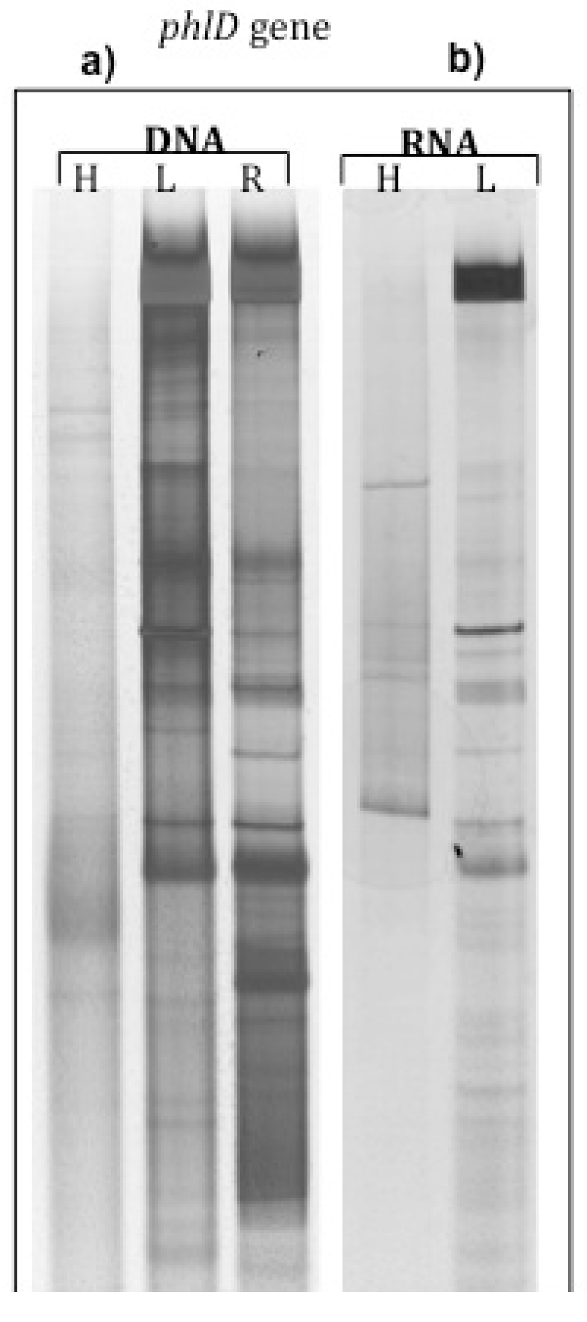
2.1. phlD Gene Expression in the Rhizosphere of A. Thaliana Grown Under Natural Conditions

Assimilation of 13C root exudates by soil microorganisms inhabiting A. thaliana rhizosphere following 27 days of 13CO2 labeling was evidenced by ∂13C measurements of fractions along the gradient obtained by isopycnic centrifugation of DNA and RNA derived from rhizosphere soil. The most 13C enriched DNA and RNA fractions corresponding to microbial communities involved in root exudates assimilation and the less enriched one corresponding to microbial communities involved in soil organic matter degradation (SOM) as well as root DNA and RNA were used to investigate the presence and expression of phlD gene in A. thaliana rhizosphere. PCR and RT-PCR products were analyzed by DGGE (Figure 1a,b).
phlD gene fragments were successfully amplified from heavy-, light- and root-DNA fractions and DGGE profiles from root DNA and light DNA fractions were more complex than those obtained from the heavy DNA fraction (Figure 1a,b). The expression of phlD gene was detected in 12C- and 13C- RNA from rhizosphere soil. However, no phlD transcript could be detected with cDNA from root RNA (Figure 1a). These data suggest a negative control by the plant on the production of the antifungal metabolite DAPG by the bacteria. Certain phlD bands from DNA and cDNA were identified by sequencing, and showed high homology to Pseudomonas species (Table 1), such as Pseudomonas sp., P. fluorescens and P. brassicacearum. This is compatible with what we know about the diversity of Pseudomonas species colonizing A. thaliana plant [15] and producing DAPG antibiotics [6].
Beneficial bacteria are known to produce many diverse, bioactive secondary metabolites, such as cyanide and 2,4-diacetylphloroglucinol (DAPG), which act to alter development of neighboring fungal species. Several studies have analyzed the genetic diversity of DAPG-producing rhizobacterial populations by isolating them from different plant roots, including maize [8], wheat, sugar beet, potato, and lily [9]. However, to our knowledge, this is the first attempt to analyze the genetic diversity, at the transcriptional level, of DAPG-producing Pseudomonas spp. in situ using DNA- and mRNA-SIP approaches.
Bacteria containing phlD gene were located in the three studied compartments, however the expression of this gene was evidenced only in the rhizosphere soil of A. thaliana.
The expression of phlD gene seemed repressed on the roots of A. thaliana. It is worth noting, to remind, that phlD is involved in DAPG synthesis and that the latter is not only toxic toward bacteria, fungi, and nematodes, but also toward plants [16]. At concentrations reported to exist in situ, Brazelton et al. [17] observed that DAPG caused drastic changes in root physiology and morphology of tomato plants. These changes included the development of brown and shriveled root tips and swollen root zones, inhibition of primary root growth, and stimulation of root branching. They indicated also that at least some phlD + Pseudomonas strains may be able to produce sufficiently high DAPG concentrations near root tips to alter root development, suggesting that DAPG’s effects on plant roots may be universal [17]. The plant may repress the expression of phlD by producing certain metabolites such as salicylic acid. Indeed, de Werra et al., [12] tested the impact of 63 different low-molecular-weight compounds, most of them plant derived, on the in vitro expression of phlA gene involved in DAPG biosynthesis by P. fluorescens CHA0 and demonstrated that some of them repress phlA expression such as salicylate, jasmonate, and methyl jasmonate, whereas, the plant hormone indole-3-acetic acid, for example, induced the expression of this antifungal gene.
Bacterial metabolites such as salicylate, pyoluteorin, and fungal metabolites such as fusaric acid, produced by the phytopathogenic fungus Fusarium oxysporum, were also shown to strongly repress DAPG production in P. fluorescens CHA0 [18,19]. In all likelihood, the signals to downregulate phlD gene expression may either be of microbial origin or plant origin.
Schnider-Keel et al. [19] showed that autoinduction of 2,4-DAPG biosynthesis can be countered by certain bacterial (and fungal) metabolites. This mechanism, which depends on phlF repression function, is strongly improved by the bacterial extracellular metabolites, such as salicylate [19].

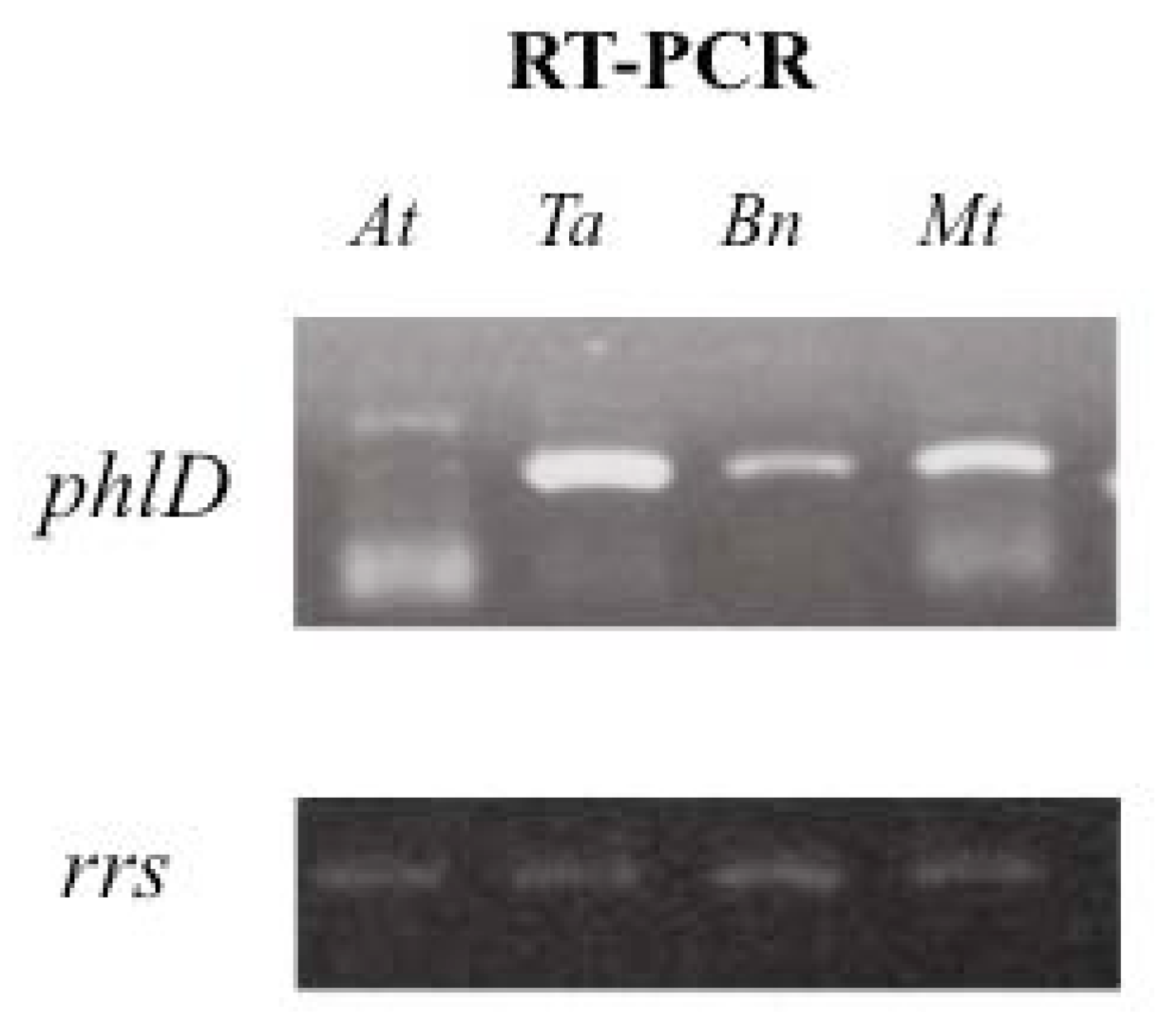
2.2. Impact of Root Exudates of Different Plant Species on phlD Gene Expression with Pseudomonas brassicacearum

P. brassicacearum has been described as the major root-associated bacteria of A. thaliana and Brassica napus plants [20,21,22]. It has the ability to suppress plant pathogens [23] by producing antifungal compounds, such as 2,4-diacetylphloroglucinol and cyanide.
We used the strain NFM421 to analyze the expression of phlD gene under the influence of root exudates of four plant species, A. thaliana, B. napus, Triticum aestivum and Medicago truncatula. This study was conducted under controlled monoxenic conditions, where three-weeks old seedling and bacteria interacted for 24 h. A high expression level was observed on the root system of wheat and M. truncatula and to a lesser extent on that of B. napus, while no expression was noticed on the root system of A. thaliana (Figure 2).
Phillips et al. [24] reported that DAPG might be considered as a potential enhancer of root exudation as it triggers amino-acids exudation by plants, and diminishes their influx by plants, suggesting, hence, that certain bacteria are able to manipulate plant exudates level to favor their proliferation in the rhizosphere. DAPG producing Pseudomonads may potentially increase their access to plant carbon resources. P. brassicacearum phlD gene expression was highly activated in the rhizosphere of wheat. It is worth mentioning that Phillips et al [24] demonstrated that wheat was not very sensitive to DAPG, in addition the DAPG can play a key role in the suppression of important root diseases such as take-all of wheat caused by Gaeumannomyces graminis var. tritici [25,26]. The high discrepancy in the expression level in the four plant species rhizosphere suggests that certain plant species may have developed strategies to shut down the expression of phl operon to preclude the deleterious effect of DAPG.
Each plant species may have developed strategies to balance the cost and benefits of interaction with soil bacteria. Thus, optimization of plant bacteria interaction during evolution may occur in coordination with that of plant development. In the absence of pathogens, the maintenance of a manipulating system through DAPG production by bacteria is costly for the plant because metabolic resources are limited. For this reason, plants may have evolved a system to inactivate the expression of certain bacterial phytobeneficial genes, when not required.
Table 1. Phylogenetic affiliation of phlD sequences corresponding to Denaturing Gradient Gel Electrophoresis (DGGE) prominent bands retrieved from root DNA (R-DNA) and RNA (R-RNA), heavy and light DNA fractions and 12C- and 13C-RNA fractions. No amplification was detected from RNA extracted from root system. Accession numbers for the BlastN matches are indicated in brackets.
Rhizosphere soilRoot DNARhizosphere soil
Light-DNAHeavy-DNA12C-RNA13C-RNA
-
Pseudomonas sp. K93.31 [AF396852]
-
Pseudomonas fluorescens strain 11-18 [AF396848]
-
Pseudomonas fluorescens strain 19-41 [AF396852]
-
Pseudomonas sp. S7-42 [EF554345]
-
Pseudomonas fluorescens strain 19-7 [AF396850]
-
Pseudomonas brassicacearum [CP002585]
-
Pseudomonas sp. K93.31 [AF396852]
-
Pseudomonas fluorescens strain 11-18 [AF396848]
-
Pseudomonas sp. K93.31 [AF396852]
-
Pseudomonas fluorescens strain 3-1 [AF396845]
-
Pseudomonas sp. C6-9 [EF554343]
-
Pseudomonas sp. K93.31 [AF396852]
-
Pseudomonas sp. K93.31 [AF396852]
Figure 1. DGGE patterns of phlD gene fragments amplified from (a) heavy, light, and root DNA fractions and (b) 12C- & 13C-RNA fractions retrieved from the rhizosphere of A. thaliana after 27 days of 13CO2 labeling.
Agronomy 03 00621 g001
Figure 2. Modulation of phlD gene expression of Pseudomonas brassicacearum NFM421 inoculated in vitro in the rhizosphere of A. thaliana (At), M. truncatula (Mt), B. napus (Bn) and T. astivum (Ta). rrs gene was amplified to check that the same amount of C-DNA was used to amplified phlD gene.
Agronomy 03 00621 g002

3. Experimental Section

3.1. Plant Growth & 13C Labeling

The experiment was carried out in the laboratory with A. thaliana (ecotype Columbia) on Eutric cambisol soil [27]. Forty grams of dry weight soil was then placed into polypropylene cylinders. Seeds of A. thaliana were sterilized according to Achouak et al. [15] and germinated on half-strength Hoagland medium and 0.8% phytagel (Sigma, St Louis) plates at 25 °C for 48 h. After germination, one seedling was planted per pot. Plants were grown in triplicate in a growth chamber (developed and managed by Group of applied Research in Phytotechnologies (GRAP), CEA Cadarache, France) as previously described [3]. Triplicate cylinders containing unplanted soil (bulk soil treatment) were also incubated under the same conditions.
Continuous labeling started one week after seeds were sown in pots, and A. thaliana plants and soil microcosms (bulk soil) were harvested in triplicate after 27 days of 13C labeling according to Haichar et al. [2].

3.2. DNA Extraction and Gradient Fractionation

At the end of labeling, A. thaliana roots system were manually and carefully separated from the rhizosphere soil fraction and root fractions, and rhizosphere soil fractions were frozen in liquid N2 and stored at −80 °C. We considered the rhizosphere soil as the soil adhering to the root system after manual shaking. DNA was extracted from 5 g rhizosphere soil and from 500 mg of root system from each plant, as described by Haichar et al. [2]. DNA extracted from the rhizosphere soil was fractionated by CsCl equilibrium density gradient centrifugation [28]. Nucleic acids were purified from CsCl salts using Geneclean Kit (Qbiogene, Montreal, QC) and for each gradient, one fraction representative of 13C-labeled (“heavy”, fraction number 10) DNA and one fraction representative of unlabeled (“light”, fraction number 5) DNA were chosen based on δ13C and density measurements according to previous studies in the laboratory [2,29].

3.3. RNA Extraction and CsTFA Centrifugation

RNA was extracted from the rhizosphere soil and root tissues, and treated to remove DNA traces according to Haichar et al. [3]. The quality of RNA was checked by agarose gel electrophoresis and measured by Nanodrop instrument. Two micrograms of RNA from each rhizosphere soil and at each date was fractionated by cesium trifluoroacetate (CsTFA) equilibrium density gradient centrifugation [30]. For each gradient, one fraction representative of 13C-labeled RNA and one fraction representative of unlabeled 12C-RNA were chosen according to Rangel-Castro et al. [30]. Nucleic acids were purified from CsTFA salts by isopropanol precipitation and RNA pellets were air-dried and resuspended in 20 µL of RNase-free sterile water.

3.4. PCR and RT-PCR Amplification of phlD Gene

PCR amplification of phlD gene was performed from heavy, light and root DNA fractions using the primers 5’-CCAGTTGCAGGACCAGTTCATC-3’ and 5’-TGCTATCAACCCCAGGACACC-3’ developed by Bergsma-Vlami et al. [14] and targeting DAPG producing Pseudomonads in the plant rhizosphere.
Reverse transcription of RNA to complementary DNA (cDNA) was performed according to Rangel-Castro et al. [30]. Five microliters of RT-PCR product from 12C-, 13C- and root RNA was used as template to amplify phlD gene with the same primers used for PCR amplification from DNA.

3.5. Denaturing Gradient Gel Electrophoresis (DGGE) Fingerprinting and the Recovery of DNA Template from DGGE Bands

DGGE analysis of PCR products from root DNA and RNA, light and heavy DNA, and RNA fractions was carried out using the Dcode Universal Mutation Detection System (BIO-Rad Laboratories, France) according to Haichar et al. [29]. Following electrophoresis, the gels were silver-stained and scanned [31].
Bands of interest in heavy and light DNA and RNA fractions, and DNA and RNA root fractions were excised from DGGE gels and DNA was eluted, amplified, and purified according to Haichar et al. [29]. The correct migration positions of PCR products of purified bands were confirmed by DGGE analysis of these products and environmental PCR products on the same gel.

3.6. Sequencing and Phylogenetic Analysis

PCR products were sequenced at Genome express (Meylan, France) and sequences were analyzed by the BLASTN search tool [32] to determine sequence homology and to search for similar sequences in the GenBank database.
Sequences of phlD gene obtained form A. thaliana rhizosphere were deposited in GenBank under the accession numbers: GQ323767 to GQ323773.

3.7. Seeds Sterilization and Plant Growth in vitro

A. thaliana (ecotype Columbia), B. napus, and Medicago truncatula seeds were surface sterilized according to Achouak et al. [15]. Following sterilization, seeds were washed three times in ethanol 95% and air-dried. Triticum aestivum seeds were sterilized in saturated Ca hypochlorite solution for 2 h under vacuum, extensively washed with sterilized ultrapure water, incubated in 3% hydrogen peroxide for 20 min under vacuum, then extensively washed with sterilized ultrapure water. A. thaliana, T. aestivum, B. napus, and M. truncatula were routinely grown in 12 × 12 cm square petri dishes containing half strength Hoagland medium solidified with 0.8% gelrite (Duchefa, Haarlem, NL). Plant growth was routinely done in a phytotron under alternating 14 h of day light at 21 °C (at 100 mmol m−2 s−1 of photon) and 10 h of night at 18 °C, as described by Achouak et al. [15]. After three weeks of growth, plants were inoculated with a bacterial suspension of P. brassicacearum (107 cfu per seedling) and incubated for 24 h. Bacteria were collected from growth medium and from roots by vortexing, pelleted, and then RNA was extracted according to manufacturer instruction (Qiagen RNasy kit, France).

3.8. RT-PCR Amplification of phlD Gene

RNA was extracted using the “RNAprotect Bacteria Reagent” and “RNeasy Mini Kit” (Qiagen). RT-PCR assays were done using the “Transcriptor First Strand cDNA Synthesis Kit” (Roche).
RT-PCR amplification of phlD gene was performed by using the following primers phlD-F (5’-ctccatcatcggtgcgacatc-3’), and phlD-R (5’-catatcagccgcgttattcgg-3’) targeting phlD gene from P. brassicacearum NFM421.

4. Conclusions

Given the richness of bacterial species interacting in the rhizosphere and competing for the same resources, it is likely that communication through chemical exchanges may occur.
These data highlight the multifarious regulatory cascades that mediate cross talks between microorganisms and plants that may occur in complex environments such as the rhizosphere.
The coevolution of interacting plants and microbes has given rise to a diverse array of exchanged signals and responses. Plants have evolved to select certain bacterial populations from the reservoir, the soil and probably also to optimize their costs relative to their benefits by adjusting functions of coevolving populations.

Acknowledgments

We thank the Group of Applied Research in Phytotechnologies (GRAP) of the iBEB/SBVME in CEA Cadarache for labeling facilities. This work was supported by a CNRS PhD grant.

Conflicts of Interest

The authors declare no conflict of interest.

References

  1. Bais, H.P.; Weir, T.L.; Perry, L.G.; Gilroy, S.; Vivanco, J.M. The role of root exudates in rhizosphere interactions with plants and other organisms. Annu. Rev. Plant Biol. 2006, 57, 233–266. [Google Scholar] [CrossRef]
  2. Haichar, F.Z.; Roncato, M.A.; Achouak, W. Stable isotope probing of bacterial community structure and gene expression in the rhizosphere of Arabidopsis thaliana. FEMS Microb. Ecol. 2012, 81, 291–302. [Google Scholar] [CrossRef]
  3. Haichar, F.Z.; Marol, C.; Berge, O.; Rangel-Castro, J.; Prosser, J.I.; Balesdent, J.; Heulin, T.; Achouak, W. Plant host habitat and root exudates shape soil bacterial community structure. ISME J. 2008, 2, 1221–1230. [Google Scholar] [CrossRef]
  4. Jousset, A.; Rochat, L.; Lanoue, A.; Bonkowski, M.; Keel, C.; Scheu, S. Plants respond to pathogen infection by enhancing the antifungal gene expression of root-associated bacteria. MPMI. 2011, 24, 352–358. [Google Scholar] [CrossRef]
  5. Haas, D.; Défago, G. Biological control of soil-borne pathogens by Fluorescent pseudomonads. Nat. Rev. Microbiol. 2005, 3, 307–319. [Google Scholar] [CrossRef]
  6. Almario, J.; Moenne-Loccoz, Y.; Muller, D. Monitoring of the relation between 2,4-diacetylphloroglucinol-producing Pseudomonas and Thielaviopsis basicola populations by real-time PCR in tobacco black root-rot suppressive and conducive soils. Soil Biol. Biochem. 2012, 57, 44–155. [Google Scholar]
  7. Achouak, W.; Sutra, L.; Heulin, T.; Meyer, J.-M.; Fromin, N.; Degraeve, S.; Christen, R; Gardan, L. Description of Pseudomonas brassicacearum sp. nov. and Pseudomonas thivervalensis sp. nov., two root-associated bacteria isolated from Arabidopsis thaliana and Brassica napus. Int. J. Syst. Evol. Microbiol. 2000, 50, 9–18. [Google Scholar] [CrossRef]
  8. Persello-Cartieaux, F.; David, P.; Sarrobert, C.; Thibaud, MC.; Achouak, W.; Robaglia, C.; Nussaume, L. Utilization of mutants to analyze the interaction between Arabidopsis thaliana and its naturally root-associated Pseudomonas. Planta 2001, 212, 190–198. [Google Scholar] [CrossRef]
  9. Lalaouna, D.; Fochesato, S.; Sanchez, L.; Schmitt-Kopplin, P.; Haas, D.; Heulin, T.; Achouak, W. Phenotypic switching involves GacS/GacAdependent Rsm small RNAs in Pseudomonas brassicacearum. Appl. Environ. Microbiol. 2012, 78, 1658–1665. [Google Scholar] [CrossRef]
  10. Haas, D.; Keel, C. Regulation of antibiotic production in root-colonizing Pseudomonas spp. and relevance for biological control of plant disease. Annu. Rev. Phytopathol. 2003, 41, 117–153. [Google Scholar] [CrossRef]
  11. Laville, J.; Voisard, C.; Keel, C.; Mauhofer, M.; Défago, G.; Haas, D. Global control in Pseudomonas fluorescens mediating antibiotic synthesis and suppression of black root rot of tobacco. Proc. Natl. Acad. Sci. USA. 1992, 89, 1562–1566. [Google Scholar]
  12. de Werra, P.; Huser, A.; Tabacchi, R.; Keel, C.; Maurhofer, M. Plant- and microbe-derived compounds affect the expression of genes encoding antifungal compounds in a pseudomonad with biocontrol activity. Appl. Environ. Microbiol. 2011, 77, 2807–2812. [Google Scholar] [CrossRef]
  13. Picard, C.; Di Cello, F.; Ventura, M.; Fani, R.; Guckert, A. Frequency and biodiversity of 2,4-diacetylphloroglucinol producing bacteria isolated from the maize rhizosphere at different stages of plant growth. Appl. Environ. Microbiol. 2000, 66, 948–955. [Google Scholar] [CrossRef]
  14. Bergsma-Vlami, M.; Prins, M.E.; Raaijmakers, J.M. Influence of plant species on population dynamics, genotypic diversity and antibiotic production in the rhizosphere by indigenous Pseudomonas spp. FEMS Microbiol. Ecol. 2005, 52, 59–69. [Google Scholar] [CrossRef]
  15. Achouak, W.; Conrod, S.; Cohen, V.; Heulin, T. Phenotypic variation of Pseudomonas brassicacearum as a plant root-colonisation strategy. MPMI. 2004, 17, 872–879. [Google Scholar] [CrossRef]
  16. Keel, C.; Schnider, U.; Maurhofer, M.; Voisard, C.; Laville, J.; Burger, P.; Wirthner, P.; Haas, D.; Défago, G. Suppression of root diseases by Pseudomonas fluorescens CHA0, importance of the bacterial secondary metabolite 2,4-diacetylphloroglucinol. MPMI. 1992, 5, 4–13. [Google Scholar] [CrossRef]
  17. Brazelton, J.N.; Pfeufer, E.E.; Sweat, T.A.; Gardener, B.B.; Coenen, C. 2,4-Diacetylphloroglucinol alters plant root development. Mol. Plant-Microb. Interact. 2008, 21, 1349–1358. [Google Scholar] [CrossRef]
  18. Duffy, B.K.; Défago, G. Zinc improves biocontrol of Fusarium crown and root rot of tomato by Pseudomonas fluorescens and represses the production of pathogen metabolites inhibitory to bacterial antibiotic biosynthesis. Phytopathology 1997, 87, 1250–1257. [Google Scholar] [CrossRef]
  19. Schnider-Keel, U.; Seematter, A.; Maurhofer, M.; Blumer, C.; Duffy, B.; Gigot-Bonnefoy, C.; Reimmann, C.; Notz, R.; Défago, G.; Haas, D.; Keel, C. Autoinduction of 2,4-diacetylphloroglucinol biosynthesis in the biocontrol agent Pseudomonas fluorescens CHA0 and repression by the bacterial metabolites salicylate and pyoluteorin. J. Bact. 2000, 182, 1215–1225. [Google Scholar] [CrossRef]
  20. Chabeaud, P.; de Groot, A.; Bitter, W.; Tommassen, J.; Heulin, T.; Achouak, W. Phase-variable expression of an operon encoding extracellular alkaline protease, serine protease homologue and lipase in Pseudomonas brassicacearum. J. Bacteriol. 2001, 183, 2117–2120. [Google Scholar] [CrossRef]
  21. Fromin, N.; Achouak, W.; Thiery, J.M.; Heulin, T. The genotypic diversity of Pseudomonas brassicacearum populations isolated from roots of Arabidopsis thaliana, influence of plant genotype. FEMS Microbiol. Ecol. 2001, 37, 21–29. [Google Scholar] [CrossRef]
  22. Ross, I.; Alami, Y.; Harvey, P.R.; Achouak, W.; Ryder, M. Genetic diversity and biological control activity of novel species related to pseudomonads isolated from wheat field soils in South Australia. Appl. Environ. Microbiol. 2000, 66, 1609–1616. [Google Scholar] [CrossRef]
  23. Weller, D.M.; Mavrodi, D.V.; van Pelt, J.A.; Pieterse, C.M.J.; van Loon, L.C.; Bakker, P.A.H.M. Induced systemic resistance in Arabidopsis thaliana against Pseudomonas syringae pv. tomato by 2,4-diacetylphloroglucinolproducing Pseudomonas fluorescens. Phytopathology. 2012, 102, 403–412. [Google Scholar]
  24. Phillips, D.A.; Fox, T.C.; King, M.D.; Bhuvaneswari, T.V.; Teuber, L.R. Microbial products trigger amino acid exudation from plant roots. Plant Physiol. 2004, 136, 2887–2894. [Google Scholar] [CrossRef]
  25. Raaijmakers, J.M.; Weller, D.M. Natural plant protection by 2,4-diacetylphloroglucinol-producing Pseudomonas spp. in take-all decline soils. MPMI. 1998, 11, 144–152. [Google Scholar] [CrossRef]
  26. De Souza, J.T.; Raaijmakers, J.M. Polymorphisms within the prnD and pltC genes from pyrrolnitrin and pyoluteorin-producing Pseudomonas and Burkholderia spp. FEMS Microbiol. Ecol. 2003, 43, 21–34. [Google Scholar]
  27. Derrien, D.; Marol, C.; Balesdent, J. The dynamics of neutral sugars in the rhizosphere of wheat, an approach by 13C pulse-labelling and GC/C/IRMS. Plant Soil 2004, 267, 243–253. [Google Scholar] [CrossRef]
  28. Lueders, T.; Manefield, M.; Friedrich, M.W. Enhanced sensitivity of DNA-and rRNA-based stable isotope probing by fractionation and quantitative analysis of isopycnic centrifugation gradients. Environ. Microbiol. 2004, 6, 73–78. [Google Scholar]
  29. Haichar, F.Z.; Achouak, W.; Christen, R.; Heulin, T.; Marol, C.; Marais., M.F.; Mougel, C.; Ranjard, L.; Balesdent, J.; Berge, O. Identification of cellulolytic bacteria in soil by stable isotope probing. Environ. Microbiol. 2007, 9, 625–634. [Google Scholar] [CrossRef]
  30. Rangel-Castro, J.I.; Killham, K.; Ostle, N.; Nicol, G.W.; Anderson, I.C.; Scrimgeour, C.M.; Killham, K.; Meharg, A.A. Stable isotope probing analysis of the influence of liming on root exudates utilization by soil microorganisms. Environ. Microbiol. 2005, 7, 828–838. [Google Scholar] [CrossRef]
  31. McCaig, A.E.; Glover, L.A.; Prosser, J.I. Numerical analysis of grassland bacterial community structure under different land management regimes by using 16S ribosomal DNA sequence data and denaturing gradient gel electrophoresis banding patterns. Appl. Environ. Microbiol. 2001, 67, 4554–4559. [Google Scholar] [CrossRef]
  32. Altschul, S.F.; Gish, W.; Miller, W.; Myers, W.; Lipman, D.J. Basic local alignment search tool. J. Mol. Biol. 1990, 215, 403–410. [Google Scholar]

