Article

Brassinosteroids Regulate Antioxidant System and Protect Chloroplast Ultrastructure of Autotoxicity-Stressed Cucumber (Cucumis sativus L.) Seedlings

1
College of Life Science and Technology, Honghe University, Mengzi 661100, China
2
Department of Horticulture, College of Agriculture, University of Sargodha, Sargodha Punjab 40100, Pakistan
*
Author to whom correspondence should be addressed.
Submission received: 21 March 2019 / Revised: 19 May 2019 / Accepted: 21 May 2019 / Published: 26 May 2019
(This article belongs to the Special Issue Functional Metabolism in Crops/Agronomy)

Abstract

:
Autotoxicity is a common problem being faced in protected vegetable cultivation system. Phytoremediation of plant autotoxicity is an emerging concept to minimize deterioration of soil environment and reduction of yield and quality of vegetable crops. Brassinosteroids (BRs) have been reported as a potential phytohormone to assist phytoremediation. However, the effects of BRs-induced autotoxicity stress on plant growth, photosynthesis and antioxidant defense system are poorly understood. Hence, we focused on the changes in physiological characteristics and ultrastructure of cucumber leaves in response to the application of 24-epibrassinolide (EBR) under autotoxicity stress conditions. The results showed that leaf area, plant height, fresh weight and dry weight of cucumber were obviously decreased under autotoxicity stress conditions. EBR application obviously improved the phenotypic characteristics of cucumber seedlings. Chlorophyll content, net photosynthetic rate, stomatal conductance and transpiration rate of cucumber leaves were markedly reduced under autotoxicity stress conditions. Application of EBR improved the photosynthetic pigments (chlorophyll a by 15.80%, chlorophyll b by 18.70% and total chlorophyll content by 17.30%), net photosynthetic rate by 36.40% and stomatal opening of leaves under autotoxicity stress conditions. EBR application also maintained the integrity of chloroplast and thylakoid structures under autotoxicity stress conditions. The activity of catalase (CAT), peroxidase (POD) and ascorbate peroxidase (APX) and antioxidative compounds ascorbate (AsA) and reduced glutathione (GSH) contents were markedly decreased, however, these were obviously increased after EBR application under autotoxicity stress. EBR application also increased the soluble sugar and protein, and proline concentration by 59.70%, 7.22% and 36.58%, respectively in the leaves of cucumber, decreased malondialdehyde by 24.13% and reactive oxygen species contents (H2O2 by 35.17%, O2 by 12.01% and OH by 16.59%), and reduced the relative permeability of the cell membrane by 14.31%. These findings suggest that EBR application enhanced the photosynthetic capacity of leaves, maintained the integrity of chloroplast and thylakoid structures, and effectively alleviated the damage of membrane caused by lipid peroxidation and root damage under autotoxicity stress conditions. The growth inhibition effect of autotoxicity stress on cucumber was reduced by EBR application.

1. Introduction

The rapid developments in protected cultivation and intensive cropping pattern have led to an increased index of multiple cropping [1,2]. The continuous cropping results in the deterioration of soil environment, serious diseases and pests attack on vegetables, and reduction of yield and quality of vegetable crops [3,4]. Continuous cropping has become the main factor limiting vegetable’s production [5]. If the same type of vegetable is cultivated continuously, it affects the species and quantity of soil microbes. The root system of the vegetable secrete the same substances (roots exudates) for a long time that may affect microbial diversity and microbial population and increase the incidence of soil-borne diseases [6]. Autotoxicity is a common problem being faced in protected vegetable cultivation system because after harvesting the roots remains in the soil [7]. A range of secondary metabolites in the residual root of vegetables, such as cinnamic acid (CA), flavones and terpenoids are the potential autotoxins [8,9].
Cucumber (Cucumis sativus L.) is one of the main vegetable cultivated at a commercial scale across the World. Likewise, cucumber is widely cultivated in solar greenhouses and plastic greenhouses in northern China [10]. Principally, farmers annually grow the same crop for years to maximize economic return [4,11,12]. Continuous cultivation of cucumber on the same piece of land is a major hindrance for the further development of cucumber industry [12]. Grafting is an important technique in a modern vegetable production system because it provides resistance against soil-borne diseases leading towards increased yield [13,14,15]. At present, grafting is widely used to alleviate the obstacles caused by continuous cropping of cucumber, but the production of grafted transplants takes time, resources, and it is labor intensive [16,17]. Additionally, the use of rootstock during the process of grafting has certain negative impacts on fruit shape, flavor, and quality of cucumber [18]. Thus, there is a need to find a simple and labor-saving method that protects cucumber plants from stress without altering the flavor and quality of the cucumber.
Brassinosteroids (BRs) widely exist in plant species and are involved in the regulation of plant growth and development [19]. Brassinosteroids are involved in plant metabolism; promote cell division and cell elongation, and key physiological processes, such as photosynthesis and antioxidant system leading towards improved plant growth and development [20,21,22,23]. BRs maintain a high level of chlorophyll content and improve the photosynthetic performance of plants [24]. BRs enhance the activity of protective enzymes in plants and help reduce oxidative damage caused by reactive oxygen species (ROS) thereby improving resistance against disease and other kinds of environmental stresses [25,26]. Additionally, BRs promote water absorption capacity of plants under drought stress, maintain the stability of the cell membrane, regulate physiological and biochemical process within cells, and improve the yield and quality of crops [27]. According to a report, the application of BRs alleviates hypoxia stress on mitochondria and ultrastructure of melon roots [28].
Liu et al. [29] found that 24-epibrassinolide (EBR) promotes the growth of bamboo by improving the activity of antioxidants, enhancing the antioxidant capacity of plants, and increasing the proline and soluble protein contents. BRs also alleviate membrane damage caused by lipid peroxidation under low temperature stress conditions.
The alternate techniques to improve stress tolerance of plants, such as the use of BRs are getting attention plant biologist [21]. BRs play an important role in hormonal homeostasis particularly under stress conditions [20]. Cinnamic acid (CA) is a major autotoxin secreted from the roots in cucumber plant. CA has an inhibitory effect on the regulation of plant morphogenesis and development [30]. The information regarding the physiological changes in cucumber under autotoxicity stress conditions, particularly in the antioxidant system and photosynthetic characteristics is limited. Here, we hypothesized that EBR alleviates the CA-induced stress by improving photosynthesis and strengthening antioxidant systems. The findings reported in this study may provide a theoretical basis for BRs as a potential phytohormone that can be utilized to alleviate autotoxicity stress of cucumber.

2. Materials and Methods

EBR was purchased from Sigma-Aldrich (St.Louis, MO, USA), and dissolved in a small quantity of ethanol and then the final volume was made by adding distilled water. The seeds of cucumber cultivar Changlu No. 1 were soaked in water at 28–30 °C for 6 h and then placed in an incubator for germination. After germination, the seeds were planted in seeding trays and the trays were placed in a growth chamber. The temperature of growth chamber was set at 28 ± 1 °C for day and the night temperature was maintained at 18 ± 1 °C having a 12 h photoperiod (500 μmol m−2 s−1) and relative humidity was adjusted to 70–80%. After the second leaf emerged, uniform seedlings were transferred to a container filled with half-strength Hoagland solution. The plants were kept in the growth chamber for seven days. Plants were exposed to four treatments: 1/2 Hoagland’s nutrient solution as the control group (C); application of 0.l μM EBR in 1/2 Hoagland’s nutrient solution (C + EBR); application of 100 mM cinnamic acid in 1/2 Hoagland’s nutrient solution (CA); application of 100 mM cinnamic acid and 0.1 μM EBR in 1/2 Hoagland’s nutrient solution (CA + EBR). In this study, 27 plants were used for every treatment. The concentration of CA and EBR were determined according to the procedure described in our previous studies [31]. The nutrient solution was replaced every three days to avoid deficiency of any specific ion.

2.1. Determination of Morphological Characteristics

Three cucumber seedlings were selected from each treatment; ruler and vernier caliper were used to measure the plant height and stem diameter, respectively. The leaf area was assessed by using leaf area meter. Second true leaf from the top was selected to measure leaf area. The data for all these parameters were measured after seven days of treatment.

2.2. Measurement of Biomass

Three uniform cucumber seedlings were harvested from all treatments. Plants were washed with clear water and surface dried by an absorbent paper, the fresh weight of shoot (above ground part of the plant) and root (underground part of the plant) was measured by an electric balance. The samples were placed in paper bags, labeled and dried in an oven initially at 105 °C for 15 min and then at 80 °C, until the samples maintained a constant dry weight. The dry weight of shoot and root was measured by electronic balance. The data were measured after seven days of treatment.

2.3. Determination of Root Morphology and Root Activity

The root length from root tip to rhizome junction was measured with a ruler and the root morphology analysis was performed using root scanner (Epson Expression 1180XL, Seiko Epson Corp., Nagano-ken, Japan) and WinRHIZO root analysis system (Regent Instruments, Quebec, Canada). The root activity of cucumber seedlings was determined by TTC chloride method with 0.5 g fresh sample after 7 days of treatment.

2.4. Determination of Photosynthetic Pigments and Observation of Stomatal Opening

The acetone (80%) extraction method was used to determine the photosynthetic pigments [32]. The stomatal opening of leaves was observed using a light microscope at 40× magnification.

2.5. Determination of Gas Exchange Parameters

The net photosynthetic rate (Pn), stomatal conductance (Gs), transpiration rate (Tr) and intercellular CO2 concentration (Ci) of the cucumber leaves were determined by using a portable photosynthesis analysis system (Li-6400, Li-COR Company, Lincoln, NE, USA) on a sunny day at 09:00 to 11:00. The temperature of the leaf chamber was controlled at 25 ± 1 °C, PPFD was 600 μmol m−2 s−1, and the CO2 concentration was adjusted to 360 ± 10 μmol mol−1.

2.6. Ultrastructure of Chloroplast and Thylakoid

The ultrastructure of chloroplast and thylakoid of cucumber leaves were observed using transmission electron microscope (TEM-100CX, JEOL Company, Tokyo, Japan) at an accelerating voltage of 75 kV as explained by Zeng et al. [33]. The cucumber leaf was cut into 1 mm × 5 mm pieces in the midsection on both sides of the mid rib of the leaf, and the cut rectangular pieces were immersed into the 4% glutaraldehyde at 4 °C and fixed for 12 h. After washing with phosphoric acid buffer, the samples were fixed with 1% osmic acid at 4 °C for 6 h. Then, the samples were dehydrated by ethanol with different concentration gradients and then immersed and embedded in epoxy resin. The samples were cut into 1 μm slices by an ultrathin microtome. The sections were stained with uranyl acetate and lead citrate and then observed and photographed using a transmission electron microscope.

2.7. ROS and Lipid Peroxidation Detection and Quantification

The stained images of H2O2 and O2 were taken according to the procedure described by Ma et al. [34]. For H2O2, the cucumber leaves were infiltrated with 0.1 mg mL−1 DAB working solution in 50 mM Tris-acetate (pH 3.8) under dark conditions and incubated for 24 h at 25 °C. Then the leaves were faded with the 80% ethanol for 15 min at 90 °C. Finally, the cucumber leaves were mounted with a mixture of ethanol/acetic acid/glycerol (3:1:1, v/v) at 4 °C and photographed. For O2, the cucumber leaves were infiltrated with 0.1 mg mL−1 NBT working solution in 25 mM K-HEPES buffer (pH 7.8) under dark conditions and incubated for 2 h at 25 °C. The steps for fading and mounting were the same as described for H2O2. Schiff’s reagent was utilized for histochemical detection and quantification of aldehydes produced from the lipid peroxide that was the malondialdehyde (MDA). Their contents were quantified using spectrophotometer by a peroxidase-coupled assay. The chopped leaves in ice bath were ground with 0.1% trichloroacetic acid (TCA). The homogenates were centrifuged at 12,000× g for 15 min at 4 °C. The centrifugal supernatant with 0.1 M phosphate buffer solution (PBS) (pH 7.0) was placed in darkness for 1 h and then determined by spectrophotometer at 390 nm to calculate the concentration of H2O2 in the leaves. The O2 production rate (nitrite formation from hydroxylamine) was monitored in the presence of O2 and calculated as a standard curve with NO2. For the quantification of OH, the samples were homogenized in 50 mM PBS buffer (pH 7.0) and centrifuged at 10,000× g for 10 min at 4 °C. The supernatant with 2.5 mM 2-deoxyribose was developed at 35 °C in the dark for 1 h. The mixture with 1% (w/v) thiobarbituric acid (TBA) and acetic acid was boiled for 30 min and immediately cooled for 10 min on ice, and then absorbance was measured at 532 nm. MDA content was measured as an end product of lipid peroxidation via 2-thiobarbituric acid (TBA) reaction. The absorbance of the red adduct was recorded at 450, 532, and 600 nm to calculate the concentration of MDA in the leaves.

2.8. Activity of Antioxidant Enzymes and Contents of Proline, Soluble Sugar, and Protein

The antioxidant enzyme activities of superoxide dismutase (SOD), peroxidase (POD), catalase (CAT), and ascorbate peroxidase (APX) were measured using the procedure described by Li et al. [35]. To measure the antioxidant activities and contents, frozen leaf samples (0.5 g) were ground and homogenized using 5 mL ice-cold 25 mM HEPES buffer with a pH of 7.8 containing 0.2 mM ethylene diamine tetraacetic acid (EDTA), 2 mM AsA, and 2% polyvinylpolypyrrolidone at 4 °C. This homogenate was centrifuged at 4 °C at 12,000× g and supernatant was utilized for enzymes analysis and quantification. The SOD activity was measured by assessing its ability to inhibit the photochemical reduction of nitroblue tetrazolium (NBT). One unit of SOD was considered when it inhibited the photochemical reduction of NBT by 50%. For the measurement of POD activity, guaiacol method was followed, and one unit of POD activity was 0.01 increases per minute at 470 nm. The CAT activity was measured by adding H2O2, and immediately measuring the decrease rate of H2O2 at 240 nm. One unit of CAT activity was the amount of enzyme required to decompose 1 μmol H2O2 in 1 min. APX was measured by calculating the rate of decrease in absorbance at 290 nm, and one unit of APX activity was the reduction in AsA in 1 min.
AsA and GSH contents were measured from the leaf samples. Samples were ground and homogenized in a cold mortar placed on ice using 5% ice-cold meta-phosphoric acid. AsA was measured by grinding leaf samples in liquid nitrogen, and 1 gram of grounded powder was mixed with 600 µL of 6% trichloroacetic acid. Samples were then centrifuged at 12,000× g for 15 min at 4 °C. AsA was calculated using standard curve prepared according to the method described by Stevens et al. [36]. The GSH content was measured on the basis of reduction of 2-nitrobenzoic acid (DTNB) according to the method of Griffith [37].
After seven days of treatment, proline, soluble sugar and protein contents of the leaves were measured. Briefly, proline content was measured using 3% 5-sulphosalicylic acid as an extraction solution at room temperature according to the method of Bates et al. [38]. Soluble sugars were determined using anthrone colorimetry and estimated on fresh weight basis according to the method of Buysse and Merckx [39]. The soluble protein was measured on a fresh weight basis using the method described by Bradford [40].

2.9. Statistical Analysis

The data were subjected to ANOVA using SPSS statistical package (13.0, SPSS Institute Ltd, New York, NY, USA). The treatment means were compared by using Duncan’s multiple range test (p < 0.05). The data were expressed as means of three replicates (± standard error (SE) 3).

3. Results

3.1. Morphological Changes

No difference for morphological characteristics, such as plant height, stem diameter and leaf area of cucumber seedlings was observed with or without the exogenous EBR application under normal growth conditions. Application of EBR improved plant height and leaf area by 31.7% and 44.4%, respectively compared with CA treatment under autotoxicity stress conditions (Figure 1 and Table 1). The fresh weight and dry weight of shoot were improved by 32.90% and 30.10%, respectively in EBR treated plants compared with CA treatment. Fresh weight and dry weight of the root (underground part) of EBR-treated cucumber plants were higher compared with CA treatment; however, the differences were non-significant (Table 2). Under normal growth conditions, there was no significant difference in fresh and dry weight of aboveground and underground parts between the EBR-treated and non-EBR treated cucumber seedlings.

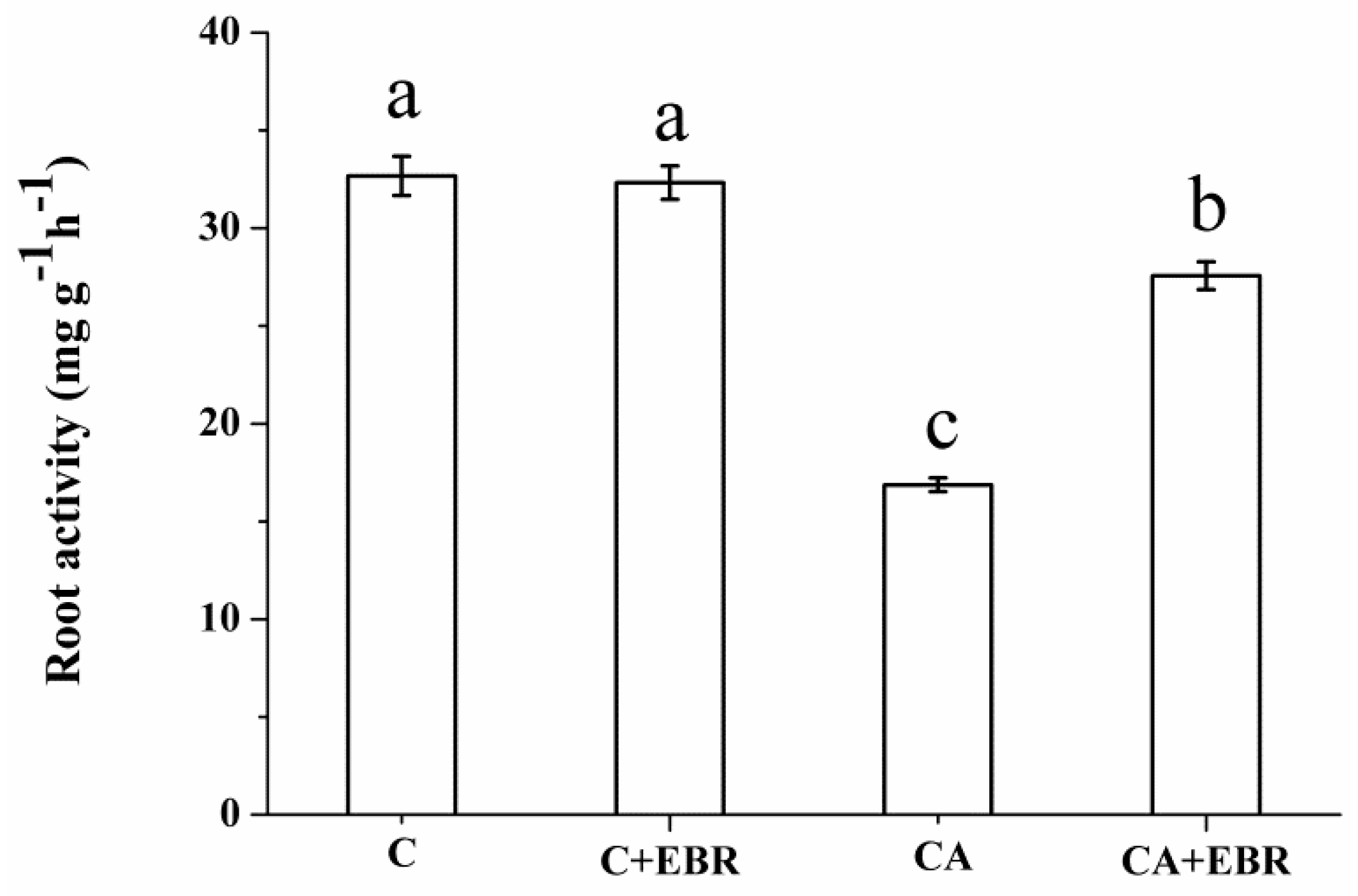
3.2. Root Morphology and Root Activity

Under normal growth conditions, no significant differences for root morphology (total root length, root surface area, root volume, and root tips) were observed between EBR-treated and non- EBR treated cucumber seedlings. The total root length and root surface area under CA + EBR treatment were increased by 41.50% and 34.20%, respectively, and the root volume and root tips were decreased compared with CA treatment (Figure 1, Table 3). Under normal growth conditions, the root activity of cucumber seedlings treated with EBR was not affected compared with control. The root activity in CA-treated plants was decreased by 45.80% compared with control. However, the application of EBR obviously increased the root activity (35.40%) compared with CA-treated cucumber plants under autotoxicity stress (Figure 2).

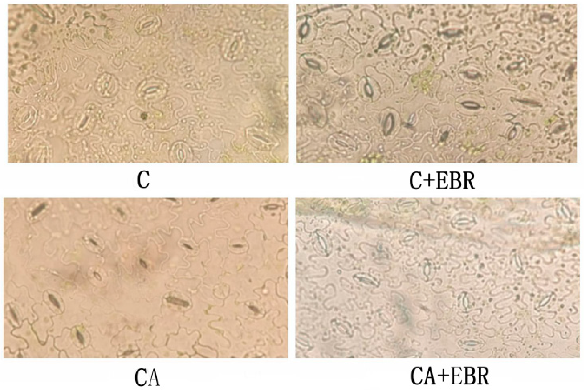
3.3. Photosynthetic Pigments and Stomatal Opening

EBR application improved the chlorophyll a (15.80%), chlorophyll b (18.70%) and total chlorophyll contents (17.30%) compared with non-EBR treated cucumber plants under autotoxicity stress conditions (Table 4). Under normal growth conditions, there was no significant difference regarding stomatal opening between the control and EBR-treated cucumber plants. The stomatal opening of cucumber leaves was obviously increased by the application of EBR compared with CA-treated plants under autotoxicity stress (Figure 3).

3.4. Gas Exchange

Under normal growth conditions, no difference was observed for photosynthetic parameters (Pn, Ci, Gs, and Tr) between the control and EBR-treated cucumber plants. However, exogenous EBR application obviously increased the Pn by 36.40% compared with CA-treated cucumber plants under autotoxicity stress. Ci, Gs and Tr were not affected by the application of EBR under autotoxicity stress conditions (Table 5).

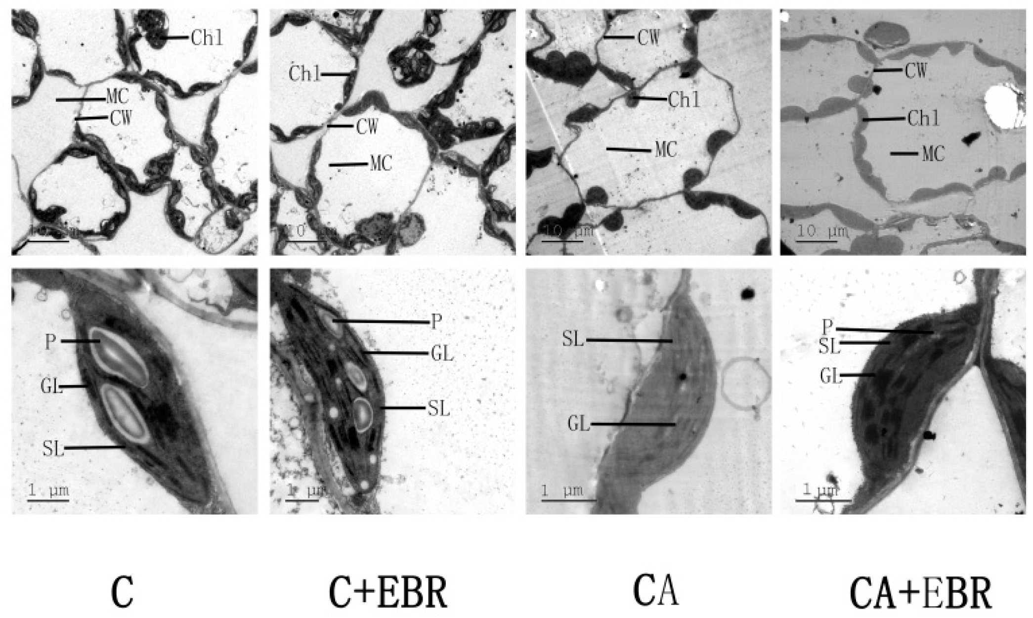
3.5. Ultrastructure of Chloroplast and Thylakoid

The ultrastructural difference of chloroplast and thylakoid between the C and C + EBR treatment was not obvious, chloroplast was fusiform, the outer membrane was clear, starch granule was clear and plump, and thylakoid grana lamellae were clear and folded regularly. Under autotoxicity stress conditions, chloroplast swelling, chloroplast outer membrane degradation, starch granulocyte blurring, and thylakoid granulocyte lamellae disappeared. Under CA + EBR treatment, the chloroplast presented fuzzy thylakoid granulocyte lamellae. Exogenous EBR markedly alleviated the damage caused by autotoxicity to chloroplast and thylakoid. Autotoxicity induced the degradation of the cell membrane; however, EBR application improved the cell membrane stability (Figure 4).

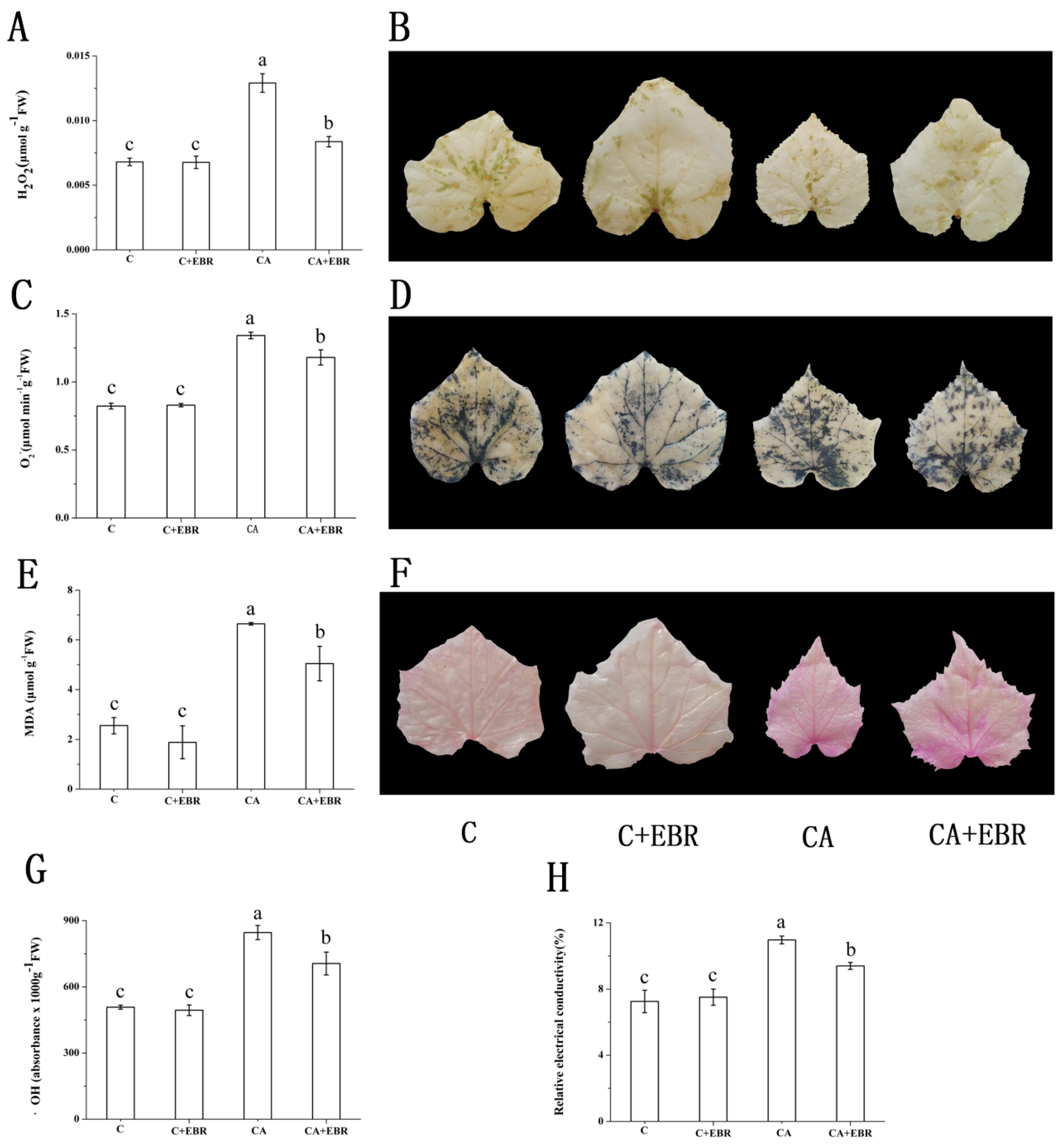
3.6. ROS, MDA and Relative Membrane Permeability

Exogenous EBR application did not affect the ROS (i.e., H2O2, O2, OH) and MDA contents of cucumber seedlings under normal growing conditions. CA treatment significantly increased ROS and MDA contents and relative electrical conductivity compared with control. However, EBR application markedly reduced ROS and MDA contents and relative electrical conductivity compared with CA treatment under autotoxicity stress conditions (Figure 5). Similar effects of EBR on leaf H2O2 accumulation, O2 production rate and lipid peroxidation were observed by an independent staining method (Figure 5B,D,F).

3.7. Antioxidant System

Under normal growing conditions, no significant differences were observed for POD, CAT and APX activity in cucumber seedlings by the application of EBR compared with control. However, EBR application improved the SOD, POD, APX and CAT activities by 4.40%, 11.10%, 17.50% and 35.80%, respectively, in EBR-treated cucumber plants compared with CA-treated plants (Figure 6A,B,C,D). The contents of AsA and GSH in CA-treated cucumber seedlings were decreased by 26.40% and 30.10%, respectively, compared with control. However, EBR application improved the AsA and GSH contents by 8.40% and 23.00%, respectively compared with CA treatment under autotoxicity stress conditions (Figure 6E,F).

3.8. Proline, Soluble Sugar and Protein Contents

Under normal growing conditions, there was no significant difference in the proline, soluble sugar and protein contents among the EBR-treated and non-EBR treated cucumber plants. However, under autotoxicity stress conditions, EBR application markedly improved soluble protein, soluble sugar and proline contents (Table 6).

4. Discussion

BRs act as a positive regulator under stressful environmental conditions [19]. BRs are reported to alleviate temperature stress [41], drought stress, salinity stress [42], and heavy metals stress [43]. However, the role of BRs to alleviate autotoxicity stress for cucumber seedlings was not reported. In our study, we observed the effect of BRs on cucumber seedling under autotoxicity stress conditions. Root, as an important organ to absorb water and nutrients from the soil and transport them to aboveground plant parts, directly affects the morphogenesis and biomass accumulation of aboveground plant parts [44]. Under stress conditions, plant roots sense the stress signal, plants make adjustments in the metabolic pathways, change the carbon distribution ratio and direction of carbon assimilation products, and finally, root morphology is altered to adapt to the environmental stress [45]. Müssig et al. [46] reported that exogenous EBR application at 0.05–0.1 nM stimulated the growth of the taproot of Arabidopsis thaliana, however, at higher concentration root growth inhibition was observed. In our study, exogenous application of EBR at 0.1 μM improved the growth and biomass of root (Table 2 and Table 3). Li et al. [47] showed that 0.1 μM EBR treatment promoted the accumulation of rhoptry protein 2 (ROP2) in the root system from the central column to the elongation zone to respond to gravity and improved the polar distribution of ROP2 and actively regulated lateral root development. In the present study, exogenous application of 0.1 μM EBR increased leaf area, plant height (Table 1) and aboveground biomass accumulation (Table 2), root activity (Figure 2), and root morphology (Table 3) as an important indicator of direct contact between roots and environmental conditions and reflected plant growth, suggesting that BRs can effectively promote the growth of cucumber seedlings under autotoxicity stress conditions. EBR increased root absorption area exposed to the nutrient medium, and enhanced the nutrients and water absorption ability to maintain their functional behavior.
In this study, we provided evidence that endogenous EBR played a positive role and improved photosynthetic pigments under autotoxicity stress in cucumber (Table 4). Chlorophyll is the main photosynthetic pigment which has the function of absorbing, transferring and converting light energy into chemical energy, and changes in photosynthetic pigments can be used as photosynthesis indicators [48]. Interestingly, application of exogenous EBR apparently increased the total chlorophyll, chlorophyll a, and chlorophyll b contents of cucumber leaves under autotoxicity stress. The increased photosynthetic pigments are beneficial to capture more light energy and increase the rate of conversion of light energy to chemical energy [49]. We also observed increased stomatal opening by EBR application, indicating that BRs could help improve the availability of carbon for photosynthesis (Figure 3). Meanwhile, our evidence for gas exchange parameters showed the importance of BRs in regulating photosynthesis, an apparent increase in the net photosynthetic rate under autotoxicity stress conditions was observed (Table 5). We also found that autotoxicity caused damage to membrane system and organelles of cucumber leaves, and eventually, the chloroplasts were swollen, the outer membrane of chloroplast was degraded, the starch granules were fuzzy, thylakoids lamellae disappeared, and cell membrane degradation was increased (Figure 4). These damages reduced the photosynthetic efficiency of cucumbers and also caused damage to the antioxidant system. In this study, exogenous EBR apparently reduced the damage of chloroplast and thylakoids in cucumber leaves caused by autotoxicity, and maintained the relative stability of the organelle system (Figure 4).
It was reported that the level of ROS in the plants remains lower [50]. Plants could clean up excess reactive oxygen species through the antioxidant system and maintain the active oxygen homeostasis under normal circumstances. ROS are considered important for the growth and development of plants [51]. Under stress environment, plants produce ROS that causes normal active oxygen metabolism disorder in vivo [52]. In this study, we observed that EBR positively regulated H2O2, superoxide radical (O2) and the hydroxyl radical (OH) under autotoxicity stress conditions (Figure 5). It proved that BRs could remove excessive ROS produced by autotoxicity stress in order to reduce ROS damage to the membranes. It was confirmed that antioxidant enzymes in plants work together and a single protective enzyme did not maintain the balance of active oxygen metabolism in cells [50]. Under stress conditions, because of changes in physiological and biochemical reactions, the activities of antioxidant enzymes, including POD, SOD, and CAT are reduced and excess reactive oxygen species, MDA, and other harmful substances generated in the plant are not removed in a timely fashion [53]. CAT was a major H2O2 scavenging enzyme, and SOD acted as an early defense against ROS, particularly H2O2 [22,54]. According to a report, EBR-mediated resistance could be attributed to the up-regulation of antioxidants-related genes (SOD, CAT, POD, GR, and APX) [22]. In our study, cucumber seedlings treated with EBR under autotoxicity conditions obviously enhanced the activities of antioxidant enzymes, such as SOD, CAT, POD, and APX (Figure 6). AsA and GSH are two important non-enzymatic antioxidants, they participate in the removal of free radicals and peroxidation products in plants, quench active oxygen, and protect enzymes and structural proteins to protect cell membrane of plants under stressful conditions [35]. Our findings showed that BR increased the AsA and GSH contents and helped cells to reduce ROS damage (Figure 6). The findings of MDA content and relative conductivity of cucumber leaves suggested that BRs have a positive effect and decrease cell membrane injury (Figure 6).
In order to further understand the mechanism of BRs alleviating the damage of cell membrane induced by autotoxicity, we measured the concentration of osmotic substances (soluble protein, soluble sugar and proline). In this study, we observed that EBR application enhanced the soluble protein, soluble sugar and proline contents in the cells indicating that EBR improved plant capacity to withstand autotoxicity stress. One of the major responses of autotoxicity on plant damage was the increased permeability of the cell membrane. In this experiment, autotoxicity stress significantly increased the membrane permeability of cucumber leaves. However, EBR treatment improved soluble protein, soluble sugar and proline contents of cucumber leaves, and improved membrane stability of cucumber (Table 6).

5. Conclusions

Autotoxicity affects normal metabolism of plants because of the accumulation of ROS and lipid peroxidation under autotoxicity stress conditions. ROS accumulation and lipid peroxidation leads to the destruction of cells structure and reduced plant growth. Under autotoxicity stress conditions, EBR application improves antioxidant contents (AsA and GSH) and the activities of antioxidant enzymes (POD, APX and CAT) and proline, soluble sugar, and soluble protein contents; resultantly OH, H2O2 and MDA content are reduced, thus root damage is reduced and the integrity of chloroplast and thylakoid structure is maintained. Photosynthetic pigments and photosynthetic capacity of the cucumber leaves is improved. All these events lead to improved plant growth and development of cucumber seedlings under autotoxicity stress conditions. To further clarify the mechanism of EBR application to alleviate autotoxixity stress, gene expression analysis and genome wise transcriptome profiling can be considered. Based on the findings of this study, EBR application can help improve plant growth and development of cucumber under protected cultivation system.

Author Contributions

J.L. conceived and designed the experiments. P.Y., F.L. and L.B. performed the experiments and analyzed the data. M.A.N. helped perform the analysis with constructive discussions and writing this manuscript. All authors have read and approved the final manuscript.

Funding

The financial support for this study was provided by National Natural Science Foundation of China (31760592); Scientific Research Fund Project Yunnan Provincial Educational Department (2017ZZX119); Special Scientific Research Fund for PhD of Honghe University (XJ17B07; XJ17B08) and Yunnan Province Applied Basic Research Plan of China (2018FD087).

Acknowledgments

We are thankful to the staff of the Experimental Station of Honghe University for their assistance during this study.

Conflicts of Interest

The authors declare no conflict of interest.

References

  1. Zhang, Y.; Gu, M.; Xia, X.; Shi, K.; Zhou, Y.; Yu, J. Alleviation of autotoxin-induced growth in hibition and respiration by sucrose in Cucumis sativus (L.). Allelopathy J. 2010, 25, 147–154. [Google Scholar]
  2. Paudel, M.N. Multiple Cropping for Raising Productivity and Farm Income of Small Farmers. J. Nepal Agric. Res. Counc. 2016, 2, 37–45. [Google Scholar] [CrossRef] [Green Version]
  3. Chou, C. Roles of allelopathy in plant biodiversity and sustainable agriculture. Crit. Rev. Plant Sci. 1999, 18, 609–636. [Google Scholar] [CrossRef]
  4. Bie, Z.; Nawaz, M.A.; Huang, Y.; Lee, J.M.; Colla, G. Introduction of vegetable grafting. In Vegetable Grafting, Principles and Practices; Colla, G., Alfocea, F.P., Schwarz, D., Eds.; CABI Publishing: Wallingford, UK, 2017; pp. 1–21. [Google Scholar]
  5. Callaway, R.; Vivanco, J. Can plant biochemistry contribute to understanding of invasion ecology? Trends Plant Sci. 2006, 11, 574–580. [Google Scholar] [CrossRef]
  6. Qiao, Y. Study on Different Responses to Autotoxin Toxicity in Cucumber and Figleaf Gourd Seedlings. Ph.D. Thesis, China Agricultural University, Beijing, China, 2013. [Google Scholar]
  7. Li, J.; Li, Y.; Tian, Y.; Qu, M.; Zhang, W.; Gao, L. Melatonin has the potential to alleviate cinnamic acid stress in cucumber seedlings. Front. Plant Sci. 2017, 8, 1193. [Google Scholar] [CrossRef]
  8. Seal, A.; Haig, T.; Pratley, J. Evaluation of putative allelochemicals in rice root exudates for their role in the suppression of arrowhead root growth. J. Chem. Ecol. 2004, 30, 1663–1678. [Google Scholar] [CrossRef]
  9. Belz, R. Allelopathy in crop/weed interactions-an update. Pest Manag. Sci. 2007, 63, 308–326. [Google Scholar] [CrossRef]
  10. FAO. FAOSTAT Domains/Production/Crops. Available online: http://www.fao.org/faostat/en/#data/QC (accessed on 19 December 2016).
  11. Xie, J.; Yu, J.; Chen, B.; Feng, Z.; Li, J.; Zhao, C.; Lyu, J.; Hu, L.; Gan, Y.; Kadambot, H. Facility cultivation systems “设施农业”: A Chinese model for the planet. Adv. Agron. 2017, 145, 1–42. [Google Scholar] [CrossRef]
  12. Bennett, A.; Bending, G.; Chandler, D.; Hilton, S.; Mills, P. Meeting the demand for crop production: The challenge of yield decline in crops grown in short rotations. Biol. Rev. 2012, 87, 52–71. [Google Scholar] [CrossRef] [PubMed]
  13. Al-Harbi, A.; Al-Omran, A.; Alharbi, K. Grafting improves cucumber water stress tolerance in Saudi Arabia. Saudi J. Biol. Sci. 2018, 25, 298–304. [Google Scholar] [CrossRef] [PubMed]
  14. Nawaz, M.A.; Shireen, F.; Huang, Y.; Bie, Z.; Waqar, A.; Basharat, A. Perspectives of vegetable grafting in Pakistan, current status, challenges and opportunities. Int. Agric. Biol. 2017, 19, 1165–1174. [Google Scholar] [CrossRef]
  15. Nawaz, M.A.; Muhamamd, I.; Kong, Q.; Cheng, F.; Waqar, A.; Huang, Y.; Bie, Z. Grafting: A technique to modify ion accumulation in horticultural crops. Front. Plant Sci. 2016, 7, 1457. [Google Scholar] [CrossRef] [PubMed]
  16. Huang, Y.; Jiao, Y.; Nawaz, M.A.; Chen, C.; Liu, L.; Lu, Z.; Kong, Q.; Cheng, F.; Bie, Z. Improving magnesium uptake, photosynthesis and antioxidant enzyme activities of watermelon by grafting onto pumpkin rootstock under low magnesium. Plant Soil 2016, 409, 229–246. [Google Scholar] [CrossRef]
  17. Alfocea, P.; Schwarz, D. (Eds.) Vegetable Grafting. Principles and Practices; CABI Publishing: Wallingford, UK, 2017; pp. 1–21. [Google Scholar]
  18. Velkov, N.; Pevicharova, G. Effects of cucumber grafting on yield and fruit sensory characteristics. Zemdirbyste 2016, 103, 405–410. [Google Scholar] [CrossRef] [Green Version]
  19. Vriet, C.; Russinova, E.; Reuzeau, C. Boosting crop yields with plant steroids. Plant Cell 2012, 24, 842–857. [Google Scholar] [CrossRef]
  20. Krishna, P. Brassinosteroid-mediated stress responses. J. Plant Growth Regul. 2003, 22, 289–297. [Google Scholar] [CrossRef] [PubMed]
  21. Bajguz, A.; Hayat, S. Effects of brassinosteroids on the plant responses to environmental stresses. Plant Physiol. Biochem. 2009, 47, 1–8. [Google Scholar] [CrossRef] [PubMed]
  22. Cui, J.; Zhou, Y.; Ding, J.; Xia, X.; Shi, K.; Chen, S.; Asami, T.; Chen, Z.; Yu, J. Role of nitric oxide in hydrogenperoxide-dependent induction of abiotic stress tolerance by brassinosteroids in cucumber. Plant Cell Environ. 2011, 34, 347–358. [Google Scholar] [CrossRef]
  23. Xia, X.; Fang, P.; Guo, X.; Qian, X.; Zhou, J.; Shi, K.; Zhou, Y.; Yu, J. Brassinosteroid-mediated apoplastic H2O2-glutaredoxin 12/14 cascade regulates antioxidant capacity in response to chilling in tomato. Plant Cell Environ. 2018, 41, 1052–1064. [Google Scholar] [CrossRef]
  24. Hu, W.; Wu, Y.; Zeng, J.; He, L.; Zeng, Q. Chill-induced inhibition of photosynthesis was alleviated by 24-epibrassinolide pretreatment in cucumber during chilling and subsequent recovery. Photosynthetica 2010, 48, 537–544. [Google Scholar] [CrossRef]
  25. Eremina, M.; Unterholzner, S.; Rathnayake, A.; Castellanos, M.; Khan, M.; Kugler, K.; Poppenberger, B. Brassinosteroids participate in the control of basal and acquired freezing tolerance of plants. Proc. Natl. Acad. Sci. USA 2016, 113, E5982–E5991. [Google Scholar] [CrossRef] [PubMed] [Green Version]
  26. Jiang, Y.; Huang, L.; Cheng, F.; Zhou, Y.; Xia, X.; Mao, W.; Shi, K.; Yu, J. Brassinosteroids accelerate recovery of photosynthetic apparatus from cold stress by balancing the electron partitioning, carboxylation and redox homeostasis in cucumber. Physiol. Plant. 2013, 148, 133–145. [Google Scholar] [CrossRef] [PubMed]
  27. Todorova, D.; Talaat, N.B.; Katerova, Z.; Alexieva, V.; Shawky, B. Polyamines and brassinosteroids in drought stress responses and tolerance in plants. In Water Stress and Crop Plants, 1st ed.; Ahmad, P., Ed.; John Wiley & Sons, Ltd.: Hoboken, NY, USA, 2016; pp. 608–627. [Google Scholar]
  28. Liu, Z.; Li, L.; Luo, Z.; Zeng, F.; Jiang, L.; Tang, K. Effect of brassinolide on energy status and proline metabolism in postharvest bamboo shoot during chilling stress. Postharvest Biol. Technol. 2016, 111, 240–246. [Google Scholar] [CrossRef]
  29. Kang, Y.; Guo, S.; Li, J.; Duan, J. Effect of root applied 24-epibrassinolide on carbohydrate status and fermentative enzyme activities in cucumber (Cucumis sativus L.) seedlings under hypoxia. Plant Growth Regul. 2008, 57, 259–269. [Google Scholar] [CrossRef]
  30. Bu, R.; Wang, R.; Wei, Q.; Hu, H.; Sun, H.; Song, P.; Yu, Y.; Liu, Q.; Zheng, Z.; Li, T.; et al. Silencing of glycerol-3-phosphate acyltransferase 6 (GPAT6) gene using a newly established virus induced gene silencing (VIGS) system in cucumber alleviates autotoxicity mimicked by cinnamic acid (CA). Plant Soil. 2019, 438, 329–346. [Google Scholar] [CrossRef]
  31. Li, J.; Yang, P.; Xie, J.; Yu, J. Effects of 24-epibrassinolide on growth and antioxidant enzymes system in pepper roots under chilling stress. J. Nucl. Agric. Sci. 2015, 29, 1001–1008. [Google Scholar] [CrossRef]
  32. Arnon, D. Copper enzymes in isolated chloroplasts polyphenoloxidase in beta vulgaris. Plant Physiol. 1949, 24, 1–15. [Google Scholar] [CrossRef] [PubMed]
  33. Zeng, Y.; Zhang, Y.; Xiang, J.; Wu, H.; Chen, H.; Zhang, Y.; Zhu, D. Effects of chilling tolerance induced by spermidine pretreatment on antioxidative activity, endogenous hormones and ultrastructure of indica-japonica hybrid rice seedlings. J. Integr. Agric. 2016, 15, 295–308. [Google Scholar] [CrossRef] [Green Version]
  34. Ma, N.; Zuo, Q.; Liang, X.; Yin, B.; Wang, Q.; Meng, D. The multiple stress-responsive transcription factor SlNAC1 improves the chilling tolerance of tomato. Physiol. Plant. 2013, 149, 474–486. [Google Scholar] [CrossRef]
  35. Li, J.; Yang, P.; Gan, Y.; Yu, J.; Xie, J. Brassinosteroid alleviates chilling-induced oxidative stress in pepper by enhancing antioxidation systems and maintenance of photosystem II. Acta Physiol. Plant. 2015, 37, 222. [Google Scholar] [CrossRef]
  36. Stevens, R.; Page, D.; Gouble, B.; Garchery, C.; Zamir, D.; Causse, M. Tomato fruit ascorbic acid content is linked with monodehydroascorbate reductase activity and tolerance to chilling stress. Plant Cell Environ. 2008, 31, 1086–1096. [Google Scholar] [CrossRef]
  37. Griffith, O.W. Determination of glutathione and glutathione disulphide using glutathione reductase and 2-vinylpyridine. Anal. Biochem. 1980, 106, 207–212. [Google Scholar] [CrossRef]
  38. Bates, L.S.; Waldren, R.P.; Teare, I.D. Rapid determination of free proline for water-stress studies. Plant Soil 1973, 39, 205–207. [Google Scholar] [CrossRef]
  39. Buysse, J.; Merckx, R. An improved colorimetric method to quantify sugar content of plant tissue. J. Exp. Bot. 1993, 44, 1627–1629. [Google Scholar] [CrossRef]
  40. Bradford, M. A rapid and sensitive method for the quantitation of microgram quantities of protein utilizing the principle of protein dye binding. Anal. Biochem. 1976, 72, 248–254. [Google Scholar] [CrossRef]
  41. Sadura, I.; Janeczko, A. Physiological and molecular mechanisms of brassinosteroid-induced tolerance to high and low temperature in plants. Biol. Plant. 2018, 62, 601–616. [Google Scholar] [CrossRef] [Green Version]
  42. Shahbaz, M.; Ashraf, M.; Athar, H. Does exogenous application of 24-epibrassinolide ameliorate salt induced growth inhibition in wheat (Triticum aestivum L.)? Plant Growth Regul. 2008, 55, 51–64. [Google Scholar] [CrossRef]
  43. Sharma, I.; Pati, P.; Bhardwaj, R. Effect of 24-epibrassinolide on oxidative stress markers induced by nickel-ion in Raphanus sativus L. Acta Physiol. Plant. 2011, 33, 1723–1735. [Google Scholar] [CrossRef]
  44. Cheng, J.; Dai, T.; Jing, Q.; Jiang, D.; Pan, X.; Cao, W. Root morphological and physiological characteristics in relation to nitrogen absorption efficiency in different rice genotypes. Acta Pedol. Sin. 2007, 44, 266–272. [Google Scholar] [CrossRef]
  45. Pierret, A.; Doussan, C.; Capowiez, Y.; Bastardie, F.; Pagès, L. Root functional architecture: A framework for modeling the interplay between roots and soil. Vadose Zone J. 2007, 6, 269–281. [Google Scholar] [CrossRef]
  46. Müssig, C.; Shin, G.; Altmann, T. Brassinosteroids promote root growth in Arabidopsis. Plant Physiol. 2003, 133, 1261–1271. [Google Scholar] [CrossRef]
  47. Li, L.; Xu, J.; Xu, Z.; Xue, H. Brassinoeroids stimulate plant tropisms through modulation of polar auxin transport in Brassica and Arabidopsis. Plant Cell 2005, 17, 2738–2753. [Google Scholar] [CrossRef] [PubMed]
  48. Ahammed, G.; Yuan, L.; Ogweno, J.; Zhou, Y.; Xia, X.; Mao, W.; Shi, K.; Yu, J. Brassinosteroid alleviates phenanthrene and pyrene phytotoxicity by increasing detoxification activity and photosynthesis in tomato. Chemosphere 2012, 86, 546–555. [Google Scholar] [CrossRef] [PubMed]
  49. Jiang, L.; Wen, L. 2-Photonic sensitive switchable materials A2-Meglinski, Igor. In Biophotonics for Medical Applications; Woodhead Publishing: Cambridge, UK, 2015; pp. 25–51. [Google Scholar]
  50. Xia, X.; Wang, Y.; Zhou, Y.; Tao, Y.; Mao, W.; Shi, K.; Asami, T.; Chen, Z.; Yu, J. Reactive oxygen species are involved in brassinosteroid-induced stress tolerance in cucumber. Plant Physiol. 2009, 150, 801–814. [Google Scholar] [CrossRef]
  51. Xia, X.; Huang, L.; Zhou, Y.; Mao, W.; Shi, K.; Wu, J.; Asami, T.; Chen, Z.; Yu, J. Brassinosteroids promote photosynthesis and growth by enhancing activation of Rubisco and expression of photosynthetic genes in Cucumis sativus. Planta 2009, 230, 1185–1196. [Google Scholar] [CrossRef]
  52. Cao, S.; Xu, Q.; Cao, Y.; Qian, K.; An, K.; Zhu, Y.; Hu, B.; Zhao, H.; Kuai, B. Loss of function mutations in DET2 gene lead to an enhanced resistance to oxidative stress in Arabidopsis. Physiol. Plant. 2005, 123, 57–66. [Google Scholar] [CrossRef]
  53. Muhammad, A.; Chen, C.; Fareeha, S.; Zheng, Z.; Jiao, Y.; Hamza, S.; Muhammad, A.; Muhammad, I.; Muhammad, A.; Huang, Y.; et al. Improving vanadium stress tolerance of watermelon by grafting onto bottle gourd and pumpkin rootstock. Plant Growth Regul. 2018, 85, 41–56. [Google Scholar] [CrossRef]
  54. Wang, P.; Sun, X.; Li, C.; Wei, Z.; Liang, D.; Ma, F. Long-term exogenous application of melatonin delays drought-induced leaf senescence in apple. J. Pineal Res. 2013, 54, 292–302. [Google Scholar] [CrossRef]
Figure 1. Phenotypic changes (A) in autotoxicity-stressed cucumber seedlings with or without the application of 24-epibrassinolide (EBR); the root phenotype (B) after 7 d of stress treatment. C, 1/2 Hoagland’s nutrient solution; C + EBR, application of 0.l μM EBR in 1/2 Hoagland’s nutrient solution; CA, application of 100 mM cinnamic acid in 1/2 Hoagland’s nutrient solution; CA + EBR, application of 100 mM cinnamic acid and 0.1 μM EBR in 1/2 Hoagland’s nutrient solution.
Agronomy 09 00265 g001
Figure 2. Root activity of cucumber seedlings exposed to autotoxicity conditions with or without the application of EBR. Values are means ± SE (n = 6). Letters in the column diagram indicate significant differences at p < 0.05. C, 1/2 Hoagland’s nutrient solution; C + EBR, application of 0.l μM EBR in 1/2 Hoagland’s nutrient solution; CA, application of 100 mM cinnamic acid in 1/2 Hoagland’s nutrient solution; CA + EBR, application of 100 mM cinnamic acid and 0.1 μM EBR in 1/2 Hoagland’s nutrient solution.
Agronomy 09 00265 g002
Figure 3. Stomatal opening of cucumber leaves exposed to autotoxicity conditions with or without the application of EBR. C, 1/2 Hoagland’s nutrient solution; C + EBR, application of 0.l μM EBR in 1/2 Hoagland’s nutrient solution; CA, application of 100 mM cinnamic acid in 1/2 Hoagland’s nutrient solution; CA + EBR, application of 100 mM cinnamic acid and 0.1 μM EBR in 1/2 Hoagland’s nutrient solution.
Agronomy 09 00265 g003
Figure 4. Chloroplast and thylakoid ultrastructure of cucumber leaves exposed to autotoxicity conditions with or without the application of EBR. MC, Mesophyll cell; Chl, chloroplast; CW, cell wall; GL, grana lamellae; SL, stroma lamellae; P, plastoglobules. C, 1/2 Hoagland’s nutrient solution; C+EBR, application of 0.l μM EBR in 1/2 Hoagland’s nutrient solution; CA, application of 100 mM cinnamic acid in 1/2 Hoagland’s nutrient solution; CA+EBR, application of 100 mM cinnamic acid and 0.1 μM EBR in 1/2 Hoagland’s nutrient solution.
Agronomy 09 00265 g004
Figure 5. Change of ROS (A,C,G), lipid peroxidation (E) and relative conductivity (relative membrane permeability) (H) in control condition, autotoxicity-stressed treatment, and autotoxicity-stress with EBR of cucumber seedlings. (A,C,E,G) are the quantitative measurements of H2O2, O2, MDA and OH levels, respectively, in cucumber leaves under different treatments. Values are means of 6 replicates ± SE (n = 6). Letters in the column diagram indicate significant differences at p < 0.05. (B,D,F) are in situ detection of H2O2, O2 and lipid peroxidation in cucumber leaves via histochemical detection respectively. C, 1/2 Hoagland’s nutrient solution; C + EBR, application of 0.l μM EBR in 1/2 Hoagland’s nutrient solution; CA, application of 100 mM cinnamic acid in 1/2 Hoagland’s nutrient solution; CA + EBR, application of 100 mM cinnamic acid and 0.1 μM EBR in 1/2 Hoagland’s nutrient solution.
Agronomy 09 00265 g005
Figure 6. Changes of antioxidant system in leaves of cucumber seedlings under control or autotoxicity stress conditions with or without EBR. Letters in the column diagram indicate significant differences at p < 0.05. (AF) respectively is the activity of superoxide dismutase (SOD), peroxidase (POD), catalase (CAT), ascorbate peroxidase (APX) and antioxidative compounds ascorbate (AsA) and reduced glutathione (GSH) contents. C, 1/2 Hoagland’s nutrient solution; C + EBR, application of 0.l μM EBR in 1/2 Hoagland’s nutrient solution; CA, application of 100 mM cinnamic acid in 1/2 Hoagland’s nutrient solution; CA+EBR, application of 100 mM cinnamic acid and 0.1 μM EBR in 1/2 Hoagland’s nutrient solution.
Agronomy 09 00265 g006
Table 1. Morphological changes of cucumber seedlings after treatment with EBR under autotoxicity stress conditions.
TreatmentPlant Height (cm)Stem Diameter (cm)Leaf Area (mm2)
C16.07 ± 0.53 a0.65 ± 0.14 a2434.31 ± 25.94 a
C + EBR16.97 ± 0.23 a0.59 ± 0.01 a2545.29 ± 67.98 a
CA9.65 ± 0.61 c0.40 ± 0.02 b879.72 ± 35.41 c
CA + EBR14.13 ± 0.94 b0.49 ± 0.03 ab1583.58 ± 29.81 b
Different lowercase letters in the same column indicate a significant difference between the treatments at p ˂ 0.05. Data are the mean ± standard error of the mean of at least three different replicates of each treatment. C, 1/2 Hoagland’s nutrient solution; C + EBR, application of 0.l μM EBR in 1/2 Hoagland’s nutrient solution; CA, application of 100 mM cinnamic acid in 1/2 Hoagland’s nutrient solution; CA + EBR, application of 100 mM cinnamic acid and 0.1 μM EBR in 1/2 Hoagland’s nutrient solution.
Table 2. Effects of exogenous EBR on the biomass of cucumber seedlings under autotoxicity stress conditions.
TreatmentFresh Weight (g)Dry Weight (g)
Above-GroundUndergroundAbove-GroundUnderground
C6.67 ± 0.69 a0.74 ± 0.12 a0.49 ± 0.03 a0.054 ± 0.008 a
EBR7.14 ± 0.70 a0.83 ± 0.31 a0.48 ± 0.06 a0.065 ± 0.012 ab
CA3.74 ± 0.24 c0.63 ± 0.04 a0.28 ± 0.03 c0.041 ± 0.007 ab
CA+EBR5.57 ± 0.37 b0.73 ± 0.06 a0.40 ± 0.01 b0.052 ± 0.003 b
Different lowercase letters in the same column indicate a significant difference between the treatments at p ˂ 0.05. Data are the mean ± standard error of the mean of at least three different replicates of each treatment. C, 1/2 Hoagland’s nutrient solution; C + EBR, application of 0.l μM EBR in 1/2 Hoagland’s nutrient solution; CA, application of 100 mM cinnamic acid in 1/2 Hoagland’s nutrient solution; CA + EBR, application of 100 mM cinnamic acid and 0.1 μM EBR in 1/2 Hoagland’s nutrient solution.
Table 3. Effect of EBR on root morphology of cucumber seedlings under autotoxicity stress conditions.
TreatmentTotal Root Length
(mm)
Root Surface
(cm2)
Root Volume
(cm3)
Root Tip Number
C1642.6 ± 162.0 a354.9 ± 27.3 a1.42 ± 0.14 a161 ± 12 a
C + EBR1770.3 ± 198.8 a331.1 ± 27.3 a1.25 ± 0.33 a150 ± 22 a
CA588.2 ± 41.1 c169.0 ± 29.2 c0.69 ± 0.07 b100 ± 7 b
CA + EBR1005.0 ± 8.5 b257.0 ± 40.5 b0.32 ± 0.08 c69 ± 8 c
Different lowercase letters in the same column indicate a significant difference between the treatments at p ˂ 0.05. Data are the mean ± standard error of the mean of at least three different replicates of each treatment. C, 1/2 Hoagland’s nutrient solution; C + EBR, application of 0.l μM EBR in 1/2 Hoagland’s nutrient solution; CA, application of 100 mM cinnamic acid in 1/2 Hoagland’s nutrient solution; CA+EBR, application of 100 mM cinnamic acid and 0.1 μM EBR in 1/2 Hoagland’s nutrient solution.
Table 4. Effect of EBR on photosynthetic pigments of cucumber seedlings under autotoxicity stress conditions (mg/g FW).
TreatmentChl a ContentChl b ContentTotal Chl ContentCarotenoid Content
C27.19 ± 0.07 a31.70 ± 1.03 a58.89 ± 1.07 a9.93 ± 1.03 a
C + EBR27.30 ± 0.58 a31.80 ± 2.18 a59.10 ± 1.83 a10.41 ± 1.45 a
CA21.14 ± 0.05 c22.83 ± 1.74 c43.97 ± 1.69 c6.64 ± 0.47 b
CA + EBR25.11 ± 1.77 b28.09 ± 0.61 b53.19 ± 1.54 b8.64 ± 0.32 ab
Different lowercase letters in the same column indicate a significant difference between the treatments at p ˂ 0.05. Data are the mean ± standard error of the mean of at least three different replicates of each treatment. C, 1/2 Hoagland’s nutrient solution; C + EBR, application of 0.l μM EBR in 1/2 Hoagland’s nutrient solution; CA, application of 100 mM cinnamic acid in 1/2 Hoagland’s nutrient solution; CA + EBR, application of 100 mM cinnamic acid and 0.1 μM EBR in 1/2 Hoagland’s nutrient solution.
Table 5. Effect of EBR on the Pn, Ci, Gs and Tr of cucumber leaf under autotoxicity stress conditions.
TreatmentPn (μmol (CO2) m−2 s−1)Ci (μmol mol−1)Gs (mmol (H2O) m2 s)Tr (mmol m−2 s−1)
C12.7 ± 1.0 a341.3 ± 4.7 a32.2 ± 3.9 a443.3 ± 31.0 a
C + EBR11.3 ± 1.1 a306.7 ± 43.4 a23.9 ± 16.3 a319.3 ± 146.0 a
CA4.2 ± 0.3 c202.3 ± 51.3 b3.5 ± 0.7 b71.0 ± 4.2 b
CA + EBR6.6 ± 1.4 b167.0 ± 36.3 b4.5 ± 1.4 b115.3 ± 20.3 b
Different lowercase letters in the same column indicate a significant difference between the treatments at p ˂ 0.05. Data are the mean ± standard error of the mean of at least three different replicates of each treatment. Pn, net photosynthetic rate; Gs, stomatal conductance; Tr, transpiration rate; Ci, intercellular CO2 concentration. C, 1/2 Hoagland’s nutrient solution; C + EBR, application of 0.l μM EBR in 1/2 Hoagland’s nutrient solution; CA, application of 100 mM cinnamic acid in 1/2 Hoagland’s nutrient solution; CA + EBR, application of 100 mM cinnamic acid and 0.1 μM EBR in 1/2 Hoagland’s nutrient solution.
Table 6. The changes in proline, soluble sugar and protein contents of cucumber leaves under autotoxicity stress conditions.
TreatmentSoluble Protein
(mg g−1 Fresh weight)
Soluble Sugar
(mg g−1 Fresh weight)
Proline
(μg g−1 Fresh weight)
C15.90 ± 0.48 c0.44 ± 0.02 c19.66 ± 3.16 c
C + EBR17.76 ± 0.74 c0.46 ± 0.02 c16.37 ± 1.80 c
CA23.67 ± 0.25 b0.67 ± 0.08 b68.83 ± 3.16 b
CA + EBR25.38 ± 1.07 a1.07 ± 0.10 a94.01 ± 2.89 a
Different lowercase letters in the same column indicate a significant difference between the treatments at p ˂ 0.05. Data are the mean ± standard error of the mean of at least three different replicates of each treatment. C, 1/2 Hoagland’s nutrient solution; C + EBR, application of 0.l μM EBR in 1/2 Hoagland’s nutrient solution; CA, application of 100 mM cinnamic acid in 1/2 Hoagland’s nutrient solution; CA + EBR, application of 100 mM cinnamic acid and 0.1 μM EBR in 1/2 Hoagland’s nutrient solution.

