Article

The Effect of Drying Temperature on the Phenolic Content and Functional Behavior of Flours Obtained from Lemon Wastes

by
Jesús Patrón-Vázquez
1,
Lizzie Baas-Dzul
1,
Nelly Medina-Torres
1,
Teresa Ayora-Talavera
1,
Ángeles Sánchez-Contreras
1,
Ulises García-Cruz
2 and
Neith Pacheco
1,*
1
Food Safety Laboratory, Center for Research and Assistance in Technology and Design of Jalisco state A. C. Southeast Unit, Science and Technological Park of Yucatan, Cadastral Planking 31264, Sierra Papacal-Chuburna Port Highway Km.5.5, Merida 97302, Yucatan, Mexico
2
Center for Research and Advanced Studies of the Institute National Polytechnic, Unity Merida. Km. 6 Old road to Progreso Postal mail 73, Cordemex, Merida 97310, Yucatan, Mexico
*
Author to whom correspondence should be addressed.
Submission received: 19 July 2019 / Revised: 8 August 2019 / Accepted: 15 August 2019 / Published: 21 August 2019

Abstract

:
Lemon processing generates thousands of tons of residues that can be preserved as flours by thermal treatment to obtain phenolic compounds with beneficial bioactivities. In this study, the effect of different drying temperatures (40, 50, 60, 70, 80, 90, 100 and 110 °C) on the Total Phenolic Content (TPC), antioxidant and antimicrobial activities of phenolic compounds present in Citrus. lemon (L.) Burn f waste was determined. Identification and quantification of phenolic compounds were also performed by UPLC-PDA and UPLC-ESI-MS analysis. Eriocitrin (19.79–27.29 mg g−1 DW) and hesperidin (7.63–9.10 mg g−1 DW) were detected as the major phenolic compounds in the flours by UPLC-PDA and confirmed by UPLC-ESI-MS. Antimicrobial activity determined by Minimum Inhibitory Concentration (MIC) against Salmonella typhimurium, Escherichia coli and Staphylococcus aureus was observed. Accordingly, a stable functional flour as a source of bioactive phenolic compounds obtained from lemon residues at 50 °C may be produced as a value-added product useful in various industrial sectors.

1. Introduction

Currently, Mexico is one of the main producers of lemon, with a harvest estimated at 2.5 million tons in 2018 [1]. Approximately 350 thousand tons are processed to obtain essential oils and juices [2]. Unfortunately, industrial transformation generates thousands of tons of by-products with no commercial value, which are regularly discarded in adjacent areas, becoming an increasing problem of environmental pollution [3]. A part of the lemon by-products has been used for various purposes, including animal feed, fertilizer, charcoal, pectin source, and as a biofuel substrate [4,5,6]. Interestingly, the phytochemical compounds present in the lemon residues make it a potential source for food additives or supplements with high nutraceutical value. Lemon residues are rich in phenolic substances such as phenolic acids and flavonoids, which are responsible for a variety of helpful biological effects [7]. Antioxidant capacity has been reported for eriocitrin and hesperidin: two glycosylated flavanones found in these residues. Eriocitrin is found almost exclusively in Citrus limon and its varieties, showing the greatest antioxidant capacity of all glycosylated flavonoids present in lemon fruit, this compound is used in numerous multivitamin complexes and, due to its high stability, can be used in the preparation of nutritional products [8]. Hesperidin has manifested high antioxidant capacity mainly attributed to its more active 3′-OH group on ring B of the flavonoid, antimicrobial activity against Salmonella typhi and Salmonella typhimurium and anti-inflammatory properties produced by the inhibition of the synthesis of different pro-inflammatory mediators mainly the arachidonic acid derivatives, besides it influences vascular permeability and produces certain anticancer effects [8,9,10,11]. Hydroxycinnamic acids, such as caffeic, chlorogenic, synapic, ferulic and p-coumaric acids, are other compounds obtained from lemon by-product which have demonstrated antioxidant, anticancer, and antimicrobial activities [3,12]. The antimicrobial activity of citrus by-products has been reported by Sanz-Puig et al. [13] against Escherichia coli (E. coli) and Salmonella typhimurium (S. typhimurium), who studied mandarin extracts and their anti-proliferative potential in oat juice matrices.
The high moisture content (˃70%) of lemon residue makes it more vulnerable to decomposition caused by microorganisms which, consequently, also causes the loss of phytochemicals. Therefore, technological treatment is required to achieve microbiological stability, thereby extending shelf life. Thermal processing (drying) is a strategy aimed at reducing water activity, thus inhibiting the development of microorganisms [14]. Several methods for drying plant material and agricultural by-products have been reported, such as open sun drying, convective drying, and microwave-convective drying methods [15,16,17]. However, the convective drying process is the most common method with a favorable cost-efficient ratio which allows for economical drying of large quantities of residues [18]. Additionally, drying processes, when used in combination with grinding, favors the reduction in the volume of the material, minimizing the necessary requirements for packing, storage and shipping [19]. Although thermal treatment is a valid method of preservation, the process may cause a reduction or loss of bioactive metabolites. Specifically, phenolic compounds may undergo degradation or changes within their chemical structure, which could reduce or inhibit the functional properties of said phenolic compounds. Accordingly, the evaluation of factors, such as temperature and drying time, is important in decreasing the loss of bioactive substances [20]. Therefore, this work aimed to: (a) evaluate the effects of different drying temperatures on the antioxidant and antimicrobial activities of the phenolic compounds present in stable functional flours obtained from Citrus limon (L.) Burn f residues, (b) to identify and quantify the principal phenolic compounds related to the biological activities in the resulting functional flour using UPLC-PDA and UPLC-ESI-MS analysis and (c) to determine the shelf life of functional dried lemon residues.

2. Materials and Methods

2.1. Biological Material

Residues of C. lemon (L.) Burn f free of essential oil were provided by the Akil juicer from Akil, Yucatan, Mexico in 2012. The essential oils were removed by mechanical means (scraping) and the sample residues were transported to the research center (CIATEJ) and stored in separate two-kilogram black packages at −4 °C prior to the drying process.

2.2. Reagent

Folin-Ciocalteu’s phenol, p-iodonitrotetrazolium chloride, 2,2-diphenyl-1-picrylhydrazyl (DPPH), 6-hydroxy-2,5,7,8-tetramethylchroman-2-carboxylic acid (Trolox), gallic acid, analytical standards of chlorogenic acid, caffeic acid, rutin trihydrate, eriocitrin, ellagic acid, p-coumaric acid, sinapic acid, diosmin, naringin, hesperidin, neohesperidin, morin hydrate and quercetin, formic acid, methanol and acetonitrile (HPLC grade) were purchased from Sigma Aldrich (San Luis, MO, USA). Ultra-pure water was prepared in a Milli-Q water filtration system (Millipore, Bedford, MA, USA). Mueller-Hinton broth, Mueller-Hinton medium, and nutrient broth by BD Difco were purchased from a local distributor (Franklin Lakes, NJ, USA).

2.3. Drying of Biological Material

Residues of C. lemon (L.) Burn f. were stored at room temperature and cut with a Hobart food processor (FP350, Troy, OH, USA) into one-centimeter slices. Then, the samples were placed in a Novatech laboratory scale electric convection oven (HS60-AID, Novatech, Tlaquepaque, JAL, MX) and dried at 40, 50, 60, 70, 80, 90, 100 and 110 °C ± 1 °C until the moisture content percentage was less than 15%. Sampling was performed at predetermined intervals to measure the moisture content. Dried lemon was finely grounded (200 grinder, Pulvex, Mexico) and passed through a < 0.500 mm metal sieve to obtain lemon flours. Samples were stored at room temperature in black polypropylene bags until further analysis.

2.4. Analytical Methods

The moisture and ash content were determined according to the AOAC official methods at 934.06 and 923.03 (1990) [21], respectively. Fat, protein, carbohydrates and vitamin C were also determined according to the AOAC official methods at 960.39 (1960), 955.04 (1990), 986.25 (1990) and 967.21 (1990) [21], respectively. The raw fiber was determined in accordance with the Mexican codex NMX-F-613-NORMEX-2004. Calcium and potassium content were analyzed using the Mexican codex NOM-117-SSA1-1994 for digestion, where 20 g of the sample was used with 10 mL of concentrated nitric acid in a Foss Kjeldahl equipment for at least 3 h. For the analytical determination, an inductively coupled plasma-atomic emission spectrometer was used according to the EPA 6010B method.

2.5. Drying Kinetic Parameters Determination

Drying kinetic parameters were analyzed by the transformation of the moisture content into the percentage of water loss. The values were fitted to a Gompertz model using a non-linear regression program (Origin Pro 9, Northampton, MA, USA, 2016) according to Equation (1):
y ( t ) = a e e ( k ( t x c ) )
where y ( t ) is the percentage of water loss over time (t), a is the maximum water loss (as a percent) and t , x c is a constant related to the initial conditions when t = 0 . k is the percentage water loss rate constant or drying rate constant (H2O h−1).

2.6. Extraction and Determination of Total Phenolic Content (TPC)

The phenolic compounds extraction from lemon peels were performed using the methodology reported in Mx/2012/014554 patent: a cryogenic method. Briefly, 10 g of lemon flour were mixed with 20 mL of methanol and 10 g of solid CO2 and agitated for 60 min at 110 rpm. The TPC was determined by Folin-Ciocalteu’s phenol method [22] with slight modifications. A sample of the lemon residue extract (25 µL) was mixed with 250 µL of Folin-Ciocalteu’s phenol reagent (1 N), 1250 µL of Na2CO3 (7.5% w/v), and 475 µL of distilled water. The reacted mixture was incubated at room temperature for two hours in dark conditions. As a blank, 25µL of distilled water was used instead of extract. The absorbance was measured in a spectrophotometer (Thermo Scientific, Biomate 3S, Madison, WI, USA) at 760 nm. Estimation of TPC was carried out using gallic acid as a standard with the results expressed such as mg gallic acid equivalent per gram in dry weight (mg GAE g−1 DW).

2.7. UPLC-PDA Analysis

The chromatographic profiles were performed according to the methodology reported by Covarrubias et al. [23], using a Waters UPLC Acquity H Class (Milford, MA, USA) equipped with a quaternary pump (UPQSM), autosampler injector (UPPDALTC) and PDA eλ photodiode array detector (UPPDALTC). Empower 3 software was used for data acquisition and processing. Chromatographic separation of the phenolic compounds was carried out using a Waters Acquity UPLC BEH C18 column, 1.7 µm, 100 × 2.1 mm I. D. (Milford, MA, USA) at room temperature, with flow rate at 0.2 mL min−1 and injection volume of 2 µL. The photodiode array detector was set at 290 nm with a resolution of 4.8 nm for detection and the identification of each analyte. The mobile phase consisted in 0.1% formic acid in ultra-pure water (solvent A) and 0.1% formic acid in acetonitrile (solvent B), with the following gradient: from 0.0–2.0 min, 100% A; from 2.0–4.0 min, 90.0% A; from 4.0–6.0 min, 77.0% A; from 6.0–7.0 min, 77.0% A, from 7.0–17.5 min, 76.5% A, from 17.5–18.0 min, 0.0% A; from 18.0–24.0 min, 50% A; and from 24.0–30, 100% A. Stock solutions of phenolic compounds standards were prepared in methanol:dimethyl sulfoxide (50:50 v/v) and the calibration curve in a range of 0.5 to 40 µg/mL was made by appropriated dilutions with methanol from the stock solutions. Calibration curves were obtained by plotting the peak area of each standard versus their concentration and a linear regression model was applied to obtain the equation of the line, the correlation coefficient was calculated (0.95 ≤ R2 ≤ 0.99). Concentrations of phenolic compounds in samples were determined by the application of the obtained equation.

2.8. UPLC-ESI-MS Analysis

The chromatographic analysis was performed with the same instrument described in Section 2.7, linked a Waters Xevo TQ-S micro mass spectrometer detector (MS). MassLynx V4.1 software (Waters, Milford, MA, USA, 2014) was used for data acquisition and processing. The mass spectrometer detector was used in negative electrospray ionization mode (ESI), with a capillary voltage of 4.0 kV, cone voltage of 98 V, desolvation temperature at 350 °C and source temperature at 150 °C. The nitrogen flow of desolvation was held at 650 L h−1. The mass analysis was conducted in selected ion recording (SIR) mode using the follow mass fragments: Caffeic acid (178.96/134.95 m/z), eriocitrin (595.16/287.04 m/z), p-coumaric acid (162.97/118.97 m/z), sinapic acid (223.03/207.98 m/z), naringin (579.09/459.10 m/z), hesperidin (609.17 m/z) and neohesperidin (609.16/579.09 m/z). All previous mass fragments were optimized with IntelliStartTM by Waters. The chromatographic separation of phenolic compounds was achieved under the same conditions as previously described in Section 2.7.

2.9. Antioxidant Capacity

Antioxidant activity was evaluated using the 2,2-diphenyl-1-picrylhydrazyl (DPPH) scavenging method [16] with slight modifications and ABTS Test [16]. For DPPH method a sample of the lemon residue extract (100 µL) with standardized content of TPC of 25 mg EAG g−1 DW in each treatment was mixed with 2900 µL of 0.1 mM DPPH solution (prepared with methanol) followed by homogenization and incubated in dark conditions under ambient temperature for 30 min. For the blank, 100 µL of distilled water was used instead of the extract. Absorbance was measured in a spectrophotometer (Thermo Scientific, Biomate 3S, Madison, WI, USA) at 512 nm. The DPPH scavenging activity was evaluated based on the percentage of DPPH radical scavenged with the Equation (2):
S D P P H = S b ( S c S s ) / S b × 100
where SDPPH is DPPH radical scavenging activity expressed as a percentage, S b is the A512 nm of the blank treatment, S c is the A512 nm of sample solution, and S s is the A512 nm of the background of the sample.
For the ABTS Test, a solution of ABTS at 2 Mm was prepared, potassium persulfate 70 mM was used to activate the radical. ABTS solution was adjusted at an absorbance of 0.700 ± 0.02 (λ = 734 nm), diluting the solution with Phosphate buffer (pH = 7), then 10 μL of the lemon residue extract was added to 990 µL of the ABTS solution and the absorbance was measured at room temperature after 6 min of incubation. The results were expressed as μM Trolox Equivalent/g of dry material according to the slope generated from a calibration curve (R2 = 0.992 for DPPH and R2 = 0.990 for ABTS). All analyses were carried out in triplicates.

2.10. Antimicrobial Qualitative Test (AQT)

The AQT was carried out against Salmonella typhimurium ATCC 14028, Escherichia coli ATCC 25,922 and Staphylococcus aureus ATCC 25923, which was acquired from the strain collection at Centro de Investigación y Asistencia en Tecnología y Diseño del Estado de Jalisco A. C. Unidad Sureste (CIATEJ-Sureste). The strains were reactivated in nutrient broth and cultured on plates to assess their viability and purity. Samples of lemon residue extract were adjusted at a concentration of 27 mg GAE g−1 D W and 50 µL and were distributed in sterile microdilution plates of 96 well. Mueller-Hinton medium (100 µL) and 50 µL of bacterial inoculum (106 CFU mL−1) were added to the microplates which were then incubated at 37 °C for 24 h. Next, 50 µL of each sample was cultured in plates with Mueller-Hinton agar. The plates were incubated at 37 °C for 24 h to observe the presence of microbial growth. The scale of activity was determined as follows: no identifiable bacterial growth indicated strong activity (+++), a presence of 2–4 Colony Forming Units (CFU) corresponded to Moderate activity (++), 5–12 CFU expressed weak activity (+), and a presence of 12 CFU or more indicated no activity (−).

2.11. Minimum Inhibitory Concentration (MIC)

The MIC tests were carried out against the same types of bacteria evaluated in the AQT test. Samples of lemon residue extracts adjusted to known concentrations (25.00, 23.30, 11.70, 5.80, 2.90, 1.50 and 0.75 mg GAE g−1 D W) with isotonic solution, were distributed (100 µL) in sterile microdilution plates of 96 well, then 50 µL of Mueller-Hinton broth and 50 µL of bacterial inoculum (106 CFU mL−1) were added in each well. Amikacin was used as positive control while a salt solution was used as a negative control. Microplates were incubated at 37 °C for 24 h. after incubation, 30 µL of p-iodonitrotetrazolium chloride (0.2 mg mL−1) was added to each well for color development. The MIC of each extract was considered as the lowest concentration at which bacterial growth was inhibited, indicated by the absence of a pink color assessed visually after 1 h at 37 °C of incubation [24].

2.12. Color Measurement

The color test was evaluated using a MiniScan EZ 4500L colorimeter (HunterLab, Reston, VA, USA). The hunter meter was calibrated before use with a white reference tile and a light trap (black tile). The HunterLab coordinates L * (lightness, 0 for black to 100 for white) a * red-green) and b * (yellow-blue) were determined and the results were expressed as hue coordinate ( h * ) according to Equation (3):
h * = tan 1 ( b * a * )

2.13. Shelf-Life Testing

To determinate the shelf-life of lemon residue flours, several grams of flour samples were stored in polyethylene black bags and incubated at 4 °C, 25 °C, 35 °C and 60 °C. Antioxidant activity and h * were measured and determinations were performed on the samples every thirty days for fourteen months on the flours stored at 4 °C; every twelve days for seven months on the flours stored at 25 °C; every seven days for three and a half months on the flours incubated at 35 °C; and every three days for one and a half month on flours incubated at 60 °C. Values of Antioxidant Activity (AA) at 0% and color (expressed as h * ) at 68.09° (obtained previously in a work performed at our laboratory) were established as a critical value for shelf life determination. Due to the AA critical parameter was not reached during the experimental work, only the color parameter was used to determine shelf life. The h * values were plotted as a function of storage time and zero-order and first-order equations were applied, separately, to determine the reaction order. Considering statistical analysis, particularly r, it was found the h * values followed a zero-order kinetic Equation (4):
h * = h 0 * k t
where h * is hue value, h 0 * is the intercept, k is the slope, and t is the storage time. Accordingly, the h * data produced as a function of storage time was associated with a linear regression for each temperature where the slope corresponds to the value k (reaction rate). The temperature dependence of the color reaction was simulated by Arrhenius Equation (5):
ln k = ln A E a R T
where E a is the activation energy of reaction (J mol−1), R is the universal gas constant (8.314 J mol−1 K−1), T is absolute temperature (K) and A is pre-exponential factor (day−1).
Due to the color parameter was followed by a zero-order kinetic, Equation (6) was used to predict the shelf life
t p r e d = Δ h * k
where Δ h * is the change in hue value, t p r e d is the time predicted to incur hue change (considered shelf-life) and k is the specific constant obtained by the Arrhenius equation at a specific temperature.

2.14. Statistical Analysis

All experiments were conducted in triplicate and the data was reported as the mean ± standard deviation. Sample injections for chromatographic analysis were conducted in triplicate from each of the three analysis performed. The data was analyzed by the analysis of variance (ANOVA) and significant differences were assessed by LSD-Fisher at p < 0.05 using the Statgraphics Centurion XVI 32-bits software program (Statpoint Technologies Inc., Warrenton, VA, USA). The graphs were performed using the OriginPro 2016 64-bits software program (OriginLab Corporation, Northampton, MA, USA).

3. Results and Discussion

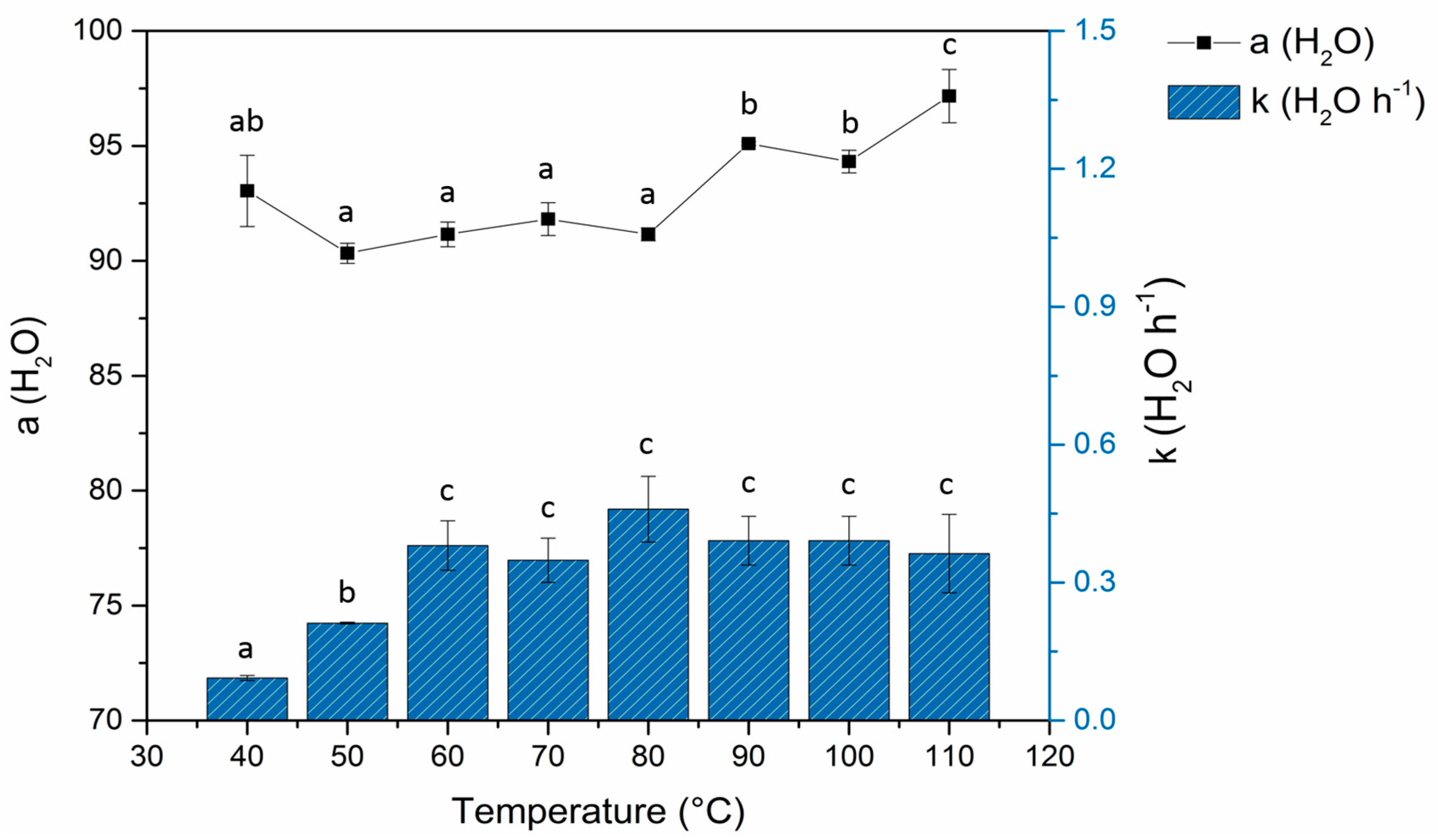
3.1. Effect of Temperature on Drying Kinetics Parameters and Moisture in Lemon Residues

Table 1 shows the moisture content before and after drying lemon residues at the different temperatures evaluated. The moisture content of the raw material was 84.64 ± 0.13% (Table 1). The drying kinetics parameters were obtained by transforming the moisture content in water loss percentage and fitted to a non-linear Gompertz model with R2 ≥ 0.997. The results of the kinetic parameters (Figure 1) showed significant differences (p < 0.05) in maximum water loss (parameter a ) within the temperature interval of the study. The highest a value (96.62 ± 1.37%) was achieved with a temperature of 110 °C, which was significantly different to the others. The minimum a value was obtained at a temperature of 50 °C. Nevertheless, no significant differences were observed within the temperatures of 40 °C to 80 °C. In the samples dried at 40 °C and 50 °C, moisture values below 15% were reached at 48 and 24 h, respectively. For the samples exposed to drying temperatures from 60 °C to 110 °C, a moisture content value lower than 15% (value require to the preservation of the product) was reached at 12 h. On the other hand, the lower drying rate (parameter k ) was presented at 40 °C. Nevertheless, an increase was observed when the drying temperature was increased to 60 °C, above 60 °C, the drying rates were not significantly different (p > 0.05) among the temperatures evaluated. These results suggest that the residues dried at higher temperatures suffered a hardening phenomenon, which prevented the release of water, thereby decreasing the drying rate. Consequently, temperatures higher than 60 °C did not promote a significant increase in the drying rate. Several authors also reported similar phenomena during the dehydration of other foods, such as banana and orange peels [19,25,26]. Panyawong and Devahastin [27] attributed the hardening to the accumulation of solutes (such as sugars and salts) which migrate to the outer layer along with the water during drying and then solidifies to create an impermeable film that prevents the mobilization of the remaining water.

3.2. Total Phenolic Content, Antimicrobial Activity and Antioxidant Capacity of Lemon Residues Flour Obtained at Different Temperatures

Flours obtained at the end of the drying period at each temperature evaluated showed variations in the total phenolic content (Table 1). Initially, fresh residues showed a TPC of 55.62 ± 0.14 mg GAE g−1 D W, however, after drying at all temperatures evaluated, this value decreased significantly (p < 0.05). Flours obtained from 40 °C to 110 °C showed a TPC content on the interval of 30 to 39 mg GAE g−1 D W, which indicates that the phenolic compounds in the flours suffered thermal degradation during drying. Several studies related to the dehydration of citrus residues have reported similar significant reductions in the total polyphenolic content due to the exposure of the material to high temperatures in combination with long drying times [28]. This reduction in phenolic compounds is thought to be associated with the union of the phenolic compounds to proteins, or to the alteration in the chemical structure of the phenol which prevents its extraction [16,29].
TPC values obtained from temperatures of 40 to 90 °C presented the higher reduction compared to the fresh extract as reported by Manqin Fua et al. [28] who observed a reduction in TPC from Citri Reticulata samples dried at temperatures above 35 °C. However, TPC results of 100 and 110 °C presented an increment compared to the lower temperatures. It is assumed that the variations in phenol concentration during drying processes are probably due to the early degradation of phenolic compounds favored by high temperatures and prolonged exposure to heat due to the release of phenolic substances caused by the rupture of the ether, ester or acetal covalent bonds. Thermal degradation may occur at the same time that the phenolic substances are released, but its concentration may be observed when maximal degradation is reached at the temperatures evaluated [29]. Salvatore Mutari et al. [30] suggests that higher temperatures can improve the solubility of phenolic compounds leading to the breakdown of cellular structures and improving the release of phenolic compounds such phenolic acids (ferulic, galic, and vanillic acid) previously bound to the macromolecules of the cell wall. These results suggest the possibility to develop drying protocols to favor the obtaining of specific phenolic compounds.
The results of qualitative antimicrobial and antioxidant capacity tests are shown in Table 1. The fresh lemon peel residues showed strong antimicrobial activity against S. aureus, moderate activity against E. coli and weak activity against S. typhimurium. Flours showed strong activity against S. aureus but weak activity against S. typhimurium and E. coli. However, the flours obtained at temperatures of 50 °C and 110 °C showed moderate activity against E. coli and S. typhimurium respectively. These last results could be related to a higher concentration of phenolic compounds in the sample obtained at 110 °C, suggesting the existence of a major concentration of those compounds that presented antimicrobial activity against gram-negative bacteria that were also presented in the fresh sample. For sample obtained at 50 °C a possible difference in the phenolic profile could favor the presence of antimicrobial compounds that effect in a higher way E. coli and S. typhimurium bacteria.
The antioxidant capacity of the fresh lemon peel residues was considerably low, with a DPPH and ABTS radical inhibition value around 10% and 20%, respectively. Lemon residue flours, on the other hand, presented significantly (p < 0.05) higher inhibition values among the temperatures evaluated. The products obtained under dehydration temperatures of 40 °C and 50 °C showed the highest values for DPPH analysis, and 50 °C, 100 °C y 110 °C for ABTS test. In both antioxidant tests, 80 °C samples presented the lowest antioxidant capacity. Flours obtained at temperatures from 60 °C to 110 °C showed values in the range of 835.18 to 493.26 mM TE g−1 DW by DPPH analysis. While samples obtained from 60 °C to 90 °C showed values in the range of 885.62, 887.92 mM TE g−1 DW according to the ABTS test.
The results of ABTS were higher to DPPH-scavenging potential. Levels of antioxidant capacity were higher to the reported by Esparza-Martínez et al. [30] in lime wastes (122–683 and 540–839 μm Trolox/g DW for DPPH and ABTS, respectively) and Ubando-Rivera in Mexican lime peel (11.3–35.5% inhibition DPPH). The antioxidant activity of phenolic compounds, especially flavonoids, is strongly influenced by their chemical structure, mainly due to the presence of three functional groups: ortho-dihydroxy (catechol) structure in the B-ring; the 2,3-double bond, in conjugation with a 4-oxo function; and the presence of both 3-(a)-and 5-(b)-hydroxyl groups. These groups confer greater stability to the aroxyl radicals by dislocating the electron from the B ring [31].

3.3. Phenolic Profile in Lemon Residue Flours Obtained from 40 °C to 110 °C

Phenolic extracts of flours obtained from lemon residues at temperatures from 40 °C to 110 °C were analyzed by UPLC-PDA chromatography. The phenol composition of the extracts (profile) and the quantity of each phenol detected are presented in Table 2. TPC determined by UPLC-PDA ranged from 30.16 ± 0.27 to 61.47 ± 0.06 mg g−1 DW. These results are higher than the range of TPC reported by Ubando-Rivera et al. [32] (10.55–19.90 mg g− 1 DW), who studied the TPC content on the peel of Mexican variety of lime (Citrus Aurantifolia (C) Swingle ). TPC values obtained by Folin Ciaocalteu Test were higher than chromatographic results, this may be due to nonphenolic reducing substances, such as reducing sugar and ascorbic acid presents in the samples [33]. The TPC values obtained from temperatures of 40 to 70 °C indicated a decrease compared to the fresh extract as reported by Fu et al. [34] who observed a reduction in TPC from Citri Reticulata samples dried at temperatures above 35 °C. However, TPC results indicated that from 90 to 110 °C an increment on TPC is observed. Mutari et al. [35] suggest that higher temperatures can improve the solubility of phenolic compounds leading to the breakdown of cellular structures and improving the release of phenolic compounds previously bound to the macromolecules of the cell wall. The two major compounds present in the fresh sample were eriocitrin and hesperidin, followed by naringin and naringenin. Del Río et al. [8] reported similar concentrations of eriocitrin and hesperidin in C. lemon cv Fino with values of 22.5 mg g−1 D W (flavedo + albedo) and 18.3 mg g−1 D W (flavedo + albedo), respectively. These compounds were also found in various citrus juices with concentrations ranging from 2 to 150 mg L−1 of eriocitrin and 4 to 56 mg L−1 of hesperidin. The concentrations depend on variety, the maturity of the fruits and the soil conditions of crops [34,35].
The concentration of individual phenolic compounds was affected by drying temperatures showing statistical differences (p < 0.05) (Table 2). The results indicated that hesperidin was the only compound detected in extracts drying at all different temperatures. While, caffeic acid and sinapic acid, were detected in extracts obtained at drying temperatures above 60 and 80 °C, respectively. In contrast, eriocitrin was not observed at temperatures above 90 °C. A different effect was observed for naringin and naringenin, which is not presented just at a temperature of 100 °C. Additionally, in fresh and dried samples obtained below 90 °C one non indentified chromatographic peak was observed, then at temperatures from 90 to 110 °C one more chromatographic peak appeared indicating that this compound could be a degradation product from eriocitrin (Table 2). These results reinforce the suggestion that the establishment of drying protocols of lemon waste using specific temperatures is of great interest to obtain particular phenolic compounds from the residues in order to increase its value. The variation on phenolic compounds in samples dried at different temperature was reported by Xu et al. [36]. in peels of huyou (Citrus sinensis (L.) × C. grandis (L.)) and by Esparza-Martínez et al. [30] in lime wastes. Shyi-Neng Lou et al. [37]. reported that flavonoids compounds such as eriocitrin and hesperidin could be degraded at temperatures over 130 °C. Likewise, M’hiri et al. [38] explained that in dried materials, the components in the cells adhere to each other making the solvent extraction more difficult, and as a result, the recovery of compounds decrease. Conversely, the formation of cafeic and sinapic acids might be due to the availability of precursors of phenolic compounds that can be converted by the non-enzymatic process to phenolic molecules, which could explain the increments of this compounds as drying temperature increased [39]. Additionally, Salvatore Mutari et al. [30] suggests that the solubility of phenolic compounds increase at higher extraction temperatures promoting the liberation of phenolic acids. This results could infer that the major antioxidant activity in samples obtained from 40° to 80 °C is related to the flavonoids eriocitrin and hesperidin and in samples obtained from 90 to 110 °C is related to the hesperidin and the phenolic acids as caffeic and sinapic acids. The UPLC-ESI-MS confirmed the presence of hesperidin in all samples and the presence of the 6 reported compounds (caffeic acid, eriocitrin, sinapic acid, naringin, hesperidin, and naringenin) among the samples.
The drying temperatures of 50 °C and 110 °C were selected for evaluation of the changes of TPC, antimicrobial activity and antioxidant capacity related to drying time. The above based on the phenolic profile that indicated a major concentration of hesperidin and eriocitin in the sample obtained at 50 °C, and major TPC by UPLC-DAD of 110 °C as well as the results of the biological properties evaluation.

3.4. Changes of TPC during the Drying Process in Lemon Residue Flour at 50 °C and 110 °C

The changes of phenolic content during the drying process of lemon residues at temperatures of 50 °C and 110 °C showed significant variations (p < 0.05) (Table 3). Both treatments displayed degradation of phenols during the first hours of drying, which continued until reaching a maximum reduction of ~ 50% at twelve hours in the 50 °C treatment and ~ 40% at six hours in the 110 °C drying treatment. Surprisingly, an unusual phenomenon occurred in the concentration of phenolic substances in both drying treatments after reaching their maximum degradation points; the highest value of TPC for 50 °C was presented at 15 h, and for 110 °C at the end of the process after 12 h. Both conditions had an impact on the concentration of phenolic compounds, which included degradation and an apparent liberation of phenolic compounds. As described in Section 3.2, the combination of temperature with long drying time caused the degradation of polyphenol compounds. Conversely, there are other studies that report increments in TPC as a result of maintaining the plant material (citrus peels) at high temperatures in conjunction with short drying times [36,40,41]. The increments are products of the breakdown of either ester or acetal covalent bonds that keep the phenolic compounds bound with proteins and arabinoxylans. It has been reported that this breakdown starts to occur at the beginning of the drying process, showing an accumulation of phenolic compounds within minutes of starting the process [40]. Studies of the citrus residue drying process indicate degradation of TPC throughout the process or increment of TPC from the beginning of the drying process [35]. The drying process at 50 °C produces a combination of both phenomena with a clear TPC degradation during the first hours of drying until a minimal point is reached. Later an increase of compounds may be observed during the final hour of the drying process. With regards to the drying process at 110 °C, the TPC degradation is faster, after which the development of phenolic compounds is even more pronounced. Thermal degradation may occur at the same time the phenolic substances are released, but its concentration may be observed when maximal degradation is reached at the temperatures evaluated.

3.5. Changes on Antimicrobial and Antioxidant Capacity during the Drying Process in Lemon Residue Flour at 50 °C and 110 °C

The influence of drying time on the antimicrobial properties of lemon peels against the pathogens Salmonella typhimurium ATCC 14028, Escherichia coli ATCC 25,922 and Staphylococcus aureus ATCC 25,923 are shown in Table 3. Before any dehydration treatment, the minimum inhibitory concentrations were calculated from the fresh material revealing concentrations at 11.70 ± 0.00 mg GAE g−1 D W for S. typhimurium and S. aureus and 17.50 ± 8.20 mg GAE g−1 D W for E. coli.
Exposure of the lemon residues to a temperature of 50 °C caused some variations in the MIC, which depended on the drying time and the pathogen evaluated. The values of the antimicrobial inhibition of S. typhimurium obtained during the dehydration process remained constant during the first 18 h. At the end of dehydration, a decrease in MIC (5.80 ± 0.00 mg GAE g−1 D W) was observed, thus indicating potentiation of antimicrobial activity. However, the inhibition concentration of the S. aureus pathogen remained unchanged during and after dehydration. The antimicrobial activity against E. coli was more affected by the drying process revealing inhibition values between 17.50 mg GAE g−1 D W to 23.00 mg GAE g−1 D W during the drying process and later decreasing to an inhibition concentration value of 11.70 ± 0.00 mg GAE g1 D W, showing a clear improvement in antimicrobial activity.
The exposure of lemon residues to a temperature of 110 °C also produced some variations in inhibition concentrations. MIC values for S. typhimurium, S. aureus and E. coli fluctuated during the dehydration process, showing concentrations ranging from 17.50 mg GAE g−1 D W to 23.30 mg GAE g−1 D W. Antimicrobial activity improved after the drying process, with MIC values of 5.80 ± 0.00 mg GAE g−1 D W for S. typhimurium and 11.70 ± 0.00 mg GAE g−1 D W for S. aureus and E. coli.
The inhibition concentrations for the S. typhimurium, S. aureus and E. coli pathogens which was manifested in the lemon residue flours at the end of the thermal dehydration process at 50 °C were the same as those obtained in dehydrated residues at 110 °C (5.80 ± 0.00 mg GAE g−1 D W for S. typhimurium and 11.70 ± 0.00 mg GAE g−1 D W for S. aureus and E. coli). In addition, the MIC values for S. typhimurium and E. coli found in the dried residues were lower compared to those obtained in fresh material, revealing that the dehydration process improved the antimicrobial activity against these particular pathogens. The lowered MIC values may have been caused by the activation or formation of active phenolic species produced from heating as indicated in Section 3.2.
The antimicrobial activity of dried lemon residues is probably related to the presence of the glycosylated flavanone hesperidin, which has inhibitory effects against S. aureus, S. hemolyticus, B. subtilis, E. coli, Klebsiella species, P. aeruginosa, S. typhi, S. dysenteriae, S. flexneri and V. cholera [42]. Yi et al. [43] also reported that hesperidin had inhibitory effects on S. aureus, E. faecals, and S. epidermidis bacteria at higher levels than other flavonoids such as nobiletin and tangeretin.
The results of the drying time related to the antioxidant capacity of the lemon residues are shown in Table 3. The percentage of inhibition for the DPPH radical in the fresh material was 10.59 ± 0.91%. The lemon residues exposed to the temperatures of 50 °C and 110 °C showed significant increases (p < 0.05) in the percentage of DPPH radical inhibition as the drying time increased. The higher inhibition percentages were recorded at 50 °C drying temperature with maximum inhibition values of 73.73 ± 1.12% and 50.07 ± 5.15%, at 50 °C and 110 °C respectively. Both maximum inhibition values were obtained at the end of the drying process and based on the chromatographic results, it can be inferred that antioxidant capacity may be attributed in the case of 50 °C to both eriocitrin and hesperidin compounds and to hesperidin for the sample obtained at 110 °C.
The flavonoids hesperidin and eriocitrin have been reported as the main compounds which demonstrate antioxidant capacities in citrus fruits [8]. Goulas and Manganaris [10] explained that the potent antioxidant activity found in the Valeria and Mandora varieties of Citrus sinensis was mainly attributed to the presence of hesperidin, a compound that has a radical uptake activity similar to Trolox. Recent studies have also shown that hesperidin can act as an antioxidant agent in vivo systems. For example, Bharathi and Jagedeesan [44] demonstrated that an oral dose of 5 mg kg−1 of hesperidin can significantly decrease levels of oxidation in the liver tissue of rats that had been administered toxic doses of mercury chloride. The administration of hesperidin also recovered normal levels of antioxidant and non-antioxidant enzymes such as glutathione peroxidase (GPX), catalase (CAT) and superoxide dismutase (SOD) [44].
Eriocitrin has also shown important antioxidant effects in vivo systems. Minato et al. [45] reported that intragastrical administration of eriocitrin 600 mg kg−1 in rats prior to an exercise routine significantly suppressed an increase in thiobarbituric acid reactive substances (TBARS) caused by lipid peroxidation during exercise. Certain markers of oxidative stress such as Nε-(hexanoyl) lysine (HEL), O-dityrosine (DT) and nitrotyrosine (NT) were significantly suppressed by the administration of eriocitrin prior to exercise [45].
According to our previous work [46], where a phenolic extract of Citrus aurantium was partially purified, a synergic effect of phenolic compounds was suggested after the higher antioxidant activity found in the crude extract in comparison with the purified fractions at a specific TPC. These results were also reported by Shiraishi et al., [47] who evaluated the antioxidant activity of the phenolic compounds in table grapes.
Due to the higher antioxidant activity obtained in lemon residue flours at a drying temperature of 50 °C and the major presence of eriocitrin this flour was selected for proximal analysis and testing for shelf-life stability of bioactive compounds.

3.6. Proximal Composition of Lemon Peel Residues Flours Obtained from Thermal Dehydration at 50 °C

Proximal composition analysis of lemon residue flour was performed. The content of protein in the lemon residue flours was 5.97%, while the content of ash was 6.39%, revealing values comparable with other citrus residues [48]. The lemon residue flours contained a low percentage of fat (1.29%) but a very high content of carbohydrates (79.15%). The flours showed 15.55% of raw fiber, which makes it a potentially valuable source of fiber for human consumption. Vitamin C, which is useful for collagen and connective tissue formation while helping absorb inorganic iron and carrying antioxidant properties, was measured at 3.53 mg/100 g in the lemon residue flours. Calcium and potassium were also found in the lemon residue flours at values of 8705 mg/kg and 6706 mg/kg, respectively, which shows that the flour is a good source of those minerals.

3.7. The Shelf Life of Lemon Residue Flours Obtained from Thermal Dehydration at 50 °C

The lemon residues flour obtained under thermal treatment at 50 °C were subjected to a shelf life test to evaluate the stability of the flours as a source of polyphenol compounds. Accordingly, Antioxidant Activity (AA) and color parameter (as h * coordinate) were used to indicate flour quality as parameters for shelf life determination. Values at 0 % of AA and 68.09° of h * were established as critical parameters. Due to the AA critical value was not reached during the shelf life experimental work, there were no significant changes during the shelf life study, therefore the h * coordinate results were used to establish a shelf life. Although antioxidant activity decreases during storage because of the polyphenol degradation, the results obtained from AA throughout time indicated that the functional properties of the flour remained stable under test conditions, rendering that the particular lemon residues such as flour, a good and stable source of phenolic compounds with high antioxidant capacity and antimicrobial activity.
Color values showed decreases over the time of the test, likely caused by a degradation of pigments, acid ascorbic oxidation and the Maillard reaction [28]. Color values related to storage time were fitted to a linear regression model and reaction rate ( k ) values were determined for the four temperatures evaluated (Figure 2A). Arrhenius Equation (5) was used to analyze the temperature dependence of reactions’ rate constants. k values were transformed to ln k values and then drawn versus 1/absolute temperature (Figure 2B). The activation energy E a and pre-exponential factor A were calculated according to the above equation, resulting in values of 67273.39 J mol−1 and 1.16 × 10¹⁰ days−1, respectively. The rate constant at 25 °C ( k 25   ° C ) and 35 °C ( k 35   ° C ) was also calculated with a value of 0.0190 days−1 and 0.0458 days−1, respectively.
Based on the critical parameter of h * coordinate, previously established at 68.09° ( L * = 50.92 , a * = 11.51 , b * = 28.62 ), when the flour develops a brown color, making it less appropriate for use as a food additive, as color is an important consumer consideration with flours. Thus with the shelf-life at k 25   ° C , at room temperature, was estimated to have a duration of 889 days ( ~ 30 months). The lemon residue flours stored in regions where the temperature is hotter k 35   ° C , resulted in a 369 days ( ~ 12 months) duration. Nevertheless, it is important to specify the final use of the flour to determine the shelf life. If the flours will be used as an additive itself, the values using the color as a critical parameter expressed herein may be used to estimate shelf life. If the flour will be used as a source of bioactive compounds, the results obtained here in suggested that drying process favors the preservation of the biological activity of compounds for a larger period and a longer shelf life experiment may be conducted.

4. Conclusions

The effects of different temperatures on the drying process of lemon residues showed a combined phenomenon of degradation of polyphenolic substances during the first few hours of drying and a concentration of them at the end of the process. This previously unreported phenomenon allowed the presentation of the biological activity of the extracted phenols at different drying temperatures. MIC values were slightly affected during the course of drying, presenting higher activity against S. typhimurium. During the drying of the lemon residues, the antioxidant capacity experienced significant changes. Drying temperatures of 50 °C and 110 °C showed substantial increases in the inhibition percentage of the DPPH radical, whereby as the drying time increased, the highest value for samples was obtained at 50 °C. The biological activities observed may be attributed to the presence of hesperidin and eriocitrin since they were the only flavonoids detected by PDA and confirmed by MS in SIR mode. This study recommends using a temperature of 50 °C in drying as a technological treatment to obtain stable nutraceutical flours, which act as a source of hesperidin and eriocitrin carrying antimicrobial activity and antioxidant capacity. Additionally the results suggest the possibility to develop drying protocols to favor the obtaining of specific phenolic compounds. Using color changes as a critical parameter, the flours presented an extended shelf life of around 26 months at room temperature (25 °C). This work provides an alternative to reduce the accumulation of lemon residues as well as obtaining a value-added product.

Author Contributions

J.P.-V. and L.B.-D. for the performance of experiments, N.M.-T. for the development of the chromatographic techniques, T.A.-T. for the analysis of the results and supervision of experiments performed in his laboratory, Á.S.-C., U.G.-C., and N.P. for the approach of the research, the topic expertise, supervision of the experimental work, analysis of the information and the structure of the paper.

Funding

This research was funding by CONACYT-Yucatán government through Project Fomix Yuc-2011-C09-0169165 and the APC was funded by CIATEJ Own Resources.

Acknowledgments

The authors thanks CONACYT to the grant of Lizzie Baas-Dzul and Ingrid Rodriguez-Buenfil and Manuel Ramirez-Sucre for the comments provided in the Lizzie Baas thesis development and Jamie Pruitt, Esq. for the English editing.

Conflicts of Interest

The authors declare no conflict of interest.

References

  1. SIAP Agri-food and Fisheries Information Service. 2018. Available online: https://nube.siap.gob.mx/cierreagricola/ (accessed on 5 August 2019).
  2. USDA. U.S. Department of Agriculture. 2017. Available online: http://apps.fas.usda.gov/psdonline/circulars/citrus.pdf (accessed on 23 April 2017).
  3. González-Molina, E.; Domínguez-Perles, R.; Moreno, D.A.; García-Viguera, C. Natural bioactive compounds of Citrus limon for food and health. J. Pharm. Biomed. Anal. 2010, 51, 327–345. [Google Scholar] [CrossRef] [PubMed]
  4. Rezzadori, K.; Benedetti, S.; Amante, E.R. Proposals for the residues recovery: Orange waste as raw material for new products. Food Bioprod. Process. 2012, 90, 606–614. [Google Scholar] [CrossRef]
  5. Ayora-Talavera, T.; Ramos-Chan, C.; Covarrubias-Cárdenas, A.; Sánchez-Contreras, A.; García-Cruz, U.; Pacheco, L.N. Evaluation of Pectin Extraction Conditions and Polyphenol Profile from Citrus x lantifolia Waste: Potential Application as Functional Ingredients. Agriculture 2017, 7, 28. [Google Scholar] [CrossRef]
  6. Volpe, M.; Panno, D.; Volpe, R.; Messineo, A. Upgrade of citrus waste as a biofuel via slow pyrolysis. J. Anal. Appl. Pyrolysis 2015, 115, 66–76. [Google Scholar] [CrossRef]
  7. Casquete, R.; Marilia, S.; Martín, A.; Ruiz-moyano, S.; Saraiva, J.A.; Córdoba, M.G.; Teixeira, P. Evaluation of the effect of high pressure on total phenolic content, antioxidant and antimicrobial activity of citrus peels. Innov. Food Sci. Emerg. Technol. 2015, 31, 37–44. [Google Scholar] [CrossRef]
  8. Del Río, J.A.; Fuster, M.D.; Gómez, P.; Porras, I.; García-Lidón, A.; Ortuño, A. Citrus limon: A source of flavonoids of pharmaceutical interest. Food Chem. 2004, 84, 457–461. [Google Scholar] [CrossRef]
  9. Benavente-García, O.; Castillo, J. Update on uses and properties of citrus flavonoids: New findings in anticancer, cardiovascular, and anti-inflammatory activity. J. Agric. Food Chem. 2008, 56, 6185–6205. [Google Scholar] [CrossRef] [PubMed]
  10. Goulas, V.; Manganaris, G.; Manganaris, G. Exploring the phytochemical content and the antioxidant potential of Citrus fruits grown in Cyprus. Food Chem. 2012, 131, 39–47. [Google Scholar] [CrossRef]
  11. Khan, M.K.; Dangles, O. A comprehensive review on flavanones, the major citrus polyphenols. J. Food Compos. Anal. 2014, 33, 85–104. [Google Scholar] [CrossRef]
  12. Wang, Y.C.; Chuang, Y.C.; Hsu, H.W. The flavonoid, carotenoid and pectin content in peels of citrus cultivated in Taiwan. Food Chem. 2008, 106, 277–284. [Google Scholar] [CrossRef]
  13. Sanz-Puig, M.; Pina-Pérez, M.C.; Martínez-López, A.; Rodrigo, D. Escherichia coli O157: H7 and Salmonella Typhimurium inactivation by the effect of mandarin, lemon, and orange by-products in reference medium and in oat-fruit juice mixed beverage. LWT Food Sci. Technol. 2016, 66, 7–14. [Google Scholar] [CrossRef]
  14. Dutta, D.; Dutta, A.; Raychaudhuri, U.; Chakraborty, R. Rheological characteristics and thermal degradation kinetics of beta-carotene in pumpkin puree. J. Food Eng. 2006, 76, 538–546. [Google Scholar] [CrossRef]
  15. Mirzabeigi Kesbi, O.; Sadeghi, M.; Mireei, S.A. Quality assessment and modeling of microwave-convective drying of lemon slices. Eng. Agric. Environ. Food 2016, 9, 216–223. [Google Scholar] [CrossRef]
  16. Chen, M.L.; Yang, D.J.; Liu, S.C. Effects of drying temperature on the flavonoid, phenolic acid and antioxidative capacities of the methanol extract of citrus fruit (Citrus sinensis (L.) Osbeck) peels. Int. J. Food Sci. Technol. 2011, 46, 1179–1185. [Google Scholar] [CrossRef]
  17. Papoutsis, K.; Pristijono, P.; Golding, J.B.; Stathopoulos, C.E.; Bowyer, M.C.; Scarlett, C.J.; Vuong, Q.V. Effect of vacuum-drying, hot air-drying and freeze-drying on polyphenols and antioxidant capacity of lemon (Citrus limon) pomace aqueous extracts. Int. J. Food Sci. Technol. 2017, 52, 880–887. [Google Scholar] [CrossRef]
  18. Michalska, A.; Wojdyło, A.; Łysiak, G.P.; Lech, K.; Figiel, A. Functional relationships between phytochemicals and drying conditions during the processing of blackcurrant pomace into powders. Adv. Powder Technol. 2017, 28, 1340–1348. [Google Scholar] [CrossRef]
  19. Simal, S.; Femenía, A.; Llull, P.; Rosselló, C. Dehydration of aloe vera: Simulation of drying curves and evaluation of functional properties. J. Food Eng. 2000, 43, 109–114. [Google Scholar] [CrossRef]
  20. Kuljarachanan, T.; Devahastin, S.; Chiewchan, N. Evolution of antioxidant compounds in lime residues during drying. Food Chem. 2009, 113, 944–949. [Google Scholar] [CrossRef]
  21. AOAC. Official Methods of Analyses of the Association of Official Analytical Chemists; Association of Official Analytical Chemists: Washington, DC, USA, 1990. [Google Scholar]
  22. Vasco, C.; Ruales, J.; Kamal-Eldin, A. Total phenolic compounds and antioxidant capacities of major fruits from Ecuador. Food Chem. 2008, 111, 816–823. [Google Scholar] [CrossRef]
  23. Covarrubias-Cárdenas, A.; Martínez-Castillo, J.; Medina-Torres, N.; Ayora-Talavera, T.; Espinosa-Andrews, H.; García-Cruz, N.; Pacheco, N. Antioxidant Capacity and UPLC-PDA ESI-MS Phenolic Profile of Stevia rebaudiana Dry Powder Extracts Obtained by Ultrasound Assisted Extraction. Agronomy 2018, 8, 170. [Google Scholar] [CrossRef]
  24. Lagnika, L.; Anago, E.; Atindehou, M.; Adjahoutonon, B.; Dramane, K.; Sanni, A. Antimicrobial activity of Crataeva religiosa Forst against bacteria isolated from Thryonomys swinderianus Temminck. Afr. J. Biotechnol. 2011, 10, 10034–10039. [Google Scholar] [Green Version]
  25. Garau, M.C.; Simal, S.; Rosselló, C.; Femenia, A. Effect of air-drying temperature on physico-chemical properties of dietary fibre and antioxidant capacity of orange (Citrus aurantium v. Canoneta) by-products. Food Chem. 2007, 104, 1014–1024. [Google Scholar] [CrossRef]
  26. Demirel, D.; Turhan, M. Air-drying behavior of Dwarf Cavendish and Gros Michel banana slices. J. Food Eng. 2003, 59, 1–11. [Google Scholar] [CrossRef]
  27. Panyawong, S.; Devahastin, S. Determination of deformation of a food product undergoing different drying methods and conditions via evolution of a shape factor. J. Food Eng. 2007, 78, 151–161. [Google Scholar] [CrossRef]
  28. Nesrine, G.R.; Catherine, B.; Nabil, K.; Nourhène, B.M. Effect of Air-Drying Temperature on Kinetics of Quality Attributes of Lemon (Citrus limon cv. lunari) Peels. Dry. Technol. 2015, 33, 1581–1589. [Google Scholar]
  29. Martín-Cabrejas, M.A.; Aguilera, Y.; Pedrosa, M.M.; Cuadrado, C.; Hernández, T.; Díaz, S.; Esteban, R.M. The impact of dehydration process on antinutrients and protein digestibility of some legume flours. Food Chem. 2009, 114, 1063–1068. [Google Scholar] [CrossRef]
  30. Esparza-Martínez, F.J.; Miranda-López, R.; Guzman-Maldonado, S.H. Effect of air-drying temperature on extractable and non-extractable phenolics and antioxidant capacity of lime wastes. Ind. Crop. Prod. 2016, 84, 1–6. [Google Scholar] [CrossRef]
  31. Tripoli, E.; La Guardia, M.; Giammanco, S.; Di Majo, D.; Giammanco, M. Citrus flavonoids: Molecular structure, biological activity and nutritional properties: A review. Food Chem. 2007, 104, 466–479. [Google Scholar] [CrossRef]
  32. Ubando-Rivera, J.; Navarro-Ocaña, A.; Valdivia-López, M.A. Mexican lime peel: Comparative study on contents of dietary fibre and associated antioxidant activity. Food Chem. 2005, 89, 57–61. [Google Scholar] [CrossRef]
  33. Zhao, G.; Zhang, R.; Liu, L.; Deng, Y.; Wei, Z.; Zhang, Y.; Ma, Y.; Zhang, M. Different thermal drying methods affect the phenolic profiles, their bioaccessibility and antioxidant activity in Rhodomyrtus tomentosa (Ait.) Hassk berries. LWT Food Sci. Technol. 2017, 79, 260–266. [Google Scholar] [CrossRef]
  34. Fu, M.; An, K.; Xu, Y.; Chen, Y.; Wu, J.; Yu, Y.; Zou, B.; Xiao, G.; Ti, H. Effects of different milling methods on physicochemical properties of common buckwheat fl our. LWT Food Sci. Technol. 2018, 92, 220–226. [Google Scholar]
  35. Multari, S.; Marsol-Vall, A.; Keskitalo, M.; Yang, B.; Suomela, J.P. Effects of different drying temperatures on the content of phenolic compounds and carotenoids in quinoa seeds (Chenopodium quinoa) from Finland. J. Food Compos. Anal. 2018, 72, 75–82. [Google Scholar] [CrossRef]
  36. Xu, G.; Ye, X.; Chen, J.; Liu, D. Effect of Heat Treatment on the Phenolic Compounds and Antioxidant Capacity of Citrus Peel Extract. J. Agric. Food Chem. 2007, 55, 330–335. [Google Scholar] [CrossRef]
  37. Lou, S.N.; Lai, Y.C.; Huang, J.D.; Ho, C.T.; Ferng, L.H.A.; Chang, Y.C. Drying effect on flavonoid composition and antioxidant activity of immature kumquat. Food Chem. 2015, 171, 356–363. [Google Scholar] [CrossRef] [PubMed]
  38. M’hiri, N.; Ghali, R.; Ben Nasr, I.; Boudhrioua, N. Effect of different drying processes on functional properties of industrial lemon byproduct. Process. Saf. Environ. Prot. 2018, 116, 450–460. [Google Scholar] [CrossRef]
  39. Que, F.; Mao, L.; Fang, X.; Wu, T. Comparison of hot air-drying and freeze-drying on the physicochemical properties and antioxidant activities of pumpkin (Cucurbita moschata Duch.) flours. Int. J. Food Sci. Technol. 2008, 43, 1195–1201. [Google Scholar] [CrossRef]
  40. Jeong, S.M.; Kim, S.Y.; Kim, D.R.; Jo, S.C.; Nam, K.C.; Ahn, D.U.; Lee, S.C. Effect of heat treatment on the antioxidant activity of extracts from citrus peels. J. Agric. Food Chem. 2004, 52, 3389–3393. [Google Scholar] [CrossRef]
  41. Kim, S.Y.; Jeong, S.M.; Kim, S.J.; Jeon, K.I.; Park, E.; Park, H.R.; Lee, S.C. Effect of heat treatment on the antioxidative and antigenotoxic activity of extracts from persimmon (Diospyros kaki L.) peel. Biosci. Biotechnol. Biochem. 2006, 70, 999–1002. [Google Scholar] [CrossRef] [PubMed]
  42. Iranshahi, M.; Rezaee, R.; Parhiz, H.; Roohbakhsh, A.; Soltani, F. Protective effects of flavonoids against microbes and toxins: The cases of hesperidin and hesperetin. Life Sci. 2015, 137, 125–132. [Google Scholar] [CrossRef] [PubMed]
  43. Yi, Z.; Yu, Y.; Liang, Y.; Zeng, B. In vitro antioxidant and antimicrobial activities of the extract of Pericarpium Citri Reticulatae of a new Citrus cultivar and its main flavonoids. LWT Food Sci. Technol. 2008, 41, 597–603. [Google Scholar] [CrossRef]
  44. Bharathi, E.; Jagadeesan, G. Antioxidant potential of hesperidin and ellagic acid on renal toxicity induced by mercuric chloride in rats. Biomed. Prev. Nutr. 2014, 4, 131–136. [Google Scholar] [CrossRef]
  45. Minato, K.I.; Miyake, Y.; Fukumoto, S.; Yamamoto, K.; Kato, Y.; Shimomura, Y.; Osawa, T. Lemon flavonoid, eriocitrin, suppresses exercise-induced oxidative damage in rat liver. Life Sci. 2003, 72, 1609–1616. [Google Scholar] [CrossRef]
  46. Ana, C.C.; Jesús, P.V.; Hugo, E.A.; Teresa, A.T.; Ulises, G.C.; Neith, P. Antioxidant capacity and UPLC–PDA ESI–MS polyphenolic profile of Citrus aurantium extracts obtained by ultrasound assisted extraction. J. Food Sci. Technol. 2018, 55, 5106–5114. [Google Scholar] [CrossRef] [PubMed]
  47. Shiraishi, M.; Shinomiya, R.C.H. Varietal differences in polyphenol contents, antioxidant activities and their correlations in table grape cultivars bred in Japan. Sci. Hortic. 2018, 227, 272–277. [Google Scholar] [CrossRef]
  48. Boluda-aguilar, M.; López-gómez, A. Production of bioethanol by fermentation of lemon (Citrus limon L.) peel wastes pretreated with steam explosion. Ind. Crop. Prod. 2013, 41, 188–197. [Google Scholar] [CrossRef]
Figure 1. The kinetic parameters of drying lemon residues estimated by Gompertz model.
Agronomy 09 00474 g001
Figure 2. Change of hue coordinate as a function of storage time in accelerated shelf-life test in lemon residues flours obtained by thermal process at 50 °C, and fit to linear regression.
Agronomy 09 00474 g002
Table 1. Moisture content (%), total phenolic content (TPC), antimicrobial qualitative test activity (AQT) and antioxidant capacity of lemon residues dried at 40–110 °C.
T (°C)/Drying Time (h)Moisture Content (%)Energy (Watts)TPC 2 (mg GAE g−1 DW)AQT 1Antioxidant Capacity 3
123DPPH: TE 4 (mM TE g−1 DW)ABTS: TE 4 (mM TE g−1 DW)
Initial/084.64 ± 0.13 e055.62 ± 0.14 e++++++4.48 ± 0.38 a11.82 ± 0.21 a
40/249.73 ± 0.13 c28,80030.11 ± 0.32 a,b+++++742.82 ± 46.04 f880.77 ± 10.76 b
50/2010.46 ± 1.54 d28,80030.62 ± 1.13 a,b+++++++835.18 ± 37.49 g945.64 ± 20.00 e
60/2010.52 ± 0.09 d24,00030.47 ± 1.13 a,b,+++++516.31 ± 19.36 d885.62 ±7.58 b,c
70/2010.62 ± 0.32 d24,00030.89 ± 1.13 a,b+++++260.70 ± 63.13 b900.18 ± 2.17 b,c,d
80/169.52 ± 0.06 c19,20031.77 ± 2.59 b+++++389.93 ± 85.45 c761.26 ± 15.35 a
90/167.64 ± 0.33 b19,20029.58 ± 1.06 a++++597.32 ± 68.36 g887.92 ± 9.57 b
100/127.22 ± 0.22 b14,40035.03 ± 0.29 c+++++466.99 ± 3.49 d914.48 ± 13.39 d
110/125.05 ± 0.52 a14,40039.31 ± 2.57 d+++++++493.26 ± 3.66 d910.40 ± 8.44 c,d
1 Antimicrobial quality test activity against to (1) S. typhimurium, (2) S. aureus and (3) E. coli. +++ Strong activity (no bacterial growth), ++ Moderate activity (2–4 CFU), + Weak activity (5–12 CFU)-No activity (>12 CFU). 2 Total Phenolic Conten. 3 Radical scavenging activity. 4 Trolox Equivalent. All data were reported as mean ± standard deviation, n = 3. Radical scavenging activity Antimicrobial qualitative test activity was determined from the observation of three samples. Values with the same letter in the same column were not significantly different at p < 0.05 determined by multiple comparisons of means by LSD-Fisher test.
Table 2. Phenolic compounds profile of extracts from lemon residues flours obtained at different temperatures.
T (°C)Phenolic Compounds (mg/g DW)Uid*. PeaksTPC (mg/g DW)
Caffeic AcidEriocitrinSinapic AcidNaringinHesperidinNaringenin
Freshn/d23.17 ± 0.07 b n/d5.37 ± 0.06 d 20.62 ± 0.69 f 1.46 ± 2.33 f 150.62 ± 0.74 c
40 °Cn/d12.54 ± 0.10 an/d2.24 ± 0.13 a15.22 ± 0.18 d0.19 ± 0.02 a130.19 ± 0.21 a
50 °Cn/d13.67 ± 1.59 an/d2.96 ± 0.18 a,b16.14 ± 0.07 e 0.90 ± 0.01 d 133.67 ± 0.64 b
60 °C2.33 ± 0.12 a 11.59 ± 0.43 a n/d2.35 ± 0.11 a 13.37 ± 0.07 c 0.52 ± 0.01 b 130.16 ± 0.27 a
70 °C5.89 ± 0.10 b 12.75 ± 2.19 a n/d3.81 ± 0.24 b,c 11.28 ± 0.17 b 0.81 ± 0.02 c 134.54 ± 1.08 b
80 °C17.06 ± 0.16 c11.89 ± 0.39 a 2.43 ± 0.11a 4.16 ± 0.14 b,c,d 11.33 ± 0.20 b 2.71 ± 0.01 h 149.58 ± 2.41 c
90 °C29.32 ± 0.06 d n/d8.92 ± 0.03d 9.45 ± 1.62 e11.25 ± 0.06 b 2.06 ± 0.01 g 261.00 ± 1.86 d
100 °C39.07 ± 0.15 f n/d4.69 ± 0.02b n/d8.94 ± 0.08 an/d252.70 ± 0.25 c
110 °C37.93 ± 0.04 e n/d6.68 ± 0.07c 4.55 ± 0.10 c,d 8.39 ± 0.34 a 1.07 ± 0 e 261.47 ± 0.06 d
Results are presented as mean ± SD and represent means of four independent measurements. Values with different letters (a–h) within the same column differ significantly (p < 0.05). n/d = not detected (i.e., below the detection level). Uid* = unidentified.
Table 3. Changes on moisture content, TPC, antimicrobial activity and antioxidant capacity during the drying process of lemon residues at 50 °C.
T (°C)/Drying Time (h)Moisture Content (%)TPC (mg GAE g−1 D W)Antimicrobial Activity (MIC) 1
(mg GAE g−1 D W)
Antioxidant Capacity
S. typhimuriumS. aureusE. coliDPPH RSA 2 (%) InhibitionTE 3 (mM TE g−1 DW)
Initial/084.64 ± 0.13 h55.62 ± 0.2 f11.70 ± 0 b11.70 ± 0 a17.50 ± 8.20 b10.59 ± 0.91 a4.48 ± 0.38
50 °C/1263.4 ± 2.2 g27.82 ± 0.9 a11.70 ± 0 b11.70 ± 0 a23.30 ± 0 c30.27 + 2.35 d298.96 + 56.13
50 °C/1559.85 ± 1.0 f32.09 ± 0.5 b,c11.70 ± 0 b11.70 ± 0 a23.30 ± 0.00 c55.76 ± 1.99 d,e561.44 ± 34.7
50 °C/1840.12 ± 1.2 e29.41 ± 1.3 a,b11.70 ± 0 b11.70 ± 0 a23.30 ± 0 c61.73 ± 1.55 e635.06 ± 27.03
50 °C/2119.25 ± 0.7 c28.32 ± 0.6 a5.80 ± 0 a11.70 ± 0 a17.50 ± 8.20 b71.50 ± 1.18 f755.52 ± 20.58
50 °C/2410.46 ± 1.5 b31.40 ± 0.1 b,c5.80 ± 0 a11.70 ± 0 a11.70 ± 0 a73.73 ± 1.12 f783.02 ± 19.53
110 °C/424.12 + 1.3 d34.17 + 0.6 d23.30 ± 0.00 d23.30 ± 0.0 c23.30 ± 0.00 c21.70 + 0.92 b141.47 + 16.04
110 °C/621.58 + 1.5 c31.85 + 0.8 b,c17.50 ± 8.20 c17.50 ± 8.2 b17.50 ± 8.20 b37.69 + 1.72 c338.63 + 29.99
110 °C/86.96 + 2.1 a33.12 + 1.1 c,d17.50 ± 8.20 c17.50 ± 8.2 b11.70 ± 0.00 a47.43 + 2.29 d458.73 + 39.93
110 °C/125.62 + 1.3 a39.05 + 0.9 e5.80 ± 0.00a11.70 ± 0.0 a11.70 ± 0.00 a50.07 + 5.15 d491.28 + 89.81
1 The Antimicrobial activity was evaluated as Minimum Inhibitory Concentration, 2 Radical scavenging activity. 3 Trolox Equivalent. All data were expressed as mean ± standard deviation, n = 3. Values with the same letter in the same block were not significantly different at p < 0.05 determined by multiple comparisons of means by LSD-Fisher test.

