Article

Redox Balance Differentially Affects Biomechanics in Permeabilized Single Muscle Fibres—Active and Passive Force Assessments with the Myorobot

1
Institute of Medical Biotechnology, Department of Chemical and Biological Engineering, Friedrich-Alexander-University Erlangen-Nürnberg, Paul-Gordan-Str. 3, 91052 Erlangen, Germany
2
Erlangen Graduate School in Advanced Optical Technologies (SAOT), Paul-Gordan-Str. 6, 91052 Erlangen, Germany
3
College of Health & Human Performance, University of Florida, 1864 Stadium Road, Gainesville, FL 32611, USA
*
Author to whom correspondence should be addressed.
Submission received: 26 August 2022 / Revised: 16 November 2022 / Accepted: 17 November 2022 / Published: 22 November 2022
(This article belongs to the Special Issue Redox Control of Cell Signaling in Cardiac and Skeletal Muscle)

Abstract

:
An oxidizing redox state imposes unique effects on the contractile properties of muscle. Permeabilized fibres show reduced active force generation in the presence of H2O2. However, our knowledge about the muscle fibre’s elasticity or flexibility is limited due to shortcomings in assessing the passive stress–strain properties, mostly due to technically limited experimental setups. The MyoRobot is an automated biomechatronics platform that is well-capable of not only investigating calcium responsiveness of active contraction but also features precise stretch actuation to examine the passive stress–strain behaviour. Both were carried out in a consecutive recording sequence on the same fibre for 10 single fibres in total. We denote a significantly diminished maximum calcium-saturated force for fibres exposed to ≥500 µM H2O2, with no marked alteration of the pCa50 value. In contrast to active contraction (e.g., maximum isometric force activation), passive restoration stress (force per area) significantly increases for fibres exposed to an oxidizing environment, as they showed a non-linear stress–strain relationship. Our data support the idea that a highly oxidizing environment promotes non-linear fibre stiffening and confirms that our MyoRobot platform is a suitable tool for investigating redox-related changes in muscle biomechanics.

1. Introduction

It is well known that reactive oxygen species (ROS) play a crucial role in many cellular processes and influence the performance of muscles [1,2]. As free radicals, ROS shift the cell’s microenvironment towards a more oxidized state, which, in muscle fibres, was found to impact fatiguability, muscle damage, and active force generation [3,4,5,6]. Such deviation from its native state, or rather changes to the cell’s reductive or oxidative environment, were enclosed in the term redox balance [4]. Ever since then, studies have sought to unravel the effects, the origin, and the mechanisms of a shifted redox balance in muscle contraction. Even up to date, there is debate on how ROS are effectively produced, which species contributes to what extent, and what intracellular signalling pathways are involved in their action [1]. In that setting, hydrogen peroxide (H2O2) has attracted major interest, as it is naturally produced in mitochondria, an abundant organelle to be found in muscle cells [7,8]. Nowadays, the diverse effects attributed to H2O2 (e.g., altered Ca2+ sensitivity in intact fibres [9] or decreased maximum Ca2+-activated force in permeabilized fibres [5,10]) were found to be rather mediated by dismuted superoxide or hydroxyl radicals, both being produced in the presence of H2O2 [1,9]. Interestingly, more regular and frequent muscle activation accelerates the production of ROS [11], which leads to performance adaptation [4] but also cellular damage [12]. These effects also seem to increase with temperature [13].
So far, an oxidative redox balance due to H2O2 and/or related radicals was found to influence muscle fatigue, excitation–contraction coupling, maximum Ca2+ activated force, and the Ca2+ sensitivity in numerous studies [1,5,6,10]. While the findings from those studies improve our understanding of the redox balance in muscle, study outcomes vary (e.g., inhibiting effect of H2O2 to sarcoplasmic Ca2+-release [14] vs. a large insensitivity of the Ca2+-release towards externally applied H2O2 [6]). These differences, however, seem to relate to the use of either intact single muscle fibres or permeabilized muscle fibres.
From that perspective, particularly the influence of ROS on the Ca2+ sensitivity of the contractile apparatus is well studied. Intriguingly, ROS seem to have little effect on the Ca2+ sensitivity of the contractile apparatus in permeabilized muscle fibres [5,10,14], whereas intact fibre studies suggest a decreased Ca2+ sensitivity under oxidizing conditions (presence of H2O2) [1,4]. This is why it was hypothesized that ROS affect the Ca2+ sensitivity by pathways that seem to be absent/disrupted once the fibre is permeabilized and the cytosol is replaced by an externally applied solution [1]. The decrease in Ca2+ sensitivity is believed to occur from (cellularly) generated hydroxyl radicals or superoxide [9,15] that attack methionine or cysteine residues [16,17]. Therefore, ROS-related performance adaptations could at least be partially reversed by the anti-oxidant agent Dithiothreitol (DTT), predominantly in intact muscle fibres [1,4,18]. Additionally, in intact muscle fibres, DTT causes a further reduction in maximum force production [4], an effect that was not detected in permeabilized fibre preparations [3]. Consistently in permeabilized fibres, many studies have independently reported a decrease in the maximum Ca2+-activated force in the presence of DTT [5,10,15], which, in intact fibres, is suspected to originate from oxidized methionine residues [18]. Whether these potential interaction sites for DTT and H2O2 translate to the effects seen in permeabilized muscle fibres and how they align with the proposed pathways in intact muscle fibres remains yet to be resolved. Further, it is unclear from the literature how and to what degree these ROS-mediated changes impact the axial elasticity of the fibre, as an important parameter for damage prevention, structural memory, and the ability to resist large strains [19].
Therefore, we systematically assessed muscle mechanics at different redox balances in permeabilized muscle fibres during active contraction and passive force recordings with the automated MyoRobot biomechatronics platform [20]. We investigated the maximum Ca2+ activated force, the Ca2+ sensitivity of the contractile apparatus, and axial elasticity from stress–strain curves. All recordings were successively performed on the same fibre (sarcomere length (SL) adjusted in the range of 2.3–2.4 µm) in a fully automated sequence, both in the presence and absence of H2O2 (100 µM, 500 µM, 1000 µM), as well as in the reduced state (after 60 s of 1000 µM DTT incubation). Our findings of decreased Ca2+ activated force under oxidative stress (H2O2 presence) and unaltered Ca2+ sensitivity support previously proposed hypotheses and observations in permeabilized fibres. Based on these, we hypothesized that ROS exposure would increase the passive axial stiffness of muscle fibres, for which we carried out technically challenging passive stress–strain relationship recordings with our automated biomechatronics platform. Here, we indeed denote a significant fibre stiffening at high external concentrations of H2O2 that seem to affect the linear elastic deformation regime, potentially through inducing molecular stiffening. The technology presented here delivers consistent and reliable results and is, thus, well-suited to study the complex effects of ROS on the contractile properties of muscle.

2. Materials and Methods

2.1. Animal Handling and Single Fibre Preparation

All animal experiments were conducted in accordance with the approved animal experimentation guidelines at the Friedrich-Alexander-Universität Erlangen-Nürnberg (TS06/2016), the German regulations for the care of laboratory animals, and the guidelines of the Federation of European Laboratory Animal Sciences Associations. All animals for this study were male CS57BL/6 mice of approx. 30 g body weight and aged between 15–21 weeks. The samples were obtained through a tissue-sharing collaboration with local institutes. Anesthesia was induced by inhalation of 5% isoflurane, followed by cervical dislocation and subsequent cutting off of the hind limb. From here on, M. extensor digitorum longus (EDL) isolation was performed with physiological Ringer’s solution, which, after EDL isolation, was replaced by high K+ solution (HKS, see below) for manually tethering the single muscle fibres under a stereomicroscope (Olympus SZ-X7) using fine forceps (Dumont #5) and scissors (FST). HKS permanently depolarizes the membrane and inactivates Na+ channels after an initial contraction. During that procedure, the muscle was constantly pinned onto a Polydimethylsiloxane (PDMS, Sylgard, Dow Corning)-coated Petri dish under slight pre-stretch. Isolated single muscle fibres were then tied to two micro silk knots (each end) and transferred to the MyoRobot for fibre mounting in an HKS droplet. Each fibre was tied with one end to the pin of a force transducer (FT) and the other end to a voice–coil actuator (VC) before lowering it in the idle well underneath. Please note that prior to any other solution exposure, the single fibre was chemically permeabilized for 20 s in 0.01% (w/v) saponin. This permeabilization ensures complete diffusional access to the intracellular myoplasm, in particular, to clamp the myoplasmic free Ca2+ to any desired levels and to initially wash out endogenously produced ROS from the preparation. Additionally, since the Ca2+ solutions were highly EGTA-buffered, SR Ca2+ release would not affect the steady-state pCa-force data [21,22].
High-Potassium Solution (HKS): 140 mM K-glutamate, 1 mM MgCl2, 10 mM HEPES buffer, 1 mM EGTA, 10 mM glucose, pH: 7.0, osmolality: 275 mosmol/kg.

2.2. System Electronics and Software

The MyoRobot, described in more detail in [20], is a fully automated biomechatronics platform to assess a plethora of active and passive biomechanics properties. For this, it is equipped with a piezo-optical FT (TR5S, Myotronic UG, Heidelberg, Germany) and a linear VC actuator (CAL12-010-51-B5A, SMAC) acting as a counter pin for fibre mounting. A rack housing 32 individual wells to be filled with distinct bioactive solutions is actuated via a bi-axial linear stage configuration to allow automated exposure to different environmental conditions (see Active and Passive Force Assessment Protocols). The FT data are sampled at 100 Hz during each recording and read and displayed in a LabVIEW-written (National Instruments) user interface. The latter is likewise used to operate the entire setup digitally, as well as load and execute the biomechanics recording protocols. The rack, to whose solutions the sample is exposed, features a glass bottom with a miniaturized ∼25× magnifying optics system. This allows for capturing the morphological sample parameters, such as the SL and fibre diameter (cross section assumed as circular) online.

2.3. Bioactive Solutions

The solutions for fibre manipulation (activation or relaxation) were used and composed as given in [23]. Their summarized physiological purpose is mentioned in the table below (Table 1). In general, High Activating solution (HA) is an EGTA-buffered Ca2+-rich solution that activates troponin-C. High Relaxing solution (HR) is a Ca2+-free, EGTA-buffered solution to relax the fibre. Low Relaxing solution (LR) relies on the mild Ca2+ buffering agent HDTA and is likewise Ca2+ free. All of the control (CTRL) experiments were carried out in the absence of H2O2, while any recordings at oxidizing conditions were achieved by supplementing the base solutions with 100, 500, or 1000 µM H2O2. Reducing conditions were achieved by incubation in dithiothreitol (DTT) for 60 s before returning to the base solutions, free of H2O2.

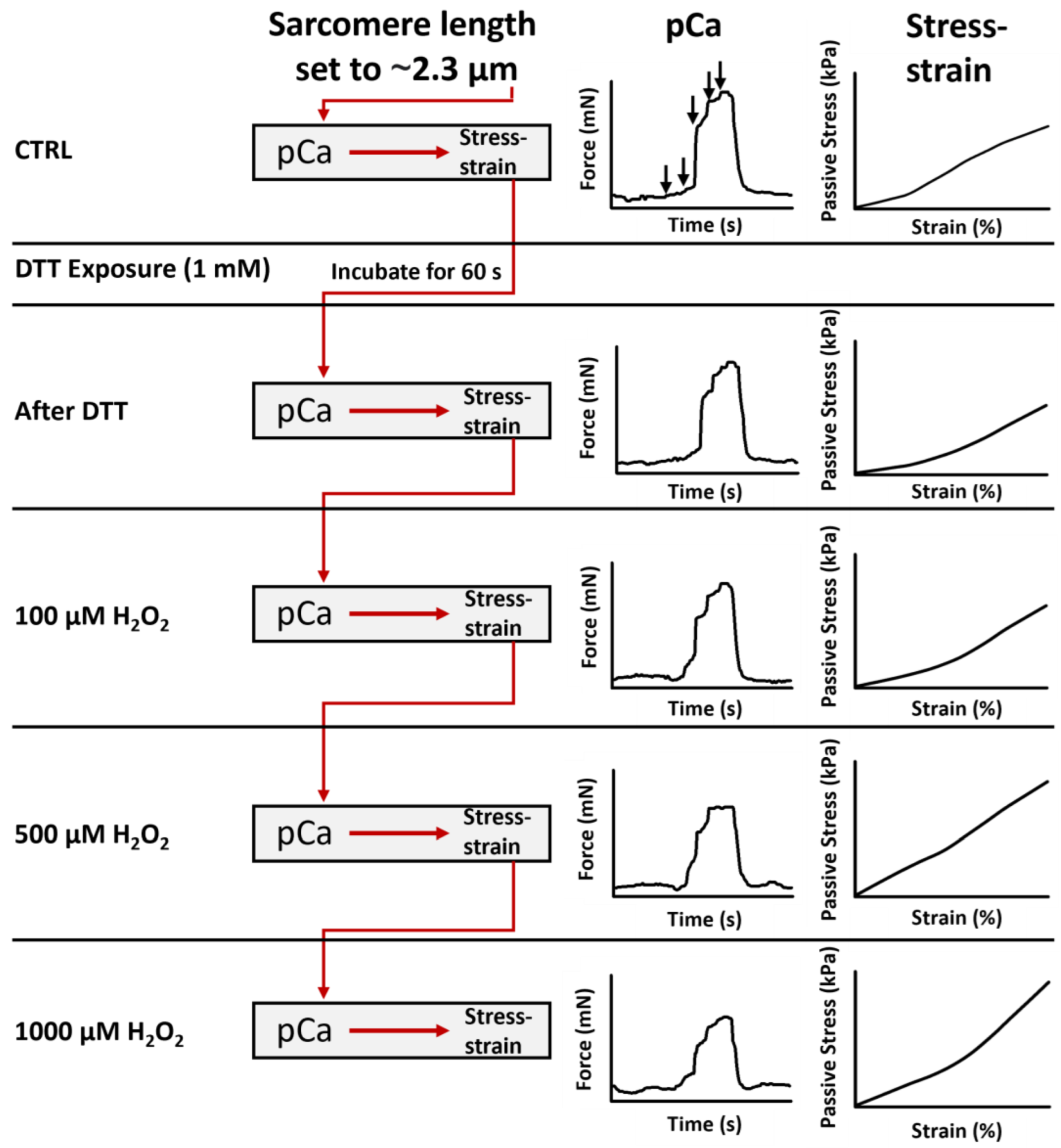
2.4. Active and Passive Force Assessment Protocols

In the present study, we utilized our MyoRobot technology to study the effects of different redox environments on permeabilized single muscle fibre biomechanics. Being equipped with a 32-well rack allows our biomechatronics system to perform all active and passive force recordings under five different conditions (CTRL, DTT (reduced), 100 µM H2O2, 500 µM H2O2, and 1000 µM H2O2) in the same single fibre. Each experiment took approx. 3 h. The sequence of recordings is depicted in a flow chart in Figure 1.
First, single EDL muscle fibres were chemically permeabilized by immersion for 20 s in saponin 0.01% (w/v). The so-called ‘skinned’ fibre preparation is known to be very robust, and experimental sessions on a single fibre can robustly involve repetitive sets of complete pCa-force activation cycles [24,25]. Consequently, the fibre’s average sarcomere length (SL) was set to ∼2.3 µm by adjusting the position of the VC (equivalent to elongating the fibre), and its diameter was read out. Then, we recorded the pCa-force relationships and carried out stress–strain recordings under CTRL conditions after 60 s DTT incubation and in solutions supplemented with 100 µM H2O2, 500 µM H2O2, and 1000 µM H2O2, as given in Figure 1.
Active Force—Ca2+ sensitivity of the contractile apparatus & maximum Ca2+ activated force. Single fibres were exposed to wells with solutions of increasing Ca2+ concentration (correspondingly decreasing pCa values) to assess the Ca2+ sensitivity at the chemical–mechanical interface of troponin C. At a pCa value of 4.92 (Ca2+ saturated conditions), force generation was assumed to be maximum. Force generation followed a staircase-like pattern, as seen in Figure 1. The analysis included extracting the steady-state forces for each pCa step and plotting them as a pCa-force graph. Fitting a sigmoidal curve to the data ( y = 10 b x c b + 10 b x , with x being pCa, and y being force, [26]) allowed for the extraction of the pCa50 value (−log10(c)) and the Hill parameter (b). Maximum Ca2+ saturated force was calculated according to the steady-state force at pCa 4.92.
Passive Force—Stress–strain relationships via slow, linear fibre elongation. The stress–strain relationships were recorded in LR solution to maintain relaxing conditions during the protocol. The VC elongated the fibre at an optimized velocity of 1 µm/s from 100% to 140% of its resting length (=40% strain). Knowing the force and the fibre diameter allowed us to convert forces to the more robust parameter stress, also taking the cross-sectional area into account. The maximum restoration stress was computed as the stress exerted at 40% strain. The axial stiffness was extracted from the force–elongation curves, as linear fits were applied to each section of 10% strain. The increase in the respective fit curve equals the passive axial stiffness [27].

3. Data Analysis

All of the data were analysed for normality by performing a Shapiro–Wilk test to determine the appropriate testing procedure. When a data set was not normally distributed, nonparametric tests were adopted. In this case, a Kruskal–Wallis test was performed based on the rank sum of the data, followed by post hoc analysis running Tukey’s method to test for a difference of means to a significance level of 0.05. When a data set was normally distributed, a parametric one-way ANOVA was carried out. This was followed by an all-pairwise comparison of means based on the Holm–Sidak method to a significance level of 0.05. If the statistical tests confirmed any significant difference, this was indicated as vertical bars in all plots shown, including an asterisk to indicate the magnitude as follows: * = p < 0.05, ** = p < 0.01, *** = p < 0.005. Any absence of such indicators is equivalent to “no significant difference confirmed”.

4. Results

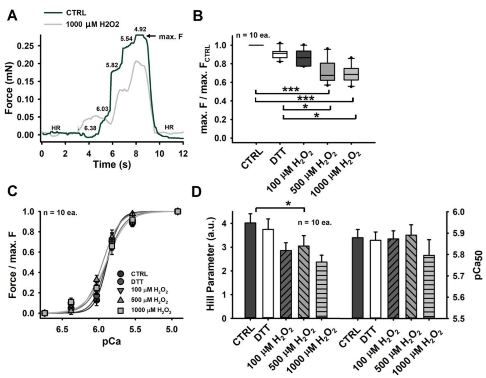
4.1. Maximum Ca2+ Saturated Force Is Diminished in Permeabilized Single Fibres in an Oxidizing Environment

Investigating the active contractility in the presence of Ca2+ ions in different redox states yielded a significantly reduced maximum force generation with increasing amounts of H2O2 (≥500 µM). Although a reducing environment (after 60 s of DTT incubation) and the presence of 100 µM H2O2 also seemed to slightly reduce contractile performance, their effects were not confirmed to be significant (Figure 2B).
According to our observations, this decline in contractile force does not correlate with reduced Ca2+ sensitivity of the contractile apparatus, which was consistently unaltered in all differently imposed redox states. This can be seen in Figure 2C, where all pCa-force curves matched very closely (similar inflection points and, thus, similar pCa50 values). The only marked difference we could observe was a gradual decrease in the curves’ steepness, which is reflected by a reduced Hill parameter, as shown in Figure 2D, displaying a single significant difference between the CTRL fibres and the fibres exposed to 500 µM H2O2 (roughly 30% less than for CTRL levels).
Please note that the pCa-force relationship is explicitly given in force values, as the most common notation. The corresponding stress values are given in Table 2. Additionally, note that any prior conversion from force to stress would not affect the well-known and routinised analysis procedure applied here.

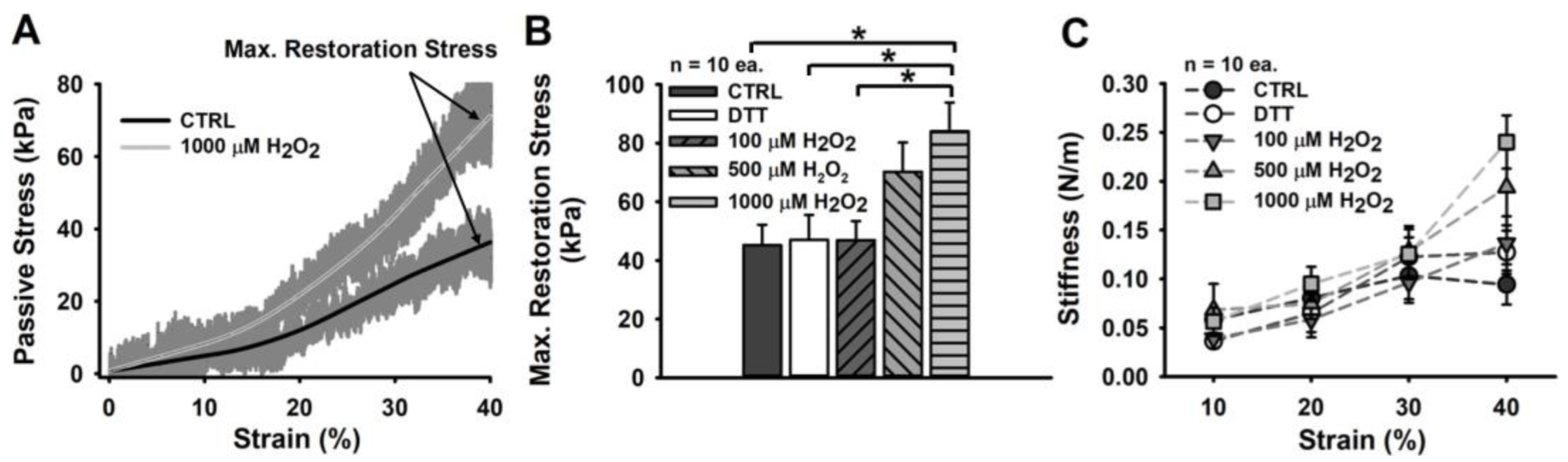
4.2. An Oxidizing Redox Balance Imposes a Non-Linear Fibre Stiffening onto Its Passive Strain Resistance

In contrast to what we observed for the active contractile properties, large concentrations of H2O2 correlate with an increased passive axial stiffness in permeabilized single muscle fibres. As seen in Figure 3B, maximum restoration stress at 40% strain appears to double for fibres exposed to ≥500 µM H2O2, which was supported by statistical analysis.
The observed stiffening under oxidizing conditions is reflected by a strong, non-linear increase that can be seen in the average stress–strain plot (Figure 3A). Here, particularly fibres exposed to 1000 µM H2O2 (light grey curve) display a much more exponential incline commencing at strain levels of ∼20% than the CTRL fibres. Exposure to the reducing agent DTT had no marked effects on passive restoration stress.
The non-linear stiffening, which is much more apparent under oxidizing conditions, was likewise confirmed by stiffness analysis, performed by applying linear fits to each section of 10% strain (Figure 3C). Here, the entity of the samples presents a gradual but constant stiffness increase. However, the fibres exposed to ≥500 µM H2O2 strongly deviate from this relationship, displaying notably enlarged stiffness values above 20–30% strain. Such effects were not detectable for CTRL fibres, fibres exposed to DTT, or immersed in a mildly oxidizing environment (100 µM H2O2).

5. Discussion

5.1. Reduced Maximum Ca2+ Activated Force Does Not Originate from an Altered Ca2+ Sensitivity Inpermeabilized Muscle Fibres

Intact muscle fibres, exposed or subjected to H2O2 (100–500 µM) present with reduced tetanic force, which is suggested to arise from Ca2+ desensitization [28,29]. Intriguingly, the maximum Ca2+-activated force generation in permeabilized muscle fibres was also found to be reduced, however, entirely independent from the fibre’s Ca2+ sensitivity being unchanged [3,30]. This is likewise reflected in our analysis of active single-fibre biomechanics recordings being carried out in a more robust and highly automated robotics system. Particularly for larger H2O2 concentrations (≥500 µM), we observed a significant reduction in the maximum Ca2+-activated force (Figure 2B). Overall, our mean absolute force value of ∼0.26 mN for CTRL fibres matches well with maximum Ca2+ saturated force levels of other studies [31,32]. A conversion to stress, assuming a circular cross-section and taking the average CTRL fibre diameter of 44.3 µm into account, yields 170 kPa—a reasonable stress value for murine EDL single fibres (∼200 kPa [33]) that also translates well to the whole muscle (∼300 kPa [34]), still containing their ECM. Similar to Plant et al. (2000) and Lamb and Posterino (2003) [3,30], we also detected no change in Ca2+ sensitivity (pCa50)and observed values ranging from 5.8 to 5.9 in our experiments. In additional agreement with the literature, our Hill parameter likewise declined by over 30% (Figure 2D).
The diverse findings in intact muscle fibres (tetanic force reduced due to the Ca2+ desensitization of the contractile apparatus) and permeabilized fibres (Ca2+ activated force reduced without altered Ca2+ sensitivity) suggest that ROS influence force generation via a pathway that is disrupted when the muscle fibre is permeabilized [1]. This would involve a cytosolic protein as a key player, which, after skinning, is replaced by an externally applied solution. This theory finds support in studies involving the reducing agent DTT, which is capable of ‘shielding’ the fibre against aggressive, oxidizing radicals [1,3,4]. DTT is a membrane-permeant thiol donor that reverses disulphide bonds to regenerate reduced thiol (-SH) moieties. DTT was shown to reverse the Ca2+-desensitizing effects of H2O2 in intact muscle fibres [1,4]. Intriguingly, this ‘protective’, or sometimes even described as ‘preventive’ [1] effect, appears to be absent in permeabilized fibres—as shown by others [3] and confirmed by our study in a highly automated system. In this setting, the MyoRobot allowed us to reproduce previous findings on the Ca2+-responsiveness of the contractile apparatus with ease, in due time, and in an automated sequence that can be carried out consecutively without interchanging the sample. While the findings from previous studies contribute valuable knowledge, often with sophisticated self-tinkered systems, many of them suffered from mentionable specimen losses within their setups or during their procedure (e.g., over 50% of fibres in [28]). Others needed to estimate important structural parameters, e.g., fibre diameter, due to engineering shortcomings [30]. These challenges must have tremendously prolonged experiment time and required tight supervision, ranking even some of the most extensive biomechanics studies on redox changes in the order of having processed less than ten samples [3,10,30]. Within two weeks, after concluding the experiment design and performing protocol optimizations and adaptations, we were able to record 10 entire data sets from single muscle fibres for all mentioned conditions with our MyoRobot (two–three mouse legs each week, two–three single fibres recorded each day, each recording sequence taking approx. 3 h). This confirms that our technology is a resourceful tool for investigating the redox-related changes in active force generation (e.g., the Ca2+-sensitivity of the contractile apparatus) in permeabilized muscle fibres with ease.

5.2. An Oxidized Redox Balance Promotes Increased Stress with Strain in Permeabilized Muscle Fibres

In contrast to multiple studies that assessed the active biomechanics of skeletal muscle fibres at different redox states, to the best of our knowledge, an assessment of the fibre’s passive mechanical properties has neither been initiated nor performed. This likely originates from the technical complexity and precision–actuation requirements that are neither met by commercial single-fibre biomechanics systems nor by most custom-engineered devices. However, our MyoRobot is well-capable of performing such experiments via extremely slow, truly linear stretches that minimize any viscous relaxation [20]. The passive stiffness of muscle (fibres) plays an important role in many human diseases and conditions that are accompanied by increased ROS activity and decreased muscle function, such as Duchenne Muscular Dystrophy [35,36], fibrosis [37,38,39], sepsis [40,41], and cancer cachexia [42,43].
In the literature, H2O2 is described as a potential modulator of cysteine and methionine residues, acting as an important redox sensor in selected proteins [44]. Given the proposed role of methionine [9,18] and cysteine [45], it is plausible that both contribute to protein function and may constitute a mechanism for protein regulation [46]. Under oxidizing conditions, methionine is oxidized to methionine sulfoxide through the addition of an oxygen atom to its side chain. According to Hoshi et al. (2001), this increases the stiffness and the polarity of the molecule, potentially augmenting dipole–dipole forces [46].
In addition, methionine sulfoxide is involved in the conversion from catalytic cysteine, in its protonated form (Cys-SH), to cysteine sulfenic acid (Cys-SOH) via a reductase (see [47], Figure 1) [47]. The presence of both forms then allows the formation of strong disulphide bonds that, in vitro, can only be reversed by an external reducing agent (e.g., DTT) [47]. As such, the prolonged effect of H2O2 on two of its potential targets (cysteine and methionine) would suggest a marked increase in passive restoration stress through additionally introduced disulphide bonds.
Interestingly, this idea finds support in our results, postulating an almost two-fold, significant increase in passive restoration stress of samples exposed to 1000 µM H2O2 over CTRL single fibres (Figure 3B). This effect was already seen at 500 µM H2O2, yet, in a less pronounced fashion, while lower concentrations or reducing conditions (after 60 s incubation in DTT) imposed no detectable change in passive restoration stress at 140% of the fibre’s resting length (SL: ∼3.2 µm). The here-reported maximum stress values of ∼40 kPa for the CTRL fibres are slightly larger than the values obtained from comparable experiments on murine EDL (e.g., ∼30 kPa at ∼3.2 µm SL [48], and ∼21 kPa at 40% strain [20]), yet, still approx. half the stress reported in M. soleus single fibres (∼60 kPa [49]). The latter are known to display twice the stiffness of EDL single fibres in rats [50] and similarly in mice [27].
The marked increase in stiffness observed under highly oxidizing conditions (≥500 µM H2O2) was also expressed by an exponentially increasing stiffness and resistance to strain (restoration force development in response to elongation changes) (Figure 3C), while the CTRL fibres displayed a more linear relationship here. Similar effects were detected when comparing single muscle fibres-to-fibre bundle preparations, with the latter still being surrounded by an extracellular matrix. While single fibres present with an almost linear stress–strain relationship, the presence of an extracellular matrix in fibre bundles contributed to rather exponential stress–strain relationships and, thus, non-linear effects [48,51,52]. As such, we hypothesize that a highly oxidizing environment promotes a non-linear stress–strain behaviour through a potentially reduced linear elastic stretch regime. However, if such a mechanism can solely be attributed to disulphide bond formation through ROS acting upon cysteine and methionine residues or if it involves post-translational modifications to structural proteins, e.g., actin [53] or titin [54,55,56], remains to be addressed in future studies.
In that regard, a previous study on mechanically ventilated rats in an ICU model associated with increased ROS production has confirmed the presence of multiple post-translational modifications of the myosin-heavy chain [57]. Perspectively, such protein modifications could be directly (e.g., ROS oxidation of regulatory sulfhydryls [58]) or indirectly imposed (e.g., phosphorylation via redox-sensitive kinases [53]). However, it remains to be clarified if the major targets of either potential mechanism truly align with the proteins that would normally determine passive stiffness to a major extent (e.g., titin [59,60], connectin [61], desmin [31], etc.).

6. Conclusions

We provide the first data that examine the passive elastic properties in the form of stress–strain curves performed in single, permeabilized muscle fibres at an oxidizing redox balance. Therefore, our study presents the first link between active and passive biomechanics properties in the field of muscle redox biology. By detecting a significant decrease in maximum Ca2+ activated force in an oxidizing environment, e.g., the presence of H2O2, we could easily reproduce data from previous studies. In addition, we observed an increased passive axial stiffness, significantly enhanced in the fibres exposed to larger concentrations of H2O2 (≥500 µM), which supports a potentially new mechanism for passive stiffness modulation that can be of clinical relevance. However, it is still unclear whether these findings may arise from a rising formation of disulphide bonds on certain amino acids in an oxidizing environment or post-transcriptional modification to structural proteins, which remains to be addressed in future experiments. Nevertheless, our MyoRobot is capable of delivering passive stress–strain data with ease in feasible and repetitive experiments and, thus, will provide a resourceful tool to study redox balance biomechanics in muscles.

Author Contributions

Conceptualization, M.M., L.K., M.B.R., O.F. and M.H.; methodology, M.M., P.R. and L.K.; software, M.M. and M.H.; validation, M.M. and M.H.; formal analysis, M.M. and M.H.; investigation, M.M. and L.K.; resources, O.F.; data curation, O.F. and M.H.; writing—original draft preparation, M.M., O.F. and M.H.; writing—review and editing, M.B.R. and O.F.; visualization, M.M. and M.H.; supervision, M.B.R., O.F. and M.H.; project administration, M.H.; funding acquisition, M.M. and P.R. All authors have read and agreed to the published version of the manuscript.

Funding

This research was funded by the Erlangen Graduate School in Advanced Optical Technologies (SAOT, graduate school GSC 80) throughout a scholarship to M.M. We acknowledge financial support by Deutsche Forschungsgemeinschaft and Friedrich-Alexander-Universität Erlangen-Nürnberg within the funding programme “Open Access Publication Funding”.

Institutional Review Board Statement

Not applicable.

Informed Consent Statement

Not applicable.

Data Availability Statement

The data that support the findings of this study are available from the corresponding author, M.H., upon request.

Conflicts of Interest

The authors declare no conflict of interest. The funders had no role in the design of the study; in the collection, analyses, or interpretation of data; in the writing of the manuscript; or in the decision to publish the results.

Abbreviations

The following abbreviations are used in this manuscript:
EDLM. Extensor Digitorum Longus
CTRLControl
FTForce Transducer
VCVoice Coil
HAHigh Activating (Solution)
HRHigh Relaxing (Solution)
LRLow Relaxing (Solution)
HKSHigh-Potassium Solution
EDTAEthylene glycol-bis(β-aminoethylether))-N,N,N′,N′-tetraacetic acid
HDTAHexamethylenediaminetetraacetic acid
DTTDithiothreitol

References

  1. Moopanar, T.R.; Allen, D.G. The activity-induced reduction of myofibrillar Ca2+ sensitivity in mouse skeletal muscle is reversed by dithiothreitol. J. Physiol. 2006, 571, 191–200. [Google Scholar] [CrossRef] [PubMed]
  2. Reid, M.B. Invited Review: Redox modulation of skeletal muscle contraction: What we know and what we don’t. J. Appl. Physiol. 2001, 90, 724–731. [Google Scholar] [CrossRef] [PubMed] [Green Version]
  3. Lamb, G.D.; Posterino, G.S. Effects of oxidation and reduction on contractile function in skeletal muscle fibres of the rat. J. Physiol. 2003, 546, 149–163. [Google Scholar] [CrossRef] [PubMed]
  4. Andrade, F.H.; Reid, M.B.; Allen, D.G.; Westerblad, H. Effect of hydrogen peroxide and dithiothreitol on contractile function of single skeletal muscle fibres from the mouse. J. Physiol. 1998, 509, 565–575. [Google Scholar] [CrossRef] [PubMed]
  5. Darnley, G.M.; Duke, A.M.; Steele, D.S.; MacFarlane, N.G. Effects of reactive oxygen species on aspects of excitation-contraction coupling in chemically skinned rabbit diaphragm muscle fibres. Exp. Physiol. 2001, 86, 161–168. [Google Scholar] [CrossRef] [Green Version]
  6. Posterino, G.S.; Cellini, M.A.; Lamb, G.D. Effects of oxidation and cytosolic redox conditions on excitation-contraction coupling in rat skeletal muscle. J. Physiol. 2003, 547, 807–823. [Google Scholar] [CrossRef]
  7. Smith, M.A.; Reid, M.B. Redox modulation of contractile function in respiratory and limb skeletal muscle. Respir. Physiol. Neurobiol. 2006, 151, 229–241. [Google Scholar] [CrossRef]
  8. Willingham, T.B.; Ajayi, P.T.; Glancy, B. Subcellular Specialization of Mitochondrial Form and Function in Skeletal Muscle Cells. Front. Cell Dev. Biol. 2021, 9, 757305. [Google Scholar] [CrossRef] [PubMed]
  9. Murphy, R.M.; Dutka, T.L.; Lamb, G.D. Hydroxyl radical and glutathione interactions alter calcium sensitivity and maximum force of the contractile apparatus in rat skeletal muscle fibres. J. Physiol. 2008, 586, 2203–2216. [Google Scholar] [CrossRef]
  10. Callahan, L.A.; She, Z.W.; Nosek, T.M. Superoxide, hydroxyl radical, and hydrogen peroxide effects on single-diaphragm fiber contractile apparatus. J. Appl. Physiol. 2001, 90, 45–54. [Google Scholar] [CrossRef]
  11. Reid, M.B.; Haack, K.E.; Franchek, K.M.; Valberg, P.A.; Kobzik, L.; West, M.S. Reactive oxygen in skeletal muscle. I. Intracellular oxidant kinetics and fatigue in vitro. J. Appl. Physiol. 1992, 73, 1797–1804. [Google Scholar] [CrossRef] [PubMed]
  12. Ji, L.L.; Yeo, D.; Kang, C.; Zhang, T. The role of mitochondria in redox signaling of muscle homeostasis. J. Sport Health Sci. 2020, 9, 386–393. [Google Scholar] [CrossRef] [PubMed]
  13. Zuo, L.; Christofi, F.L.; Wright, V.P.; Liu, C.Y.; Merola, A.J.; Berliner, L.J.; Clanton, T.L. Intra- and extracellular measurement of reactive oxygen species produced during heat stress in diaphragm muscle. Am. J. Physiol.-Cell Physiol. 2000, 279, C1058–C1066. [Google Scholar] [CrossRef] [PubMed] [Green Version]
  14. Brotto, M.A.; Nosek, T.M. Hydrogen peroxide disrupts Ca2+ release from the sarcoplasmic reticulum of rat skeletal muscle fibers. J. Appl. Physiol. 1996, 81, 731–737. [Google Scholar] [CrossRef]
  15. MacFarlane, N.G.; Miller, D.J. Depression of peak force without altering calcium sensitivity by the superoxide anion in chemically skinned cardiac muscle of rat. Circ. Res. 1992, 70, 1217–1224. [Google Scholar] [CrossRef] [Green Version]
  16. Halliwell, B.; Gutteridge, J.M.C. Free Radicals in Biology and Medicine; Oxford University Press: Oxford, UK, 2015. [Google Scholar] [CrossRef]
  17. Van der Poel, C.; Stephenson, D.G. Reversible changes in Ca2+-activation properties of rat skeletal muscle exposed to elevated physiological temperatures. J. Physiol. 2002, 544, 765–776. [Google Scholar] [CrossRef]
  18. Prochniewicz, E.; Lowe, D.A.; Spakowicz, D.J.; Higgins, L.; O’Conor, K.; Thompson, L.V.; Ferrington, D.A.; Thomas, D.D. Functional, structural, and chemical changes in myosin associated with hydrogen peroxide treatment of skeletal muscle fibers. Am. J. Physiol.-Cell Physiol. 2008, 294, C613–C626. [Google Scholar] [CrossRef] [Green Version]
  19. Haug, M.; Ritter, P.; Michael, M.; Reischl, B.; Schurmann, S.; Prols, G.; Friedrich, O. Structure-Function Relationships in Muscle Fibres: MyoRobot Online Assessment of Muscle Fibre Elasticity and Sarcomere Length Distributions. IEEE Trans. Biomed. Eng. 2022, 69, 148–155. [Google Scholar] [CrossRef]
  20. Haug, M.; Meyer, C.; Reischl, B.; Prölß, G.; Nübler, S.; Schürmann, S.; Schneidereit, D.; Heckel, M.; Pöschel, T.; Rupitsch, S.J.; et al. MyoRobot 2.0: An advanced biomechatronics platform for automated, environmentally controlled skeletal muscle single fiber biomechanics assessment employing inbuilt real-time optical imaging. Biosens. Bioelectron. 2019, 138, 111284. [Google Scholar] [CrossRef]
  21. Launikonis, B.S.; Stephenson, D.G. Effect of saponin treatment on the sarcoplasmic reticulum of rat, cane toad and crustacean (Yabby) skeletal muscle. J. Physiol. 1997, 504, 425–437. [Google Scholar] [CrossRef] [Green Version]
  22. Posterino, G.S.; Lamb, G.D.; Stephenson, D.G. Twitch and tetanic force responses and longitudinal propagation of action potentials in skinned skeletal muscle fibres of the rat. J. Physiol. 2000, 527, 131–137. [Google Scholar] [CrossRef] [PubMed]
  23. Haug, M.; Meyer, C.; Reischl, B.; Prölß, G.; Vetter, K.; Iberl, J.; Nübler, S.; Schürmann, S.; Rupitsch, S.J.; Heckel, M.; et al. The MyoRobot technology discloses a premature biomechanical decay of skeletal muscle fiber bundles derived from R349P desminopathy mice. Sci. Rep. 2019, 9, 108. [Google Scholar] [CrossRef] [PubMed] [Green Version]
  24. Friedrich, O.; Yi, B.; Edwards, J.N.; Reischl, B.; Wirth-Huecking, A.; Buttgereit, A.; Lang, R.; Polyak, F.; Liu, I.; Wegner, F.v.; et al. IL-1α reversibly inhibits skeletal muscle ryanodine receptor. A novel mechanism for critical illness myopathy? Am. J. Respir. Cell Mol. Biol. 2014, 50, 1096–1106. [Google Scholar] [CrossRef] [PubMed] [Green Version]
  25. Lamb, G.D.; Stephenson, D.G. Measurement of force and calcium release using mechanically skinned fibers from mammalian skeletal muscle. J. Appl. Physiol. 2018, 125, 1105–1127. [Google Scholar] [CrossRef] [PubMed]
  26. Hill, A.V. The possible effects of the aggregation of the molecules of haemoglobin on its oxygen dissociation. J. Physiol. 1910, 40, 4–7. [Google Scholar]
  27. Haug, M.; Reischl, B.; Prölß, G.; Pollmann, C.; Buckert, T.; Keidel, C.; Schürmann, S.; Hock, M.; Rupitsch, S.; Heckel, M.; et al. The MyoRobot: A novel automated biomechatronics system to assess voltage/Ca2+ biosensors and active/passive biomechanics in muscle and biomaterials. Biosens. Bioelectron. 2018, 102, 589–599. [Google Scholar] [CrossRef]
  28. Moopanar, T.R.; Allen, D.G. Reactive oxygen species reduce myofibrillar Ca2+ sensitivity in fatiguing mouse skeletal muscle at 37 degrees C. J. Physiol. 2005, 564, 189–199. [Google Scholar] [CrossRef]
  29. Bruton, J.D.; Place, N.; Yamada, T.; Silva, J.P.; Andrade, F.H.; Dahlstedt, A.J.; Zhang, S.J.; Katz, A.; Larsson, N.G.; Westerblad, H. Reactive oxygen species and fatigue-induced prolonged low-frequency force depression in skeletal muscle fibres of rats, mice and SOD2 overexpressing mice. J. Physiol. 2008, 586, 175–184. [Google Scholar] [CrossRef]
  30. Plant, D.R.; Lynch, G.S.; Williams, D.A. Hydrogen peroxide modulates Ca2+-activation of single permeabilized fibres from fast- and slow-twitch skeletal muscles of rats. J. Muscle Res. Cell Motil. 2000, 21, 747–752. [Google Scholar] [CrossRef]
  31. Pollmann, C.; Haug, M.; Reischl, B.; Prölß, G.; Pöschel, T.; Rupitsch, S.J.; Clemen, C.S.; Schröder, R.; Friedrich, O. Growing Old Too Early: Skeletal Muscle Single Fiber Biomechanics in Ageing R349P Desmin Knock-in Mice Using the MyoRobot Technology. Int. J. Mol. Sci. 2020, 21, 5501. [Google Scholar] [CrossRef]
  32. Haug, M.; Reischl, B.; Nübler, S.; Kiriaev, L.; Mázala, D.A.G.; Houweling, P.J.; North, K.N.; Friedrich, O.; Head, S.I. Absence of the Z-disc protein a-actinin-3 impairs the mechanical stability of Actn3KO mouse fast-twitch muscle fibres without altering their contractile properties or twitch kinetics. Skelet. Muscle 2022, 12, 14. [Google Scholar] [CrossRef] [PubMed]
  33. Fink, R.H.; Stephenson, D.G.; Williams, D.A. Calcium and strontium activation of single skinned muscle fibres of normal and dystrophic mice. J. Physiol. 1986, 373, 513–525. [Google Scholar] [CrossRef] [PubMed]
  34. Chan, S.; Seto, J.T.; MacArthur, D.G.; Yang, N.; North, K.N.; Head, S.I. A gene for speed: Contractile properties of isolated whole EDL muscle from an alpha-actinin-3 knockout mouse. Am. J. Physiol.-Cell Physiol. 2008, 295, C897–C904. [Google Scholar] [CrossRef] [Green Version]
  35. Choi, M.H.; Ow, J.R.; Yang, N.D.; Taneja, R. Oxidative Stress-Mediated Skeletal Muscle Degeneration: Molecules, Mechanisms, and Therapies. Oxidative Med. Cell. Longev. 2016, 2016, 6842568. [Google Scholar] [CrossRef] [PubMed] [Green Version]
  36. Williams, I.A.; Allen, D.G. The role of reactive oxygen species in the hearts of dystrophin-deficient mdx mice. Am. J. Physiol. Heart Circ. Physiol. 2007, 293, H1969–H1977. [Google Scholar] [CrossRef] [PubMed] [Green Version]
  37. Cheresh, P.; Kim, S.J.; Tulasiram, S.; Kamp, D.W. Oxidative stress and pulmonary fibrosis. Biochim. Biophys. Acta 2013, 1832, 1028–1040. [Google Scholar] [CrossRef] [PubMed] [Green Version]
  38. Richter, K.; Kietzmann, T. Reactive oxygen species and fibrosis: Further evidence of a significant liaison. Cell Tissue Res. 2016, 365, 591–605. [Google Scholar] [CrossRef] [Green Version]
  39. Gonzalez-Gonzalez, F.J.; Chandel, N.S.; Jain, M.; Budinger, G.R.S. Reactive oxygen species as signaling molecules in the development of lung fibrosis. Transl. Res. J. Lab. Clin. Med. 2017, 190, 61–68. [Google Scholar] [CrossRef]
  40. Alamdari, N.; Toraldo, G.; Aversa, Z.; Smith, I.; Castillero, E.; Renaud, G.; Qaisar, R.; Larsson, L.; Jasuja, R.; Hasselgren, P.O. Loss of muscle strength during sepsis is in part regulated by glucocorticoids and is associated with reduced muscle fiber stiffness. Am. J. Physiol. Regul. Integr. Comp. Physiol. 2012, 303, R1090–R1099. [Google Scholar] [CrossRef] [Green Version]
  41. Goossens, C.; Weckx, R.; Derde, S.; van Helleputte, L.; Schneidereit, D.; Haug, M.; Reischl, B.; Friedrich, O.; van den Bosch, L.; van den Berghe, G.; et al. Impact of prolonged sepsis on neural and muscular components of muscle contractions in a mouse model. J. Cachexia Sarcopenia Muscle 2021, 12, 443–455. [Google Scholar] [CrossRef]
  42. Judge, S.M.; Nosacka, R.L.; Delitto, D.; Gerber, M.H.; Cameron, M.E.; Trevino, J.G.; Judge, A.R. Skeletal Muscle Fibrosis in Pancreatic Cancer Patients with Respect to Survival. JNCI Cancer Spectr. 2018, 2, pky043. [Google Scholar] [CrossRef] [PubMed]
  43. Bjørklund, G.; Dadar, M.; Aaseth, J.; Chirumbolo, S.; Pen, J.J. Cancer-associated Cachexia, Reactive Oxygen Species and Nutrition Therapy. Curr. Med. Chem. 2019, 26, 5728–5744. [Google Scholar] [CrossRef] [PubMed]
  44. Finkel, T. Redox-dependent signal transduction. FEBS Lett. 2000, 476, 52–54. [Google Scholar] [CrossRef] [PubMed] [Green Version]
  45. Alegre-Cebollada, J.; Kosuri, P.; Giganti, D.; Eckels, E.; Rivas-Pardo, J.A.; Hamdani, N.; Warren, C.M.; Solaro, R.J.; Linke, W.A.; Fernández, J.M. S-glutathionylation of cryptic cysteines enhances titin elasticity by blocking protein folding. Cell 2014, 156, 1235–1246. [Google Scholar] [CrossRef] [Green Version]
  46. Hoshi, T.; Heinemann, S. Regulation of cell function by methionine oxidation and reduction. J. Physiol. 2001, 531, 1–11. [Google Scholar] [CrossRef] [PubMed]
  47. Lee, E.H.; Lee, K.; Kwak, G.H.; Park, Y.S.; Lee, K.J.; Hwang, K.Y.; Kim, H.Y. Evidence for the dimerization-mediated catalysis of methionine sulfoxide reductase A from Clostridium oremlandii. PLoS ONE 2015, 10, e0131523. [Google Scholar] [CrossRef]
  48. Meyer, G.A.; Lieber, R.L. Elucidation of extracellular matrix mechanics from muscle fibers and fiber bundles. J. Biomech. 2011, 44, 771–773. [Google Scholar] [CrossRef] [Green Version]
  49. Anderson, J.; Joumaa, V.; Stevens, L.; Neagoe, C.; Li, Z.; Mounier, Y.; Linke, W.; Goubel, F. Passive stiffness changes in soleus muscles from desmin knockout mice are not due to titin modifications. Pfluegers Arch. Eur. J. Physiol. 2002, 444, 771–776. [Google Scholar] [CrossRef]
  50. Mutungi, G.; Ranatunga, K.W. The viscous, viscoelastic and elastic characteristics of resting fast and slow mammalian (rat) muscle fibres. J. Physiol. 1996, 496, 827–836. [Google Scholar] [CrossRef]
  51. Gillies, A.R.; Lieber, R.L. Structure and function of the skeletal muscle extracellular matrix. Muscle Nerve 2011, 44, 318–331. [Google Scholar] [CrossRef]
  52. Wood, L.K.; Kayupov, E.; Gumucio, J.P.; Mendias, C.L.; Claflin, D.R.; Brooks, S.V. Intrinsic stiffness of extracellular matrix increases with age in skeletal muscles of mice. J. Appl. Physiol. 2014, 117, 363–369. [Google Scholar] [CrossRef] [PubMed] [Green Version]
  53. Varland, S.; Vandekerckhove, J.; Drazic, A. Actin Post-translational Modifications: The Cinderella of Cytoskeletal Control. Trends Biochem. Sci. 2019, 44, 502–516. [Google Scholar] [CrossRef] [PubMed] [Green Version]
  54. Koser, F.; Loescher, C.; Linke, W.A. Posttranslational modifications of titin from cardiac muscle: How, where, and what for? FEBS J. 2019, 286, 2240–2260. [Google Scholar] [CrossRef] [PubMed] [Green Version]
  55. Grützner, A.; Garcia-Manyes, S.; Kötter, S.; Badilla, C.L.; Fernandez, J.M.; Linke, W.A. Modulation of Titin-Based Stiffness by Disulfide Bonding in the Cardiac Titin N2-B Unique Sequence. Biophys. J. 2009, 97, 825–834. [Google Scholar] [CrossRef] [Green Version]
  56. Loescher, C.M.; Breitkreuz, M.; Li, Y.; Nickel, A.; Unger, A.; Dietl, A.; Schmidt, A.; Mohamed, B.A.; Kötter, S.; Schmitt, J.P.; et al. Regulation of titin-based cardiac stiffness by unfolded domain oxidation (UnDOx). Proc. Natl. Acad. Sci. USA 2020, 117, 24545–24556. [Google Scholar] [CrossRef]
  57. Corpeno, R.; Dworkin, B.; Cacciani, N.; Salah, H.; Bergman, H.M.; Ravara, B.; Vitadello, M.; Gorza, L.; Gustafson, A.M.; Hedström, Y.; et al. Time course analysis of mechanical ventilation-induced diaphragm contractile muscle dysfunction in the rat. J. Physiol. 2014, 592, 3859–3880. [Google Scholar] [CrossRef]
  58. Bibli, S.I.; Fleming, I. Oxidative Post-Translational Modifications: A Focus on Cysteine S-Sulfhydration and the Regulation of Endothelial Fitness. Antioxid. Redox Signal. 2021, 35, 1494–1514. [Google Scholar] [CrossRef]
  59. Herzog, W.; Schappacher, G.; DuVall, M.; Leonard, T.R.; Herzog, J.A. Residual Force Enhancement Following Eccentric Contractions: A New Mechanism Involving Titin. Physiology 2016, 31, 300–312. [Google Scholar] [CrossRef] [Green Version]
  60. Li, Y.; Lang, P.; Linke, W.A. Titin stiffness modifies the force-generating region of muscle sarcomeres. Sci. Rep. 2016, 6, 24492. [Google Scholar] [CrossRef]
  61. Preetha, N.; Yiming, W.; Helmes, M.; Norio, F.; Siegfried, L.; Granzier, H. Restoring force development by titin/connectin and assessment of Ig domain unfolding. J. Muscle Res. Cell Motil. 2005, 26, 307–317. [Google Scholar] [CrossRef]
Figure 1. Sequence of recordings performed in one single muscle fibre. Each recording investigated five conditions: Control (CTRL), after 60 s of DTT exposure (1 mM), in 100 µM H2O2, in 500 µM H2O2 and in 1000 µM H2O2. For each condition, the single fibre was tested for active force generation in pCa-force curves and passive material properties in stress–strain recordings. While CTRL experiments represent the fibre’s native redox state, a 60 s DTT exposure (1 mM) shifts the redox balance towards a more reduced environment, whereas submersing the fibre in solutions with increasing H2O2 concentration produces a shift towards a more oxidizing environment. The vertical arrows indicate pCa solution exchanges. Axis are equally scaled for each respective recording protocol.
Cells 11 03715 g001
Figure 2. Maximum active forces decline with increasing H2O2 concentration in each single recording sequence. (A) example recording of a pCa-force curve at increasing Ca2+-concentrations (decreasing pCa). (B) maximum force (at pCa = 4.92) declined significantly for fibres exposed to 500 and 1000 µM H2O2, while a 60 s DTT incubation and exposure to low levels of H2O2 (100 µM) had no significant effect in relation to control levels. DTT seemed to present with forces similar to CTRL levels but was significantly larger when compared to forces produced in the presence of high concentrations of H2O2 (≥500 µM H2O2). (C) for each pCa level, the relative force was plotted and described by the mean reconstructed fit curve. While the inflection points (pCa50 value) match closely, we denote a gradual decrease in the curve’s steepness. (D) this decreasing steepness is reflected in a progressively declining Hill parameter, revealing a single significance between CTRL fibres and fibres exposed to 500 µM H2O2. Significance indicated as follows: * = p < 0.05; *** = p < 0.001.
Cells 11 03715 g002
Figure 3. Passive restoration stress increases for larger H2O2 concentrations. (A) average stress–strain curve for all CTRL fibres and all fibres exposed to 1000 µM H2O2. Maximum restoration stress is obtained at 40% strain. (B) while DTT and exposure to 100 µM H2O2 had no marked effect on maximum restoration stress, high levels of H2O2 (1000 µM) present with a significant increase over CTRL, DTT and 100 µM H2O2 in fibres. Particularly, 1000 µM H2O2 evoked twice the restoration stress in comparison to CTRL fibres. (C) axial stiffness ranged between 0.05 and 0.25 N/m, with CTRL fibres displaying the least increase from 0.05 N/m to 0.1 N/m, only. All other conditions revealed a marked gradual decrease in compliance and thus, a substantial increase in stiffness at higher strains (≥30%). Significance indicated as follows: * = p < 0.05.
Cells 11 03715 g003
Table 1. Setup of bioactive solutions in the MyoRobot rack.
WellBioactive SolutionPurpose
1Saponin (0.01% w/v)fibre skinning
2LRreplace EGTA with HDTA, relax
3HRbuffer excess Ca2+, relax
4pCa 6.38initiate contraction
5pCa 6.03initiate contraction
6pCa 5.82initiate contraction
7pCa 5.54initiate contraction
8pCa 4.92initiate maximum contraction
9DTTreducing environment
10LR (100 µM H2O2)replace EGTA with HDTA, relax
11HR (100 µM H2O2)buffer excess Ca2+, relax
12pCa 6.38 (100 µM H2O2)initiate contraction
13pCa 6.03 (100 µM H2O2)initiate contraction
14pCa 5.82 (100 µM H2O2)initiate contraction
15idlerelaxing environment
16pCa 5.54 (100 µM H2O2)initiate contraction
17pCa 4.92 (100 µM H2O2)initiate maximum contraction
18LR (500 µM H2O2)replace EGTA with HDTA, relax
19HR (500 µM H2O2)buffer excess Ca2+, relax
20pCa 6.38 (500 µM H2O2)initiate contraction
21pCa 6.03 (500 µM H2O2)initiate contraction
22pCa 5.82 (500 µM H2O2)initiate contraction
23pCa 5.54 (500 µM H2O2)initiate contraction
24pCa 4.92 (500 µM H2O2)initiate maximum contraction
25LR (1000 µM H2O2)replace EGTA with HDTA, relax
26HR (1000 µM H2O2)buffer excess Ca2+, relax
27pCa 6.38 (1000 µM H2O2)initiate contraction
28pCa 6.03 (1000 µM H2O2)initiate contraction
29pCa 5.82 (1000 µM H2O2)initiate contraction
30pCa 5.54 (1000 µM H2O2)initiate contraction
31pCa 4.92 (1000 µM H2O2)initiate maximum contraction
Table 2. Comparison of maximum contractile stress produced in a Ca2+ saturated environment and maximum restoration stress in response to strain. Significance indicated as follows: * = p < 0.05 vs. CTRL, and # = p < 0.05 active vs. passive.
Stress (kPa)CTRLDTT100 µM H2O2500 µM H2O21000 µM H2O2
Contractile
Restoration
stress at 140% L0 (∼3.2 µm SL)
172 ± 20 #
45 ± 7 #
156 ± 19 #
47 ± 8 #
148 ± 17 #
47 ± 6 #
130 ± 19 *#
70 ± 9 #
118 ± 14 *#
84 ± 9 *#
Publisher’s Note: MDPI stays neutral with regard to jurisdictional claims in published maps and institutional affiliations.

