Article

Comparative Genomics of Lactobacillus crispatus from the Gut and Vagina Reveals Genetic Diversity and Lifestyle Adaptation

1
State Key Laboratory of Food Science and Technology, Jiangnan University, Wuxi 214122, China
2
School of Food Science and Technology, Jiangnan University, Wuxi 214122, China
3
International Joint Research Center for Probiotics & Gut Health, Jiangnan University, Wuxi 214122, China
4
APC Microbiome Ireland, University College Cork, Cork, Ireland
5
National Engineering Research Center for Functional Food, Jiangnan University, Wuxi 214122, China
6
Wuxi Translational Medicine Research Center and Jiangsu Translational Medicine Research Institute Wuxi Branch, Wuxi 214122, China
7
Beijing Innovation Centre of Food Nutrition and Human Health, Beijing Technology and Business University (BTBU), Beijing 100048, China
*
Author to whom correspondence should be addressed.
Submission received: 13 February 2020 / Revised: 23 March 2020 / Accepted: 25 March 2020 / Published: 27 March 2020
(This article belongs to the Special Issue Genetics and Genomics of Probiotic Organisms)

Abstract

:
Lactobacillus crispatus colonizes the human feces, human vagina, and the crops and ceca of chicken. To explore the genetic characteristics and evolutionary relationships of L. crispatus isolated from different niches, we selected 37 strains isolated from the human vagina (n = 17), human feces (n = 11), and chicken feces (n = 9), and used comparative genomics to explore the genetic information of L. crispatus from the feces and vagina. No significant difference was found in the three sources of genomic features such as genome size, GC content, and number of protein coding sequences (CDS). However, in a phylogenetic tree constructed based on core genes, vagina-derived L. crispatus and feces-derived strains were each clustered separately. Therefore, the niche exerted an important impact on the evolution of L. crispatus. According to gene annotation, the L. crispatus derived from the vagina possessed a high abundance of genes related to acid tolerance, redox reactions, pullulanase, and carbohydrate-binding modules (CBMs). These genes helped L. crispatus to better adapt to the acidic environment of the vagina and obtain more nutrients, maintaining its dominance in the vagina in competition with other strains. In feces-derived bacteria, more genes encoding CRISPR/Cas system, glycoside hydrolases (GHs) family, and tetracycline/lincomycin resistance genes were found to adapt to the complex intestinal environment. This study highlights the evolutionary relationship of L. crispatus strains isolated from the vagina and feces, and the adaptation of L. crispatus to the host environment.

1. Introduction

Lactobacillus crispatus is an obligately homofermentative, catalase-negative, Gram-positive bacterium [1]. Numerous studies have shown the considerable probiotic potential of L. crispatus, such as modulating the host’s immune system [2], reducing allergic symptoms in mice [3], down-regulating the expression of cancer genes, and inhibiting the proliferation of MDA-MB-231 breast cancer cell lines [4]. In addition, as one of the most detected Lactobacillus species in the healthy female vagina [5], L. crispatus can inhibit vaginal pathogenic microorganisms, and maintain the health of the female reproductive system [6,7,8]. Moreover, clinical trials have shown the therapeutic effect of L. crispatus on female vaginitis and recurrent urinary tract infection [9,10].
In recent years, with the rapid development of gene sequencing technology, we can conduct in-depth researches on the genetic and functional diversity of bacteria at the genome level. Comparative genomics provides favorable support for understanding the basic genetic features, species evolution, and host adaptation of strains [11,12]. Teija Ojala et al. compared the pan- and core genomes of 10 L. crispatus strains, and revealed that some core genes implicated in protecting the urogenital tract from Gardnerella vaginalis colonization [13]. Van der Veer et al. found that glycosyltransferase (GT) genes were more abundant in L. crispatus isolated from vaginal samples with dysbiotic vaginal microbiota than that from the vaginal tracts of women with Lactobacillus-dominated vaginal microbiota (LVM). The vaginal niche in a dysbiotic state was under some immune pressure. The GTs regulated the surface glycogen of L. crispatus through a phase change, helping it escape immunity [14].
Other species of Lactobacillus that have been subjected to comparative genomic analysis are L. reuteri [15], L. salivarius [16], L. curvatus [17], and L. brevis [18]. The results showed that niches affected the genetic characteristics and evolutionary orientations of the given species. Meanwhile, the strains exhibited a specific host adaptability during the evolution process. However, the above studies only compared L. crispatus isolated from the same niche (vagina). Therefore, the differences in the genetic information inherent in L. crispatus isolated from different niches remain unclear. Recently, comparative genomics of two L. crispatus strains from the vagina and feces showed that the two isolates differed slightly in terms of genomic size, exopolysaccharides, and CRISPR–Cas system [19]. However, this work did not reveal the genetic relationships of strains from different habitats, and only focused on limited number of strains.
In this study, we analyzed 37 L. crispatus strains isolated from the human vagina, human feces, and chicken feces. Comparative genomics was applied to analyze whether feces-derived strains and vagina-derived strains differed significantly in basic genetic features and evolutionary relationships. In addition, the genes of L. crispatus associated with intestinal or vaginal adaptation were highlighted.

2. Materials and Methods

2.1. L. crispatus Strain Isolation

The human and chicken feces, and the vaginal swabs from healthy people were plated on the Lactobacillus-selective medium (LBS) agar under anaerobic conditions for 48 h at 37 °C [20]. Colonies with morphology (white, round, central raised, irregular edges) similar to L. crispatus were picked and streaked on LBS agar. One single colony was chosen from each plate and amplified using L. crispatus-specific primers (forward: 5′-ATTGATCGGAAGCGCAGTCT-3′, reverse: 5′-CAGTTGGAGTGCGTGAAAGG-3′). PCR was performed as follows: 95 °C for 5 min, 34 cycles (95 °C for 30 s, 58 °C for 40 s, 72 °C for 40 s), and 72 °C for 5 min. The products were then subjected to 1% agarose gel electrophoresis at 120 V for 30 min. The strains with the target band were 16S-rRNA-sequenced to further identify the species. In total, 37 strains of L. crispatus were separated from different samples, including 17 strains from human vaginal samples, 11 from human feces, and 9 from chicken feces (Table 1). These strains were incubated in de Man, Rogosa, and Sharpe (MRS) broth for 24 h at 37 °C. After centrifugation at 8000 rpm for 5 min, the bacterial cells were washed three times with sterilized water and stored at −80 °C before sequencing. Genomic DNA was extracted using a bacterial DNA extraction kit (OMEGA, Norcross, GA, USA) according to the manufacturer’s instructions. The quality of the extracted genomic DNA was determined by agarose gel electrophoresis (1% gel concentration), and the purity of the DNA was examined using an ultra-micro ultraviolet spectrophotometer. The concentration of DNA was determined using a QubitTM4 fluorometer and Qubit® DNA Assay Kit (Life Technologies, USA).
The study (Ethical No. KS202006) was approved by the clinical new technology and scientific research ethics committee of the Wuxi People’s Hospital, Wuxi, Jiangsu province, China.

2.2. Genome Sequencing, Assembly, and Annotation

The draft genomes of L. crispatus were sequenced using Illumina Hiseq×10 platform (Majorbio BioTech Co, Shanghai, China). Each sample provided an amount of raw sequencing data with no less than 100× coverage depth of the genome. The genome sequences were assembled using the software SOAP denovo2 (v2.0) [21]. The protein-coding sequences (CDS) were predicted using Glimmer according to Delcher et al. [22]. The tRNAscan-SE (v2.0) [23] and Barrnap were used to predict the tRNA and rRNA of genomes, respectively. The CDS of predicted genes were annotated across the Kyoto Encyclopedia of Genes and Genomes (KEGG) database to obtain functional information [24].

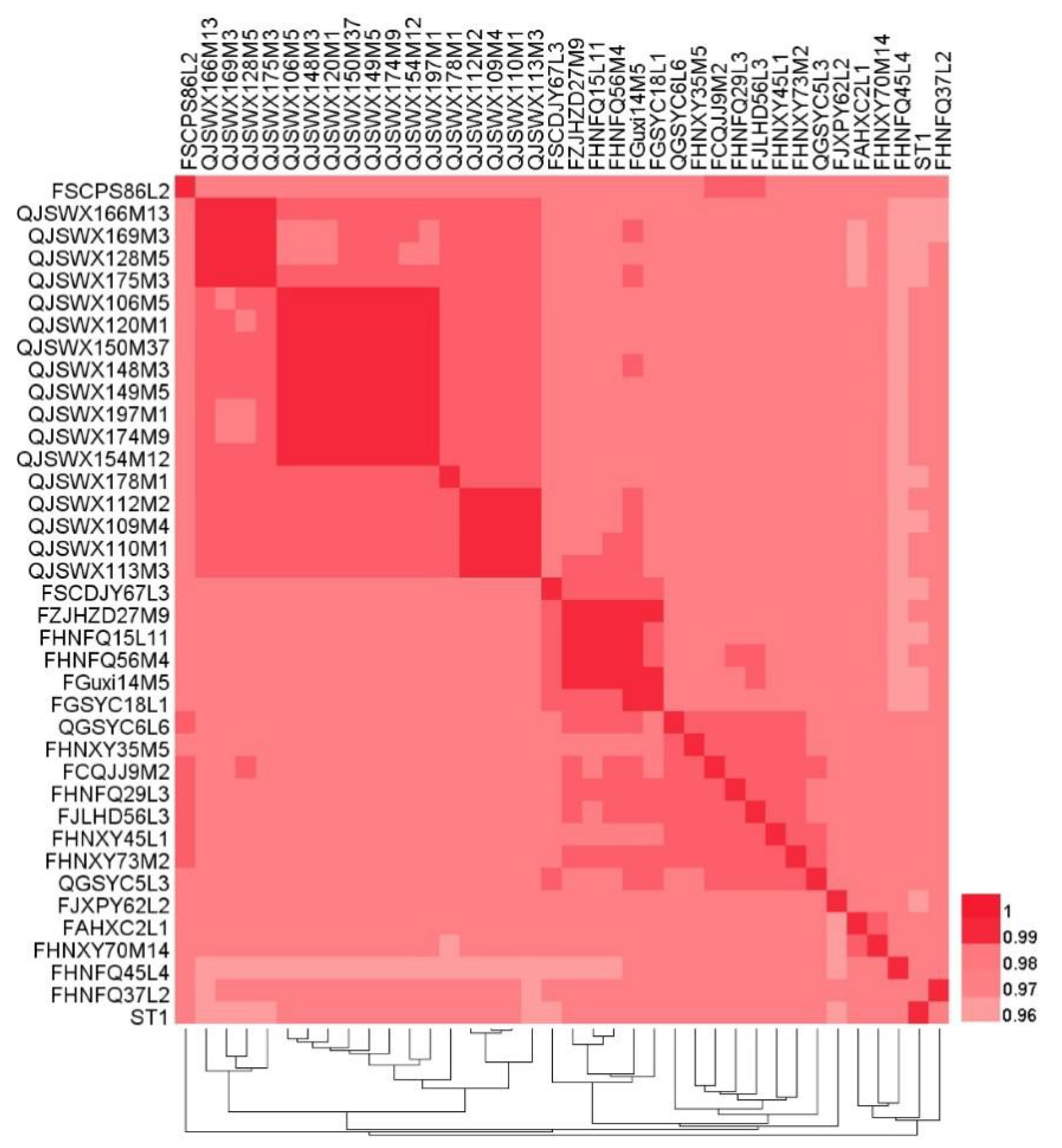
2.3. Average Nucleotide Identity Calculation

The average nucleotide identity (ANI) value between any two genomes was calculated using ANI perl script. The resulting matrices were clustered and visualized using R-packages’ heat map software (v1.0.8).

2.4. Pangenome and Core Genome Analysis

The pangenome and core genome of the 37 L. crispatus genomes were calculated using the PGAP1.21 [25], and the pangenome model was analyzed according to Heap’s law.

2.5. Phylogenetic Analysis

The protein sequences of the 37 L. crispatus were analyzed by OrthoMCL1.4 [26]. The protein families with the same function were then clustered using the Markov cluster algorithm (MCL). Core genes and specific genes were shown in a Venn plot. The core genes were aligned using MAFFT-7.313 [27], clustering analysis was performed using the neighbor-joining (NJ) method [28], and phylogenetic trees were constructed using PHYLIP (v3.6).

2.6. Carbohydrate Active Enzyme Analysis

The strain’s carbohydrate metabolism genes were annotated using the Carbohydrate Active Enzyme Database (CAZy) [29].

2.7. Statistical Analysis

Difference between two groups was evaluated using an independent samples t-test (SPSS 19.0), and a p value of < 0.05 was considered to indicate a significant difference.

3. Results

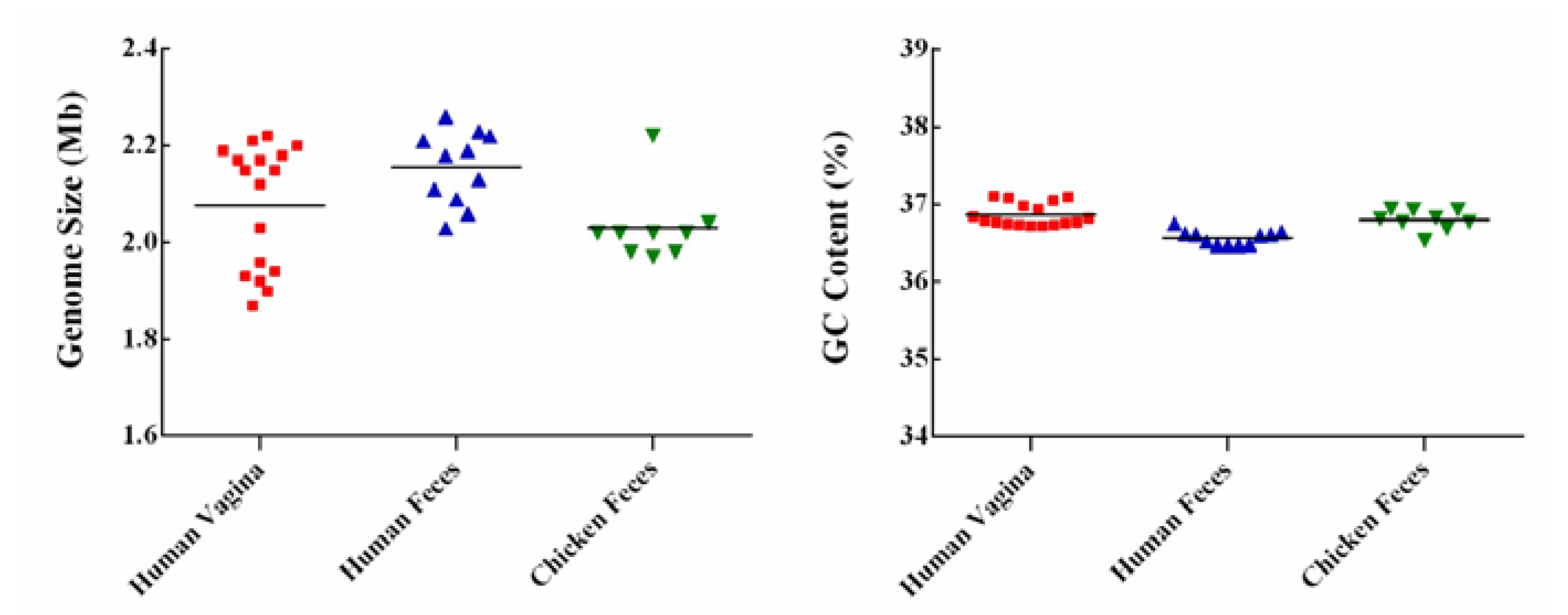
3.1. General Genome Features of the L. crispatus Strains

The 37 L. crispatus genomes ranged in size from 1.87 Mb to 2.26 Mb, with an average length of 2.09 Mb (Figure 1). Genomic sizes of vaginal-source bacteria (range 1.87–2.22 Mb, ~2.08 Mb) were similar to those of strains isolated from chicken (range 1.97–2.22 Mb, ~2.03 Mb). The genomes of the human-fecal-origin L. crispatus (range 2.03–2.26 Mb, ~2.16 Mb) were the largest. The number of CDS varied from 1869 to 2325, with an average of 2110 (Table 1). The GC content of the genomes was on average 36.76%, and no significant difference was found among the three sources (Figure 1). The ANI value of the 37 strains compared with the reference strain L. crispatus ST1 from NCBI and between any two strains was 96.27% to 99.96% (Figure 2), i.e., greater than 96%, indicating that they belonged to the same species [30]. As shown in Figure 2, the genomes of vaginal L. crispatus strains clustered separately from the feces isolates based on hierarchal clustering of ANI values. However, the human fecal-origin L. crispatus did not form a separate cluster from the chicken isolates. The feces-derived strains had a closer genetic relationship.

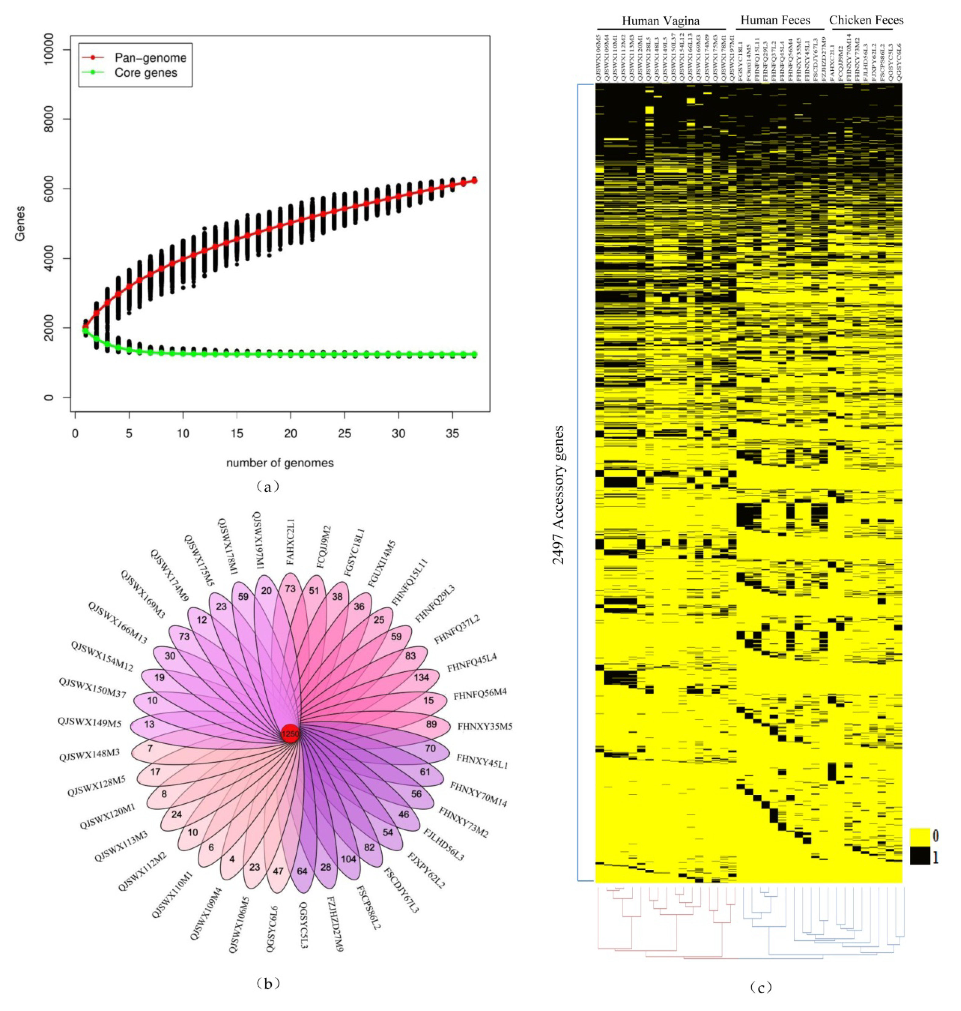
3.2. Pan-genome and Core Genome Analysis of L. crispatus

The pan-genome of 37 strains contained 5014 genes. As the number of strains increased, the pan-genome curve gradually converged. The derived mathematical function had an index value of less than 0.5, indicating that L. crispatus has a closed pan-genome [16]. However, the core genome only consisted of 1250 genes. The number of core genes was saturated and the gene curve trend was stable after the first 30 strains were analyzed (Figure 3a). The results showed that the 37 strains of L. crispatus in this study fully represented this species. According to the result of MCL clustering, the core genes, accessory genes, and specific genes of 37 strains were obtained. An accessory gene is defined as a gene shared by at least two strains. A specific gene is a gene that only exists in a certain strain. The number of specific genes in L. crispatus varied from 4 to 134 (Figure 3b). The average number of specific genes of L. crispatus isolated from chicken and human feces was 61 and 57 respectively. However, L. crispatus strains isolated from vaginal samples had an average number of specific genes of only 21.
Accessory genes contribute to species diversity. They are not important for bacterial growth, but confer selective advantages such as adaptation to different niches, drug resistance, or colonization of new hosts [31]. The distribution of the 2497 accessory genes was different in the 37 strains, as visualized by heat map (Figure 3c). It was shown that the 37 L. crispatus strains were divided into two large branches; one branch was vaginal-derived strains and the other was strains from feces. This indicated that the accessory genes were affected by the habitat. Meanwhile, strains isolated from chicken or human feces were similar in the composition of their accessory genes.

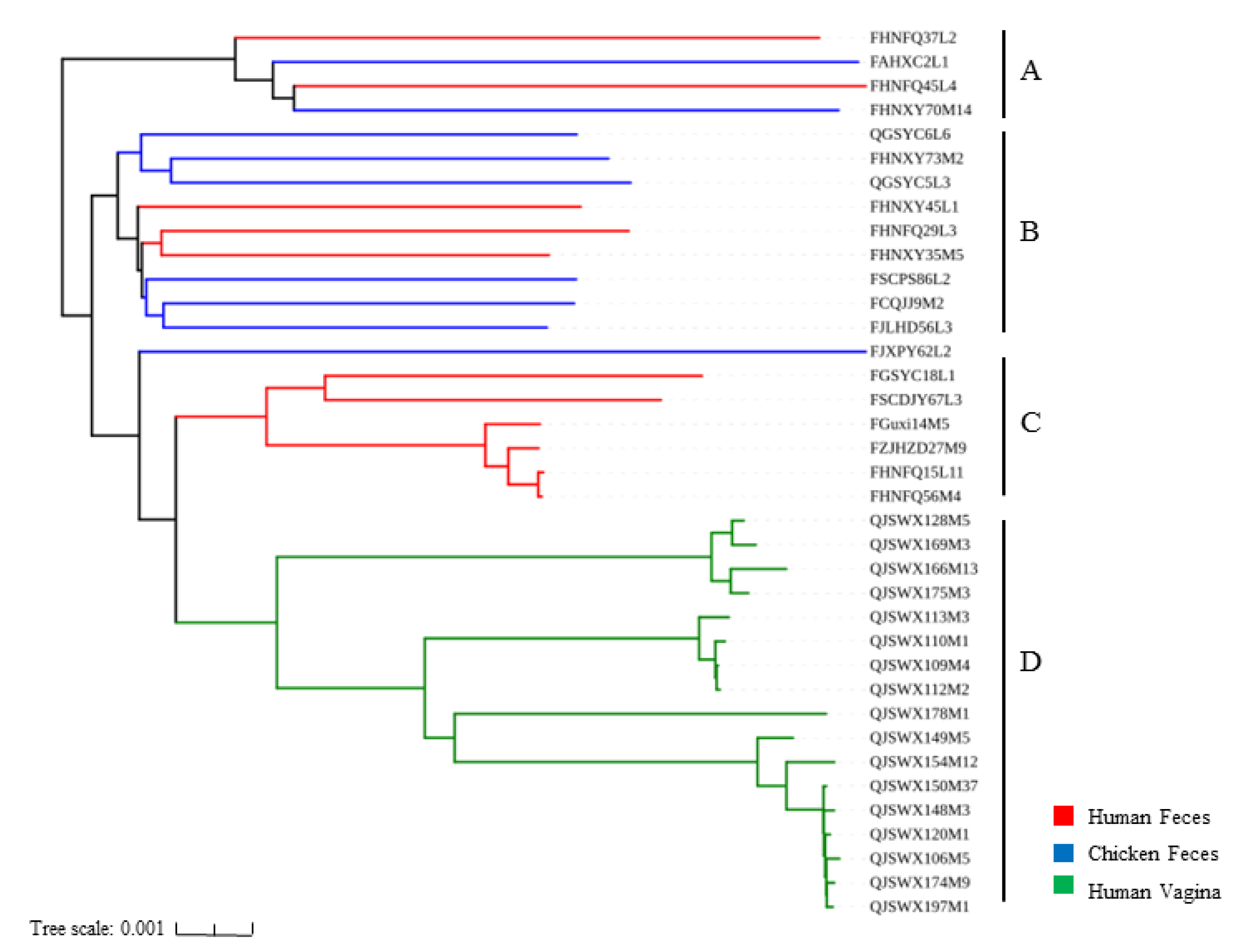
3.3. Phylogenetic Analysis of L. crispatus Strains

To explore the effects of different niches on the phylogeny of the L. crispatus genome, 1091 single-copy orthologous genes were used to construct a phylogenetic tree (Figure 4) against the 37 strains via the neighbor-joining method. The feces-derived strains and the vagina-derived strains were each clustered into different clusters. The phylogenetic tree was divided into four branches, including Branches A, B, C, and D. All 17 vagina-isolated strains were clustered into Branch D. However, chicken and human isolates were distributed in either Branch A or Branch B. Branch C contained only six L. crispatus strains, all from human feces. Therefore, different habitats might relate to differences in the genetic evolution of the strains.

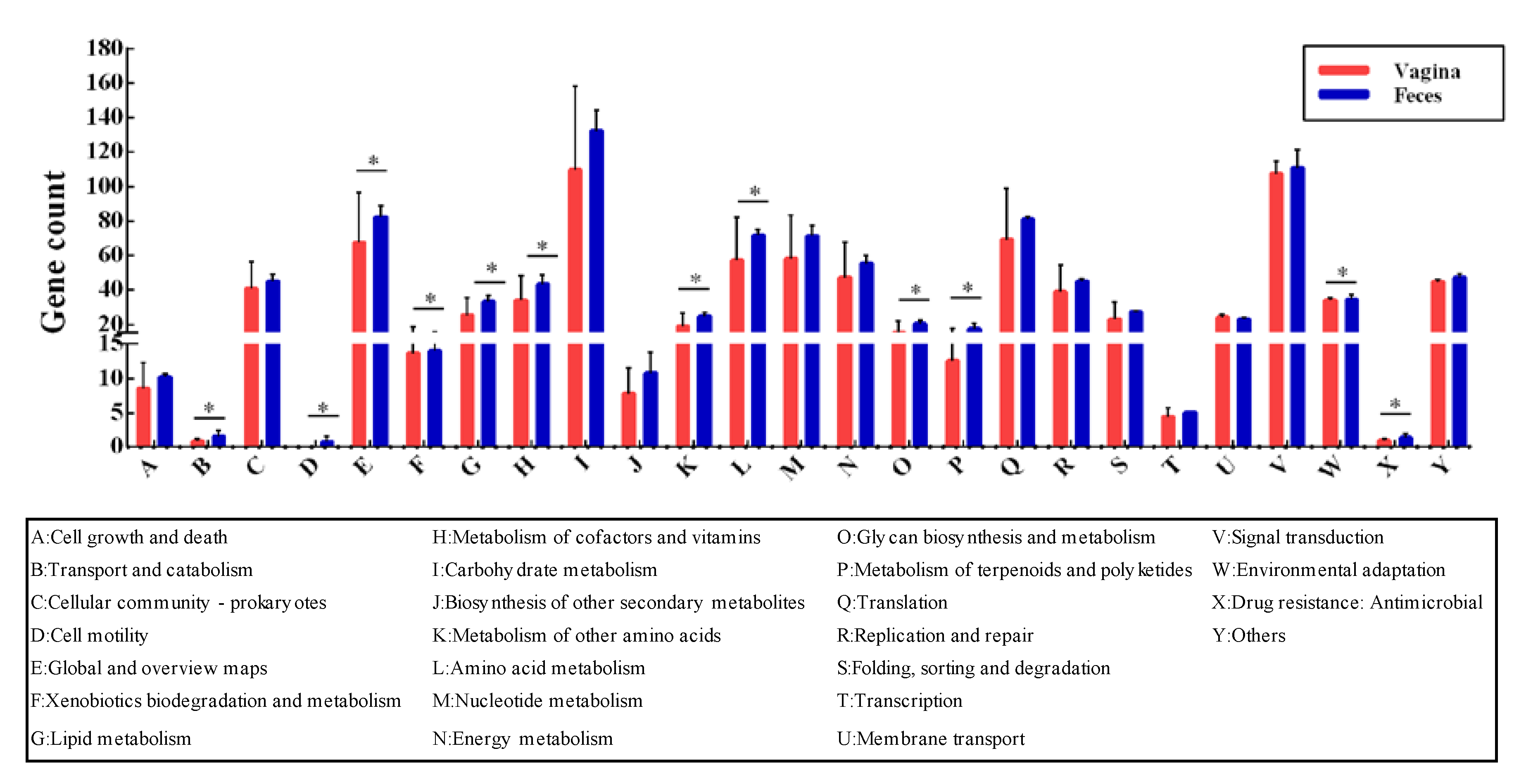
3.4. Evolution and Adaptation to Environment

To investigate whether L. crispatus showed some adaptability to different habitats in terms of gene function, we performed KEGG annotation for all 37 strains. However, based on the results of ANI value, phylogenetic tree, and the distribution of the accessory genes, we found that the feces-derived (human/chicken) strains had a high degree of similarity. Therefore, in terms of subsequent habitat adaptation analysis, we mainly focused on the adaptation of fecal-derived strains and vagina-derived strains to their respective niches. Across the two niches, genes associated with carbohydrate metabolism (~13%) and membrane transport (~11%) occupied a large proportion. The genes encoding transport and catabolism, cell motility, global and overview maps, xenobiotics biodegradation and metabolism, lipid metabolism, metabolism of cofactors and vitamins, metabolism of other amino acids, amino acid metabolism, glycan biosynthesis and metabolism, metabolism of terpenoids and polyketides, and drug resistance were annotated. Significant differences (p < 0.05) were observed between the two niches in genes involved with antimicrobial and environmental adaptation (Figure 5).
To further explore genes that might be adaptive to the vaginal and intestinal environment, we analyzed the genes with significant differences (p < 0.05) in gene abundance of each KEGG orthology between the two sources. Meanwhile, if a gene was present in more than half of the fecal strains, but in fewer than half in the vaginal strains, this gene was considered adapted to the gut. The genes associated with vaginal adaptation were also analyzed according to the same method. Based on this criterion, 69 important genes were found. Among the feces-derived L. crispatus isolates, 31 genes were analyzed, including 11 fecal-specific genes, (i.e., only present in feces). Of 38 genes found in the vagina-derived strains, 7 were vagina-specific genes.
Of the 31 genes in the feces-derived strains, 22% were related to the CRISPR–Cas system, and 19% to carbohydrate metabolism (deoxyribose-phosphate aldolase, sucrose phosphorylase, α-galactosidase). Other genes were linked to biosynthesis of other secondary metabolites and to amino acid metabolism. In the 38 genes of vagina-derived L. crispatus, 31% were related to carbohydrate metabolism (galactitol-specific IIC component, pullulanase, hexulose-6-phosphate isomerase). In addition, there were many genes involved in redox reactions and acid tolerance in the vagina, such as iron–sulfur protein and manganese ion transporters (Tables S1 and S2).

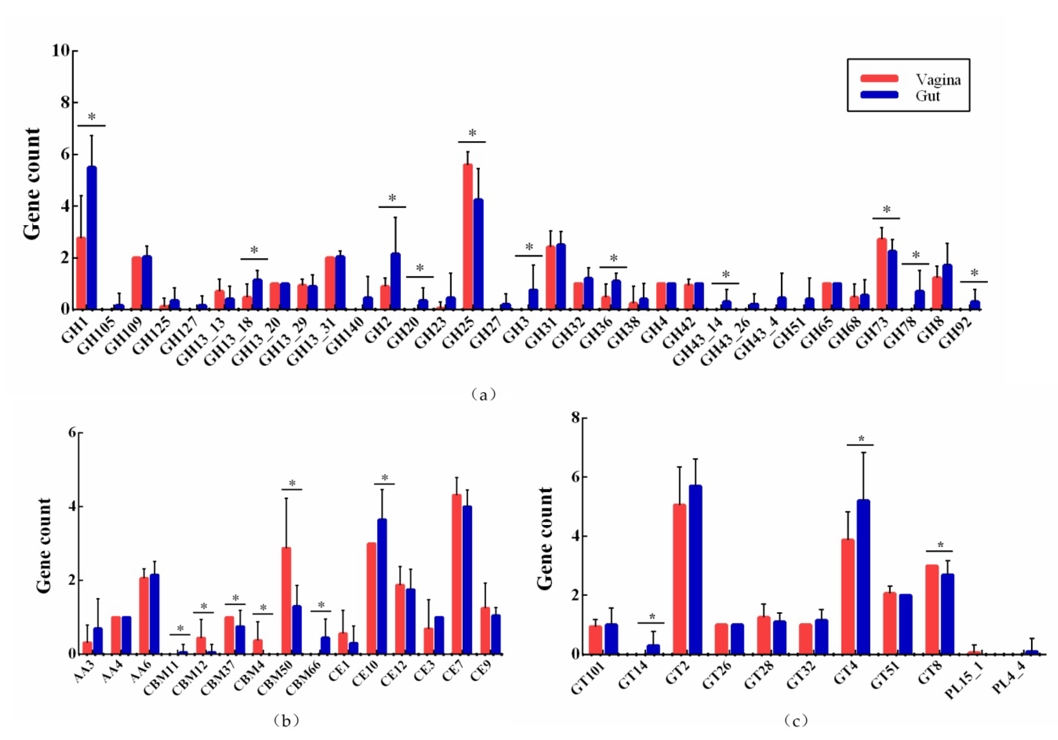
3.5. Active Carbohydrate Enzymes

The carbohydrate-utilization-related genes occupied the largest proportion in each genome by KEGG annotation. Therefore, we further compared the number of genes encoding carbohydrate-active enzymes in strains from feces and vagina.
The 37 strains had genes encoding 33 glycoside hydrolases (GHs) families. The GH family was the most important type of carbohydrate enzyme in L. crispatus, accounting for 55.9%. The abundance of GH1, GH13_18, GH2, GH20, GH25, GH3, GH36, GH43_14, GH73, GH78, and GH92 differed significantly (p < 0.05) between strains isolated from the feces and vagina. L. crispatus strains of fecal origin contained more types of carbohydrate enzyme than did L. crispatus of vaginal origin. Twelve GH families were only present in strains derived from feces. The GH140 and GH43_4 only existed in human feces isolates, whereas GH105 were only present in strains isolated from chicken feces (Figure 6a).
Additionally, nine glycosyl transferases (GTs), three auxiliary activities (AAs), six carbohydrate-binding modules (CBMs), six carbohydrate esterases (CEs), and two polysaccharide lyases (PL) were detected in 37 strains. The abundances of GT14, GT4, GT8, CE10, and CBM families were significantly different between the two groups. The CBM4 and PL15_1 were present only in vaginal strains (Figure 6b,c).

4. Discussion

Comparative genomic analysis of 37 L. crispatus strains from fecal and vaginal sources revealed the host’s influence on the genetic characteristics and evolution of the strain, as well as the adaptability of the strain to the intestine and vagina.
The general genomic characteristics in terms of size, GC content, and number of CDS (Table 1) showed that there was no significant difference between the L. crispatus strains derived from the three sources. However, when compared with chicken feces and human vaginal isolates, the genome size of strains isolated from human feces was larger. A possible explanation is that the human gut placed more environmental pressure on the bacteria [32]. To survive in a complicated environment, the L. crispatus could have obtained additional genes through horizontal transfer [33].
The phylogenetic tree results showed that the fecal isolates and the vaginal isolates were each clustered separately (Figure 4). This distinction indicated that the niche exerted an important impact on the evolution of the L. crispatus [15]. This result provided the insight that we might be able to determine the niche of an unknown source L. crispatus based on the clustering results of strains in the phylogenetic tree. A previous study revealed that Bifidobacteria isolated from the vagina and feces had no tendency to host in the phylogenetic tree [34]. In this case, it might have been that Bifidobacteria were not the main strain in the vagina. Therefore, the effect of the source on the evolutionary relationship of Bifidobacteria was not obvious.
Compared with the gut, the vagina has a lower pH (4 ± 0.5) [35]. Low pH helps reduce the risk of gynecological diseases and maintain female reproductive health [36]. Therefore, L. crispatus from the vagina require more defense mechanisms to respond to the low pH. The gene encoding manganese transport protein was presented only in vagina-sourced strains. This protein can take up Mn2+ and expel protons out of the cell to take part in the acid response, maintaining intracellular pH homeostasis [35]. Meanwhile, Mn2+ can also help in protecting bacteria against oxidative stress [37].
Lactobacilli in the vagina produce high levels of hydrogen peroxide [38,39]. Hydrogen peroxide is an important antibacterial substance. It can inhibit such pathogens as Gardnerella vaginalis and Neisseria gonorrhoeae in the vagina, and help maintain the normal flora of the vagina [40]. Meanwhile, hydrogen peroxide also causes oxidative stress in L. crispatus. Therefore, we observed that genes associated with oxidative stress were significantly enriched in vaginally derived bacteria, as described in Table S1. The two-component system is a broad signal transduction pathway in bacteria. It plays a major role in adapting to changing environmental conditions such as cell envelope stress response, phosphate regulation, and oxidative stress [37,41]. The OmpR family significantly increases the resistance of bacteria to hydrogen peroxide [42]. The genes encoding Fe-S cluster assembly protein were more abundant in the vagina (Table S1), and these genes are critical in the catalysis of electron transfer or metabolic support reactions [43]. Meanwhile, the Suf protein complex helps to assemble or repair oxygen-labile Fe-S clusters under oxidative stress [44,45]. In addition, hydrogen peroxide could further react with some Fe2+ iron to result in high-activity oxidants through the Fenton reaction [46]. This highly active oxidant might have a stronger inhibitory effect on pathogens in the vagina and a more positive effect on reproductive health, but this requires further testing to verify.
Pullulanase is an enzyme that breaks down glycogen [14]. In the vagina, glycogen is the main source of carbohydrates for microorganisms [47]. Genes encoding pullulanase were more abundant in the vagina-derived L. crispatus strains than in strains of fecal origin (Table S1). L. crispatus uses this enzyme to break down glycogen and produce lactic acid, which could maintain the low pH and inhibit pathogenic bacteria in the vagina [48]. The genes coding clumping factor A and A GntR family transcription factor were only present in the vagina-derived L. crispatus strains (Table S1).
Due to the variety and high density of bacteria, the intestine is an ideal niche for phage survival [49]. The L. crispatus isolated from feces were more susceptible to phage infection. The CRISPR/Cas system is known as the prokaryotic immune system and resists the invasion of foreign genetic material such as phage viruses and foreign plasmids [50]. To protect against damage to cells by foreign DNA, more genes related to the CRISPR–Cas system were observed in feces-derived L. crispatus (Table S2).
Tetracycline and lincomycin resistance genes were present only in the fecal strains (Table S2). Antibiotics have been used extensively to treat diseases caused by bacterial infections in humans and animals. Correspondingly, bacteria have acquired certain resistance genes through horizontal transfer to improve antibiotic resistance and their own survival ability [51]. Gene transfer at the level of drug-resistance genes often occurs in the human gastrointestinal tract [52]. In addition, genes related to streptomycin biosynthesis, such as rfbA, rfbB, and rfbC, were significantly enriched in the feces-derived bacteria (Table S2). Meanwhile, some genes associated with galactose metabolism, cysteine and methionine metabolism, and arginine biosynthesis were found only in the fecal strains.
To increase our understanding of the ability of L. crispatus to use host-derived glycogen fermentation, the genomes of 37 L. crispatus were analyzed and compared using CAZymes. The feces-derived strain was enriched in various GH family enzymes, including GH140, GH20, GH3, GH43_14, GH78, and GH92, which are involved in the utilization of a variety of carbon sources (Figure 6a). This might be because the gut contains more types of carbohydrate than the vagina. L. crispatus would need more enzymes to break down these carbohydrates to provide them with energy. The abundance of GH2 (β-galactosidase) and GH 36 (α-galactosidase) isolates in human feces was significantly higher than human vagina and chicken feces. Milk and other dairy products are an important part of human diet. GH2 and GH36 can hydrolyze galactose in dairy products, which is beneficial to the growth of strains in the intestine [53]. However, the abundance of CBM12, CBM37, CBM4, CBM66, GH25, and GH73 family in the vagina was higher than in the gut. CBM is a non-catalytic member of the cellulolytic enzymes. It is thought to assist in the synergy of enzymes through proximity and targeting, and plays an important role in the degradation of insoluble substrates [54,55]. Cellulose is the main component of plant cell walls. Humans and chickens consume cellulose-containing vegetables, but the feces isolates had a lower CBM abundance than the vagina (Figure 6b). This might require more in-depth exploration of the specific role of CBM in vagina-derived L. crispatus. GH25 has a role in facilitating cell division and in defense, as a lysozyme involved in cell wall and peptidoglycan catabolism [56]. GH73 encodes β-N-acetylglucosaminidases that cleave the β-1,4 glycosidic linkage between the N-acetylglucosamine (GlcNAc) and N-acetylmuramic acid (MurNAc) residues of bacterial cell wall peptidoglycan (PG) [57]. These two GH family lysozymes might provide a bacterial strategy to improve the competitive advantage of L. crispatus by destroying the cell walls of other pathogenic bacteria in the vagina and lysing them [58]. In summary, the above results demonstrate that L. crispatus is adaptable to different environments to improve its ability to survive.

5. Conclusions

In this study, we selected the genomes of 37 L. crispatus strains isolated from the human vagina, human feces, and chicken feces. The results of comparative genomics demonstrated that niche affected the evolution of L. crispatus to a large extent. Moreover, the L. crispatus strains showed adaptability to different environments to improve their own survival ability during the evolution of the strain.

Supplementary Materials

The following are available online at https://0-www-mdpi-com.brum.beds.ac.uk/2073-4425/11/4/360/s1, Table S1: Higher abundance of 38 genes in the vagina, Table S2: Higher abundance of 31 genes in the feces.

Author Contributions

Conceptualization, Q.Z. and J.Z.; methodology, L.Z. and J.Z.; software, H.Z.; validation, L.Z., Q.Z.; formal analysis, L.Z., Q.Z., and P.R.; investigation, J.Z.; resources, Q.Z., J.Z. and W.C.; data curation, L.Z. and J.Z.; writing-original draft preparation, L.Z. and Q.Z.; writing-review and editing, Q.Z., P.R., and J.Z.; visualization, Q.Z.; supervision, H.Z. and W.C.; project administration, W.C.; funding acquisition, W.C. All authors have read and agreed to the published version of the manuscript.

Funding

This work was supported by the National Natural Science Foundation of China (No. 31820103010, 31530056), National First-class Discipline Program of Food Science and Technology (JUFSTR20180102), and Collaborative innovation center of food safety and quality control in Jiangsu Province.

Acknowledgments

We would like to thank Weiyan Wu (Wuxi People’s hospital, Wuxi, Jiangsu Province) for collecting samples and processing data.

Conflicts of Interest

The authors declare no conflict of interest.

References

  1. Ruan, J. Bergey’s manual of systematic bacteriology (second edition) volume 5 and the study of Actinomycetes systematic in China. Wei Sheng Wu Xue Bao Acta Microbiol. Sin. 2013, 53, 521–530. [Google Scholar]
  2. Eslami, S.; Hadjati, J.; Motevaseli, E.; Mirzaei, R.; Bonab, S.F.; Ansaripour, B.; Khoramizadeh, M.R. Lactobacillus crispatus strain SJ-3C-US induces human dendritic cells (DCs) maturation and confers an anti-inflammatory phenotype to DCs. Apmis Acta Pathol. Microbiol. Immunol. Scand. 2016, 124, 697–710. [Google Scholar] [CrossRef]
  3. Tobita, K.; Yanaka, H.; Otani, H. Anti-allergic effects of Lactobacillus crispatus KT-11 strain on ovalbumin-sensitized BALB/c mice. Anim. Sci. J. 2010, 81, 699–705. [Google Scholar] [CrossRef] [PubMed]
  4. Azam, R.; Ghafouri-Fard, S.; Tabrizi, M.; Modarressi, M.H.; Ebrahimzadeh-Vesal, R.; Daneshvar, M.; Mobasheri, M.B.; Motevaseli, E. Lactobacillus acidophilus and Lactobacillus crispatus culture supernatants downregulate expression of cancer-testis genes in the MDA-MB-231 cell line. Asian Pac. J. Cancer Prev. 2014, 15, 4255–4259. [Google Scholar] [CrossRef] [PubMed] [Green Version]
  5. Humphries, C. Detecting diversity. Nature 2017, 550, S12–S14. [Google Scholar] [CrossRef] [PubMed]
  6. Wang, S.; Wang, Q.Y.; Yang, E.C.; Yan, L.; Li, T.; Zhuang, H. Antimicrobial compounds produced by vaginal lactobacillus crispatus are able to strongly inhibit candida albicans growth, hyphal formation and regulate virulence-related gene expressions. Front. Microbiol. 2017, 8. [Google Scholar] [CrossRef] [PubMed] [Green Version]
  7. Antonio, M.A.; Petrina, M.A.; Meyn, L.A.; Hillier, S.L. Women colonised by lactobacillus crispatus have a lower risk of acquisition of bacterial vaginosis (Bv) than women colonised by other lactobacilli. Sex. Transm. Infect. 2013, 89, A232. [Google Scholar] [CrossRef] [Green Version]
  8. Li, T.; Liu, Z.; Zhang, X.; Chen, X.; Wang, S. Local probiotic lactobacillus crispatus and lactobacillus delbrueckii exhibit strong antifungal effects against vulvovaginal candidiasis in a rat model. Front. Microbiol. 2019, 10, 1033. [Google Scholar] [CrossRef]
  9. Bohbot, J.M.; Darai, E.; Bretelle, F.; Brami, G.; Daniel, C.; Cardot, J.M. Efficacy and safety of vaginally administered lyophilized Lactobacillus crispatus IP 174178 in the prevention of bacterial vaginosis recurrence. J. Gynecol. Obstet. Hum. Reprod. 2018, 47, 81–86. [Google Scholar] [CrossRef] [Green Version]
  10. Stapleton, A.E.; Au-Yeung, M.; Hooton, T.M.; Fredricks, D.N.; Roberts, P.L.; Czaja, C.A.; Yarova-Yarovaya, Y.; Fiedler, T.; Cox, M.; Stamm, W.E. Randomized, placebo-controlled phase 2 trial of a Lactobacillus crispatus probiotic given intravaginally for prevention of recurrent urinary tract infection. Clin. Infect. Dis. 2011, 52, 1212–1217. [Google Scholar] [CrossRef]
  11. Binnewies, T.T.; Motro, Y.; Hallin, P.F.; Lund, O.; Dunn, D.; La, T.; Hampson, D.J.; Bellgard, M.; Wassenaar, T.M.; Ussery, D.W. Ten years of bacterial genome sequencing: Comparative-genomics-based discoveries. Funct. Integr. Genom. 2006, 6, 165–185. [Google Scholar] [CrossRef] [PubMed]
  12. Field, D.; Wilson, G.; van der Gast, C. How do we compare hundreds of bacterial genomes? Curr. Opin. Microbiol. 2006, 9, 499–504. [Google Scholar] [CrossRef] [PubMed]
  13. Ojala, T.; Kankainen, M.; Castro, J.; Cerca, N.; Edelman, S.; Westerlund-Wikstrom, B.; Paulin, L.; Holm, L.; Auvinen, P. Comparative genomics of Lactobacillus crispatus suggests novel mechanisms for the competitive exclusion of Gardnerella vaginalis. BMC Genom. 2014, 15. [Google Scholar] [CrossRef] [PubMed] [Green Version]
  14. van der Veer, C.; Hertzberger, R.Y.; Bruisten, S.M.; Tytgat, H.L.P.; Swanenburg, J.; Angelino-Bart, A.D.; Schuren, F.; Molenaar, D.; Reid, G.; de Vries, H.; et al. Comparative genomics of human Lactobacillus crispatus isolates reveals genes for glycosylation and glycogen degradation: Implications for in vivo dominance of the vaginal microbiota. Microbiome 2019, 7. [Google Scholar] [CrossRef] [PubMed] [Green Version]
  15. Yu, J.; Zhao, J.; Song, Y.; Zhang, J.; Yu, Z.; Zhang, H.; Sun, Z. Comparative genomics of the herbivore gut symbiont lactobacillus reuteri reveals genetic diversity and lifestyle adaptation. Front. Microbiol. 2018, 9, 1151. [Google Scholar] [CrossRef] [PubMed]
  16. Lee, J.Y.; Han, G.G.; Kim, E.B.; Choi, Y.J. Comparative genomics of Lactobacillus salivarius strains focusing on their host adaptation. Microbiol. Res. 2017, 205, 48–58. [Google Scholar] [CrossRef]
  17. Eisenbach, L.; Janssen, D.; Ehrmann, M.A.; Vogel, R.F. Comparative genomics of Lactobacillus curvatus enables prediction of traits relating to adaptation and strategies of assertiveness in sausage fermentation. Int. J. Food Microbiol. 2018, 286, 37–47. [Google Scholar] [CrossRef]
  18. Feyereisen, M.; Mahony, J.; Kelleher, P.; Roberts, R.J.; O’Sullivan, T.; Geertman, J.M.A.; van Sinderen, D. Comparative genome analysis of the Lactobacillus brevis species. BMC Genom. 2019, 20. [Google Scholar] [CrossRef] [Green Version]
  19. Pan, M.C.; Hidalgo-Cantabrana, C.; Goh, Y.J.; Sanozky-Dawes, R.; Barrangou, R. Comparative analysis of lactobacillus gasseri and lactobacillus crispatus isolated from human urogenital and gastrointestinal tracts. Front. Microbiol. 2020, 10. [Google Scholar] [CrossRef]
  20. Beasley, S.S.; Takala, T.M.; Reunanen, J.; Apajalahti, J.; Saris, P.E.J. Characterization and electrotransformation of Lactobacillus crispatus isolated from chicken crop and intestine. Poult. Sci. 2004, 83, 45–48. [Google Scholar] [CrossRef]
  21. Luo, R.B.; Liu, B.H.; Xie, Y.L.; Li, Z.Y.; Huang, W.H.; Yuan, J.Y.; He, G.Z.; Chen, Y.X.; Pan, Q.; Liu, Y.J.; et al. SOAPdenovo2: An empirically improved memory-efficient short-read de novo assembler. Gigascience 2012, 1. [Google Scholar] [CrossRef] [PubMed]
  22. Delcher, A.L.; Bratke, K.A.; Powers, E.C.; Salzberg, S.L. Identifying bacterial genes and endosymbiont DNA with Glimmer. Bioinformatics 2007, 23, 673–679. [Google Scholar] [CrossRef] [PubMed]
  23. Besemer, J.; Borodovsky, M. GeneMark: Web software for gene finding in prokaryotes, eukaryotes and viruses. Nucleic Acids Res. 2005, 33, W451–W454. [Google Scholar] [CrossRef] [PubMed] [Green Version]
  24. Kanehisa, M.; Goto, S. KEGG: Kyoto encyclopedia of genes and genomes. Nucleic Acids Res. 2000, 28, 27–30. [Google Scholar] [CrossRef]
  25. Zhao, Y.B.; Wu, J.Y.; Yang, J.H.; Sun, S.X.; Xiao, J.F.; Yu, J. PGAP: Pan-genomes analysis pipeline. Bioinformatics 2012, 28, 416–418. [Google Scholar] [CrossRef] [Green Version]
  26. Li, L.; Stoeckert, C.J.; Roos, D.S. OrthoMCL: Identification of ortholog groups for eukaryotic genomes. Genome Res. 2003, 13, 2178–2189. [Google Scholar] [CrossRef] [Green Version]
  27. Katoh, K.; Rozewicki, J.; Yamada, K.D. MAFFT online service: Multiple sequence alignment, interactive sequence choice and visualization. Brief. Bioinform. 2019, 20, 1160–1166. [Google Scholar] [CrossRef] [Green Version]
  28. Kumar, S.; Gadagkar, S.R. Efficiency of the neighbor-joining method in reconstructing deep and shallow evolutionary relationships in large phylogenies. J. Mol. Evol. 2000, 51, 544–553. [Google Scholar] [CrossRef] [Green Version]
  29. Wang, S.; Yang, B.; Ross, R.P.; Stanton, C.; Zhao, J.X.; Zhang, H.; Chen, W. Comparative genomics analysis of lactobacillus ruminis from different niches. Genes 2020, 11, 70. [Google Scholar] [CrossRef] [Green Version]
  30. Ciufo, S.; Kannan, S.; Sharma, S.; Badretdin, A.; Clark, K.; Turner, S.; Brover, S.; Schoch, C.L.; Kimchi, A.; DiCuccio, M. Using average nucleotide identity to improve taxonomic assignments in prokaryotic genomes at the NCBI. Int. J. Syst. Evol. Microbiol. 2018, 68, 2386–2392. [Google Scholar] [CrossRef]
  31. Medini, D.; Donati, C.; Tettelin, H.; Masignani, V.; Rappuoli, R. The microbial pan-genome. Curr. Opin. Genet. Dev. 2005, 15, 589–594. [Google Scholar] [CrossRef] [PubMed]
  32. O’Sullivan, O.; O’Callaghan, J.; Sangrador-Vegas, A.; McAuliffe, O.; Slattery, L.; Kaleta, P.; Callanan, M.; Fitzgerald, G.F.; Ross, R.P.; Beresford, T. Comparative genomics of lactic acid bacteria reveals a niche-specific gene set. BMC Microbiol. 2009, 9, 50. [Google Scholar] [CrossRef] [PubMed] [Green Version]
  33. Broadbent, J.R.; Neeno-Eckwall, E.C.; Stahl, B.; Tandee, K.; Cai, H.; Morovic, W.; Horvath, P.; Heidenreich, J.; Perna, N.T.; Barrangou, R.; et al. Analysis of the Lactobacillus casei supragenome and its influence in species evolution and lifestyle adaptation. BMC Genom. 2012, 13, 533. [Google Scholar] [CrossRef] [PubMed] [Green Version]
  34. Freitas, A.C.; Hill, J.E. Bifidobacteria isolated from vaginal and gut microbiomes are indistinguishable by comparative genomics. PLoS ONE 2018, 13, e0196290. [Google Scholar] [CrossRef] [PubMed] [Green Version]
  35. Shabayek, S.; Spellerberg, B. Acid stress response mechanisms of group B streptococci. Front. Cell. Infect. Microbiol. 2017, 7, 395. [Google Scholar] [CrossRef] [PubMed]
  36. Miller, E.A.; Beasley, D.E.; Dunn, R.R.; Archie, E.A. Lactobacilli dominance and vaginal pH: Why is the human vaginal microbiome unique? Front. Microbiol. 2016, 7, 1936. [Google Scholar] [CrossRef]
  37. Cotter, P.D.; Hill, C. Surviving the acid test: Responses of gram-positive bacteria to low pH. Microbiol. Mol. Biol. Rev. MMBR 2003, 67, 429–453. [Google Scholar] [CrossRef] [Green Version]
  38. Xu, H.Y.; Tian, W.H.; Wan, C.X.; Jia, L.J.; Wang, L.Y.; Yuan, J.; Liu, C.M.; Zeng, M.; Wei, H. Antagonistic potential against pathogenic microorganisms and hydrogen peroxide production of indigenous lactobacilli isolated from vagina of Chinese pregnant women. Biomed. Environ. Sci. 2008, 21, 365–371. [Google Scholar] [CrossRef]
  39. Balkus, J.E.; Mitchell, C.; Agnew, K.; Liu, C.; Fiedler, T.; Cohn, S.E.; Luque, A.; Coombs, R.; Fredricks, D.N.; Hitti, J. Detection of hydrogen peroxide-producing Lactobacillus species in the vagina: A comparison of culture and quantitative PCR among HIV-1 seropositive women. BMC Infect. Dis. 2012, 12, 188. [Google Scholar] [CrossRef] [Green Version]
  40. Pascual, L.M.; Daniele, M.B.; Pajaro, C.; Barberis, L. Lactobacillus species isolated from the vagina: Identification, hydrogen peroxide production and nonoxynol-9 resistance. Contraception 2006, 73, 78–81. [Google Scholar] [CrossRef]
  41. Stock, A.M.; Robinson, V.L.; Goudreau, P.N. Two-component signal transduction. Annu. Rev. Biochem. 2000, 69, 183–215. [Google Scholar] [CrossRef] [PubMed] [Green Version]
  42. Zhao, Z.; Peng, T.; Oh, J.I.; Glaeser, J.; Weber, L.; Li, Q.; Klug, G. A response regulator of the OmpR family is part of the regulatory network controlling the oxidative stress response of Rhodobacter sphaeroides. Environ. Microbiol. Rep. 2019, 11, 118–128. [Google Scholar] [CrossRef]
  43. Kiley, P.J.; Beinert, H. The role of Fe-S proteins in sensing and regulation in bacteria. Curr. Opin. Microbiol. 2003, 6, 181–185. [Google Scholar] [CrossRef]
  44. Selbach, B.P.; Chung, A.H.; Scott, A.D.; George, S.J.; Cramer, S.P.; Dos Santos, P.C. Fe-S cluster biogenesis in Gram-positive bacteria: SufU is a zinc-dependent sulfur transfer protein. Biochemistry 2014, 53, 152–160. [Google Scholar] [CrossRef] [PubMed] [Green Version]
  45. Nachin, L.; Loiseau, L.; Expert, D.; Barras, F. SufC: An unorthodox cytoplasmic ABC/ATPase required for [Fe-S] biogenesis under oxidative stress. EMBO J. 2003, 22, 427–437. [Google Scholar] [CrossRef] [Green Version]
  46. Maresca, D.; Zotta, T.; Mauriello, G. Adaptation to aerobic environment of lactobacillus johnsonii/gasseri strains. Front. Microbiol. 2018, 9, 157. [Google Scholar] [CrossRef]
  47. Mirmonsef, P.; Hotton, A.L.; Gilbert, D.; Gioia, C.J.; Maric, D.; Hope, T.J.; Landay, A.L.; Spear, G.T. Glycogen levels in undiluted genital fluid and their relationship to vaginal ph, estrogen, and progesterone. PLoS ONE 2016, 11, e0153553. [Google Scholar] [CrossRef] [Green Version]
  48. Tachedjian, G.; Aldunate, M.; Bradshaw, C.S.; Cone, R.A. The role of lactic acid production by probiotic Lactobacillus species in vaginal health. Res. Microbiol. 2017, 168, 782–792. [Google Scholar] [CrossRef]
  49. Stern, A.; Mick, E.; Tirosh, I.; Sagy, O.; Sorek, R. CRISPR targeting reveals a reservoir of common phages associated with the human gut microbiome. Genome Res. 2012, 22, 1985–1994. [Google Scholar] [CrossRef] [Green Version]
  50. Barrangou, R.; Marraffini, L.A. CRISPR-Cas systems: Prokaryotes upgrade to adaptive immunity. Mol. Cell 2014, 54, 234–244. [Google Scholar] [CrossRef] [Green Version]
  51. Zhang, F.; Gao, J.; Wang, B.; Huo, D.; Wang, Z.; Zhang, J.; Shao, Y. Whole-genome sequencing reveals the mechanisms for evolution of streptomycin resistance in Lactobacillus plantarum. J. Dairy Sci. 2018, 101, 2867–2874. [Google Scholar] [CrossRef] [PubMed] [Green Version]
  52. McKeegan, K.S.; Borges-Walmsley, M.I.; Walmsley, A.R. The structure and function of drug pumps: An update. Trends Microbiol. 2003, 11, 21–29. [Google Scholar] [CrossRef]
  53. Miguez Amil, S.; Jimenez-Ortega, E.; Ramirez-Escudero, M.; Talens-Perales, D.; Marin-Navarro, J.; Polaina, J.; Sanz-Aparicio, J.; Fernandez-Leiro, R. The cryo-EM structure of thermotoga maritima beta-galactosidase: Quaternary structure guides protein engineering. ACS Chem. Biol. 2020, 15, 179–188. [Google Scholar] [CrossRef] [PubMed]
  54. Novy, V.; Aissa, K.; Nielsen, F.; Straus, S.K.; Ciesielski, P.; Hunt, C.G.; Saddler, J. Quantifying cellulose accessibility during enzyme-mediated deconstruction using 2 fluorescence-tagged carbohydrate-binding modules. Proc. Natl. Acad. Sci. USA 2019, 116, 22545–22551. [Google Scholar] [CrossRef] [PubMed] [Green Version]
  55. Duan, C.J.; Huang, M.Y.; Pang, H.; Zhao, J.; Wu, C.X.; Feng, J.X. Characterization of a novel theme C glycoside hydrolase family 9 cellulase and its CBM-chimeric enzymes. Appl. Microbiol. Biotechnol. 2017, 101, 5723–5737. [Google Scholar] [CrossRef] [PubMed]
  56. O’Riordan, N.; O’Callaghan, J.; Butto, L.F.; Kilcoyne, M.; Joshi, L.; Hickey, R.M. Bovine glycomacropeptide promotes the growth of Bifidobacterium longum ssp. infantis and modulates its gene expression. J. Dairy Sci. 2018, 101, 6730–6741. [Google Scholar] [CrossRef]
  57. Zaloba, P.; Bailey-Elkin, B.A.; Derksen, M.; Mark, B.L. Structural and biochemical insights into the peptidoglycan hydrolase domain of FlgJ from Salmonella typhimurium. PLoS ONE 2016, 11. [Google Scholar] [CrossRef] [Green Version]
  58. Morais, S.; Cockburn, D.W.; Ben-David, Y.; Koropatkin, N.M.; Martens, E.C.; Duncan, S.H.; Flint, H.J.; Mizrahi, I.; Bayer, E.A. Lysozyme activity of the Ruminococcus champanellensis cellulosome. Environ. Microbiol. 2016, 18, 5112–5122. [Google Scholar] [CrossRef]
Figure 1. Genome size and GC content of L. crispatus. The red square, blue triangle, and green inverted triangle represent L. crispatus isolated from the human vagina, human feces, and chicken feces, respectively.
Genes 11 00360 g001
Figure 2. Heatmap showing the Average Nucleotide Identity (ANI) value among 38 L. crispatus strains.
Genes 11 00360 g002
Figure 3. Pan-genome and core genome of L. crispatus. (a) The number of new genes accumulated in the L. crispatus pan-genome was plotted against the number of new genomes added and the number of accumulated genes attributed to the core genome relative to the number of added genomes. (b) Venn diagram representing the core and specific genes of 37 L. crispatus strains obtained by MCL clustering. The number in the middle red circle represents the number of core genes of the 37 strains. The numbers on the petals indicate the specific genes of each strain. (c) A heatmap representation of accessory genes against sequenced L. crispatus genomes. Each row represents an accessory gene. Each column represents a strain.
Genes 11 00360 g003
Figure 4. Phylogenetic tree of 37 L. crispatus strains. Based on 1091 single-copy core genes of 37 strains, the phylogenetic tree was constructed using the neighbor-joining method. According to the clustering results of the phylogenetic tree, the phylogenetic tree is divided into 4 branches, namely Branch A, B, C and D.
Genes 11 00360 g004
Figure 5. Annotation for genes of L. crispatus isolated from vagina and gut using the KEGG database. * p < 0.05.
Genes 11 00360 g005
Figure 6. Distribution and abundance of active carbohydrate enzyme family genes in L. crispatus strains isolated from feces and vagina. * p < 0.05.
Genes 11 00360 g006
Table 1. General genome features of the 38 L. crispatus strains.
StrainsSourceGenome Size (Mb)GC Content (%)CDS No.tRNA No.rRNA No.Accession No.
ST1Chicken Feces2.0436.920236412SAMEA2272191
FGSYC18L1Human Feces2.1836.472177373SAMN12869300
FGuxi14M5Human Feces2.1936.472191421SAMN12869301
FHNFQ15L11Human Feces2.2636.482241443SAMN12869302
FHNFQ29L3Human Feces2.0336.752053563SAMN12869303
FHNFQ37L2Human Feces2.0936.602091451SAMN12869304
FHNFQ45L4Human Feces2.1336.642190443SAMN12869305
FHNFQ56M4Human Feces2.2336.522218403SAMN12869306
FHNXY35M5Human Feces2.1136.612121403SAMN12869307
FHNXY45L1Human Feces2.0636.622066350SAMN12869308
FSCDJY67L3Human Feces2.2136.612190451SAMN12869309
FZJHZD27M9Human Feces2.2236.472243411SAMN12869310
FAHXC2L1Chicken Feces2.0436.812023464SAMN12869311
FCQJJ9M2Chicken Feces1.9736.931988443SAMN12869312
FHNXY70M14Chicken Feces1.9836.921985453SAMN12869313
FHNXY73M2Chicken Feces2.0236.691989493SAMN12869314
FJLHD56L3Chicken Feces2.0236.771970383SAMN12869315
FJXPY62L2Chicken Feces2.0236.921977413SAMN12869316
FSCPS86L2Chicken Feces2.2236.532203493SAMN12869317
QGSYC5L3Chicken Feces2.0236.822003453SAMN12869318
QGSYC6L6Chicken Feces1.9836.772004433SAMN12869319
QJSWX106M5Human Vagina2.1536.822194724SAMN12869320
QJSWX109M4Human Vagina2.1836.722231533SAMN12869321
QJSWX110M1Human Vagina2.1736.722227414SAMN12869322
QJSWX112M2Human Vagina2.1736.732241533SAMN12869323
QJSWX113M3Human Vagina2.2236.772309463SAMN12869324
QJSWX120M1Human Vagina2.1536.772206684SAMN12869325
QJSWX128M5Human Vagina1.9436.731972410SAMN12869326
QJSWX148M3Human Vagina1.8737.051869432SAMN12869327
QJSWX149M5Human Vagina1.9637.081981533SAMN12869328
QJSWX150M37Human Vagina1.9036.981914403SAMN12869329
QJSWX154M12Human Vagina2.0337.12108422SAMN12869330
QJSWX166M13Human Vagina1.9236.751962421SAMN12869331
QJSWX169M3Human Vagina2.2136.742325667SAMN12869332
QJSWX174M9Human Vagina1.9337.091969493SAMN12869333
QJSWX175M3Human Vagina2.1236.792112502SAMN12869334
QJSWX178M1Human Vagina2.1936.842243716SAMN12869335
QJSWX197M1Human Vagina2.2036.942275433SAMN12869336

