Article

Genetic Polymorphisms Associated with Prothrombin Time and Activated Partial Thromboplastin Time in Chinese Healthy Population

1
Department of Pharmacy, Peking University First Hospital, Beijing 100034, China
2
School of Pharmaceutical Sciences, Peking University Health Science Center, Beijing 100191, China
3
Institute of Clinical Pharmacology, Peking University, Beijing 100191, China
*
Authors to whom correspondence should be addressed.
These authors contributed equally to this work.
Submission received: 5 September 2022 / Revised: 30 September 2022 / Accepted: 12 October 2022 / Published: 15 October 2022
(This article belongs to the Special Issue Pharmacogenomics of Cardiovascular Disease)

Abstract

:
(1) Background: The purpose of this study was to evaluate the effect of gene polymorphisms on prothrombin time (PT) and activated partial thromboplastin time (APTT) in a healthy Chinese population. (2) Methods: A total of 403 healthy volunteers from a series of novel oral anticoagulants (NOACs) bioequivalence trials in China were included. Coagulation tests for PT and APTT were performed in the central lab at Peking University First Hospital. Whole-exome sequencing (WES) and genome-wide association analysis were performed. (3) Results: In the correlation analysis of PT, 105 SNPs from 84 genes reached the genome-wide significance threshold (p < 1 × 10⁻⁵). Zinc Finger Protein 594 (ZNF594) rs184838268 (p = 4.50 × 10⁻¹⁹) was most significantly related to PT, and Actinin Alpha 1 (ACTN1) was found to interact most with other candidate genes. Significant associations with previously reported candidate genes Aurora Kinase B (AURKB), Complement C5(C5), Clock Circadian Regulator (CLOCK), and Histone Deacetylase 9(HDAC9) were detected in our dataset (p < 1 × 10⁻⁵). PiggyBac Transposable Element Derived 2(PGBD2) rs75935520 (p = 4.49 × 10⁻⁶), Bromodomain Adjacent To Zinc Finger Domain 2A(BAZ2A) rs199970765 (p = 5.69 × 10⁻⁶) and Protogenin (PRTG) rs80064850 (p = 8.69 × 10⁻⁶) were significantly correlated with APTT (p < 1 × 10⁻⁵). The heritability values of PT and APTT were 0.83 and 0.64, respectively; (4) Conclusion: The PT and APTT of healthy populations are affected by genetic polymorphisms. ZNF594 and ACTN1 variants could be novel genetic markers of PT, while PRTG polymorphisms might be associated with APTT levels. The findings could be attributed to ethnic differences, and need further investigation.

Graphical Abstract

1. Introduction

Prothrombin time (PT) and activated partial thromboplastin time (APTT) are commonly used parameters for screening coagulation disorders and monitoring anticoagulant therapy [1,2]. APTT is widely used in the screening of inherited and acquired coagulation factor deficiencies in intrinsic and common coagulation pathways, while the PT test is more sensitive for extrinsic coagulation system function [3,4]. In addition, PT and APTT monitoring are essential for clinical decision-making in critical situations, such as bleeding, thromboembolism, and emergency surgery, and may also predict the perioperative bleeding risk of patients taking anticoagulants undergoing elective surgery [5,6].
Except for the influence of sample preparation and detection methods, PT and APTT levels are affected by some physiological factors. It is well known that the hemostatic system exhibits dynamic age-related evolution and thus can present differences in PT and APTT at different ages [7]. In addition, genetic factors may influence PT and APTT levels. A European genome-wide association study found that KNG1, HRG, F11, F12, and ABO are associated with APTT, while F7 and PROCR/EDEM2 are related to PT [8,9]. A recent study also found a positive correlation between the allele load of the JAK2V617F mutation and APTT [10]. Polymorphisms in PT and APTT levels also vary greatly in healthy people. Some people have a low baseline level; even if the PT or APTT is doubled, it may still be within the normal range. However, some people have a high baseline level, and a slight extension (less than 3 s) will exceed the normal range. This indicates a potential impact of genetic factors on PT and APTT in healthy people. However, the overall effects of gene polymorphisms on these coagulation indexes are still poorly understood. Moreover, there is no large-scale genome-wide association analysis (GWAS) on PT and APTT levels among Asians.
In this study, we conducted whole-exome sequencing (WES) and association analysis in a healthy Chinese population to further explore genetic markers and their heritability affecting PT and APTT.

2. Materials and Methods

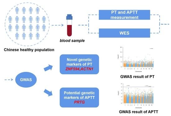
2.1. Study Population

The study population for genome-wide association analysis comprised a series of novel oral anticoagulants (NOACs) bioequivalence trial participants in China. Healthy participants were defined according to the general inclusion criteria of BE trials, including history, physical examination, vital signs, 12-lead electrocardiogram (ECG), ultrasound and imaging examination, laboratory examination, and alcohol exhalation, etc. A total of 424 healthy volunteers aged 18–60 years were enrolled in 10 centers. None of the participants enrolled in the study had taken any drug for at least 4 weeks before the start of the study. Blood samples were collected at baseline for genotyping and coagulation tests. All protocols and informed consent were reviewed and approved by the independent ethics committee of Peking University First Hospital and all participating centers. Before the start of the study, all subjects were informed of the purpose, duration, and potential risks of the study and provided written informed consent. This study was registered on ClinicalTrial.org with the registration number NCT03161496.

2.2. PT and APTT Measurement

Blood samples were collected in sodium citrate test tubes (3.2% v/v) and centrifuged at 2500× g at room temperature for 15 min within 60 min of sampling. Plasma samples were transferred to cryovials and stored at −70 °C. Coagulation tests for PT and APTT were performed in the central lab in Peking University First Hospital within 6 months after sampling [11]. Coagulation tests showed circadian rhythm [12], so all samples were tested at similar times during the day to avoid the values changing. PT and APTT were measured using validated Coagulation Method Assay Kits (Thromborel-S® and Actin®, Siemens Healthcare Diagnostics Products GmbH, Marburg, Germany) following a standard protocol. The units of the PT and APTT measurement values were seconds.

2.3. Whole-Exome Sequencing and Quality Control

Blood samples were collected in EDTA-K2 test tubes, transferred to cryovials, and stored at <−70 °C until genotyping. WES [13,14] for all included samples was conducted at CapitalBio Technology Co., Ltd. (Beijing, China). The quality of isolated genomic DNA was assessed by using these three methods in combination: (1) DNA degradation and contamination were monitored on 0.8% agarose gels. (2) DNA purity was checked using the NanoPhotometer® spectrophotometer (IMPLEN, Westlake Village, CA, USA). (3) DNA concentration was measured by Qubit® DNA Assay Kit in Qubit® 3.0 Flurometer (Invitrogen, Carlsbad, CA, USA). The sequencing library was prepared with the Agilent Sure SelectXT Human All Exon V6 Kit (Agilent Technologies, Palo Alto, CA, USA), and the genomic DNA was cut to an average fragment size of 200 bp. The ends of DNA fragments underwent a terminal repair process, and then the A-tail and adapter were connected. DNA fragments with adaptors attached at both ends were selectively enriched by polymerase chain reaction (PCR). Biotin-labeled probes and magnetic bead selection were used for library hybridization and exome capture. The captured library was enriched and labeled with PCR for sequencing. The final library was quantified using the Kapa Library Quantification Kit (Kapa Biosystems, Boston, MA, USASouth Africa) and Agilent 2100 Bioanalyzer. The final 2 × 150 bp paired-end sequencing was generated using Illumina NovaSeq 6000 sequencers (Illumina, San Diego, CA, USA).
Variant filtering and prediction were performed on 173,128 SNPs using PLINK v 1.9 [15]. SNPs with a missing rate > 10%, minor allele frequency (MAF) < 0.01 or Hardy-Weinberg equilibrium (HWE) p < 1 × 10⁻⁶ were removed. Exclusion criteria for samples included individuals with genotyping rate < 0.3%, abnormal heterozygosity values and genetic outliers. Principal component analysis (PCA) was conducted for population stratification correction using PLINK v 1.9 and R package 4.2.0 [16] (Supplementary Figure S1). A total of 101,844 SNPs in 403 samples were included for subsequent analysis according to the quality control criteria.

2.4. Data Analysis and Functional Annotation

Linear regression of PT or APTT in an additive genetic model was conducted to determine the association between SNPs and phenotypes, adjusting for age and sex. The association analyses were performed using Plink v 1.9. A genome-wide significance threshold p < 1 × 10⁻⁵ was used to correct for multiple testing. GWAS results were presented as Manhattan plots and QQ plots using the R package. The top hit SNPs were further illustrated as regional association plots using LocusZoom [17].
Haploreg v4.19 [18] was queried to investigate the corresponding functional annotation of all identified SNPs with genome-wide significance. Reactome Gene Sets and Gene Oncology (GO) pathway analyses were carried out in Metascape [19] to identify differentially enriched genes. The protein‒protein interaction (PPI) analysis was constructed on STRING [20]. GCTA 1.94.0 β [21] and the R package were used to calculate the heritability of single nucleotide polymorphisms at the genome-wide level using a genomic-relatedness-based restricted maximum-likelihood (GREML) model to estimate the proportion of phenotypic variation explained by single nucleotide polymorphisms [22,23].

3. Results

3.1. Baseline Characteristics and Coagulation Parameters

A total of 403 healthy people aged 18–60 years old were included in this study. The highest values of PT and APTT reached 3 times and 2 times the lowest values, respectively. The characteristics of the study participants are shown in Table 1.

3.2. Discovery of Candidate SNPs for PT and APTT

In the PT association analysis, 105 SNPs located in 84 genes reached the preset threshold (p < 1 × 10⁻⁵). The strongest association of PT was with rs184838268 (p = 4.50 × 10⁻¹⁹) located in an exon in Zinc Finger Protein 594 (ZNF594) on chromosome 17 (Figure 1). Detailed representations of these candidate SNPs with functional annotations are listed in supplementary Tables S1 and S2.
In the APTT association analysis, only 3 SNPs passed the GWA significance level (p < 1 × 10⁻⁵). The rs75935520 (p = 4.49 × 10⁻⁶) located in an exon in PiggyBac Transposable Element Derived 2 (PGBD2) on chromosome 12 showed the strongest correlation signal with APTT (Figure 2). The other two associated SNPs were introns rs199970765 (p = 5.69 × 10⁻⁶) located in Bromodomain Adjacent To Zinc Finger Domain 2A (BAZ2A) on chromosome 12 and rs80064850 (p = 8.69 × 10⁻⁶) located in Protogenin (PRTG) on chromosome 15.

3.3. Pathway Enrichment and PPI

Pathway and enrichment analyses were performed from the 84 annotated candidate genes of PT, and one Reactome Gene Sets pathway and three GO pathways of significance were identified (Figure 3). A total of 15 pathways were enriched, of which the Rac1 GTPase cycle pathway was associated with the coagulation pathway. PPI analysis of these candidate genes identified 23 key node genes in the PPI network (Supplementary Figure S2). Actinin Alpha 1(ACTN1) was found to be a central node gene, indicating that it might have an important role in the process of human coagulation. Eleven genes were enriched in the most significant pathways, including the ACTN1 and Aurora Kinase B (AURKB) genes, which were reported to be involved in coagulation pathways.

3.4. Heritability and Phenotypic Variance Explained

The heritability of PT and APTT are shown in Table 2. The phenotypic variation explained (PVE) of the mixed linear model (MLM) was used to estimate the genetic contribution rate of each SNP. The heritability values of PT and APTT were 0.83 and 0.64, respectively.

4. Discussion

This WES and GWAS evaluated the effect of gene polymorphisms on PT and APTT in a healthy Chinese population and identified several candidate variants. The large sample size and rigorous protocol design ensured the study quality.
In our study, ZNF594 rs184838268 was found to have the strongest association with PT (p = 4.50 × 10⁻¹⁹). ZNF594 is one of the zinc finger genes. The zinc finger proteins belong to a group of proteins that bind divalent zinc ions through a combination of cysteine and histidine. The functions of zinc finger proteins can include bifunctional RNA and DNA binding, transcriptional repression, single-stranded DNA binding, RNA-binding, stimulation of transcription, etc. [24]. Previous studies have suggested that the ZNF594 gene is associated with airway remodeling in asthma, which is characterized by the thickening of the reticular basement membrane (RBM) and regulated through DNA transcription [25]. Another study speculated that ZNF594 might be a crucial gene in human primary bronchial epithelial cells, which could impact the effectiveness of inhaled 2-adrenoceptor agonists in managing asthma [26]. Subcutaneous angiogenesis is one of the features of asthma and an initiator of coagulation. The tissue factor (TF) is also a key player in angiogenesis. In mechanical injury, bronchial epithelial cells may be a potential source of secreted TF [27] and may respond rapidly to mechanical injury by forming a cross-linked fibrin matrix [28]. Several studies have provided evidence of platelet activation in asthma, as evidenced by an increase in platelet-derived mediators, such as platelet factor 4, beta-thromboglobulin (β-TG), RANTES, and thromboxane [29]. However, there are no reports on the ZNF594 gene affecting the coagulation pathway, and further exploration is needed.
In our pathway and enrichment analysis, the most significant pathway was the Rac1 GTPase cycle. Rac is a Rho GTPase family member that has a crucial role in regulating platelet function by mobilizing the actin cytoskeleton during the activation of platelets [30,31,32]. Extracellular matrix proteins become exposed after vascular injury and send signals to the hemostasis system, leading to platelet cytoskeleton modification to an active state. Rac1 is required for the integrity of thrombosis aggregation. Rac1 controls actin polymerization on the membrane to encourage the growth and development of platelet lamellar pseudopodia and to stimulate platelet diffusion [32,33,34]. Aslan et al. [35] discovered that Rac1- and p21-activated kinases (PAKs) mediate thrombin-triggered platelet reactions after thrombin-activated platelets. Following Rac1 activation, the PAK signaling system contributes to platelet diffusion and aggregation by promoting thrombin-mediated activation of the MEK/ERK pathway, Akt, and calcium signaling. Nine genes from our dataset were found to be enriched in the most important Rac1 GTPase cycle pathway, among which ACTN1 and AURKB were previously reported to be associated with coagulation pathways. The ACTN1 gene was also presented as a core node in the subsequent PPI network.
ACTN1 encodes α-actinin-1, a member of the actin cross-linking protein superfamily, which is involved in the organization of the cytoskeleton [36,37]. A genome-wide association analysis of platelet count (PLT) in 12,491 Hispanics/Latinos revealed that ACTN1 rs117672662 was most substantially associated with PLT (p = 1.16 × 10⁻²⁸). ACTN1 was found to be correlated with hereditary thrombocytopenia [38]. A study in Japanese congenital macrothrombocytopenia (CMTP) patients revealed that ACTN1 polymorphisms might result in a half-reduction in platelet counts and a 30% increase in platelet size [39]. Thus, the impact of ACTN1 on platelet function deserves further research.
Aurora kinase is a serine/threonine kinase. Aurora members have an important role in mitosis, and AURKB (Aurora kinase B) is an important Aurora member [40]. The expression and function of AURKB may affect the production of platelets, while its inhibition of Aurora B can induce growth arrest and apoptosis of megakaryocytes (precursors of platelets) during mitosis [41,42]. A small study in Japan showed that the gene expression level of Aurora B was related to PT. The PT of patients with high expression of the Aurora B gene in nontumor liver tissues of patients with hepatocellular carcinoma was higher than that of patients with low expression of the Aurora B gene [11.6 (11.1, 12.0) vs. 12.0 (11.5, 12.9), p = 0.03] [43]. A recent study also identified upregulation of AURKB expression in platelets among STEMI cases compared with NSTEMI cases [44]. Therefore, AURKB seemed to be an appropriate genetic marker for PT.
Furthermore, significant associations with previously reported candidate genes Complement C5 (C5), Clock Circadian Regulator (CLOCK), and Histone Deacetylase 9 (HDAC9) were detected in our GWAS of PT. C5 rs2230212 showed some correlation with PT (7.74 × 10⁻⁸). Studies suggested several connections between complement cascade responses and coagulation. The blood coagulation system and the complement system are enzymatic cascades that support host defense. These two system activation mechanisms are correlated, and C5 can be activated by coagulation enzymes, including thrombin and kallikrein [45]. In venous thrombosis, C5 promotes thrombosis by activating tissue factor activation [46]. The C5b-9 terminal complement complex is assembled on cell membranes due to complement system activation [47]. A dose-dependent increase in the binding of the coagulation factors Va and Xa to the plasma membrane is caused by the membrane assembly of the complement proteins C5b-9 on human platelets, and this is accompanied by a notable rise in the activity of the enzyme platelet prothrombinase [48].
In the present study, CLOCK rs3762836 showed some correlation with PT (p = 1.105 × 10⁻⁶), and the correlation between the CLOCK gene and coagulation indexes has been reported in previous studies. The circadian rhythm governs the coagulation function of the cardiovascular system [49], and the CLOCK gene can regulate the expression of thromboproteins [50]. According to previous studies, the circulatory system’s downregulation of CLOCK impacts on coagulation and fibrinolytic factors. Mice with downregulated CLOCK gene expression were reported to have longer PT and APTT and were less likely to develop thrombosis [51]. However, some studies have found that the CLOCK mutation could affect the fibrinolytic system, but the coagulation parameters (APTT, PT) were not affected by the clock mutation [52]. More research is needed to understand how the CLOCK gene affects the coagulation system.
HDAC9 is an important determinant of vascular smooth muscle cell phenotype and calcification, and HDAC9 deficiency significantly reduces vascular calcification in mice [53]. Another study showed that HDAC9 might mediate inflammatory injury in vascular endothelial cells by regulating the phosphorylation level of P38 mitogen-activated protein kinase (P38 MAPK) [54]. Previous studies have also shown that the P38 MAPK signaling pathway is an important signaling pathway in the coagulation pathway. P38 MAPK can control thrombospondin expression by regulating RNA 3’ end processing [55]. A P38 MAPK inhibitor inhibits coagulation, fibrinolysis, and endothelial cell activation [56]. Further research is required to determine the precise impact of the HDAC9 gene on the coagulation system.
APTT was found to be inversely correlated with active platelets, suggesting that APTT might reflect platelet function [57]. In our GWAS of APTT, we identified three potential candidate genes, and PRTG and BAZ2A were found to be engaged in platelet activation and thrombosis formation. However, the subfamily of the piggyBac transposable element-derived gene PGBD2 appears to be limited, and the specific function of PGBD2 remains unclear.
PRTG expression was previously reported to be upregulated in Helicobacter pylori-infected gastric cancer tissues. PRTG activates the cGMP/PKG signaling pathway downstream of gastric cancer cells in response to the induction of the epithelial-mesenchymal transition (EMT) transcription factor ZEB1, which has an important role in the progression of gastric cancer [58]. Platelet actin polymerization is required for thrombus stability under flow, and the cGMP/PKG signaling pathway is involved in regulating platelet actin remodeling [59]. Primary hemostasis and arterial thrombosis include platelet activation and aggregation to generate thrombi, and these processes are controlled by intracellular signaling networks [60]. The cGMP/PKG pathway mediates the suppression of platelet aggregation, according to numerous studies [61,62,63].
BAZ2A, also called transcription termination factor-1 interacting protein 5 (TIP5), belongs to the bromodomain adjacent to zinc finger proteins (BAZ) family of chromatin remodeling factors, which can have a role in chromatin remodeling, DNA replication, and DNA repair [64]. Previous studies have shown that overexpression of BAZ2A can predict tprostate cancer recurrence [65]. In addition, BAZ2A can have a regulatory role in hepatocellular carcinoma, cervical cancer, and chronic lymphoblastic leukemia [66,67,68]. MicroRNA (miRNA) is a noncoding RNA that pairs with target messenger RNA (mRNA) in a sequence-specific manner to regulate the expression of target genes [69,70]. One study found that MIR-15a/16-1 at chromosome band 13q14 is downregulated in most patients with chronic lymphocytic leukemia (CLL), and by comparing the expression of MIR-15a/16-1 and computationally predicted MIR-15 a/16-1 target genes in CLL patients and normal controls, BAZ2A was identified as the MIR-15a/16-1 specific target gene, and BAZ2A was significantly upregulated in CLL patients with MIR-15a/16-1 expression (p < 0.05) [68].
In a mouse stroke model, endothelial-targeted deletion of the miR-15a/16-1 cluster bound to complementary sequences in the 3’ untranslated region (3’UTR) of mRNA, inhibiting key proangiogenic factors VEGFA, FGF2, and their receptors VEGFR2 and FGFR1, or cerebral angiogenesis was suppressed after stroke [71]. Yet, BAZ2A is a poor indicator of coagulation, and its function in the coagulation cascade requires more research.
Genetic factors are important in determining hemostasis-related phenotypic variation, with APTT and PT showing a significant genetic contribution [72,73]. We performed a heritability analysis for the genes, and the results showed that the heritability of PT and APTT were 0.83 and 0.64, respectively. This supports the idea that genetic variables have a role in APTT and PT results.
Similar studies have been previously reported. For example, an APTT genetic association study in 9719 European Americans and 2799 African Americans identified associations of the F5, HRG, KNG1, F11, F12, and ABO genes with APTT in African Americans and KNG1, HRG, F12, and ABO with APTT in European Americans [9]. In another study, the association of KNG1, HRG, F11, F12, and ABO with APTT was confirmed, and F7 and PROCR/EDEM2 were found to significantly affect PT [8]. Moreover, the results of a genome-wide association study of APTT in an elderly population included 488 samples with a mean age of 79.1 years (SD = 0.6), where SNPs on the F12, KNG1, and HRG genes showed strong associations with APTT, with F12 rs2731672 (p = 2.16 × 10⁻³⁰) showing the strongest association. All of these three genes were associated with the coagulation cascade, and their variants explained 18% of the phenotypic variation in APTT in the study cohort [74]. The aforementioned findings were not confirmed in our investigation, and racial disparities and the health of the included samples probably affect the variations.
The results of our study differ from those obtained in previous works. These differences could be due to the different ethnicity of the population studied, as well as the different inclusion and exclusion criteria used for selecting the study subjects and the sensitivity PT and APTT have towards various preanalytical factors.
The present study has some limitations. First, our study only included a Chinese population, so we did not investigate the impact of ethnicity. Second, as our analyses were based on the PT and APTT of healthy subjects, we need to further investigate the impact of genetic polymorphisms on patients with coagulation disorders or patients taking antithrombotic drugs.

5. Conclusions

Our data suggest that the PT and APTT of healthy populations are affected by genetic polymorphisms. ZNF594 and ACTN1 variants could be novel genetic markers of PT, while PRTG polymorphisms might be associated with APTT levels. The findings could be attributed to ethnic differences. Further research is required to confirm these data.

Supplementary Materials

The following supporting information can be downloaded at: https://0-www-mdpi-com.brum.beds.ac.uk/article/10.3390/genes13101867/s1. Figure S1: PCA principal component analysis diagram; Figure S2: Protein network interaction diagram; Table S1: SNPs that reached p < 1 × 10⁻⁵ from GWAS of PT; Table S2: SNPs that reached p < 1 × 10⁻⁵ from GWAS of APTT.

Author Contributions

Conceptualization, Y.C. and Q.X. (Qian Xiang); methodology, F.Z., G.M. and Q.X. (Qian Xiang); formal analysis, F.Z.; investigation, G.M., Q.X. (Qiufen Xie) and Z.L.; resources, H.Z., S.Z., Z.W. (Zhe Wang), K.H., Z.W. (Zining Wang) and Group Investigators; writing—original draft preparation, F.Z. and G.M.; writing—review and editing, X.Z., Y.C. and Q.X. (Qian Xiang); funding acquisition, X.Z., Y.C. and Q.X. (Qian Xiang). All authors have read and agreed to the published version of the manuscript.

Funding

This research was supported by grants from the National Key R&D Program of China (2016YFC0904900), National Science and Technology Major Projects for “Major New Drugs Innovation and Development” (2017ZX09101001), National Natural Science Foundation of China (82073935, 81973395, and 81872940), and Beijing Municipal Commission of Science and Technology of China Pharmaceutical Innovation Cultivation and Industry Support Platform Capacity Construction Project (Z191100007619038).

Institutional Review Board Statement

The study was conducted in accordance with the Declaration of Helsinki, and approved by an independent ethics committee and the Institutional Review Board of Peking University First Hospital and all participating research sub-central hospitals (protocol code 2016[1236] and date of approval 14 February 2017).

Informed Consent Statement

Informed consent was obtained from all subjects involved in the study.

Data Availability Statement

All data related to the study are shown in the article, and other relevant data can be obtained upon communication with the authors.

Acknowledgments

The author gratefully thanks Group Investigators (Group Investigators Information: Clinical Sites (sorted alphabetically): Peking University First Hospital (Yimin Cui); The First Affiliated Hospital of Nanchang University (Xiaohua Wei); The Affiliated Hospital of Liaoning University of Traditional Chinese Medicine (Wenping Wang); The Third Hospital of Changsha (Xin Li); The Affiliated Hospital of Qingdao University (Xin Li); Beijing Huilongguan Hospital (Yunlong Tan).). The authors gratefully thank CapitalBio Technology Co., Ltd. (Beijing, China) for the whole-exome sequencing services. The author thanks J Mao for his technical assistance.

Conflicts of Interest

The authors declare no conflict of interest.

References

  1. Chandler, E.; Kakkar, N.; Kaur, R. Comparison of Rapid Centrifugation Technique with Conventional Centrifugation for Prothrombin Time (PT) and Activated Partial Thromboplastin Time (APTT) Testing. Indian J. Hematol. Blood Transfus. 2019, 35, 161–166. [Google Scholar] [CrossRef]
  2. Samuelson, B.T.; Cuker, A.; Siegal, D.M.; Crowther, M.; Garcia, D.A. Laboratory Assessment of the Anticoagulant Activity of Direct Oral Anticoagulants: A Systematic Review. Chest 2017, 151, 127–138. [Google Scholar] [CrossRef] [PubMed] [Green Version]
  3. Favaloro, E.J.; Kershaw, G.; Mohammed, S.; Lippi, G. How to Optimize Activated Partial Thromboplastin Time (APTT) Testing: Solutions to Establishing and Verifying Normal Reference Intervals and Assessing APTT Reagents for Sensitivity to Heparin, Lupus Anticoagulant, and Clotting Factors. Semin. Thromb. Hemost. 2019, 45, 22–35. [Google Scholar] [CrossRef] [PubMed]
  4. Winter, W.E.; Flax, S.D.; Harris, N.S. Coagulation Testing in the Core Laboratory. Lab. Med. 2017, 48, 295–313. [Google Scholar] [CrossRef] [PubMed]
  5. Dunois, C. Laboratory Monitoring of Direct Oral Anticoagulants (DOACs). Biomedicines 2021, 9, 445. [Google Scholar] [CrossRef]
  6. Pipilis, A.; Makrygiannis, S.; Anagnostou, G.; Kaliampakos, S.; Tsakonas, G.; Sourlas, N.; Mallios, P.; Kostelidou, T. Dabigatran plasma levels, aPTT and thromboelastography in patients with AF: Implications for allowing early non-elective surgical procedures. J. Thromb. Thrombolysis 2017, 44, 9–13. [Google Scholar] [CrossRef] [PubMed]
  7. Achey, M.A.; Nag, U.P.; Robinson, V.L.; Reed, C.R.; Arepally, G.M.; Levy, J.H.; Tracy, E.T. The Developing Balance of Thrombosis and Hemorrhage in Pediatric Surgery: Clinical Implications of Age-Related Changes in Hemostasis. Clin. Appl. Thromb. Hemost. 2020, 26, 1076029620929092. [Google Scholar] [CrossRef]
  8. Tang, W.; Schwienbacher, C.; Lopez, L.M.; Ben-Shlomo, Y.; Oudot-Mellakh, T.; Johnson, A.D.; Samani, N.J.; Basu, S.; Gögele, M.; Davies, G.; et al. Genetic associations for activated partial thromboplastin time and prothrombin time, their gene expression profiles, and risk of coronary artery disease. Am. J. Hum. Genet. 2012, 91, 152–162. [Google Scholar] [CrossRef] [PubMed] [Green Version]
  9. Weng, L.C.; Cushman, M.; Pankow, J.S.; Basu, S.; Boerwinkle, E.; Folsom, A.R.; Tang, W. A genetic association study of activated partial thromboplastin time in European Americans and African Americans: The ARIC Study. Hum. Mol. Genet. 2015, 24, 2401–2408. [Google Scholar] [CrossRef] [PubMed] [Green Version]
  10. Hu, L.; Pu, L.; Ding, Y.; Li, M.; Cabanero, M.; Xie, J.; Zhou, D.; Yang, D.; Zhang, C.; Wang, H.; et al. Relationship between JAK2V617F mutation, allele burden and coagulation function in Ph-negative myeloproliferative neoplasms. Hematology 2017, 22, 354–360. [Google Scholar] [CrossRef]
  11. Liu, Z.; Xie, Q.; Zhang, H.; Mu, G.; Zhou, S.; Wang, Z.; Jiang, J.; Xiang, Q.; Cui, Y. Target Drug-Calibrated Anti-Xa Activity Assays and Expected Peak-Trough Levels in an Asian Population: A Multicenter Study. Am. J. Cardiovasc. Drugs 2021, 21, 669–679. [Google Scholar] [CrossRef] [PubMed]
  12. Budkowska, M.; Lebiecka, A.; Marcinowska, Z.; Woźniak, J.; Jastrzębska, M.; Dołęgowska, B. The circadian rhythm of selected parameters of the hemostasis system in healthy people. Thromb. Res. 2019, 182, 79–88. [Google Scholar] [CrossRef] [PubMed] [Green Version]
  13. Warr, A.; Robert, C.; Hume, D.; Archibald, A.; Deeb, N.; Watson, M. Exome Sequencing: Current and Future Perspectives. Genetics 2015, 5, 1543–1550. [Google Scholar] [CrossRef] [PubMed] [Green Version]
  14. Hansen, M.C.; Haferlach, T.; Nyvold, C.G. A decade with whole exome sequencing in haematology. Br. J. Haematol. 2020, 188, 367–382. [Google Scholar] [CrossRef]
  15. Chang, C.C.; Chow, C.C.; Tellier, L.C.; Vattikuti, S.; Purcell, S.M.; Lee, J.J. Second-generation PLINK: Rising to the challenge of larger and richer datasets. Gigascience 2015, 4, 7. [Google Scholar] [CrossRef] [PubMed]
  16. R Core Team. R: A Language and Environment for Statistical Computing; R Foundation for Statistical Computing: Vienna, Austria, 2022; Available online: https://www.R-project.org/ (accessed on 11 October 2022).
  17. Pruim, R.J.; Welch, R.P.; Sanna, S.; Teslovich, T.M.; Chines, P.S.; Gliedt, T.P.; Boehnke, M.; Abecasis, G.R.; Willer, C.J. LocusZoom: Regional visualization of genome-wide association scan results. Bioinformatics 2010, 26, 2336–2337. [Google Scholar] [CrossRef] [PubMed] [Green Version]
  18. Ward, L.D.; Kellis, M. HaploReg: A resource for exploring chromatin states, conservation, and regulatory motif alterations within sets of genetically linked variants. Nucleic Acids Res. 2012, 40, D930–D934. [Google Scholar] [CrossRef] [PubMed] [Green Version]
  19. Zhou, Y.; Zhou, B.; Pache, L.; Chang, M.; Khodabakhshi, A.H.; Tanaseichuk, O.; Benner, C.; Chanda, S.K. Metascape provides a biologist-oriented resource for the analysis of systems-level datasets. Nat. Commun. 2019, 10, 1523. [Google Scholar] [CrossRef] [PubMed]
  20. Szklarczyk, D.; Gable, A.L.; Nastou, K.C.; Lyon, D.; Kirsch, R.; Pyysalo, S.; Doncheva, N.T.; Legeay, M.; Fang, T.; Bork, P.; et al. The STRING database in 2021: Customizable protein-protein networks, and functional characterization of user-uploaded gene/measurement sets. Nucleic Acids Res. 2021, 49, D605–D612. [Google Scholar] [CrossRef] [PubMed]
  21. Yang, J.; Lee, S.H.; Goddard, M.E.; Visscher, P.M. GCTA: A tool for genome-wide complex trait analysis. Am. J. Hum. Genet. 2011, 88, 76–82. [Google Scholar] [CrossRef] [PubMed]
  22. Nikpay, M.; Stewart, A.F.R.; McPherson, R. Partitioning the heritability of coronary artery disease highlights the importance of immune-mediated processes and epigenetic sites associated with transcriptional activity. Cardiovasc. Res. 2017, 113, 973–983. [Google Scholar] [CrossRef] [Green Version]
  23. Zhu, H.; Zhou, X. Statistical methods for SNP heritability estimation and partition: A review. Comput. Struct. Biotechnol. J. 2020, 18, 1557–1568. [Google Scholar] [CrossRef]
  24. Abrink, M.; Aveskogh, M.; Hellman, L. Isolation of cDNA clones for 42 different Krüppel-related zinc finger proteins expressed in the human monoblast cell line U-937. DNA Cell Biol. 1995, 14, 125–136. [Google Scholar] [CrossRef] [PubMed]
  25. Bazan-Socha, S.; Buregwa-Czuma, S.; Jakiela, B.; Zareba, L.; Zawlik, I.; Myszka, A.; Soja, J.; Okon, K.; Zarychta, J.; Kozlik, P.; et al. Reticular Basement Membrane Thickness is Associated with Growth- and Fibrosis-Promoting Airway Transcriptome Profile-Study in Asthma Patients. Int. J. Mol. Sci. 2021, 22, 998. [Google Scholar] [CrossRef] [PubMed]
  26. Yan, D.; Hamed, O.; Joshi, T.; Mostafa, M.M.; Jamieson, K.C.; Joshi, R.; Newton, R.; Giembycz, M.A. Analysis of the Indacaterol-Regulated Transcriptome in Human Airway Epithelial Cells Implicates Gene Expression Changes in the Adverse and Therapeutic Effects of β(2)-Adrenoceptor Agonists. J. Pharmacol. Exp. Ther. 2018, 366, 220–236. [Google Scholar] [CrossRef] [Green Version]
  27. Park, J.A.; Sharif, A.S.; Tschumperlin, D.J.; Lau, L.; Limbrey, R.; Howarth, P.; Drazen, J.M. Tissue factor-bearing exosome secretion from human mechanically stimulated bronchial epithelial cells in vitro and in vivo. J. Allergy Clin. Immunol. 2012, 130, 1375–1383. [Google Scholar] [CrossRef] [PubMed] [Green Version]
  28. Perrio, M.J.; Ewen, D.; Trevethick, M.A.; Salmon, G.P.; Shute, J.K. Fibrin formation by wounded bronchial epithelial cell layers in vitro is essential for normal epithelial repair and independent of plasma proteins. Clin. Exp. Allergy 2007, 37, 1688–1700. [Google Scholar] [CrossRef] [PubMed]
  29. Idzko, M.; Pitchford, S.; Page, C. Role of platelets in allergic airway inflammation. J. Allergy Clin. Immunol. 2015, 135, 1416–1423. [Google Scholar] [CrossRef]
  30. Watson, S.P. Platelet activation by extracellular matrix proteins in haemostasis and thrombosis. Curr. Pharm. Des. 2009, 15, 1358–1372. [Google Scholar] [CrossRef] [PubMed] [Green Version]
  31. Aslan, J.E.; McCarty, O.J. Rho GTPases in platelet function. J. Thromb. Haemost. 2013, 11, 35–46. [Google Scholar] [CrossRef] [PubMed]
  32. Sit, S.T.; Manser, E. Rho GTPases and their role in organizing the actin cytoskeleton. J. Cell Sci. 2011, 124, 679–683. [Google Scholar] [CrossRef] [Green Version]
  33. Delaney, M.K.; Liu, J.; Kim, K.; Shen, B.; Stojanovic-Terpo, A.; Zheng, Y.; Cho, J.; Du, X. Agonist-induced platelet procoagulant activity requires shear and a Rac1-dependent signaling mechanism. Blood 2014, 124, 1957–1967. [Google Scholar] [CrossRef] [PubMed] [Green Version]
  34. Aslan, J.E.; Tormoen, G.W.; Loren, C.P.; Pang, J.; McCarty, O.J. S6K1 and mTOR regulate Rac1-driven platelet activation and aggregation. Blood 2011, 118, 3129–3136. [Google Scholar] [CrossRef] [PubMed] [Green Version]
  35. Aslan, J.E.; Baker, S.M.; Loren, C.P.; Haley, K.M.; Itakura, A.; Pang, J.; Greenberg, D.L.; David, L.L.; Manser, E.; Chernoff, J.; et al. The PAK system links Rho GTPase signaling to thrombin-mediated platelet activation. Am. J. Physiol. Cell Physiol. 2013, 305, C519–C528. [Google Scholar] [CrossRef]
  36. Otey, C.A.; Carpen, O. Alpha-actinin revisited: A fresh look at an old player. Cell Motil. Cytoskelet. 2004, 58, 104–111. [Google Scholar] [CrossRef] [PubMed]
  37. Sjöblom, B.; Salmazo, A.; Djinović-Carugo, K. Alpha-actinin structure and regulation. Cell Mol. Life Sci. 2008, 65, 2688–2701. [Google Scholar] [CrossRef] [PubMed]
  38. Schick, U.M.; Jain, D.; Hodonsky, C.J.; Morrison, J.V.; Davis, J.P.; Brown, L.; Sofer, T.; Conomos, M.P.; Schurmann, C.; McHugh, C.P.; et al. Genome-wide Association Study of Platelet Count Identifies Ancestry-Specific Loci in Hispanic/Latino Americans. Am. J. Hum. Genet. 2016, 98, 229–242. [Google Scholar] [CrossRef] [PubMed] [Green Version]
  39. Kunishima, S.; Okuno, Y.; Yoshida, K.; Shiraishi, Y.; Sanada, M.; Muramatsu, H.; Chiba, K.; Tanaka, H.; Miyazaki, K.; Sakai, M.; et al. ACTN1 mutations cause congenital macrothrombocytopenia. Am. J. Hum. Genet. 2013, 92, 431–438. [Google Scholar] [CrossRef] [Green Version]
  40. Willems, E.; Dedobbeleer, M.; Digregorio, M.; Lombard, A.; Lumapat, P.N.; Rogister, B. The functional diversity of Aurora kinases: A comprehensive review. Cell Div. 2018, 13, 7. [Google Scholar] [CrossRef] [Green Version]
  41. Nguyen, H.G.; Yu, G.; Makitalo, M.; Yang, D.; Xie, H.X.; Jones, M.R.; Ravid, K. Conditional overexpression of transgenes in megakaryocytes and platelets in vivo. Blood 2005, 106, 1559–1564. [Google Scholar] [CrossRef] [PubMed]
  42. Lordier, L.; Chang, Y.; Jalil, A.; Aurade, F.; Garçon, L.; Lécluse, Y.; Larbret, F.; Kawashima, T.; Kitamura, T.; Larghero, J.; et al. Aurora B is dispensable for megakaryocyte polyploidization, but contributes to the endomitotic process. Blood 2010, 116, 2345–2355. [Google Scholar] [CrossRef] [PubMed]
  43. Tovuu, L.O.; Utsunomiya, T.; Imura, S.; Morine, Y.; Ikemoto, T.; Arakawa, Y.; Mori, H.; Hanaoka, J.; Kanamoto, M.; Sugimoto, K.; et al. The role of Aurora B expression in non-tumor liver tissues of patients with hepatocellular carcinoma. Int. J. Clin. Oncol. 2014, 19, 622–628. [Google Scholar] [CrossRef] [PubMed]
  44. Eicher, J.D.; Wakabayashi, Y.; Vitseva, O.; Esa, N.; Yang, Y.; Zhu, J.; Freedman, J.E.; McManus, D.D.; Johnson, A.D. Characterization of the platelet transcriptome by RNA sequencing in patients with acute myocardial infarction. Platelets 2016, 27, 230–239. [Google Scholar] [CrossRef] [PubMed] [Green Version]
  45. Ricklin, D.; Reis, E.S.; Lambris, J.D. Complement in disease: A defence system turning offensive. Nat. Rev. Nephrol. 2016, 12, 383–401. [Google Scholar] [CrossRef] [Green Version]
  46. Subramaniam, S.; Jurk, K.; Hobohm, L.; Jäckel, S.; Saffarzadeh, M.; Schwierczek, K.; Wenzel, P.; Langer, F.; Reinhardt, C.; Ruf, W. Distinct contributions of complement factors to platelet activation and fibrin formation in venous thrombus development. Blood 2017, 129, 2291–2302. [Google Scholar] [CrossRef] [Green Version]
  47. Vlaicu, S.I.; Tatomir, A.; Rus, V.; Rus, H. Role of C5b-9 and RGC-32 in Cancer. Front. Immunol. 2019, 10, 1054. [Google Scholar] [CrossRef] [Green Version]
  48. Wiedmer, T.; Esmon, C.T.; Sims, P.J. On the mechanism by which complement proteins C5b-9 increase platelet prothrombinase activity. J. Biol. Chem. 1986, 261, 14587–14592. [Google Scholar] [CrossRef]
  49. Rudic, R.D.; McNamara, P.; Reilly, D.; Grosser, T.; Curtis, A.M.; Price, T.S.; Panda, S.; Hogenesch, J.B.; FitzGerald, G.A. Bioinformatic analysis of circadian gene oscillation in mouse aorta. Circulation 2005, 112, 2716–2724. [Google Scholar] [CrossRef] [Green Version]
  50. Takeda, N.; Maemura, K.; Horie, S.; Oishi, K.; Imai, Y.; Harada, T.; Saito, T.; Shiga, T.; Amiya, E.; Manabe, I.; et al. Thrombomodulin is a clock-controlled gene in vascular endothelial cells. J. Biol. Chem. 2007, 282, 32561–32567. [Google Scholar] [CrossRef] [Green Version]
  51. Cheng, S.; Jiang, Z.; Zou, Y.; Chen, C.; Wang, Y.; Liu, Y.; Xiao, J.; Guo, H.; Wang, Z. Downregulation of Clock in circulatory system leads to an enhancement of fibrinolysis in mice. Exp. Biol. Med. 2011, 236, 1078–1084. [Google Scholar] [CrossRef]
  52. Ohkura, N.; Oishi, K.; Fukushima, N.; Kasamatsu, M.; Atsumi, G.I.; Ishida, N.; Horie, S.; Matsuda, J. Circadian clock molecules CLOCK and CRYs modulate fibrinolytic activity by regulating the PAI-1 gene expression. J. Thromb. Haemost. 2006, 4, 2478–2485. [Google Scholar] [CrossRef] [PubMed]
  53. Malhotra, R.; Mauer, A.C.; Lino Cardenas, C.L.; Guo, X.; Yao, J.; Zhang, X.; Wunderer, F.; Smith, A.V.; Wong, Q.; Pechlivanis, S.; et al. HDAC9 is implicated in atherosclerotic aortic calcification and affects vascular smooth muscle cell phenotype. Nat. Genet. 2019, 51, 1580–1587. [Google Scholar] [CrossRef]
  54. Kuang, X.; Chen, S.; Lao, J.; Chen, Y.; Jia, D.; Tu, L.; Ma, L.; Liao, X.; Zhao, W.; Li, Q. HDAC9 in the Injury of Vascular Endothelial Cell Mediated by P38 MAPK Pathway. J. Interferon Cytokine Res. 2021, 41, 439–449. [Google Scholar] [CrossRef] [PubMed]
  55. Danckwardt, S.; Gantzert, A.S.; Macher-Goeppinger, S.; Probst, H.C.; Gentzel, M.; Wilm, M.; Gröne, H.J.; Schirmacher, P.; Hentze, M.W.; Kulozik, A.E. p38 MAPK controls prothrombin expression by regulated RNA 3′ end processing. Mol. Cell 2011, 41, 298–310. [Google Scholar] [CrossRef]
  56. Branger, J.; van den Blink, B.; Weijer, S.; Gupta, A.; van Deventer, S.J.; Hack, C.E.; Peppelenbosch, M.P.; van der Poll, T. Inhibition of coagulation, fibrinolysis, and endothelial cell activation by a p38 mitogen-activated protein kinase inhibitor during human endotoxemia. Blood 2003, 101, 4446–4448. [Google Scholar] [CrossRef] [PubMed]
  57. Mukhopadhyay, T.; Subramanian, A.; Albert, V.; Kumar, A.; Prakash, S.; Pati, H.P. Platelet Function Analysis by Flowcytometry in Thrombocytopenic Trauma Patients. Indian J. Hematol. Blood Transfus. 2021, 37, 398–403. [Google Scholar] [CrossRef]
  58. Xiang, T.; Yuan, C.; Guo, X.; Wang, H.; Cai, Q.; Xiang, Y.; Luo, W.; Liu, G. The novel ZEB1-upregulated protein PRTG induced by Helicobacter pylori infection promotes gastric carcinogenesis through the cGMP/PKG signaling pathway. Cell Death Dis. 2021, 12, 150. [Google Scholar] [CrossRef] [PubMed]
  59. Aburima, A.; Walladbegi, K.; Wake, J.D.; Naseem, K.M. cGMP signaling inhibits platelet shape change through regulation of the RhoA-Rho Kinase-MLC phosphatase signaling pathway. J. Thromb. Haemost. 2017, 15, 1668–1678. [Google Scholar] [CrossRef] [PubMed] [Green Version]
  60. Offermanns, S. Activation of platelet function through G protein-coupled receptors. Circ. Res. 2006, 99, 1293–1304. [Google Scholar] [CrossRef] [PubMed] [Green Version]
  61. Zhang, Y.; Qian, L.; Liu, Y.; Liu, Y.; Yu, W.; Zhao, Y. CircRNA-ceRNA Network Revealing the Potential Regulatory Roles of CircRNA in Alzheimer’s Disease Involved the cGMP-PKG Signal Pathway. Front. Mol. Neurosci. 2021, 14, 665788. [Google Scholar] [CrossRef] [PubMed]
  62. Lopes-Pires, M.E.; Naime, A.C.; Almeida Cardelli, N.J.; Anjos, D.J.; Antunes, E.; Marcondes, S. PKC and AKT Modulate cGMP/PKG Signaling Pathway on Platelet Aggregation in Experimental Sepsis. PLoS ONE 2015, 10, e0137901. [Google Scholar] [CrossRef] [PubMed]
  63. Makhoul, S.; Trabold, K.; Gambaryan, S.; Tenzer, S.; Pillitteri, D.; Walter, U.; Jurk, K. cAMP- and cGMP-elevating agents inhibit GPIbα-mediated aggregation but not GPIbα-stimulated Syk activation in human platelets. Cell Commun. Signal. 2019, 17, 122. [Google Scholar] [CrossRef] [Green Version]
  64. Chen, S.; Zhou, M.; Dong, A.; Loppnau, P.; Wang, M.; Min, J.; Liu, K. Structural basis of the TAM domain of BAZ2A in binding to DNA or RNA independent of methylation status. J. Biol. Chem. 2021, 297, 101351. [Google Scholar] [CrossRef] [PubMed]
  65. Gu, L.; Frommel, S.C.; Oakes, C.C.; Simon, R.; Grupp, K.; Gerig, C.Y.; Bär, D.; Robinson, M.D.; Baer, C.; Weiss, M.; et al. BAZ2A (TIP5) is involved in epigenetic alterations in prostate cancer and its overexpression predicts disease recurrence. Nat. Genet. 2015, 47, 22–30. [Google Scholar] [CrossRef] [PubMed] [Green Version]
  66. Li, C.; Wu, W.; Ding, H.; Li, Q.; Xie, K. The transcription factor 7 like 2-binding protein TIP5 activates β-catenin/transcription factor signaling in hepatocellular carcinoma. Mol. Med. Rep. 2018, 17, 7645–7651. [Google Scholar] [CrossRef] [PubMed]
  67. Liu, Y.; Chen, J.; Zhou, L.; Yin, C. LINC00885 promotes cervical cancer progression through sponging miR-3150b-3p and upregulating BAZ2A. Biol. Direct 2022, 17, 4. [Google Scholar] [CrossRef]
  68. Hanlon, K.; Rudin, C.E.; Harries, L.W. Investigating the targets of MIR-15a and MIR-16-1 in patients with chronic lymphocytic leukemia (CLL). PLoS ONE 2009, 4, e7169. [Google Scholar] [CrossRef] [PubMed] [Green Version]
  69. Doench, J.G.; Sharp, P.A. Specificity of microRNA target selection in translational repression. Genes Dev. 2004, 18, 504–511. [Google Scholar] [CrossRef] [Green Version]
  70. Bartel, D.P. MicroRNAs: Genomics, biogenesis, mechanism, and function. Cell 2004, 116, 281–297. [Google Scholar] [CrossRef] [Green Version]
  71. Sun, P.; Zhang, K.; Hassan, S.H.; Zhang, X.; Tang, X.; Pu, H.; Stetler, R.A.; Chen, J.; Yin, K.J. Endothelium-Targeted Deletion of microRNA-15a/16-1 Promotes Poststroke Angiogenesis and Improves Long-Term Neurological Recovery. Circ. Res. 2020, 126, 1040–1057. [Google Scholar] [CrossRef] [PubMed]
  72. Vila, L.; Martinez-Perez, A.; Camacho, M.; Buil, A.; Alcolea, S.; Pujol-Moix, N.; Soler, M.; Antón, R.; Souto, J.C.; Fontcuberta, J.; et al. Heritability of thromboxane A2 and prostaglandin E2 biosynthetic machinery in a Spanish population. Arter. Thromb Vasc. Biol. 2010, 30, 128–134. [Google Scholar] [CrossRef] [Green Version]
  73. Warren, D.M.; Soria, J.M.; Souto, J.C.; Comuzzie, A.; Fontcuberta, J.; Blangero, J.; MacCluer, J.W.; Almasy, L. Heritability of hemostasis phenotypes and their correlation with type 2 diabetes status in Mexican Americans. Hum. Biol. 2005, 77, 1–15. [Google Scholar] [CrossRef] [PubMed]
  74. Houlihan, L.M.; Davies, G.; Tenesa, A.; Harris, S.E.; Luciano, M.; Gow, A.J.; McGhee, K.A.; Liewald, D.C.; Porteous, D.J.; Starr, J.M.; et al. Common variants of large effect in F12, KNG1, and HRG are associated with activated partial thromboplastin time. Am. J. Hum. Genet. 2010, 86, 626–631. [Google Scholar] [CrossRef]
Figure 1. Discovery of candidate SNPs for PT: (a) Manhattan diagram. The horizontal line in the Manhattan diagram indicates a threshold p < 1 × 10⁻⁵. (b) QQ-PLOT. (c) Regional association plot of SNPs associated with PT. SNPs on adjacent chromosomes are separated by blue and orange dots. The highest point is SNP rs184838268.
Genes 13 01867 g001
Figure 2. Discovery of candidate SNPs for APTT: (a) Manhattan diagram. The horizontal line in the Manhattan diagram indicates a threshold p < 1 × 10⁻⁵. (b) QQ-PLOT. (c) Regional association plot of SNPs associated with APTT. SNPs on adjacent chromosomes are separated by blue and orange dots. The highest point is SNP rs75935520.
Genes 13 01867 g002
Figure 3. Significant Gene Ontology biological processes in PT genome-wide association analysis.
Genes 13 01867 g003
Table 1. Baseline characteristics of the study participants.
CharacteristicsTotal
N403
Age (years)29.5 ± 8.8
Age range (years)18–60
Female (n)133 (33.0%)
BMI (kg/m2)22.6 ± 1.8
PT (s)11.6 ± 1.4
Median PT [range] (s)11.5 [9.7, 27.8]
APTT (s)29.6 ± 5.2
Median APTT [range] (s)28.7 [17.8, 46.4]
Table 2. Explaining the heritability of SNPs at the whole genome level.
Heritability of PTHeritability of APTT
SourceVarianceSESourceVarianceSE
Vg1.570.78Vg17.3310.69
Ve0.310.76Ve9.7810.50
Vp1.890.13Vp27.111.92
Vg/Vp0.830.40Vg/Vp0.640.39
Abbreviations: SE, standard error; Vg, genetic variance; Ve, residual variance; Vp, phenotypic variance; Vg/Vp, SNP heritability.
Publisher’s Note: MDPI stays neutral with regard to jurisdictional claims in published maps and institutional affiliations.

