Review

The Multi-Omic Landscape of Primary Breast Tumors and Their Metastases: Expanding the Efficacy of Actionable Therapeutic Targets

1
School of Sciences, China Pharmaceutical University, Nanjing 211121, China
2
China Grand Enterprises, Beijing 100101, China
3
State Key Laboratory of Natural Medicines, China Pharmaceutical University, Nanjing 211121, China
4
Department of Biochemistry and Molecular Medicine, University of Southern California, Norris Comprehensive Cancer Center, Los Angeles, CA 90033, USA
5
Department of Urology, University of Southern California, Norris Comprehensive Cancer Center, Los Angeles, CA 90033, USA
*
Author to whom correspondence should be addressed.
Submission received: 29 June 2022 / Revised: 8 August 2022 / Accepted: 23 August 2022 / Published: 29 August 2022
(This article belongs to the Special Issue Feature Papers in Human Genomics and Genetic Diseases)

Abstract

:
Breast cancer (BC) mortality is almost exclusively due to metastasis, which is the least understood aspect of cancer biology and represents a significant clinical challenge. Although we have witnessed tremendous advancements in the treatment for metastatic breast cancer (mBC), treatment resistance inevitably occurs in most patients. Recently, efforts in characterizing mBC revealed distinctive genomic, epigenomic and transcriptomic (multi-omic) landscapes to that of the primary tumor. Understanding of the molecular underpinnings of mBC is key to understanding resistance to therapy and the development of novel treatment options. This review summarizes the differential molecular landscapes of BC and mBC, provides insights into the genomic heterogeneity of mBC and highlights the therapeutically relevant, multi-omic features that may serve as novel therapeutic targets for mBC patients.

1. Introduction

Female breast cancer (BC) is the most diagnosed cancer worldwide, with an incidence of 2.3 million cases and 685,000 deaths in 2020. BC accounts for 30% of all women’s cancers [1]. It is estimated that one in eight women will develop invasive breast cancer in their lifetime, and the odds increase to one in four if there is a first degree relative diagnosed with BC (www.breastcancer.org/facts-statistics, accessed on 22 May 2022). Due to significant progress in screening, diagnosis and systemic adjuvant treatments in early-stage BC, patient survival outcomes have substantially improved, with 5-year survival rates approaching 90% for patients in the USA [2]. Primary breast tumors exhibit metastatic propensity to multiple distant organs, termed metastatic heterogeneity. The most common sites where breast cancer tend to spread are bone, lung, brain, and liver, as well as distal lymph nodes [3]. The 5-year survival rates decline significantly for metastatic BC (mBC) patients, with nearly 23% for patients with bone metastases, 17% for lung metastases and 9% for liver metastases (reviewed in [3]).
Targeted systemic therapies have prolonged median overall survival (OS) [4,5] for mBC patients; however, drug resistance ultimately rises in nearly all patients, thus presenting a major clinical challenge. Approximately 30% of BC patients ultimately succumb to the disease due to metastasis following a variable period of clinical remission [6]. Poor treatment response is also attributed to cancer cell population heterogeneity, which gives rise to treatment-resistant clones [7] and is reflected by the differential expression of prognostic and predictive biomarkers of tumor metastases [8,9].
Interestingly, in vivo studies have demonstrated that tumor metastasis is inherently inefficient, in which most cancer cells fail to arrive at a remote tissue. Furthermore, only a fraction of cancer cells (0.02–0.1%), successfully colonize distant organs and ultimately develop into metastatic tumors [10]. Cancer cells are not under positive selection pressure to metastasize; but rather, hallmarks of metastasis, including increased cell motility, cell invasion, invasion, tumor microenvironment remodeling, cellular plasticity, and colonization of distant sites [11], increase the likelihood of a cancer cell acquiring metastatic potential [12]. Metastatic hallmarks are derived from genetic, epigenetic, and transcriptomic reprogramming; therefore, characterizing the molecular differences between benign and metastatic BC is paramount in prolonging mBC patient survival and advancing the development of novel efficacious therapeutics.
Since cancer evolution can affect treatment strategies, there has been strong interest in investigating the dynamics of metastasis from multiple perspectives, including, but not limited to, lineage tracing [13], genomic and transcriptomic analyses [14,15,16], as well as single-cell gene expression analyses to interrogate cellular heterogeneity. In this review, we summarize the genomic and tumor microenvironment characterizations of primary breast cancer and their metastasis to provide insights into mBC biology, as well as to highlight their clinical relevance and potential actionability.

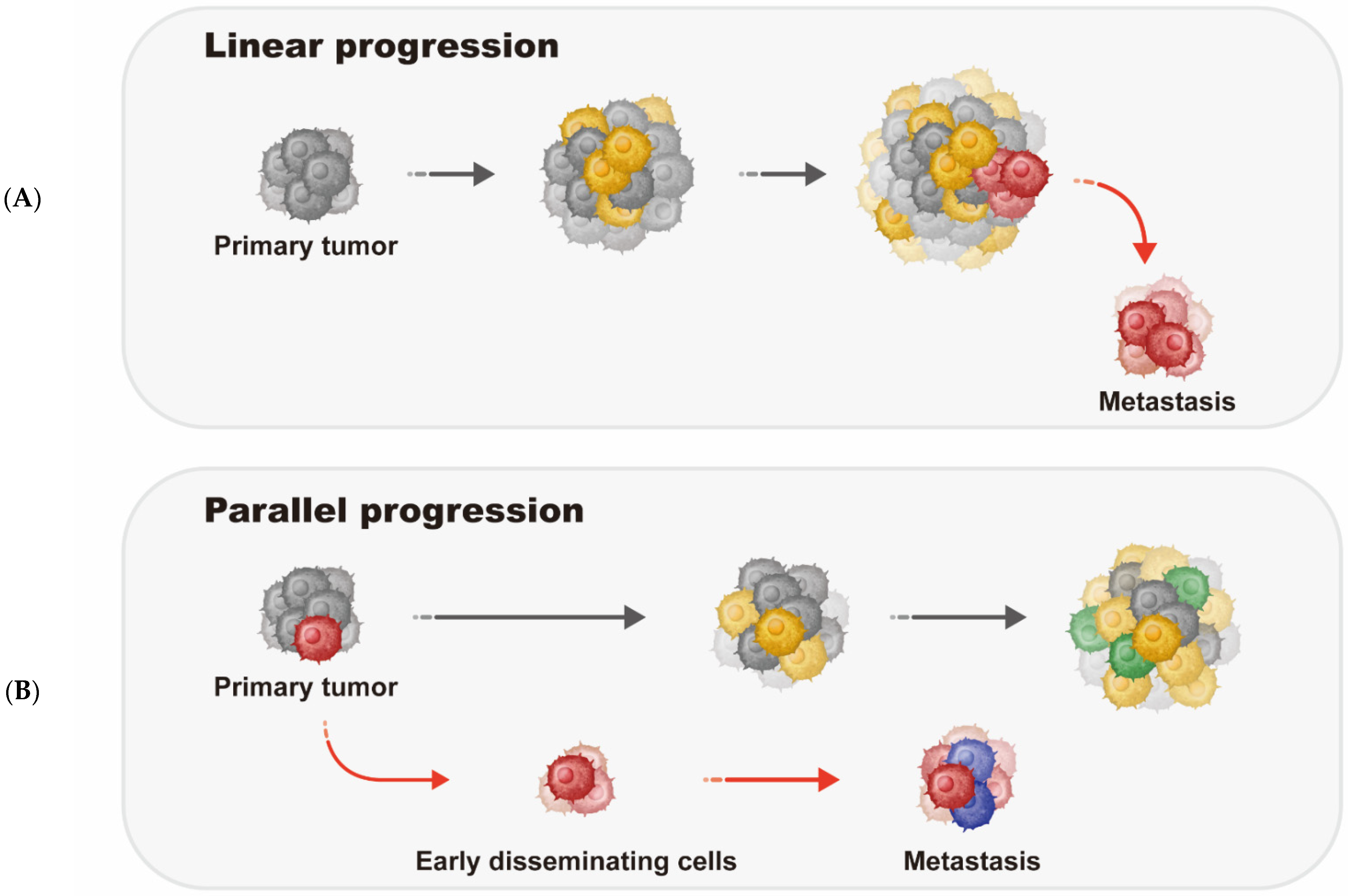
2. Metastatic Breast Cancer Progression Models

Tumor metastasis is a multi-stage evolutionary process that confers a fitness advantage upon cancer cells. Metastasis generally involves several sequential steps (reviewed in [17]) that begin with local invasion, in which cancer cells escape their primary site through stromal cells surrounding the tumor, followed by intravasation, in which the tumor cells invade the blood or lymphatic systems. Tumor cells surviving this step are known as circulating tumor cells (CTCs) [18]. CTCs can escape the circulatory system, enter distal blood vessels, and then invade distal tissues from these blood vessels in a process known as extravasation. Once these cells adapt to the local microenvironment, they are described as disseminated tumor cells (DTCs). DTCs transform into metastatic initiating cells that ultimately form metastatic lesions [3,19]. Key steps to metastasis include the epithelial to mesenchymal transition (EMT) in which tumor cells become invasive and transition into mesenchymal stem cells, and angiogenesis, a process by which the tumor generates blood vessels involving the tumor cells and the local environment.
Based on empirical findings, two models of metastasis have been proposed: the linear model and the parallel model [20,21,22,23]. In the linear model, the development of metastasis occurs relatively late after a subset of clones acquired mutational and growth capabilities that would allow them to survive and grow in distal organs. This model predicts greater genomic similarity between the metastatic and the primary tumors. In contrast, the development of metastasis occurs relatively early in the parallel model, in which the primary and metastatic tumor cells evolve independently. Therefore, owing to selection pressure and the inherent genetics of tumor cells, metastases that arose from the parallel progression model are more genetically divergent (Figure 1). Kroigard and colleagues [23] performed whole exome sequencing (WES) and targeted deep sequencing of 26 sequential tumors from six breast cancer patients. Interestingly, these data support the existence of both the linear and parallel progression models in developing metastases as well as metastasis–metastasis seeding.
Ullah and colleagues [24] performed next-generation sequencing (NGS) of sequentially collected and spatially diverse primary BC tissues, metastases, and auxiliary lymph nodes from mBC patients to identify evolutionary genetic landscapes as a function of metastasis. In agreement with the Kroigard report [23], both linear and parallel progression models of metastasis were evident. In addition, there is support for metastatic spreading and polyclonal seeding, in which metastatic subclones can originate from the primary tumor and other metastases.

3. Breast Tumor Immune Microenvironment

Breast cancer progression is controlled by both genetic and epigenetic factors that coordinate crosstalk between tumor cells and their associated immune microenvironment through interactions between immune cells, stromal cells and chemokines [25,26]. The genomic evolution of breast cancer during metastasis has been well elucidated, however, the evolution of immune cells and the tumor microenvironment is less understood. Breast cancer tumor immune microenvironment (TIME) studies have primarily focused on primary tumors [27,28,29]. In early-stage breast cancer, a positive correlation between longer survival and greater tumor infiltration lymphocyte (TIL) count, as well as upregulation of immune-related gene expression, has been observed [30,31]. These features are also associated with greater chemotherapy sensitivity in operable breast cancer patients, evident by a higher pathological complete response rate [32,33]. However, the identities of the specific genomic features associated with high TILs remain elusive.
Recently, numerous studies have profiled immune cell compositions and characterized the differences in immune microenvironment between primary and metastatic breast tumors [34,35,36]. Metastatic tumors have lower immunogenicity, evident by decreased expression of MHC class I and immune proteasome genes coupled with increased expression of HLA-E, as well as the reduction in dendritic cells. Multiple studies have demonstrated that most immune cell types and immune functions were depleted in metastatic lesions (reviewed in [37,38]). Substantially lower TIL counts and PD-L1 expression were also observed in metastatic lesions, possibly due to immune escape. Furthermore, Zhu et al. demonstrated that M2-like macrophages are enriched in metastatic lesions [27]. Their potential role in promoting metastasis warrants further investigation. Interestingly, lymph node metastases have significantly higher PD-L1 expression than visceral metastases [36].

4. Histological and Molecular Subgroups of Breast Cancer

4.1. Histological Breast Cancer Subgroups

Several histological types of human BC have been reported and characterized (reviewed in [39]). According to the World Health Organization (WHO) database, nearly all BC cases (~95%) are adenocarcinomas that are derived from the terminal duct lobular unit of the breast. The majority of BCs are invasive ductal carcinomas (50–80%). Other classifications include invasive lobular carcinomas (5–15%), mucinous carcinomas (2%), and medullary carcinomas (5–7%) [39,40]. Inflammatory breast cancer (IBC) accounts for ~1% of all BC cases. The earliest presence of breast cancer is ductal carcinoma in situ (DCIS), defined by the presence non-invasive or pre-invasive BC cells that line the breast milk ducts, but have not yet spread into surrounding tissues (www.cancer.org, accessed on 5 June 2022).
BCs are categorized into stages using the American Joint Committee on Cancer TNM system in which the size of the tumor (T), spread to lymph nodes (N) and/or metastases (M) are key defining characteristics. DCIS is commonly referred to as Stage Tis, while Stage 1 diseases are primarily defined as tumors ≤20 mm in size. Stage 2 BCs are 20–50 mm with a spread of up to three lymph nodes, while Stage 3 tumors are >50 mm in size with spread to multiple lymph nodes and tissues surrounding the breast. Finally, Stage 4 BCs are tumors that have metastasized to distal tissues (www.cancer.org, accessed on 5 June 2022).
In addition to TNM staging criteria, the status of steroid hormone receptors (HR), namely estrogen receptor (ER), progesterone receptor (PGR) and human epidermal growth factor receptor 2 (HER2), have become increasingly critical for understanding BC tumorigenesis and aggressiveness (reviewed in [41]). ER and PGR function as transcription factors following their activation by binding to estrogen and progesterone, respectively (reviewed in [42]). In their role as transcription factors, ER and PGR bind to specific DNA elements and chromatin in regulating gene expression of their target sites, thus affecting signaling pathways that involve cell growth and cell function [42]. Most BCs (60–80%) are positive for ER and PGR expression and ER-positive (ER+) BC patients show an improved response to anti-estrogen-based therapies, have longer survival and are less likely to develop recurrent diseases as compared to ER-negative or ER-negative/PGR-negative BC patients [42,43].
ER is expressed in two forms—ER-α (ERα) and ER-β (ERβ). ERα is expressed from the ESR1 gene, whereas ERβ is expressed from the ESR2 gene locus. Both ERα and ERβ bind to the estradiol ligand, dimerize and then move to the nucleus where they function as transcription factors to modulate expression of their target genes (summarized in [44]). ERα is thought to have prominent functional roles in breast and ovarian tissues [45] as well as in mammary gland development in mice [46], while ERβ is thought to function in mammary gland epithelium differentiation [47].
HER2 (ErbB2) is a member of the epidermal growth factor family that includes EGFR (HER1), ErbB3 (HER3) and ErbB4 (HER4). HER2 is an oncogene that functions as a transmembrane receptor tyrosine kinase that controls cell growth and differentiation. HER2 is over-expressed or amplified in up to 25% of BCs. HER2 overexpression correlates with poor BC patient outcome, as well as tumor metastasis, tumor aggressiveness [48], and positive lymph node status. HER2-positive (HER2+) BC patients also show poor standard chemotherapy response and = HER2-amplified BC patients display activated PI3K and EGFR (RAS/RAF) signaling and poor outcomes [49].
A subgroup of BC tumors in which ER, PGR and HER2 are negative, termed triple negative breast cancer (TNBC), accounts for 15–25% of all BC cases [50]. TNBCs are not confined to one histological subtype and have diverse molecular profiles; however, it should be noted that TNBCs show expression profiles seen in basal and myoepithelial breast cells [51,52,53]. TNBCs are an aggressive form of human breast cancer, with high rates of metastasis, tumor recurrence, and poor overall patient survival [50]. The mean TNBC patient survival is less than one year, with a dismal 5-year survival rate of only 11% for metastatic TNBC patients [50]. TNBC patients of African American or Hispanic descent have shorter OS compared to other ethnic and racial groups [50,54]. Moreover, younger women, and women of African American or Hispanic descent, are more likely to develop TNBCs, whereas HR+ BCs are more prevalent in older women [55].

4.2. Molecular Subgroups of Breast Cancer

In addition to histological BC classification, the large volume of molecular data collected has enabled for classification of molecular BC subgroups based on tumor gene expression profiles: (1) luminal A, (2) luminal B, (3) HER2-enriched and (4) basal-like [51]. Subsequently, Parker and colleagues used 50 genes from a gene expression microarray, termed Prediction-Analysis Microarray 50 (PAM50), to delineate each intrinsic BC molecular subtype [56]. It should be noted that a fifth group, termed normal-like BC, has been reported (reviewed in [52]). Normal-like BCs, as their classifier suggests, show high expression of genes specific to adipose and non-epithelial cells, as well as low expression levels of luminal epithelial cell genes [52]. These poorly understood tumors account for 5–10% of all BCs and showed the best patient prognosis of all five molecular subgroups [57].
Luminal A and B tumors are the most prevalent in ~60% of all BC cases, in which Luminal A cases comprise ~40% of all BCs, while Luminal B cases account for ~20% [58]. The Luminal BCs and are defined by high ER and PGR gene expression, as well as genes involved in ER activation and signaling [58]. Differences in Luminal A and B tumors stem from higher PGR expression and lower expression of cell cycle and proliferation genes in Luminal A tumors [58]. In this regard, the proliferation genes Ki-67 and PCNA are expressed in Luminal B tumors, but not in Luminal A BCs [58]. In addition, Luminal B tumors are enriched for TP53 somatic mutations, and Luminal B tumors are typically dedifferentiated with higher grade while Luminal A tumors are more differentiated [58]. Finally, Luminal B patients have significantly shorter OS and disease-free survival (DFS) comparted to Luminal A breast cancer patients [58].
HER2 breast tumors account for ~20% of all BCs and are primarily defined by HER2 overexpression, HER2 amplification and low ER expression [58]. In addition, HER2 BCs display amplification and overexpression of key genes surrounding the ERBB2 locus that include the GATA4 transcription factor and the GRB7 adaptor protein [52] that binds tyrosine kinases and the RAS oncogene. However, HER2-BCs also display low expression of the GATA3 transcription factor. The anti-HER2 therapy trastuzumab (Herceptin) has dramatically increased HER2-BC patient survival; however, these patients mostly die to disease progression and the development of brain metastases [59].
It should be noted that a new subgroup, termed HER2-low, was more recently described [60,61]. HER2-low BCs are defined as tumors without HER2 amplification, but show weak or intermediate HER2 detection based on immunohistochemistry (IHC), namely IHC 1+ or IHC 2+ status, respectively [61]. HER2-low BC patients comprise nearly 50–55% of all BC cases, are generally ER+, HER2-low tumors are of higher grade and display higher proliferation than HER2-negative tumors [62,63]. HER2-low patients also show a lower immune response than HER2-negative patients [62]. HER2-low patients are resistant to Herceptin therapy. HER2-low BC patients generally have poor outcome [64], as are HER2-low/ER+ BC patients who are over age 55 years old [65]. However, a meta-analysis of the TCGA BC expression data showed that HER2-low patients did not show differences in disease-free survival, progression free survival or overall survival as compared to other BC subgroups [66]. Comparing HER2-low to HER2-negative cases, Denkert and colleagues [67] showed that HER2-low tumors are distinct from HER2-negative tumors with respect to HR status, tumor grade and tumor proliferation. Interestingly, HER2-low patients displayed improved survival over HER2-negative patients as well as differences in neoadjuvant chemotherapy sensitivity. Overall these findings suggest that the clinical aspects of HER2-low BCs are unclear and require further investigation.
Basal-like breast tumors display gene expression profiles akin to basal and myoepithelial breast cells [51,52]. Since TNBCs also show similar gene expression profiles to basal-like BCs, the majority (80%) of TNBCs are indeed basal-like [51,52,53], but display a high level of molecular heterogeneity based on gene expression data [68,69]. Like TNBCs, basal-like tumors lack ER, PGR and HER2 expression. Basal-like BCs also harbor the greatest amount of tumor-specific CNV as compared to the other groups. In addition, basal-like tumors are of higher grade than the other molecular subgroups. Concurrent with this observation, basal-like BCs are aggressive tumors and basal-like BC patients have limited response to conventional therapies, poor outcome and shortened DFS and OS [69,70].

5. Genetic Landscape of Breast Cancer

5.1. Hereditary Germline Mutations

Most human cancers can develop through the accumulation of somatic mutations of key driver genes that promote cell growth, inhibit DNA repair and bypass cell growth checkpoints to achieve uncontrolled growth and division [71]. However, human cancers can also develop because of hereditary germline mutations from family members. Approximately 5–10% of all BCs have family history of breast or ovarian cancer, and women with germline mutations in breast cancer type 1 (BRCA1) and type 2 (BRCA2) are of greater risk of developing breast or ovarian cancer [72]. Indeed, BRCA1 and BRCA2 mutation carriers have risks of 57% and 49%, respectively, for developing breast cancer [73]. Moreover, BRCA1 and BRCA2 mutation carriers have risks of 60% and 55%, respectively, for developing ovarian cancer [73]. However, it should be noted that environmental factors, as well as cellular and allelic heterogeneity are also influential in determining risk of disease [74]. In addition to increased familial BC and ovarian cancer risk due to BRCA1 and BRCA2 germline mutations, approximately 40–45% of all hereditary BCs harbor BRCA1 germline mutations and 35–40% harbor BRCA2 germline mutations. In total, 80–85% of all hereditary BCs are linked to BRCA1/2 germline mutations [75,76,77], indicating that these driver mutation events confer substantial advantages in evading growth controls.
BRCA1 and BRCA2 are tumor suppressors that maintain genomic DNA stability by promoting double-strand DNA break (DSB) repair via homologous recombination and non-homologous end-joining (NHEJ) [78]. BRCA1 co-localizes with RAD51 and other DNA repair proteins, and is activated by phosphorylation from the kinases ATM, ATR and CHK2 that are also activated due to DNA damage [78]. Activated BRCA1 functions in G1-S and G2-M cell cycle arrest following ultraviolet or infrared-based DNA damage and acquiring access to specific DNA regions by interactions with histone deacetylases (HDACs) and SWI/SNF chromatin remodelers [78]. Interestingly, BRCA1 also inhibits estrogen-based transcriptional activation of ESR1 (ER α, ERα) in breast tissues, and studies are suggestive that BRCA1 directly interacts with ERα to inhibit ERα function [79]. Additional germline mutations in breast cancer include TP53, ATM and CHK2 [80]. Interestingly, all these genes are involved in DNA repair and genomic integrity.
Beyond BRCA1 and BRCA2 genetic variation, several studies have interrogated germline variation of cancer-related genes in BC and mBC patients. A report from Stuttgen and colleagues [81] summarized the frequency of germline changes of 30 genes in 100 mBC patients that varied by race/ethnicity and HR receptor status. A total of 14 patients harbored pathogenic variants, while an additional 21 patients harbored variants of unknown significance. Interestingly, 6% of patients displayed BRCA1 variation, and key additional variants were found in APC, ATM, BAP1, BARD1, BRCA2, BRIP1, CDH1 and CHEK2. Indeed, The Breast Cancer Association Consortium [82] interrogated rare germline variation of nine genes including ATM, BARD1, BRCA1, BRCA2, CHEK2, PALB2, RAD51C, RAD51D and TP53 across 42,580 BC patients and 46,387 control subjects independent of family history of disease. Overall, the 9-gene panel was associated with over 27% of all early onset TNBCs. Specifically, ATM variation was strongest for HR+/HER2-negative patients with high tumor grade. BARD1, BRCA1, PALB2, RAD51C and RAD51D genetic variation was linked to TNBC overall risk, while CHEK2 genetic variation was prevalent across all subgroups except for TNBC patients. BRCA1 was associated with increased risk of all BC subtypes, with the highest overall risk for TNBC disease. TP53 was associated with HER2-positive disease irrespective of HR status. Similarly, Couch et al. [83] evaluated associations of variation in BRCA1/2 and 19 other genes in across a study population of over 41,000 Caucasian BC patients. In addition to variations of BRCA1/2, CDH1, PTEN and TP53 that are highly associated with BC risk, variations in ATM, BARD1, CHEK2, PALB2 and RAD51D were also associated with moderate or high BC risk. However, variants of BRIP1, MLH1, MRE11A, NBN, NF1, PMS2, RAD50 and RAD51C were not linked to BC risk.
Characterizing germline variation of BC patients with varying ancestry shows interesting differences between populations. Han Chinese BRCA1/2 mutation carriers identified by next generation sequencing account for 5% of BC patients studied. BC patients with BRCA1/2 mutations are generally younger and are categorized into ER-negative and basal-like molecular subgroups [84]. Performing similar analyses on TNBC cases [85] showed that 16% of the cohort harbored at least one pathogenic variant and are associated with early onset disease. Interestingly, allele imbalances due to homologous recombination deficiency (HRD) in over 50% of cases, contributed by BRCA1, BRCA1 and RAD51D mutations in 7.1%, 2.2% and 2.8% of cases, respectively. Indeed, RAD51D germline events were more common in Chinese TNBC patients than African American or Caucasian patients and are linked to aberrancies in HRD, DNA repair, as well as sensitivity to PARP inhibitors.
A survey of 1382 Greek BC patients with extensive family history of BC or with early onset disease revealed variation in nearly one-third of the cohort, with BRCA1/2 loss of function variants found in nearly one in four cases [86]. This analysis also unveiled additional variants in ATM, CHEK2, PALB2, RAD51C and TP53. Similarly, germline analyses of early onset women from Latin America showed extensive alterations in 15% of patients [87]; however, a second report identified variants in 34% of patients studied, with BRCA2 mutations in 4% of the cohort [88].

5.2. Somatic Mutation Profiles

The development and advancement of next-generation sequencing has enabled deep coverage of whole genome sequencing (WGS) and whole exome sequencing (WES) across large numbers of primary tumors, benign tissues, as well as apparently normal and normal-adjacent cells [89]. Primary human breast tumors have been extensively profiled using WGS and WES to identify somatic mutation events that are not only drivers of disease, but also potential efficacious targets for improving patient outcomes. In 2012, the Cancer Genome Atlas (TCGA) Network Consortium reported WES data for n = 510 primary invasive ductal BCs (IDCs), of which n = 466 primary tumors were also profiled for mRNA and miRNA gene expression, DNA methylation and copy number variation (CNV) [90]. Analysis of the mutation data [90] confirmed mutations in genes previously described that include AKT1, CDH1, CDKN1B, GATA3, KTM2C(MLL3), MAP3K1, PIK3CA, PTEN, RB1 and TP53. This analyses also identified novel somatic mutations in several genes that include CCND1, NF1, RUNX1, TBX3 and others [90]. Stratified by molecular subgroupings, somatic PIK3CA mutations were enriched in the Luminal A (45% of tumors), HER2 (39%) and Luminal B (29%) subtypes. TP53 mutations were enriched in Basal-like (80%), HER2 (72%) and Luminal B (29%) subgroups. GATA3 was altered in Luminal B (15%) and Luminal A (14%) subgroups, while MAP3K1 was mutated in 13% of Luminal A tumors [90].
Stephens and colleagues from the International Cancer Genome Consortium [91] reported a separate WES analysis of 79 ER+ and 21 ER-negative BCs that confirmed the well-established somatic mutations in AKT1, CDH1, GATA3, PIK3CA, PTEN, RB1 and TP53. Driver mutations in >40 genes were identified that includes AKT2, ARID1B, CASP8, NCOR1 and SMARCD1, among others [91]. While the number of driver mutations did not correlate with tumor stage, the total number of driver and passenger mutations did positively correlate with tumor grade. In addition, the number of CpG to TpA substitutions in ER-negative BC patients correlated with patient age, whereas no correlation was identified in ER+ patients, suggesting that ER influences the mutation spectrum of BC patients [91].
In 2015, TCGA subsequently reported and compared multi-omic profiling data on n = 127 invasive lobular carcinomas (ILCs) to n = 490 IDCs and 88 mixed lobular and ductal tumors [92]. WES data for TCGA ILCs showed a high prevalence of mutations in CDH1 (63%), PIK3CA (48%), RUNX1 (10%), TBX3 (9.5%) PTEN (7%) and FOXA1 (7%) [92]. More recently, Pereira and colleagues performed DNA sequencing of the top 173 mutated genes across 2433 primary breast tumors [93]. As described in other reports, mutations in PIK3CA (40%) and TP53 (35%) were the most abundant, while MUC16 (17%), AHNAK2 (16%) SYNE1 (12%), GATA3 (11%) and KTM2C (MLL3) (11%) mutations were also identified in at least 10% of all patients [93]. Interestingly, BRCA1 and BRCA2 somatic mutations are rare, with mutation frequencies <2% for both regions, while TP53 germline mutations occurred in <1% of BCs [93]. These findings suggest that distinct profiles of germline and somatic mutations occur in driving BC tumorigenesis. By stratifying the somatic mutations by clinical tumor subgroup, ductal BCs displayed mostly TP53 and PIK3CA mutations, lobular tumors displayed CDH1 and PIK3CA mutations, and approximately 90% of medullary BCs harbored TP53 mutations [93].
The Pereira report also revealed a set of 40 BC driver gene mutations [93], most of which were also identified by TCGA [90,92] and in the Catalogue of Somatic Mutations in Cancer (COSMIC) online database (https://cancer.sanger.ac.uk/cosmic, accessed on 5 June 2022). These driver mutations are in pathways involved in AKT signaling, cell cycle regulation, chromatin modifications, DNA damage, MAP kinase signaling, tissue organization, transcriptional regulation, and others [93]. Correlating these driver mutations to clinical BC patient outcome showed that functional: (1) PIK3CA mutations in ER+ tumors were linked to lower tumor grade, (2) CBFB and GATA3 mutations were enriched for younger age patients, (3) KTM2C/MLL and CDH1 mutations occurred in older patients and (4) TP53 somatic mutations were linked to increased tumor grade irrespective of ER status [93].
Nik-Zainal and colleagues characterized WGS data for 560 primary BCs and identified 1628 protein-coding driver mutations in 93 genes [94], with at least one driver mutation in nearly every tumor analyzed. The most frequently mutated genes included CCND1, ERBB2, FGFR1, GATA3, MAP3K1, MYC, PIK3CA, PTEN, RB1 and TP53. TP53, MYC, PTEN and RB1 alterations were mostly found in ER-negative BCs, while mutations in PIK3CA, CCND1, FGFR1 and GATA3 were enriched in ER-positive BCs. Novel somatic alterations in FOXP1, MED23, MLLT4, XBP1 and ZFP36L1 were also unveiled. Inactivating germline or somatic BRCA1 or BRCA2 mutations were present in 16% of the cohort, and most of these cases also showed loss of the wild-type (WT) allele for these gene regions. As expected, BRCA1 or BRCA2 mutated BCs also included substantial genomic rearrangements that include small tandem duplications and deletions.
TCGA and the International Cancer Genome Consortium (ICGC) combined datasets to form the Pan-Cancer Analysis of Whole Genomes (PCAWG) Consortium, with the goal of summarizing genomic, epigenomic and transcriptomic variation for up to 6835 tumors across 38 cancer types [95]. With respect to BCs, PACWG showed that structural variation and point mutations are more prevalent in breast and ovarian cancers; however, BC point mutations contributed lower than colorectal cancer and B cell lymphomas [95]. Interestingly, chromothripsis, described by thousands of catastrophic mutations and rearrangements occurring in specific gene regions or chromosomes at one time, occurs most frequently in sarcomas, glioblastomas, squamous lung tumors, melanomas and breast cancers.
PCAWG also performed pathway analyses based on copy number and gene expression data from 1780 BCs stratified by molecular subgroups (basal-like, HER2, luminal A and luminal B). Pathway enrichment was most pronounced in apoptosis, chromosomal segregation, immune response and ribosomal biogenesis [96]. Specifically, apoptotic pathways were associated with patient prognosis in HER2 and Luminal A BCs. In this regard, DUSP1 expression was the strongest apoptotic marker in HER2 BCs. DUSP1 is overexpressed in BCs and functions as a phosphatase signaling protein in suppressing apoptosis, thereby contributing to poor patient outcome. The immune pathway signature was associated with improved survival in basal-like and HER2 BCs, indicating that immune-based expression inhibits tumor progression, thereby improving patient outcome. Ribosome biogenesis pathways were almost exclusive to Luminal A BCs, while Luminal B tumors had enrichment in chromosome segregation.

5.3. Copy Number Variation in Breast Tumors

Somatic mutations may result in changes to protein activity, such as the inability to bind its target, misfunction, constitutive function or catalytic inactivation. However, most somatic mutations are mono-allelic; therefore, one WT copy can still rescue the mutant genotype. According to Knudson’s Two-hit hypothesis [97], complete gene inactivation requires both alleles to be targeted. This can be achieved by bi-allelic somatic mutation or a combination of mutation, deletion and/or silencing by promoter DNA hypermethylation [98]. Gene deletions and amplifications define the copy number variation (CNV) that is common across all human cancers [99]. Deletions of tumor suppressors or amplifications of oncogenes contribute to tumorigenesis and are influential in gene expression modulation. Technologies to identify changes include fluorescence in situ hybridization (FISH), comparative genome hybridization (CGH) and SNP arrays [100,101,102].
Focal and chromosomal arm level CNV changes are abundant in primary breast tumors. Breast tumors characteristically show HER2/ERBB2 amplifications in the HER2 subgroup, chromosome (chr) 5q loss and chr10p amplifications in Basal-like BCs, chr 1q and chr 16q deletions in Luminal BCs [90]. In genes that displayed significant somatic mutations, TCGA identified focal amplifications in EGFR, FOXA1, HER2 and PIK3CA; concurrently, this analysis showed focal deletions in MAP2K4, KMT2C (MLL3), PTEN and RB1 [90]. While HER2/ERBB2 amplifications were most substantially enriched in the HER2 subgroup, HER2/ERBB2 amplifications were also evident in Luminal B tumors, and HER2/ERBB2 deletions were seen in Basal-like BCs [90]. PIK3CA amplification was detected across all four molecular subgroups; however, this enrichment was strongest in Basal-like tumors. MAP2K4, RB1 and TP53 deletion events occurred in each subgroup, whereas CDKN2A and KMT2C deletions were more focused in Luminal B tumors. Arm-level amplifications in chromosome (chr) 1q, 7p, 8p, 8q, 16p, 17q, 20p and 20q occurred mostly in Luminal and HER2 tumors. Arm-deletions were substantial in the TCGA dataset, with these events occurring in most autosomal chromosomes [90].
The Pereira report included CNV and gene expression data on 2000 primary breast tumors from the METABRIC collection [93]. These CNV-driver events in which amplification or deletion affects gene expression levels include CCND1, ERBB2, MYC, PAK and ZNF703. Stratified by ER status, ER+ BCs displayed overwhelming gene deletions with TP53, CHD1, CBFN, MAP2K4, PTEN and CTCF as the most robust, with only ERBB2 and TBL1XR1 as amplified [93]. ER-negative BCs mostly showed TP53 deletions, with lower deletion event frequencies in RB1, CDH1, BAP1 and PTEN. ER-negative tumor amplifications were also focused on ERBB2 with less frequent amplifications of AKT1, CDKN1B, FOXO3, GATA3, KRAS, PIK3CA and TBL1XR1 [93].

5.4. Genetic Profiles of Metastatic Breast Tumors

Several reports have performed somatic mutation profiling of metastatic and non-metastatic breast tumors to identify genetic changes specific to metastases (summarized in [103]). While metastases are purportedly derived from the primary tumor, empirical evidence shows profound differences in somatic mutation profiles and frequencies [103]. Thus, identifying these metastasis-derived genomic alterations is important for improving mBC patient outcome. Roy-Chowdhuri and colleagues sequenced 46 hotspot genes for somatic mutations across 415 breast tumors, of which n = 110 were recurrent or metastatic [104] (Table 1); primary and metastatic lesions were analyzed for 61 BC patients. As stated in previous reports, somatic mutations in TP53, PIK3CA, AKT1 and ATM were the most prevalent. TNBCs displayed the highest frequency of TP53 somatic mutations, while HER2 tumors displayed TP53, PIK3CA and ATM somatic mutations [104]. In comparing primary BCs to metastatic lesions, 77% of these pairs were identical. Most of the remaining pairs showed somatic mutations in the metastases that were not identified in the primary tumor, and the metastases also showed increased mutation frequencies, suggesting that these were acquired specifically in the metastatic process for genetic fitness and cell survival [105]. Other differences in mBCs versus non-metastatic tumors involve aberrant JAK/STAT signaling and SWI/SNF chromatin structure alterations in mBCs that may also serve as driver events [105].
Lefebvre et al. [106] generated WES data on 216 primary mBC—white blood cell pairs and compared the findings to non-metastatic mutation data from TCGA (Table 1). A total of 12 genes in descending order of mutation frequency (AKT1, CBFB, CDH1, CDKN2A, ESR1, GATA3, MAP2K4, MAP3K1, PIK3CA, PTEN, RB1 and TP53) were significantly mutated in mBC over non-metastatic tumors, and eight additional genes (AGR, EDC4, ESR1, FRAS1, FSIP2, IGFN1, OSBPL3 and PALB2) displayed higher mutation frequencies in mBCs than non-metastatic tumors. Interestingly, ESR1 mutations were found in ER+ metastatic tumors from patients who were resistant to endocrine therapy, suggesting that ESR1 mutations may be driver events in mBC progression. ESR1 mutations reside in the ligand binding region and occur in up to 30% of mBCs, but are only detected at low frequencies in primary non-metastatic tumors. The most frequent ESR1 mutations are gain-of-function alterations in the ligand-binding domain, resulting in constitutive enzyme activity in the absence of estrogen [107].
Ng et al. [108] performed multiregional sequencing on primary tumor and synchronous metastatic lesions in treatment-naïve patients with mBC to investigate their genomic profiles and revealed substantial difference with a median of 60% of shared somatic mutations (Table 1). Mutations were in known driver genes including, but not limited to, GATA3, PI3KCA and TP53 and were preferentially clonal in both sites [108]. Mutations in genes participating in epithelial to mesenchymal transition, such as SMAD4, TCF7L2 and TCF4, were either specific to or enriched in the metastasis.
Most driver mutations are clonal and occur on the trunk of a tumor’s phylogenetic tree [105]. Substantial evidence has supported that treatment pressure would result in the selection and the outgrowth of resistance subclones, contributing to treatment resistance [7,109]. Characterizing the genomic landscape of metastatic patients who have been treated will yield valuable insights into the effect of systemic treatment on the tumor genome and ultimately provide a basis for the development of novel therapeutic agents. Angus et al. [14] compared mutational profiles of 442 metastatic lesions to a well-characterized cohort of primary breast cancer (the BASIS cohort) and revealed that the median number of single-nucleotide variants (SNVs), insertions and deletions (Indels) and structural variants (SVs) were higher in metastatic lesions, consequently, leading to a higher tumor mutational burden (TMB), which doubles in the metastatic lesions (Table 1). TMB, a well-established predictive biomarker for immunotherapy, quantifies the total number of somatic coding mutations in a tumor. Furthermore, the mutation frequency of six well-known driver genes (ESR1, TP53, NF1, AKT1, KMT2C and PTEN) increases in metastatic lesions of ER+ breast cancer [14]. Conflicting data exist regarding the change of TMB in mBC. In contrast to the finding of Angus et al., one study [15] reported higher TMB only found in metastatic lesions of HR+ breast cancer and no difference in other subtypes (Table 1) and another demonstrated differential TMB in metastatic HR+ and triple negative breast cancer [110].
More recently, Rinaldi and colleagues [107] reported WES on over 11,000 primary breast tumors that include more than 5000 distal metastases. ER+ HER2-negative mBCs were more prevalent than in non-metastatic tumors; however, HER2 amplification frequencies were similar between BCs and mBCs. Somatic mutations in TP53, PIK3CA, CDH1, GATA3, ESR1 and KMT2D were as previously described in other reports [14,90,104,106,108], while amplifications of MYC, CCND1 and HER2 and deletions of PTEN, CDKN2A, CDKN2B and RB1 were the most frequent in non-metastatic breast tumors [107]. In addition to ESR1 mutations in mBCs over non-metastatic tumors, CTCF driver mutations were discovered in 2% of mBCs, and amplification rates of the FGFR ligands FGF3, FGF4 and FGF9 were increased in mBCs.
Paul and colleagues [16] performed WES and WGS on 28 pairs of primary BCs and mBCs, as well as 38 unpaired mBCs, to identify somatic mutation and CNVs specific for mBCs (Table 1). Seven genes, ESR1, EVC2, MYLK, PALB2, PEAK1 and SLC2A4RG, were preferentially mutated in mBCs and were not identified as mutated in TCGA BRCA tumors [16,90]. Comparing these genes to TCGA mutation data for over 30 tumor types showed that ESR1, EVC2, MYLK, PEAK1 and SLC2A4RG were not significantly mutated in any other human cancer type [16]. CNV analyses showed metastatic-specific loss of CDKN2A/B and STK11 concomitant with gains of PTK6 and PAQR8 [16]. Stratifying mutation, CNV and immunohistochemical data by pathways identified activation of mTOR, WNT and cAMP/PKA pathways and CDK/RB inactivation, as well as focal adhesion pathway changes [16]. Of note, mTOR signaling has been implicated as a mechanism of escape from therapies that target ER and HER2, as well as in clinical settings [111,112]. The preferential activation of mTOR in endocrine therapy-treated mBC patients provides a genomic basis for utilizing agents to inhibit the mTOR pathway for the treatment of mBC [111].
A meta-analysis of mutation and CNV data of 261 BC driver genes for 4268 mBCs and 5217 non-metastatic BCs [113] revealed that while most mutation frequencies were similar between mBC and non-mBC tumors, ARID1A, ESR1 and NF1 displayed significantly higher mutation frequencies in mBCs. Incorporating SNP-based analyses of insertions and deletions revealed mBC-enriched or mBC-specific alterations in ARID1A, ATRX, AURKA, ESR1, FGFR4, NF1, PARP1, SMARCA4, STAG2 and TSC2 [113]. Of these, ESR1 displayed the highest frequency of amplifications and odds ratio of metastasis, and ESR1 somatic mutations were enriched in liver metastases, but absent in positive lymph nodes. Interestingly, TP53 mutations were enriched in positive lymph nodes, but absent in liver metastases [113].

5.5. Chromosomal Instability (CIN)

Chromosomal abnormalities have been reported in tumor progression in several cancers, including breast cancer [20]. Chromosomal instability (CIN), results from errors in chromosome segregation during mitosis, is a hallmark of cancer, and has been proposed to act as a driver in metastasis. CIN equips tumors with enhanced evolutionary capabilities and facilitates the development of treatment resistance by generating intra-tumoral heterogeneity [114]. WGS analyses have demonstrated that cancer genomes acquire CIN to evade oncogene addiction [115]. CIN is a robust feature in several cancers, including mBC [116]. Numerous studies have demonstrated a poorer treatment and/or survival outcome of patients with higher CIN in multiple cancer types, including breast cancer [115,117,118].
Analysis of a pan-cancer cohort (Memorial Sloan Kettering-Metastatic Events and Tropisms) consisting of more than 25,000 metastatic patients with tumor profiling and clinical information revealed that metastatic lesions were more chromosomally unstable than primary tumors for several cancers including HR+ mBC, evident by a higher fraction of genome altered (FGA) and a higher TMB [119]. They further investigated the clinical significance of TMB by comparing the percentage of patients with TMB high (≥10 mutations/Mb) metastatic tumors vs. primary tumors. They showed that a significantly higher percentage of TMB high tumors were observed in metastases of HR + HER2- patients [119]. Furthermore, the frequency of whole-genome duplication (WGD) was higher in the metastatic tumor of HR + HER2- compared to primary tumors [119]. The major differences between primary and metastatic lesions are summarized in Figure 2.
Collectively, studies on genomic landscapes and chromosomal aberrations have demonstrated that although metastases are clonally related to the primary tumor, evident by the sharing of many driver mutations, they are biologically distinct from the primary tumors, evident by increased genomic instability, the presence of unique mutations and harboring a specific HR or HER2 status, highlighting the need to treat metastases as a distinct clinical and biological entity.

6. DNA Methylation Alterations in Breast Cancer

In addition to somatic mutations and copy number alterations driving carcinogenesis, epigenetic mechanisms are important regulators of gene expression in human cancers. While somatic mutations alter gene product function, epigenetic changes modulate gene expression levels [98]. Epigenetic modifier genes acquire somatic mutations in human cancer, and the epigenetic mechanisms of DNA methylation, chromatin modifications, nucleosome positioning are also early and ubiquitous across nearly all forms of human cancer [120]. The most well studied epigenetic mechanism is DNA methylation, the covalent modification of the C-5 position of cytosines in a 5′-CpG-3′ (CpG) DNA sequence context [121]. Human cancers generally display global DNA hypomethylation that is represented by repetitive element and CpG poor DNA hypomethylation [122]. Alternately, CpG rich promoter regions, termed promoter CpG islands, are generally unmethylated in normal somatic cells, but display DNA hypermethylation in cancers that may be associated with reduced gene expression levels [122]. Gene body regions are methylated in actively transcribed genes, and gene body DNA hypomethylation correlates with reduced gene activity [123]. The presence of DNA methylation marks may serve as beacons for the recruitment of co-repressors to silence genes or co-activators; concurrently, the absence of DNA methylation may allow for the recruitment of transcription factors to activate gene expression [122].
DNA methylation changes do not occur in isolation, but correlate with modifications (methylation, acetylation, phosphorylation, etc.) of specific amino acid residues on the N-terminal tails of histones H2A, H2B, H3 and H4 [124]. Since genomic DNA wraps around the histone core octamer, histone modifications are instrumental in opening or closing chromatin to regulate gene expression [124]. For example, Histone H3 lysine 4 trimethylation (H3K4me3) is located at promoters of actively transcribed genes, while H3K9me3 is a marker of heterochromatin and silencing of repetitive elements [124]. H3K36me3 is found in gene bodies of actively expressed genes [124]. In addition, H3K27 acetylation (H3K27Ac) is a marker of active gene expression, however, H3K27me3 is linked to silenced genes [122]. H3K4me1 is associated with enhancers that distally control gene expression, while chromatin regions with both H3K4me3 and H3K27me3 marks are associated with poised enhancers [124]. Both DNA methylation and histone modifications co-exist with nucleosome positioning in regulating gene expression [125]. Actively expressed genes are noted by the absence of nucleosomes to allow RNA polymerase and related transcription machinery. Alternatively, repressed genes show highly compacted nucleosomes that ultimately block the access of transcription machinery to these regions [125].
Human BC is a rich source of cancer-specific epigenetic alterations. Interestingly, while BRCA1 germline mutations are frequent in BC, BRCA1 somatic mutations are much less frequent. Notably, BCs display loss of heterozygosity (LOH) of BRCA1 as well as reduced BRCA1 mRNA expression due to promoter DNA methylation of the remaining allele [126,127,128,129,130]. In addition to BRCA1 gene regulation, ERα expression is an important facet of breast tumor biology and patient outcome. ERα expression is based in the activity of the ESR1 gene, which is unmethylated in normal breast epithelium that correlates with active ERα expression [131].
Human primary breast tumors display ESR1 promoter DNA hypermethylation that is inversely correlated with ESR1 gene expression. ESR1 DNA hypermethylation correlates with poor patient outcome and correlates with response to tamoxifen [132,133,134,135]. A subset (37%) of BC patients with ESR1 promoter DNA hypermethylation developed loss of ER expression as a function of metastasis. Primary tumors in which ESR1 was unmethylated never displayed ER expression loss in metastases [131], implicating ESR1 DNA methylation as a potential driver of breast cancer metastasis.
In addition to BRCA1 and ESR1 driver events, DNA hypermethylation changes in BC occur in genes that are involved in key cellular processes [136,137,138] that include: (1) apoptotic regulation—APC, BCL2, DAPK, DCC, HOXA5, HIC1, TWIST and TMS1; (2) cell cycle regulation—CCND2, CDH1, CDKN2A, FOXA2, SFRP1, WIF1, WRN, WT1 and SFN (14-3-3σ); (3) cellular homeostasis—GPC3, HOXD11, LAMA3, LAMB3, LAMC2 and ROBO1; (4) DNA repair—ATM, BRCA1, MGMT and MLH1; (5) hormone and receptor signaling—ESR1, PGR, RARB2, RASSF1; (6) inhibition of angiogenesis—SFRP5, THBS1; and (7) negative regulation of tumor invasion and metastasis—CDH1, CDH13, CST6, SYK, TIMP3. Concurrently, DNA hypomethylation of genes involved in positively modulating metastasis and tumor aggressiveness include BCSG1, CDH3, NAT1 and UPA [137,138]. de Ruijter et al. [139] performed a meta-analysis of DNA methylation data on 87 loci across 72 reports to identify biomarkers correlative to patient outcome. DNA hypermethylation of RASSF1, BRCA1, PITX2, CDH1, RARB, PCDH10, GSTP1 and PGR was associated with poor patient outcome [139]. Comparing DCIS to invasive ductal carcinoma [140] has revealed increased DNA methylation frequencies of APC, CACNA1A, CDH1, HOXA10, MGMT, TRFAP2A and TWIST1 in IDC over DCIS specimens. CDH1, MGMT, and SFRP1 displayed increased DNA methylation levels in IDC over DCIS. Stratifying DCIS by HR status revealed DNA hypermethylation of ABCB1, FOXC1, GSTP1, and RASSF1 in ER+ DCIS. GSTP1 also showed increased DNA methylation levels in PR+ DCIS [140].
Identifying aberrant DNA methylation profiles in human cancers has been propelled due to technological advancements, and the most paramount is the use of bisulfite to delineate between methylated and unmethylated cytosines (reviewed in [141]). Bisulfite treatment of genomic DNA converts unmethylated cytosines to uracil (replaced by thymine in PCR applications), while methylated cytosines are refractory to treatment; thus, generating a DNA sequence change that can be exploited using PCR and sequencing technologies [141]. Interrogating individual loci by PCR and methylation-sensitive restriction enzyme methods provided only a limited picture of cancer methylomes, and profiling DNA methylation levels on a genome-scale (microarrays) [142,143,144] or in a genome-wide manner (whole genome bisulfite sequencing (WGBS)) [142,145,146,147] has allowed for the discovery of DNA methylation changes across the genome. DNA methylation microarrays have been used on large numbers of primary tumors by TCGA [90,92,148] and other groups [149,150,151] to identify patient subgroups, correlate DNA methylation events to patient outcome and response to treatment, as well as comparisons to mutation and CNV profiles.
Hill and colleagues [150] generated Illumina DNA methylation array data on 39 BCs and four tumor/normal-adjacent pairs and compared these data to clinical features of breast cancer patients. Unsupervised two-dimensional clustering of selected probes categorized BCs into three groups: Group 1 tumors displayed high DNA methylation and are associated with relapse and are positive for ER and PGR. Alternatively, Group 3 tumors exhibited low DNA methylation and were enriched for TNBCs. Probe-based analyses identified six genes (ACADL, ITR, RECK, SFRP2, UAP1L1 and UGT3A1) in which promoter DNA hypermethylation of each gene was associated with disease relapse and shortened survival after diagnosis [150].
A separate promoter DNA methylation array analysis of 14,000 genes across 236 infiltrating BCs [151] showed similar clustering based on ER status; however, the unmethylated group clustered with the ER-enriched subgroup. Clustering of the entire tumor collection yielded six DNA methylation subgroups, in which HER2-positive tumors were enriched in Group 2, basal-like tumors in Group 3 and Luminal A tumors in Group 6 [151]. The remaining groups were mixes of expression subgroups: Group 1 contained HER2, basal-like and Luminal B tumors, Group 4 contained HER2 and Luminal B tumors, and Group 5 contained Luminal A and B tumors [151]. Comparison of DNA methylation and gene expression subgroup genes revealed that the DNA methylation signatures reflect the T cell lymphocytic infiltration of tumor tissues, implicating not only the tumor microenvironment, but also an immune component reflected in the DNA methylation data [151].
TCGA reported five BC subgroups based on the unsupervised clustering of Illumina DNA methylation array data of gene promoters and gene bodies for all genes in the human genome across 466 breast tumors [90]. Of note, Group 2 and Group 5 tumors were generally HER2-negative. Group 5 tumors were identified by the lack of cancer-specific DNA methylation an overlapped with Basal-like expression subgrouping, extensive TP53 mutations, and the absence of ER and PGR expression [90]. Group 3 tumors displayed a DNA hypermethylated phenotype that was enriched for Luminal B expression subgrouping, the absence of MAP2K4, MAP3K21 and PIK3CA mutations [90]. A separate analysis by Fang and colleagues [148] identified DNA hypermethylated tumors as belonging to a CpG island methylator phenotype (CIMP), defined as cancer-specific DNA hypermethylation of a subset of CpG islands in a subset of tumors. CIMP was first presented in 1999 for colorectal cancer [152] and CIMPs have been subsequently identified in endometrial, brain and gastric tumors (reviewed in [153]). Breast-CIMP (B-CIMP) tumors display a DNA methylation profile that determines metastatic potential. B-CIMP was associated with low risk of metastasis and improved patient outcome indicating that DNA hypomethylation is correlated with metastasis in breast cancer [148].
A comparison of promoter DNA methylation profiles between inflammatory BCs (IBCs) and non-IBCs identified only four regions with differential DNA methylation: AGT, TJP3, MOGAT2 and NTSR2 [149]. All four loci displayed DNA hypermethylation in IBCs as compared to non-IBCs. Unsupervised clustering analyses of the entire data set revealed three DNA methylation subgroups: normal-like, high DNA methylation and low DNA methylation [149]. Tumors in the high-DNA methylation group were metastatic and these patients had poor outcome [149]. Pathway analyses of genes displaying differential DNA methylation between the low and high DNA methylation subgroups showed differences in genes related to chemokine signaling, cytokine receptor activation, focal adhesion, Wnt signaling and metabolism of pyruvate, galactose, fructose and mannose [149]. Overall, the differential DNA methylation between IBCs and non-IBCs indicates the potential role of DNA methylation in metastasis and subsequent poor patient outcome.

7. Therapeutic Options for Breast Cancer Patients

Current established BC treatment options include chemotherapy (CT), endocrine therapy (ET), radiation therapy (RT), as well as targeted therapies (reviewed in [55,154]), which are selected based on HR and HER2 status. ER+/PGR+/HER2-negative BC patients mostly receive endocrine therapies such as tamoxifen, letrozole, anastrozole or exemestane; however, cytotoxic chemotherapies such as adriamycin (Doxorubicin), cyclophosphamide, paclitaxel, docetaxel or carboplatin are administered in selected patients. Tamoxifen functions to inhibit estrogen binding to ER to modulate ER function, while letrozole, anastrozole and exemestane are aromatase inhibitors that inhibit the conversion of androgens to estrogen. Cyclophosphamide is a DNA alkylating agent that complicates DNA replication, adriamycin is a DNA intercalator that interferes with topoisomerase-mediated DNA repair, docetaxel and paclitaxel inhibit microtubule function to disrupt mitosis and carboplatin functions by facilitating DNA crosslinks to inhibit DNA replication [55]. HER2+ patients usually receive chemotherapies as well as the HER2-targeted monoclonal antibody therapies trastuzumab (Herceptin) or pertuzumab [55]. If the HER2+ BC patient is HR+, then endocrine therapies are administered. Finally, TNBC patients mostly receive chemotherapies that include adriamycin/cyclophosphamide, adriamycin/cyclophosphamide/paclitaxel or docetaxel/cyclophosphamide.

8. Established Treatment Schemes for Metastatic Breast Cancer Patients

Historically, mBC was treated with the assumption that key biological features were shared between primary and metastatic lesions. However, in-depth analyses of primary and metastatic lesions of breast cancer have revealed substantial differences in genomic landscapes and significantly advanced our understanding of the effects of systemic treatments on the tumor genome, and ultimately broadens the treatment options for mBC patients. Cancer cells seeding metastases continue to evolve by acquiring novel somatic and epigenetic alterations. However, the role of genomic alterations in driving the metastatic process is not well-understood.
Therapeutic options for mBC patients are also guided by HR and HER2 status [55]. ER+/PR+/HER2– mBC patients receive CDK4/6 and aromatase inhibitors as first line therapy [55]. Subsequent lines of therapy involve hormone-based therapies such as tamoxifen or targeted therapies based on menopause status. If the mBC patient also has BRCA1 or BRCA2 germline mutations, the PARP inhibitors olaparib or talazoparib are administered. Subsequent lines of therapy are usually single-agent cytotoxic chemotherapies [55]. HER2+ mBC patients receive (1) taxane, Herceptin and pertuzumab as first line therapy; (2) endocrine therapy with HER2-based therapeutics if the patient is ER+/PGR+/HER2+, or (3) ado-trastuzumab emtansine, which is herceptin covalently linked to mertansine (DM1) [55]. DM1 inhibits microtubule assembly by interacting with tubulin to inhibit DNA replication and cell division. Subsequent therapy lines for HER2+ mBC patients involve Herceptin with chemotherapy or endocrine therapy if the tumor is ER+/PGR+. Finally, metastatic TNBC (mTNBC) patients initially receive single-agent chemotherapies including taxane, platinum or anthracycline; however, the response rates for these agents are <40% with PFS of 3–5 months [55]. Subsequent lines of therapy for mTNBC patients include single agent drugs such as capecitabine (a prodrug that is metabolized to 5-fluorouracil to inhibit DNA replication), eribulin (inhibits microtubule assembly to inhibit cell division), gemcitabine (a cytidine analog that inhibits DNA replication) or vinorelbine (inhibits mitosis) [55].

9. Novel Molecular Therapeutic Targets for mBC

Tumoral heterogeneity is observed at morphological, phenotypic, and molecular levels and represents a significant clinical challenge in accurately characterizing and treating the disease. Human cancers undergo considerable molecular and cellular evolution under therapeutic pressure as well as during development, giving rise to spatial and temporal heterogeneity. Clonal evolution supports the notion that treatment exerts pressure leading to the selection and the outgrowth of resistant subclones [7]. Unfortunately, several metastatic-specific driver mutations are related to treatment resistance rather than metastatic process [155]. mBC patient survival is dismal, since secondary lesions are often refractory to conventional therapies [155]; therefore, it is necessary to develop novel treatments for mBC.
Given the effects of BRCA1/2 alterations on DNA repair and homologous recombination in BC, treatments directed towards DNA repair are now recognized as promising targeting strategies for these patients. Poly(ADP-ribose) polymerases (PARPs) bind to DNA strand breaks as an early response to DNA damage and recruit machinery to repair the lesion, thereby promoting cell survival. Treating cancer cells with PARP inhibitors (PARPi) blocks the ability of PARP to initiate DNA repair, thus leading to accumulated DNA damage and ultimately, apoptosis. PARPi have been studied as single- and dual-agents in treating BC patients (reviewed in [156,157]). Recent meta-analyses [157,158] of clinical trials utilizing PARPi for BC patients showed efficacy in improving OS, as well as improved PFS for TNBC, HR-positive, and or HER2-negative patients, as well as those with BRCA1/2 germline mutations. PARPi showed improvement in tumor drug response, OS and PFS for patients with locally advanced or metastatic HER2-negative BC with BRCA1/2 germline variation [158], thus supporting the use of PARPi in BC patient care.
There is strong evidence for genome evolution of HR+ HER2 breast cancer, which counts for approximately 70% of all breast cancer cases, from its early stage to metastatic stage. Many of the metastasis-enriched driver mutations are a result of resistance to treatment. Mutations in RB1, a tumor suppressor regulating the G1-S phase transition through transcriptional repression of E2F1 [159], are frequently found in patients treated with CDK4/6 inhibitors [160], which in combination with an aromatase inhibitor, have become the preferred frontline therapy for endocrine-sensitive metastatic HR+/HER2 breast cancer [161]. This treatment scheme showed significant improvements in both PFS and OS observed in four clinical trials: PALOMA-2, MONARCH-3, MONALEESA-2 and MONALEESA-7 [161]. The MONALEESA trials have demonstrated that patients with RB1 mutation at baseline (pre-treatment) had a significantly shorter PFS (3.7 months), suggesting that RB1 mutation represents a primary resistance to CDK4/6 inhibition when combined with endocrine therapies [162]. Therefore, targeting RB1 deficiency has significant therapeutic potential. Preclinical data have demonstrated that RB1-decificent cells are dependent on Aurora B kinase for survival, and inhibition of Aurora kinase is synthetic lethal [163]. Therefore, Aurora kinase inhibitors are potentially efficacious for patients with RB1-mutant tumors who progressed on CDK4/6 inhibitors. In addition, preclinical studies have highlighted that a triple combination of endocrine therapy, CDK4/6 inhibition and PI3K inhibition prevents CDK4/6 inhibitor resistance [164]. In vitro studies have shown that ER+ breast cancer cells quickly adapt to CDK4/6 inhibition, which can be prevented by co-treatment with a PI3K inhibitor. Triple combination treatments in patient-derived tumor xerograph (PDX) models resulted in rapid tumor regression, resulting in superior efficacy than the double treatment scheme [165].
Multiple studies have shown that mutations in NF1, a negative regulator of Ras signaling, have been frequently observed in HR+/HER2–mBC patients and importantly, NF1 mutations were mutually exclusive with ESR1 mutations in this patient group [166]. NF1 mutations lead to the activation of the RAS/MAPK pathway [166]. Patients with NF1 somatic mutations had poor response to hormonal therapies, suggesting an attenuated ER dependence and resistance to endocrine therapy [15]. NF1 has been the focus of therapeutic intervention, the most popular and promising strategy is RAS protein targeting [167]. There is potential therapeutic efficacy in blocking critical protein–protein interactions between receptor tyrosine kinase (RTK) and RAS activation, such as GRB2, SOS or SHP2 [168]. Furthermore, NF1 loss results in sustained MAPK pathway activation, and is rescued by ERK inhibition, suggesting a potential therapeutic option [167,169,170].
The mTOR pathway has also been implicated as a mechanism of escape from therapies targeting ER and HER2 in vivo. MTOR is hypermutated in mBC patients who have been treated with endocrine therapy or had liver metastases [111,112]. Paul et al. identified that MTOR hypermutation occurs regardless of ER-therapy exposure or liver metastases, providing a genomic basis for the use of mTOR inhibitors in mBC patients [16]. In addition to somatic mutations, some studies have shown that oncogenic amplification or activation may represent a common mode of breast cancer evolution under treatment pressure [105]. Amplifications of MDM4, FGFR1 and CCND1 were observed in patients treated with endocrine therapy [105] and may also serve as potential therapeutic targets.
Epigenetic therapy, namely inhibition of DNA methylation, is a promising therapeutic agent in treating cancer patients. 5-aza-2′-deoxycytidine (5-Aza-CdR, Decitabine, DAC) and 5-azacytidine (5-Aza-CR, Vidaza, AZA) are FDA-approved for MDS [171,172,173], but DNA methylation inhibitor treatments of solid tumors as single agents have been inadequate, and may be explained by stability and toxicity of the compound to limited impact on the treatment. However, there are several new DNA methylation inhibitors, such as GSK3685032 recently published by Pappalardi et al., that show much longer half-lives and reduced toxicity compared to DAC and AZA [174], and may have better outcomes for clinical application, especially for patients with solid tumors [175,176,177]. DNA methylation inhibitors have anti-neoplastic effects including reactivation of tumor suppressor genes [121,178], down-regulation of oncogenes [123], reactivation of transposable elements (TE) to stimulate a “viral mimicry” immune response [179,180] and re-sensitization to chemotherapies [181] such as poly(ADP-ribose) Polymerase (PARP) inhibitors (PARPi) [182] and RNA splicing inhibitors [183] that hold clinical promise in treating BRCA1-mutated BCs.

10. Concluding Remarks

Scientific and clinical advancements have resulted in a substantial improvement in breast cancer morbidity and mortality. Although the advancements in the treatments for mBC have resulted in improvements in progression-free survival and/or overall survival, drug resistance inevitably rises in almost all patients. A comprehensive understanding of the genomic landscape of breast cancer metastasis could reveal the clinical impact of tumoral heterogeneity as well as facilitate the development of novel therapeutic agents. This review provides a comprehensive view on the genomic landscape of mBC and highlights potentially novel targets for therapeutic development.

Author Contributions

Conceptualization: G.Y. and G.L.; writing—original draft preparation: G.Y., D.J.W. and T.L.; writing—review and editing, G.L.; supervision: G.L. All authors have read and agreed to the published version of the manuscript.

Funding

The G.L. laboratory is supported in part by the generous contribution of George and Vicky Joseph. G.L. and DJW are supported by the National Cancer Institute (P30 CA014089).

Institutional Review Board Statement

Not Applicable.

Informed Consent Statement

Not Applicable.

Data Availability Statement

Not Applicable.

Conflicts of Interest

The authors declare no conflict of interest.

References

  1. Sung, H.; Ferlay, J.; Siegel, R.L.; Laversanne, M.; Soerjomataram, I.; Jemal, A.; Bray, F. Global Cancer Statistics 2020: GLOBOCAN Estimates of Incidence and Mortality Worldwide for 36 Cancers in 185 Countries. CA Cancer J. Clin. 2021, 71, 209–249. [Google Scholar] [CrossRef] [PubMed]
  2. Allemani, C.; Matsuda, T.; Di Carlo, V.; Harewood, R.; Matz, M.; Nikisic, M.; Bonaventure, A.; Valkov, M.; Johnson, C.J.; Esteve, J.; et al. Global surveillance of trends in cancer survival 2000–14 (CONCORD-3): Analysis of individual records for 37 513 025 patients diagnosed with one of 18 cancers from 322 population-based registries in 71 countries. Lancet 2018, 391, 1023–1075. [Google Scholar] [CrossRef]
  3. Liang, Y.; Zhang, H.; Song, X.; Yang, Q. Metastatic heterogeneity of breast cancer: Molecular mechanism and potential therapeutic targets. Semin. Cancer Biol. 2020, 60, 14–27. [Google Scholar] [CrossRef]
  4. Rocca, A.; Melegari, E.; Palleschi, M. Ribociclib and Endocrine Therapy in Breast Cancer. N. Engl. J. Med. 2019, 381, 1592. [Google Scholar] [PubMed]
  5. Swain, S.M.; Miles, D.; Kim, S.B.; Im, Y.H.; Im, S.A.; Semiglazov, V.; Ciruelos, E.; Schneeweiss, A.; Loi, S.; Monturus, E.; et al. Pertuzumab, trastuzumab, and docetaxel for HER2-positive metastatic breast cancer (CLEOPATRA): End-of-study results from a double-blind, randomised, placebo-controlled, phase 3 study. Lancet Oncol. 2020, 21, 519–530. [Google Scholar] [CrossRef]
  6. Early Breast Cancer Trialists’ Collaborative, G.; Peto, R.; Davies, C.; Godwin, J.; Gray, R.; Pan, H.C.; Clarke, M.; Cutter, D.; Darby, S.; McGale, P.; et al. Comparisons between different polychemotherapy regimens for early breast cancer: Meta-analyses of long-term outcome among 100,000 women in 123 randomised trials. Lancet 2012, 379, 432–444. [Google Scholar] [CrossRef]
  7. Greaves, M.; Maley, C.C. Clonal evolution in cancer. Nature 2012, 481, 306–313. [Google Scholar] [CrossRef]
  8. Lindstrom, L.S.; Karlsson, E.; Wilking, U.M.; Johansson, U.; Hartman, J.; Lidbrink, E.K.; Hatschek, T.; Skoog, L.; Bergh, J. Clinically used breast cancer markers such as estrogen receptor, progesterone receptor, and human epidermal growth factor receptor 2 are unstable throughout tumor progression. J. Clin. Oncol. 2012, 30, 2601–2608. [Google Scholar] [CrossRef]
  9. Karlsson, E.; Sandelin, K.; Appelgren, J.; Zhou, W.; Jirstrom, K.; Bergh, J.; Warnberg, F. Clonal alteration of breast cancer receptors between primary ductal carcinoma in situ (DCIS) and corresponding local events. Eur. J. Cancer 2014, 50, 517–524. [Google Scholar] [CrossRef]
  10. Massague, J.; Obenauf, A.C. Metastatic colonization by circulating tumour cells. Nature 2016, 529, 298–306. [Google Scholar] [CrossRef] [Green Version]
  11. Gui, P.; Bivona, T.G. Evolution of metastasis: New tools and insights. Trends Cancer 2022, 8, 98–109. [Google Scholar] [CrossRef] [PubMed]
  12. Birkbak, N.J.; McGranahan, N. Cancer Genome Evolutionary Trajectories in Metastasis. Cancer Cell 2020, 37, 8–19. [Google Scholar] [CrossRef] [PubMed]
  13. Navin, N.E.; Hicks, J. Tracing the tumor lineage. Mol. Oncol. 2010, 4, 267–283. [Google Scholar] [CrossRef] [PubMed]
  14. Angus, L.; Smid, M.; Wilting, S.M.; van Riet, J.; Van Hoeck, A.; Nguyen, L.; Nik-Zainal, S.; Steenbruggen, T.G.; Tjan-Heijnen, V.C.G.; Labots, M.; et al. The genomic landscape of metastatic breast cancer highlights changes in mutation and signature frequencies. Nat. Genet. 2019, 51, 1450–1458. [Google Scholar] [CrossRef]
  15. Bertucci, F.; Ng, C.K.Y.; Patsouris, A.; Droin, N.; Piscuoglio, S.; Carbuccia, N.; Soria, J.C.; Dien, A.T.; Adnani, Y.; Kamal, M.; et al. Genomic characterization of metastatic breast cancers. Nature 2019, 569, 560–564. [Google Scholar] [CrossRef]
  16. Paul, M.R.; Pan, T.C.; Pant, D.K.; Shih, N.N.; Chen, Y.; Harvey, K.L.; Solomon, A.; Lieberman, D.; Morrissette, J.J.; Soucier-Ernst, D.; et al. Genomic landscape of metastatic breast cancer identifies preferentially dysregulated pathways and targets. J. Clin. Investig. 2020, 130, 4252–4265. [Google Scholar] [CrossRef]
  17. Fares, J.; Fares, M.Y.; Khachfe, H.H.; Salhab, H.A.; Fares, Y. Molecular principles of metastasis: A hallmark of cancer revisited. Signal Transduct. Target. Ther. 2020, 5, 28. [Google Scholar] [CrossRef]
  18. Lin, D.; Shen, L.; Luo, M.; Zhang, K.; Li, J.; Yang, Q.; Zhu, F.; Zhou, D.; Zheng, S.; Chen, Y.; et al. Circulating tumor cells: Biology and clinical significance. Signal Transduct. Target. Ther. 2021, 6, 404. [Google Scholar] [CrossRef]
  19. Nguyen, D.X.; Bos, P.D.; Massague, J. Metastasis: From dissemination to organ-specific colonization. Nat. Rev. Cancer 2009, 9, 274–284. [Google Scholar] [CrossRef]
  20. Ding, L.; Ellis, M.J.; Li, S.; Larson, D.E.; Chen, K.; Wallis, J.W.; Harris, C.C.; McLellan, M.D.; Fulton, R.S.; Fulton, L.L.; et al. Genome remodelling in a basal-like breast cancer metastasis and xenograft. Nature 2010, 464, 999–1005. [Google Scholar] [CrossRef] [Green Version]
  21. Marusyk, A.; Almendro, V.; Polyak, K. Intra-tumour heterogeneity: A looking glass for cancer? Nat. Rev. Cancer 2012, 12, 323–334. [Google Scholar] [CrossRef]
  22. Kroigard, A.B.; Larsen, M.J.; Laenkholm, A.-V.; Knoop, A.S.; Jensen, J.D.; Bak, M.; Mollenhauer, J.; Kruse, T.A.; Thomassen, M. Clonal expansion and linear genome evolution through breast cancer progression from pre-invasive stages to asynchronous metastasis. Oncotarget 2015, 6, 5634–5649. [Google Scholar] [CrossRef] [PubMed]
  23. Kroigard, A.B.; Larsen, M.J.; Brasch-Andersen, C.; Laenkholm, A.V.; Knoop, A.S.; Jensen, J.D.; Bak, M.; Mollenhauer, J.; Thomassen, M.; Kruse, T.A. Genomic Analyses of Breast Cancer Progression Reveal Distinct Routes of Metastasis Emergence. Sci. Rep. 2017, 7, 43813. [Google Scholar] [CrossRef] [PubMed]
  24. Ullah, I.; Karthik, G.M.; Alkodsi, A.; Kjallquist, U.; Stalhammar, G.; Lovrot, J.; Martinez, N.F.; Lagergren, J.; Hautaniemi, S.; Hartman, J.; et al. Evolutionary history of metastatic breast cancer reveals minimal seeding from axillary lymph nodes. J. Clin. Investig. 2018, 128, 1355–1370. [Google Scholar] [CrossRef]
  25. Binnewies, M.; Roberts, E.W.; Kersten, K.; Chan, V.; Fearon, D.F.; Merad, M.; Coussens, L.M.; Gabrilovich, D.I.; Ostrand-Rosenberg, S.; Hedrick, C.C.; et al. Understanding the tumor immune microenvironment (TIME) for effective therapy. Nat. Med. 2018, 24, 541–550. [Google Scholar] [CrossRef] [PubMed]
  26. Salemme, V.; Centronze, G.; Cavallo, F.; Defilippi, P.; Conti, L. The crosstalk between tumore cells and the immune microenvironement in breast cancer: Implications for immunotherapy. Front. Oncol. 2021, 11, 610313. [Google Scholar] [CrossRef]
  27. Zhu, L.; Narloch, J.L.; Onkar, S.; Joy, M.; Broadwater, G.; Luedke, C.; Hall, A.; Kim, R.; Pogue-Geile, K.; Sammons, S.; et al. Metastatic breast cancers have reduced immune cell recruitment but harbor increased macrophages relative to their matched primary tumors. J. Immunother. Cancer 2019, 7, 265. [Google Scholar] [CrossRef]
  28. Schlam, I.; Church, S.E.; Hether, T.D.; Chaldekas, K.; Hudson, B.M.; White, A.M.; Maisonet, E.; Harris, B.T.; Swain, S.M. The tumor immune microenvironment of primary and metastatic HER2-positive breast cancers utilizing gene expression and spatial proteomic profiling. J. Transk Med. 2021, 19, 480. [Google Scholar] [CrossRef]
  29. Vos, H.; Lambein, K.; Richard, F.; Marien, B.; Nevelsteen, I.; Punie, K.; Wildiers, K.; Berben, L.; Laenen, A.; Floris, G.; et al. Comparison of the tumor immune microenvironment of primary hormone receptor-negative HER2-positive and triple negative breast cancer. NPJ Breast Cancer 2021, 7, 128. [Google Scholar] [CrossRef]
  30. Bianchini, G.; Qi, Y.; Alvarez, R.H.; Iwamoto, T.; Coutant, C.; Ibrahim, N.K.; Valero, V.; Cristofanilli, M.; Green, M.C.; Radvanyi, L.; et al. Molecular anatomy of breast cancer stroma and its prognostic value in estrogen receptor-positive and -negative cancers. J. Clin. Oncol. 2010, 28, 4316–4323. [Google Scholar] [CrossRef]
  31. Karn, T.; Pusztai, L.; Holtrich, U.; Iwamoto, T.; Shiang, C.Y.; Schmidt, M.; Muller, V.; Solbach, C.; Gaetje, R.; Hanker, L.; et al. Homogeneous datasets of triple negative breast cancers enable the identification of novel prognostic and predictive signatures. PLoS ONE 2011, 6, e28403. [Google Scholar] [CrossRef] [PubMed]
  32. Adams, S.; Gray, R.J.; Demaria, S.; Goldstein, L.; Perez, E.A.; Shulman, L.N.; Martino, S.; Wang, M.; Jones, V.E.; Saphner, T.J.; et al. Prognostic value of tumor-infiltrating lymphocytes in triple-negative breast cancers from two phase III randomized adjuvant breast cancer trials: ECOG 2197 and ECOG 1199. J. Clin. Oncol. 2014, 32, 2959–2966. [Google Scholar] [CrossRef] [PubMed]
  33. Denkert, C.; von Minckwitz, G.; Brase, J.C.; Sinn, B.V.; Gade, S.; Kronenwett, R.; Pfitzner, B.M.; Salat, C.; Loi, S.; Schmitt, W.D.; et al. Tumor-infiltrating lymphocytes and response to neoadjuvant chemotherapy with or without carboplatin in human epidermal growth factor receptor 2-positive and triple-negative primary breast cancers. J. Clin. Oncol. 2015, 33, 983–991. [Google Scholar] [CrossRef]
  34. Cimino-Mathews, A.; Thompson, E.; Taube, J.M.; Ye, X.; Lu, Y.; Meeker, A.; Xu, H.; Sharma, R.; Lecksell, K.; Cornish, T.C.; et al. PD-L1 (B7-H1) expression and the immune tumor microenvironment in primary and metastatic breast carcinomas. Hum. Pathol. 2016, 47, 52–63. [Google Scholar] [CrossRef]
  35. Ogiya, R.; Niikura, N.; Kumaki, N.; Bianchini, G.; Kitano, S.; Iwamoto, T.; Hayashi, N.; Yokoyama, K.; Oshitanai, R.; Terao, M.; et al. Comparison of tumor-infiltrating lymphocytes between primary and metastatic tumors in breast cancer patients. Cancer Sci. 2016, 107, 1730–1735. [Google Scholar] [CrossRef]
  36. Szekely, B.; Bossuyt, V.; Li, X.; Wali, V.B.; Patwardhan, G.A.; Frederick, C.; Silber, A.; Park, T.; Harigopal, M.; Pelekanou, V.; et al. Immunological differences between primary and metastatic breast cancer. Ann. Oncol. 2018, 29, 2232–2239. [Google Scholar] [CrossRef]
  37. Hiam-Galvez, K.J.; Allen, B.M.; Spitzer, M.H. Systemic immunity in cancer. Nat. Rev. Cancer 2021, 21, 345–359. [Google Scholar] [CrossRef]
  38. Janssen, L.M.E.; Ramsay, E.E.; Logsdon, C.D.; Overwijk, W.W. The immune system in cancer metastasis: Friend or foe? J. Immuno. Ther. Cancer 2017, 5, 79. [Google Scholar]
  39. Weigelt, B.; Reis-Filho, J.S. Histological and molecular types of breast cancer: Is there a unifying taxonomy? Nat. Rev. Clin. Oncol. 2009, 6, 718–730. [Google Scholar] [CrossRef]
  40. Weigelt, B.; Geyer, F.C.; Reis-Filho, J.S. Histological types of breast cancer: How special are they? Mol. Oncol. 2010, 4, 192–208. [Google Scholar] [CrossRef]
  41. Brouckaert, O.; Paridaens, R.; Floris, G.; Rakha, E.; Osborne, K.; Neven, P. A critical review why assessment of steroid hormone receptors in breast cancer should be quantitative. Ann. Oncol. 2013, 24, 47–53. [Google Scholar] [CrossRef] [PubMed]
  42. Campan, M.; Weisenberger, D.J.; Laird, P.W. DNA methylation profiles of female steroid hormone-driven human malignancies. Curr. Top. Microbiol. Immunol. 2006, 310, 141–178. [Google Scholar]
  43. Li, C.I.; Daling, J.R.; Malone, K.E. Incidence of invasive breast cancer by hormone receptor status from 1992 to 1998. J. Clin. Oncol. 2003, 21, 28–34. [Google Scholar] [CrossRef] [PubMed]
  44. Mal, R.; Magner, A.; David, J.; Datta, J.; Vallabhaneni, M.; Kassem, M.; Manouchehri, J.; Willingham, N.; Stover, D.; Vandeusen, J.; et al. Estrogen Receptor β (ERbeta): A Ligand Activated Tumor Suppressor. Front Oncol. 2020, 10, 587386. [Google Scholar] [CrossRef] [PubMed]
  45. Paterni, I.; Granchi, C.; Katzenellenbogen, J.A.; Minutolo, F. Estrogen receptors α (ERalpha) and β (ERbeta): Subtype-selective ligands and clinical potential. Steroids 2014, 90, 13–29. [Google Scholar] [CrossRef]
  46. Bocchinfuso, W.P.; Lindzey, J.K.; Hewitt, S.C.; Clark, J.A.; Myers, P.H.; Cooper, R.; Korach, K.S. Induction of mammary gland development in estrogen receptor-α knockout mice. Endocrinology 2000, 141, 2982–2994. [Google Scholar] [CrossRef]
  47. Forster, C.; Makela, S.; Warri, A.; Kietz, S.; Becker, D.; Hultenby, K.; Warner, M.; Gustafsson, J.A. Involvement of estrogen receptor β in terminal differentiation of mammary gland epithelium. Proc. Natl. Acad. Sci. USA 2002, 99, 15578–15583. [Google Scholar] [CrossRef]
  48. Rubin, I.; Yarden, Y. The basic biology of HER2. Ann. Oncol. 2001, 12 (Suppl. 1), S3–S8. [Google Scholar] [CrossRef]
  49. Wang, J.; Xu, B. Targeted therapeutic options and future perspectives for HER2-positive breast cancer. Signal Transduct. Target. Ther. 2019, 4, 34. [Google Scholar] [CrossRef] [PubMed]
  50. Almansour, N.M. Triple-Negative Breast Cancer: A Brief Review About Epidemiology, Risk Factors, Signaling Pathways, Treatment and Role of Artificial Intelligence. Front. Mol. Biosci. 2022, 9, 836417. [Google Scholar] [CrossRef]
  51. Perou, C.M.; Sorlie, T.; Eisen, M.B.; van de Rijn, M.; Jeffrey, S.S.; Rees, C.A.; Pollack, J.R.; Ross, D.T.; Johnsen, H.; Akslen, L.A.; et al. Molecular portraits of human breast tumours. Nature 2000, 406, 747–752. [Google Scholar] [CrossRef] [PubMed]
  52. Rakha, E.A.; Reis-Filho, J.S.; Ellis, I.O. Basal-like breast cancer: A critical review. J. Clin. Oncol. 2008, 26, 2568–2581. [Google Scholar] [CrossRef] [PubMed]
  53. Foulkes, W.D.; Smith, I.E.; Reis-Filho, J.S. Triple-negative breast cancer. N. Engl. J. Med. 2010, 363, 1938–1948. [Google Scholar] [CrossRef]
  54. Reynolds, S. Triple-negative Breast Cancer Disproportionately Affects African American and Hispanic Women. Cancer Bull. 2007, 4. [Google Scholar] [CrossRef]
  55. Waks, A.G.; Winer, E.P. Breast Cancer Treatment: A Review. JAMA 2019, 321, 288–300. [Google Scholar] [CrossRef] [PubMed]
  56. Parker, J.S.; Mullins, M.; Cheang, M.C.; Leung, S.; Voduc, D.; Vickery, T.; Davies, S.; Fauron, C.; He, X.; Hu, Z.; et al. Supervised risk predictor of breast cancer based on intrinsic subtypes. J. Clin. Oncol 2009, 27, 1160–1167. [Google Scholar] [CrossRef] [PubMed]
  57. Calza, S.; Hall, P.; Auer, G.; Bjohle, J.; Kronenwett, U.; Liu, E.T.; Miller, L.; Ploner, A.; Smeds, J.; Bergh, J.; et al. Intrinsic molecular signature of breast cancer in a population-cohort of 412 patients. Breast Cancer Res. 2006, 8, R34. [Google Scholar] [CrossRef]
  58. Johnson, K.S.; Conant, E.F.; Soo, M.S. Molecular Subtypes of Breast Cancer: A Review for Breast Radiologists. J. Breast Imaging 2021, 3, 12–24. [Google Scholar] [CrossRef]
  59. Pernas, S.; Tolaney, S.M. HER2-positive breast cancer: New therapeutic frontiers and overcoming resistance. Ther. Adv. Med. Oncol. 2019, 11, 1758835919833519. [Google Scholar] [CrossRef]
  60. Onsum, M.D.; Geretti, E.; Paragas, V.; Kudia, A.J.; Moulis, S.P.; Luus, L.; Wickham, T.J.; McDonagh, C.F.; MacBeath, G.; Hendriks, B.S. Single-cell quantitative HER2 measurement identifies heterogeneity and distinct subgreoups within traditionally defined HER2-positive patients. Am. J. Pathol 2013, 183, 1446–1460. [Google Scholar] [CrossRef]
  61. Gampenrieder, S.P.; Rinnerthaler, G.; Tinchon, C.; Petzer, C.; Balic, M.; Heibl, S.; Schmitt, C.; Zabernigg, A.F.; Egle, D.; Sandholzer, M.; et al. Landscape of HER2-low metastatic breast cancer (MBC): Results from the Austrian AGMT_MBC-Registry. Breast Cancer Res. 2021, 23, 112. [Google Scholar] [CrossRef] [PubMed]
  62. van den Ende, N.; Smid, M.; Timmermans, A.; van Brakel, J.B.; Hansum, T.; Foekens, R.; Trapman, A.M.A.C.; Heemskerk-Gerritsen, B.A.M.; Jager, A.; Martens, J.W.M.; et al. HER2-low breast cancer shows a lower immune response compared to HER2-negative vases. Sci. Rep. 2022, 12, 12974. [Google Scholar] [CrossRef] [PubMed]
  63. Migiletta, F.; Griguolo, G.; Bottosso, M.; Giarratano, T.; Lo Mele, M.; Fassan, M.; Cacciatore, M.; Genovesi, E.; De Bartolo, D.; Vernaci, G.; et al. Evolution of HER2-low expression from primary to recurrent breast cancer. NPJ Breast Cancer 2021, 7, 137. [Google Scholar] [CrossRef] [PubMed]
  64. Rossi, V.; Sarotto, I.; Maggiorotto, F.; Berchialla, P.; Kubatzki, F.; Tomasi, N.; Redana, S.; Martinello, R.; Valabrega, G.; Aglietta, M.; et al. Moderate Immunohistochemical expression of HER-2 (2+) without HER-2 gene amplification is a negative prognostic factor in early breast cancer. Oncologist 2012, 17, 1418–1425. [Google Scholar] [CrossRef]
  65. Kim, M.H.; Kim, G.M.; Kim, J.H.; Kim, J.Y.; Park, H.S.; Park, S.; Cho, Y.U.; Park, B.W.; Kim, S.I.; Sohn, J. Intermediate HER2 expression is associated with poor prognosis in estrogen receptor-positive breast cancer patients ages 55 years and older. Breast Cancer Res. Treat. 2020, 179, 687–697. [Google Scholar] [CrossRef]
  66. Agostinetto, E.; Rediti, M.; Fimereli, D.; Debien, V.; Pccart, M.; Aftimos, P.; Sotirou, C.; de Azambuja, E. HER2-low breast cancer: Molecular characteristics and prognosis. Cancers 2021, 13, 2824. [Google Scholar] [CrossRef]
  67. Denkert, C.; Seither, F.; Schneeweiss, A.; Link, T.; Blohmer, J.-U.; Just, M.; Wimberger, P.; Forberger, A.; Tesch, H.; Jackisch, C.; et al. Clinical and molecular characteristics of HER2-low-positive breast cancer: Pooled analyses of individual patient data from four prospective, neoadjuvant clinical trials. Lancet Oncol. 2021, 22, 1151–1161. [Google Scholar] [CrossRef]
  68. Cleator, S.; Heller, W.; Coombes, R.C. Triple-negative breast cancer: Therapeutic options. Lancet Oncol. 2007, 8, 235–244. [Google Scholar] [CrossRef]
  69. Milioli, H.H.; Tishchenko, I.; Riveros, C.; Berretta, R.; Moscato, P. Basal-like breast cancer: Molecular profiles, clinical features and survival outcomes. BMC Med. Genom. 2017, 10, 19. [Google Scholar] [CrossRef]
  70. Banerjee, S.; Reis-Filho, J.S.; Ashley, S.; Steele, D.; Ashworth, A.; Lakhani, S.R.; Smith, I.E. Basal-like breast carcinomas: Clinical outcome and response to chemotherapy. J. Clin. Pathol. 2006, 59, 729–735. [Google Scholar] [CrossRef]
  71. Stratton, M.R.; Campbell, P.J.; Futreal, P.A. The cancer genome. Nature 2009, 458, 719–724. [Google Scholar] [CrossRef] [PubMed]
  72. Gage, M.; Wattendorf, D.; Henry, L.R. Translational advances regarding hereditary breast cancer syndromes. J. Surg. Oncol. 2012, 105, 444–451. [Google Scholar] [CrossRef]
  73. Chen, S.; Parmigiani, G. Meta-analysis of BRCA1 and BRCA2 penetrance. J. Clin. Oncol. 2007, 25, 1329–1333. [Google Scholar] [CrossRef]
  74. Narod, S.A.; Foulkes, W.D. BRCA1 and BRCA2: 1994 and beyond. Nat. Rev. Cancer 2004, 4, 665–676. [Google Scholar] [CrossRef] [PubMed]
  75. Miki, Y.; Swensen, J.; Shattuck-Eidens, D.; Futreal, P.A.; Harshman, K.; Tavtigian, S.; Liu, Q.; Cochran, C.; Bennett, L.M.; Ding, W.; et al. A strong candidate for the breast and ovarian cancer susceptibility gene BRCA1. Science 1994, 266, 66–71. [Google Scholar] [CrossRef]
  76. Wooster, R.; Bignell, G.; Lancaster, J.; Swift, S.; Seal, S.; Mangion, J.; Collins, N.; Gregory, S.; Gumbs, C.; Micklem, G. Identification of the breast cancer susceptibility gene BRCA2. Nature 1995, 378, 789–792. [Google Scholar] [CrossRef] [PubMed]
  77. Rosen, E.M.; Fan, S.; Pestell, R.G.; Goldberg, I.D. BRCA1 gene in breast cancer. J. Cell Physiol. 2003, 196, 19–41. [Google Scholar] [CrossRef] [PubMed]
  78. Gudmundsdottir, K.; Ashworth, A. The roles of BRCA1 and BRCA2 and associated proteins in the maintenance of genomic stability. Oncogene 2006, 25, 5864–5874. [Google Scholar] [CrossRef]
  79. Rosen, E.M.; Fan, S.; Isaacs, C. BRCA1 in hormonal carcinogenesis: Basic and clinical research. Endocr.-Relat. Cancer 2005, 12, 533–548. [Google Scholar] [CrossRef]
  80. Hu, C.; Polley, E.C.; Yadav, S.; Lilyquist, J.; Shimelis, H.; Na, J.; Hart, S.N.; Goldgar, D.E.; Shah, S.; Pesaran, T.; et al. The contribution of germline predisposition gene mutations to clinical subtypes of invasive breast cancer from a clinical genetic testing cohort. J. Natl. Cancer Inst. 2020, 112, 1231–1241. [Google Scholar] [CrossRef]
  81. Stuttgen, K.; Croessmann, S.; Fetting, J.; Stearns, V.; Nunes, R.; Connolly, R.M.; Park, B.H. Pathogenic germline variants in patients with metastatic breast cancer. JAMA Oncol. 2019, 5, 10. [Google Scholar] [CrossRef] [PubMed]
  82. Breast Cancer Association Consortium. Pathology of tumors associated with pathogenic germline variants in 9 breast cancer susceptibility genes. JAMA Oncol. 2022, 22, e216744. [Google Scholar]
  83. Couch, F.J.; Shimmelis, H.; Hu, C.; Hart, S.N.; Polley, E.C.; Na, J.; Hallberg, E.; Moore, R.; Thomas, A.; Lilyquist, J.; et al. Associations between cancer predisposition testing panel genes and breast cancer. JAMA Oncol. 2017, 3, 1190–1196. [Google Scholar] [CrossRef] [PubMed] [Green Version]
  84. Li, G.; Guo, X.; Tang, L.; Chen, M.; Luo, X.; Peng, L.; Xu, X.; Wang, S.; Xiao, Z.; Yi, W.; et al. Analysis of BRCA1/2 mutation spectrum and prevalence in selected Chinese breast cancer patients by next-generation sequencing. J. Cancer Res. Clin. Oncol. 2017, 143, 2011–2024. [Google Scholar] [CrossRef] [PubMed]
  85. Ma, D.; Chen, S.-Y.; Ren, J.-X.; Pei, Y.-C.; Jiang, C.-W.; Zhao, S.; Xiao, Y.; Xu, X.-E.; Liu, G.-Y.; Hu, X.; et al. Molecular features and functional implications of germline variants in triple-negative breast cancer. J. Natl. Cancer Inst. 2021, 113, 884–892. [Google Scholar] [CrossRef]
  86. Fostira, F.; Kostantopoulou, I.; Apostolou, P.; Papamentzelopoulou, M.S.; Papadimitriou, C.; Faliakou, E.; Christodoulou, C.; Boukovinas, I.; Razis, E.; Tryfonopoulos, D.; et al. One in three highly selected Greek patients with breast cancer carries a loss-of-function variant in a cancer susceptibility gene. J. Med. Genet. 2019, 57, 53–61. [Google Scholar] [CrossRef]
  87. Urban, R.Q.; Velasquez, C.E.D.; Gitler, R.; Castillo, M.P.R.; Toporek, M.S.; Morales, A.F.; Garcia, O.M.; Esquivel, L.G.; Mejia, G.T.; Dean, M.; et al. Comprehensive analysis of germline variants in Mexican patients with hereditary breast and ovariant cancer susceptibility. Cancers 2018, 10, 361. [Google Scholar] [CrossRef]
  88. Gomez-Flores-Ramos, L.; Barraza-Arellano, A.L.; Mojar, A.; Trujillo-Martinez, M.; Grimaldo, L.; Ortiz-Lopez, R.; Trevino, V. Germline variants in cancer genes from young breast cancer Mexican patients. Cancers 2022, 14, 1467. [Google Scholar] [CrossRef]
  89. Mardis, E.M.; Wilson, R.K. Cancer genome sequencing: A review. Hum. Molec Genet. 2009, 18, R163–R168. [Google Scholar]
  90. The Cancer Genome Atlas Network. Comprehensive molecular portraits of human breast tumours. Nature 2012, 490, 61–70. [Google Scholar] [CrossRef]
  91. Stephens, P.J.; Tarpey, P.S.; Davies, H.; Van Loo, P.; Greenman, C.; Wedge, D.C.; Nik-Zainal, S.; Martin, S.; Varela, I.; Bignell, G.R.; et al. The landscape of cancer genes and mutational processes in breast cancer. Nature 2012, 486, 400–404. [Google Scholar] [CrossRef] [PubMed]
  92. Ciriello, G.; Gatza, M.L.; Beck, A.H.; Wilkerson, M.D.; Rhie, S.K.; Pastore, A.; Zhang, H.; McLellan, M.; Yau, C.; Kandoth, C.; et al. Comprehensive Molecular Portraits of Invasive Lobular Breast Cancer. Cell 2015, 163, 506–519. [Google Scholar] [CrossRef] [PubMed]
  93. Pereira, B.; Chin, S.F.; Rueda, O.M.; Vollan, H.K.; Provenzano, E.; Bardwell, H.A.; Pugh, M.; Jones, L.; Russell, R.; Sammut, S.J.; et al. The somatic mutation profiles of 2433 breast cancers refines their genomic and transcriptomic landscapes. Nat. Commun. 2016, 7, 11479. [Google Scholar] [CrossRef]
  94. Nik-Zainal, S.; Davies, H.; Staaf, J.; Ramakrishna, M.; Glodzik, D.; Zou, X.; Martincorena, I.; Alexandrov, L.B.; Martin, S.; Wedge, D.C.; et al. Landscape of somatic mutations in 560 breast cancer whole-genome sequences. Nature 2016, 534, 47–54. [Google Scholar] [CrossRef] [PubMed] [Green Version]
  95. The ICGC/TCGA Pan-Cancer Analysis of Whole Genomes Consortium. Pan-cancer analyses of whole genomes. Nature 2020, 578, 82–93. [Google Scholar] [CrossRef]
  96. Paczkowska, M.; Barenboim, J.; Sintupisut, N.; Fox, N.S.; Zhu, H.; Abd-Rabbo, D.; Mee, M.W.; Boutros, P.C.; PCAWG Drivers and Functional Interpretation Working Group; Reimand, J. PCAWG Consortium, Integrative pathway enrichment analysis of multivariate omics data. Nat. Commun. 2020, 11, 735. [Google Scholar] [CrossRef] [PubMed]
  97. Knudson, A.G., Jr. Mutation and cancer: Statistical study of retinoblastoma. Proc. Natl. Acad. Sci. USA 1971, 68, 820–823. [Google Scholar] [CrossRef]
  98. Jones, P.A.; Laird, P.W. Cancer epigenetics comes of age. Nat. Genet. 1999, 21, 163–167. [Google Scholar] [CrossRef]
  99. Shilen, A.; Malkin, D. Copy number variations and cancer. Genome Med. 2009, 1, 62. [Google Scholar] [CrossRef]
  100. Aradhya, S.; Cherry, A.M. Array-based comparative genomic hybridization: Clinical contexts for targeted and whole-genome designs. Genet. Med. 2007, 9, 553–559. [Google Scholar] [CrossRef]
  101. Dimitriadou, E.; Vermeesch, J.R.; Array, C.G.H. Fluorescence In Situ Hybridization (FISH). In Springer Protocols Handbooks; Liehr, T., Ed.; Springer: Berlin/Heidelberg, Germany, 2017. [Google Scholar] [CrossRef]
  102. Hu, L.; Ru, K.; Zhang, L.; Huang, Y.; Zhu, X.; Liu, H.; Zetterberg, A.; Cheng, T.; Miao, W. Fluorescence in situ hybridization (FISH): An increasingly demanded tool for biomarker research and personalized medicine. Biomarker Res. 2014, 2, 3. [Google Scholar] [CrossRef] [PubMed]
  103. Kok, V.C. Genomic mutational profiles of metastatic breast cancer: Obtaining them early and during the continuum of oncological care. Transl. Cancer Res. 2017, 6, S391–S394. [Google Scholar] [CrossRef]
  104. Roy-Chowdhuri, S.; de Melo Gagliato, D.; Routbort, M.J.; Patel, K.P.; Singh, R.R.; Broaddus, R.; Lazar, A.J.; Sahin, A.; Alvarez, R.H.; Moulder, S.; et al. Multigene clinical mutational profiling of breast carcinoma using next-generation sequencing. Am. J. Clin. Pathol 2015, 144, 713–721. [Google Scholar] [CrossRef] [Green Version]
  105. Yates, L.R.; Knappskog, S.; Wedge, D.; Farmery, J.H.R.; Gonzalez, S.; Martincorena, I.; Alexandrov, L.B.; Van Loo, P.; Haugland, H.K.; Lilleng, P.K.; et al. Genomic Evolution of Breast Cancer Metastasis and Relapse. Cancer Cell 2017, 32, 169–184.e7. [Google Scholar] [CrossRef]
  106. Lefebvre, C.; Bachelot, T.; Filleron, T.; Pedrero, M.; Campone, M.; Soria, J.C.; Massard, C.; Levy, C.; Arnedos, M.; Lacroix-Triki, M.; et al. Mutational Profile of Metastatic Breast Cancers: A Retrospective Analysis. PLoS Med. 2016, 13, e1002201. [Google Scholar] [CrossRef] [PubMed]
  107. Rinaldi, J.; Sokol, E.S.; Hartmaier, R.J.; Trabucco, S.E.; Frampton, G.M.; Goldberg, M.E.; Albacker, L.A.; Daemen, A.; Manning, G. The genomic landscape of metastatic breast cancer: Insights from 11,000 tumors. PLoS ONE 2020, 15, e0231999. [Google Scholar] [CrossRef]
  108. Ng, C.K.Y.; Bidard, F.C.; Piscuoglio, S.; Geyer, F.C.; Lim, R.S.; de Bruijn, I.; Shen, R.; Pareja, F.; Berman, S.H.; Wang, L.; et al. Genetic Heterogeneity in Therapy-Naive Synchronous Primary Breast Cancers and Their Metastases. Clin. Cancer Res. 2017, 23, 4402–4415. [Google Scholar] [CrossRef]
  109. Pasha, N.; Turner, N.C. Understanding and overcoming tumor heterogeneity in metastatic breast cancer treatment. Nat. Cancer 2021, 2, 680–692. [Google Scholar] [CrossRef]
  110. Razavi, P.; Chang, M.T.; Xu, G.; Bandlamudi, C.; Ross, D.S.; Vasan, N.; Cai, Y.; Bielski, C.M.; Donoghue, M.T.A.; Jonsson, P.; et al. The Genomic Landscape of Endocrine-Resistant Advanced Breast Cancers. Cancer Cell 2018, 34, 427–438.e6. [Google Scholar] [CrossRef]
  111. Beelen, K.; Hoefnagel, L.D.; Opdam, M.; Wesseling, J.; Sanders, J.; Vincent, A.D.; van Diest, P.J.; Linn, S.C. PI3K/AKT/mTOR pathway activation in primary and corresponding metastatic breast tumors after adjuvant endocrine therapy. Int. J. Cancer 2014, 135, 1257–1263. [Google Scholar] [CrossRef]
  112. Wang, Q.; Liu, P.; Spangle, J.M.; Von, T.; Roberts, T.M.; Lin, N.U.; Krop, I.E.; Winer, E.P.; Zhao, J.J. PI3K-p110alpha mediates resistance to HER2-targeted therapy in HER2+, PTEN-deficient breast cancers. Oncogene 2016, 35, 3607–3612. [Google Scholar] [CrossRef] [PubMed]
  113. Cha, S.; Lee, E.; Won, H.H. Comprehensive characterization of distinct genetic alterations in metastatic breast cancer across various metastatic sites. NPJ Breast Cancer 2021, 7, 93. [Google Scholar] [CrossRef] [PubMed]
  114. Tijhuis, A.E.; Johnson, S.C.; McClelland, S.E. The emerging links between chromosomal instability (CIN), metastasis, inflammation and tumour immunity. Mol. Cytogenet. 2019, 12, 17. [Google Scholar] [CrossRef] [PubMed] [Green Version]
  115. Salgueiro, L.; Buccitelli, C.; Rowald, K.; Somogyi, K.; Kandala, S.; Korbel, J.O.; Sotillo, R. Acquisition of chromosome instability is a mechanism to evade oncogene addiction. EMBO Mol. Med. 2020, 12, e10941. [Google Scholar] [CrossRef] [PubMed]
  116. Bakhoum, S.F.; Cantley, L.C. The multifaceted role of chromosomal instability in cancer and its microenvironment. Cell 2018, 174, 1347–1360. [Google Scholar] [CrossRef]
  117. Kou, F.; Wu, L.; Ren, X.; Yang, L. Chromosome Abnormalities: New Insights into Their Clinical Significance in Cancer. Mol. Ther. Oncolytics. 2020, 17, 562–570. [Google Scholar] [CrossRef]
  118. Mo, H.; Wang, X.; Ma, F.; Qian, Z.; Sun, X.; Yi, Z.; Guan, X.; Li, L.; Liu, B.; Xu, B. Genome-wide chromosomal instability by cell-free DNA sequencing predicts survival in patients with metastatic breast cancer. Breast 2020, 53, 111–118. [Google Scholar] [CrossRef]
  119. Nguyen, B.; Fong, C.; Luthra, A.; Smith, S.A.; DiNatale, R.G.; Nandakumar, S.; Walch, H.; Chatila, W.K.; Madupuri, R.; Kundra, R.; et al. Genomic characterization of metastatic patterns from prospective clinical sequencing of 25,000 patients. Cell 2022, 185, 563–575.e11. [Google Scholar] [CrossRef]
  120. Shen, H.; Laird, P.W. Interplay between the cancer genome and epigenome. Cell 2013, 153, 38–55. [Google Scholar] [CrossRef]
  121. Sharma, S.; Kelly, T.K.; Jones, P.A. Epigenetics in cancer. Carcinogenesis 2010, 31, 27–36. [Google Scholar] [CrossRef]
  122. Jones, P.A.; Baylin, S.B. The epigenomics of cancer. Cell 2007, 128, 683–692. [Google Scholar] [CrossRef] [PubMed]
  123. Yang, X.; Han, H.; De Carvalho, D.D.; Lay, F.D.; Jones, P.A.; Liang, G. Gene body methylation can alter gene expression and is a therapeutic target in cancer. Cancer Cell 2014, 26, 577–590. [Google Scholar] [CrossRef] [PubMed]
  124. Jones, P.A.; Issa, J.P.; Baylin, S. Targeting the cancer epigenome for therapy. Nat. Rev. Genet. 2016, 17, 630–641. [Google Scholar] [CrossRef] [PubMed]
  125. Kelly, T.K.; Liu, Y.; Lay, F.D.; Liang, G.; Berman, B.P.; Jones, P.A. Genome-wide mapping of nucleosome positioning and DNA methylation within individual DNA molecules. Genome Res. 2012, 22, 2497–2506. [Google Scholar] [CrossRef]
  126. Esteller, M.; Silva, J.M.; Dominguez, G.; Bonilla, F.; Matias-Guiu, X.; Lerma, E.; Bussaglia, E.; Prat, J.; Harkes, I.C.; Repasky, E.A.; et al. Promoter hypermethylation and BRCA1 inactivation in sporadic breast and ovarian tumors. J. Natl. Cancer Inst. 2000, 92, 564–569. [Google Scholar] [CrossRef]
  127. Rice, J.C.; Futscher, B.W. Transcriptional repression of BRCA1 by aberrant cytosine methylation, histone hypoacetylation and chromatin condensation of the BRCA1 promoter. Nucleic Acids Res. 2000, 28, 3233–3239. [Google Scholar] [CrossRef]
  128. Rice, J.C.; Ozcelik, H.; Maxeiner, P.; Andrulis, I.; Futscher, B.W. Methylation of the BRCA1 promoter is associated with decreased BRCA1 mRNA levels in clinical breast cancer specimens. Carcinogenesis 2000, 21, 1761–1765. [Google Scholar] [CrossRef]
  129. Matros, E.; Wang, Z.C.; Lodeiro, G.; Miron, A.; Iglehart, J.D.; Richardson, A.L. BRCA1 promoter methylation in sporadic breast tumors: Relationship to gene expression profiles. Breast Cancer Res. Treat. 2005, 91, 179–186. [Google Scholar] [CrossRef]
  130. Ibragimova, I.; Cairns, P. Assays for hypermethylation of the BRCA1 gene promoter in tumor cells to predict sensitivity to PARP-inhibitor therapy. Methods Mol. Biol. 2011, 780, 277–291. [Google Scholar]
  131. Kirn, V.; Strake, L.; Thangarajah, F.; Richters, L.; Eischeid, H.; Koitzsch, U.; Odenthal, M.; Fries, J. ESR1-promoter-methylation status in primary breast cancer and its corresponding metastases. Clin. Exp. Metastasis 2018, 35, 707–712. [Google Scholar] [CrossRef]
  132. Lapidus, R.G.; Nass, S.J.; Butash, K.A.; Parl, F.F.; Weitzman, S.A.; Graff, J.G.; Herman, J.G.; Davidson, N.E. Mapping of ER gene CpG island methylation-specific polymerase chain reaction. Cancer Res. 1998, 58, 2515–2519. [Google Scholar] [PubMed]
  133. Widschwendter, M.; Siegmund, K.D.; Muller, H.M.; Fiegl, H.; Marth, C.; Muller-Holzner, E.; Jones, P.A.; Laird, P.W. Association of breast cancer DNA methylation profiles with hormone receptor status and response to tamoxifen. Cancer Res. 2004, 64, 3807–3813. [Google Scholar] [CrossRef] [PubMed]
  134. Ramos, E.A.; Camargo, A.A.; Braun, K.; Slowik, R.; Cavalli, I.J.; Ribeiro, E.M.; Pedrosa Fde, O.; de Souza, E.M.; Costa, F.F.; Klassen, G. Simultaneous CXCL12 and ESR1 CpG island hypermethylation correlates with poor prognosis in sporadic breast cancer. BMC Cancer 2010, 10, 23. [Google Scholar] [CrossRef]
  135. Zurita, M.; Lara, P.C.; del Moral, R.; Torres, B.; Linares-Fernandez, J.L.; Arrabal, S.R.; Martinez-Galan, J.; Oliver, F.J.; Ruiz de Almodovar, J.M. Hypermethylated 14-3-3-sigma and ESR1 gene promoters in serum as candidate biomarkers for the diagnosis and treatment efficacy of breast cancer metastasis. BMC Cancer 2010, 10, 217. [Google Scholar] [CrossRef] [Green Version]
  136. Widschwendter, M.; Jones, P.A. DNA methylation and breast carcinogenesis. Oncogene 2002, 21, 5462–5482. [Google Scholar] [CrossRef]
  137. Jovanovic, J.; Ronneberg, J.A.; Tost, J.; Kristensen, V. The epigenetics of breast cancer. Mol. Oncol. 2010, 4, 242–254. [Google Scholar] [CrossRef]
  138. Mahmood, N.; Rabbani, S.A. DNA methylation and breast cancer: Mechanistic and therapeutic applications. Trends Cancer Res. 2017, 12, 1–6. [Google Scholar]
  139. de Ruijter, T.C.; van der Heide, F.; Smits, K.M.; Aarts, M.J.; van Engeland, M.; Heijnen, V.C.G. Prognostic DNA methylation markers for hormone receptor breast cancer: A systematic review. Breast Cancer Res. 2020, 22, 13. [Google Scholar] [CrossRef]
  140. Pang, J.M.; Dobrovic, A.; Fox, S.B. DNA methylation in ductal carcinoma in situ of the breast. Breast Cancer Res. 2013, 15, 206. [Google Scholar]
  141. Laird, P.W. The Power and the Promise of DNA methylation markers. Nat. Rev. Cancer 2003, 3, 253–266. [Google Scholar] [CrossRef]
  142. Bibikova, M.; Lin, Z.; Zhou, L.; Chudin, E.; Garcia, E.W.; Wu, B.; Doucet, D.; Thomas, N.J.; Wang, Y.; Vollmer, E.; et al. High-throughput DNA methylation using universal bead arrays. Genome Res. 2006, 16, 383–393. [Google Scholar] [CrossRef] [PubMed]
  143. Irizarry, R.A.; Ladd-Acosta, C.; Carvalho, B.; Wu, H.; Brandenburg, S.A.; Jeddeloh, J.A.; Wen, B.; Feinberg, A.P. Comprehensive high-throughput arrays for relative methylation (CHARM). Genome Res. 2008, 18, 780–790. [Google Scholar] [CrossRef] [PubMed]
  144. Bibikova, M.; Barnes, B.; Tsan, C.; Ho, V.; Klotzle, B.; Le, J.M.; Delano, D.; Zhang, L.; Schroth, G.P.; Gunderson, K.L.; et al. High density DNA methylation array with single CpG site resolution. Genomics 2011, 98, 288–295. [Google Scholar] [CrossRef]
  145. Lister, R.; Pelizzola, M.; Dowen, R.H.; Hawkins, R.D.; Hon, G.; Tonti-Filippini, J.; Nery, J.R.; Lee, L.; Ye, Z.; Ngo, Q.-M.; et al. Human DNA methylomes at base resolution show widespread epigenomic differences. Nature 2009, 462, 315–322. [Google Scholar] [CrossRef]
  146. Berman, B.P.; Weisenberger, D.J.; Aman, J.F.; Hinoue, T.; Ramjan, Z.; Liu, Y.; Noushmehr, H.; Lange, P.E.; van Dijk, C.M.; Tollenaar, R.A.E.M.; et al. Regions of focal DNA hypermethylation and long-range hypomethylation in colorectal cancer coincide with nuclear lamina-associated domains. Nat. Genet. 2011, 44, 40–46. [Google Scholar] [CrossRef]
  147. Zhou, W.; Dinh, H.Q.; Ramjan, Z.; Weisenberger, D.J.; Nicolet, C.M.; Shen, H.; Laird, P.W.; Berman, B.P. DNA methylation loss in late-replicating domains is linked to mitotic cell division. Nat. Genet. 2018, 50, 591–602. [Google Scholar] [CrossRef] [PubMed] [Green Version]
  148. Fang, F.; Turcan, S.; Rimner, A.; Kaufman, A.; Giri, D.; Morris, L.G.; Shen, R.; Seshan, V.; Mo, Q.; Heguy, A.; et al. Breast cancer methylomes establish an epigenomic foundation for metastasis. Sci. Transl. Med. 2011, 3, 75ra25. [Google Scholar] [CrossRef] [PubMed]
  149. van der Auwera, I.; Yu, W.; Suo, L.; Van Neste, L.; van Dam, P.; Van Marck, E.A.; Pauwels, P.; Vermeulen, P.B.; Dirix, L.Y.; Van Laere, S.J. Array-based DNA methylation profiling for breast cancer subtype discrimination. PLoS ONE 2010, 5, e12616. [Google Scholar] [CrossRef]
  150. Hill, V.K.; Ricketts, C.; Bieche, I.; Vacher, S.; Gentle, D.; Lewis, C.; Maher, E.R.; Latif, F. Genome-wide DNA methylation profiling of CpG islands in breast cancer identifies novel genes associated with tumorigenicity. Cancer Res. 2011, 71, 2988–2999. [Google Scholar] [CrossRef]
  151. Dedeurwaerder, S.; Desmedt, C.; Calonne, E.; Singhal, S.K.; Haibe-Kains, B.; Defrance, M.; Michiels, S.; Volkmar, M.; Deplus, R.; Luciani, J.; et al. DNA methylation profiling reveals a predominant immune component in breast cancers. EMBO Mol. Med. 2011, 3, 726–741. [Google Scholar] [CrossRef]
  152. Toyota, M.; Ahuja, N.; Ohe-Toyota, M.; Herman, J.G.; Baylin, S.B.; Issa, J.P. CpG island methylator phenotype in colorectal cancer. Proc. Natl. Acad. Sci. USA 1999, 96, 8681–8686. [Google Scholar] [CrossRef]
  153. Weisenberger, D.J. Characterizing DNA methylation alterations from The Cancer Genome Atlas. J. Clin. Investig. 2014, 124, 17–23. [Google Scholar] [CrossRef]
  154. Nounou, M.I.; ElAmrawy, F.; Ahmed, N.; Abdelraouf, K.; Goda, S.; Syed-Sha-Qhattal, H. Breast Cancer: Conventional Diagnosis and Treatment Modalities and Recent Patents and Technologies. Breast Cancer 2015, 9, 17–34. [Google Scholar] [CrossRef]
  155. Welch, D.R.; Hurst, D.R. Defining the hallmarks of metastasis. Cancer Res. 2019, 79, 3011–3027. [Google Scholar] [CrossRef]
  156. Livraghi, L.; Garber, J.E. PARP inhibitors in the management of breast cancer: Current data and future prospects. BMC Med. 2015, 13, 188. [Google Scholar] [CrossRef]
  157. Liu, X.; Wu, K.; Zheng, D.; Luo, C.; Fan, Y.; Zhong, X.; Zheng, H. Efficacy and Safety of PARP Inhibitors in Advanced or Metastatic Triple-Negative Breast Cancer: A Systematic Review and Meta-Analysis. Front Oncol. 2021, 11, 742139. [Google Scholar] [CrossRef]
  158. Taylor, A.M.; Chan, D.L.H.; Tio, M.; Patil, S.M.; Traina, T.A.; Robson, M.E.; Khasraw, M. PARP (Poly ADP-Ribose Polymerase) inhibitors for locally advanced or metastatic breast cancer. Cochrane Database Syst. Rev. 2021, 4, CD011395. [Google Scholar]
  159. Dyson, N.J. RB1: A prototype tumor suppressor and an enigma. Genes Dev. 2016, 30, 1492–1502. [Google Scholar] [CrossRef]
  160. McCartney, A.; Migliaccio, I.; Bonechi, M.; Biagioni, C.; Romagnoli, D.; De Luca, F.; Galardi, F.; Risi, E.; De Santo, I.; Benelli, M.; et al. Mechanisms of resistance to CDK4/6 inhibitors: Potential implications and biomarkers for clinical practice. Front Oncol. 2019, 9, 666. [Google Scholar] [CrossRef]
  161. Brandao, M.; Maurer, C.; Ziegelmann, P.K.; Ponde, N.F.; Ferreira, A.; Martel, S.; Piccart, M.; de Azambuja, E.; Debiasi, M.; Lambertini, M. Endocrine therapy-based treatments in hormone receptor-positive/HER2-negative advanced breast cancer: Systematic review and network meta-analysis. ESMO Open 2020, 5, e000842. [Google Scholar] [CrossRef]
  162. O’Leary, B.; Cutts, R.J.; Liu, Y.; Hrebien, S.; Huang, X.; Fenwick, K.; Andre, F.; Loibl, S.; Loi, S.; Garcia-Murillas, I.; et al. The Genetic Landscape and Clonal Evolution of Breast Cancer Resistance to Palbociclib plus Fulvestrant in the PALOMA-3 Trial. Cancer Discov. 2018, 8, 1390–1403. [Google Scholar] [CrossRef]
  163. Oser, M.G.; Fonseca, R.; Chakraborty, A.A.; Brough, R.; Spektor, A.; Jennings, R.B.; Flaifel, A.; Novak, J.S.; Gulati, A.; Buss, E.; et al. Cells Lacking the RB1 Tumor Suppressor Gene Are Hyperdependent on Aurora B Kinase for Survival. Cancer Discov. 2019, 9, 230–247. [Google Scholar] [CrossRef]
  164. Alves, C.L.; Ehmsen, S.; Terp, M.H.; Portman, N.; Tuttolomondo, M.; Gammelgaard, O.L.; Hundebol, M.F.; Kaminska, K.; Johansen, L.E.; Bak, M.; et al. Co-targeting CDK4/6 and AKT with endocrine therapy prevents progression in CDK4/6 inhibitor and endocrine therapy-resistant breast cancer. Nat. Commun. 2021, 12, 5112. [Google Scholar] [CrossRef]
  165. Herrera-Abreu, M.T.; Palafox, M.; Asghar, U.; Rivas, M.A.; Cutts, R.J.; Garcia-Murillas, I.; Pearson, A.; Guzman, M.; Rodriguez, O.; Grueso, J.; et al. Early Adaptation and Acquired Resistance to CDK4/6 Inhibition in Estrogen Receptor-Positive Breast Cancer. Cancer Res. 2016, 76, 2301–2313. [Google Scholar] [CrossRef]
  166. Zheng, Z.-Y.; Anurag, M.; Lei, J.T.; Cao, J.; Singh, P.; Peng, J.; Kennedy, H.; Nguyen, N.-C.; Chen, Y.; Lavere, P.; et al. Neurofibromin is an estrogen receptor-α transcriptional co-repressor in breast cancer. Cancer Cell 2020, 37, 387–402. [Google Scholar] [CrossRef]
  167. Tao, J.; Sun, D.; Dong, L.; Zhu, H.; Hou, H. Advancement in research and therapy of NF1 mutant malignant tumors. Cancer Cell Int. 2020, 20, 492. [Google Scholar] [CrossRef]
  168. Lemmon, M.A.; Schelssinger, J. Cell signaling by receptor-tyrosine kinases. Cell 2010, 141, 1117–1134. [Google Scholar] [CrossRef] [Green Version]
  169. Wang, Y.; Kim, E.; Wang, X.; Novitch, B.G.; Yoshikawa, K.; Chang, L.-S.; Zhu, Y. ERK inhibition rescues defects in fate specification of Nf1-deficient neural progenitors and brain abnormalities. Cell 2012, 150, 816–830. [Google Scholar] [CrossRef]
  170. Bok, S.; Shin, D.Y.; Yallowitz, A.R.; Eiseman, M.; Cung, M.; Xu, R.; Li, N.; Sun, J.; Williams, A.L.; Scott, J.E.; et al. MEKK2 mediates aberrant ERK activation in neurofibromatosis type 1. Nat. Commun. 2020, 11, 5704. [Google Scholar] [CrossRef]
  171. Plimack, E.R.; Kantarjian, H.M.; Issa, J.P. Decitabine and its role in the treatment of hematopoietic malignancies. Leuk Lymphoma 2007, 48, 1472–1481. [Google Scholar] [CrossRef]
  172. Issa, J.P. The myelodysplastic syndrome as a prototypical epigenetic disease. Blood 2013, 121, 3811–3817. [Google Scholar] [CrossRef]
  173. Navada, S.C.; Steinmann, J.; Lubbert, M.; Silverman, L.R. Clinical development of demethylating agents in hematology. J. Clin. Investig. 2014, 124, 40–46. [Google Scholar] [CrossRef]
  174. Pappalardi, M.B.; Keenan, K.; Cockerill, M.; Kellner, W.A.; Stowell, A.; Sherk, C.; Wong, K.; Pathuri, S.; Briand, J.; Steidel, M.; et al. Discovery of a first-in-class reversible DNMT1-selective inhibitor with improved tolerability and efficacy in acute myeloid leukemia. Nat. Cancer 2021, 2, 1002–1017. [Google Scholar] [CrossRef]
  175. Matei, D.E.; Nephew, K.P. Epigenetic therapies for chemoresensitization of epithelial ovarian cancer. Gynecol. Oncol. 2010, 116, 195–201. [Google Scholar] [CrossRef]
  176. Juo, Y.Y.; Gong, X.J.; Mishra, A.; Cui, X.; Baylin, S.B.; Azad, N.S.; Ahuja, N. Epigenetic therapy for solid tumors: From bench science to clinical trials. Epigenomics 2015, 7, 215–235. [Google Scholar] [CrossRef]
  177. Parker, W.B.; Thottassery, J.V. 5-Aza-4′-thio-2′-deoxycytidine, a New Orally Bioavailable Nontoxic “Best-in-Class”: DNA Methyltransferase 1-Depleting Agent in Clinical Development. J. Pharmacol. Exp. Ther. 2021, 379, 211–222. [Google Scholar] [CrossRef]
  178. Baylin, S.B.; Jones, P.A. A decade of exploring the cancer epigenome—biological and translational implications. Nat. Rev. Cancer 2011, 11, 726–734. [Google Scholar] [CrossRef]
  179. Chiappinelli, K.B.; Strissel, P.L.; Desrichard, A.; Li, H.; Henke, C.; Akman, B.; Hein, A.; Rote, N.S.; Cope, L.M.; Snyder, A.; et al. Inhibiting DNA Methylation Causes an Interferon Response in Cancer via dsRNA Including Endogenous Retroviruses. Cell 2015, 162, 974–986. [Google Scholar] [CrossRef]
  180. Roulois, D.; Loo Yau, H.; Singhania, R.; Wang, Y.; Danesh, A.; Shen, S.Y.; Han, H.; Liang, G.; Jones, P.A.; Pugh, T.J.; et al. DNA-Demethylating Agents Target Colorectal Cancer Cells by Inducing Viral Mimicry by Endogenous Transcripts. Cell 2015, 162, 961–973. [Google Scholar] [CrossRef]
  181. Mehdipour, P.; Marhon, S.A.; Ettayebi, I.; Chakravarthy, A.; Hosseini, A.; Wang, Y.; de Castro, F.A.; Loo Yau, H.; Ishak, C.; Abelson, S.; et al. Epigenetic therapy induces transcription of inverted SINEs and ADAR1 dependency. Nature 2020, 588, 169–173. [Google Scholar] [CrossRef]
  182. Muvarak, N.E.; Chowdhury, K.; Xia, L.; Robert, C.; Choi, E.Y.; Cai, Y.; Bellani, M.; Zou, Y.; Singh, Z.N.; Duong, V.H.; et al. Enhancing the Cytotoxic Effects of PARP Inhibitors with DNA Demethylating Agents—A Potential Therapy for Cancer. Cancer Cell 2016, 30, 637–650. [Google Scholar] [CrossRef]
  183. Bowling, E.A.; Wang, J.H.; Gong, F.; Wu, W.; Neill, N.J.; Kim, I.S.; Tyagi, S.; Orellana, M.; Kurley, S.J.; Dominguez-Vidana, R.; et al. Spliceosome-targeted therapies trigger an antiviral immune response in triple-negative breast cancer. Cell 2021, 184, 384–403.e21. [Google Scholar] [CrossRef]
Figure 1. Models of cancer metastasis. Two models of cancer metastasis have been proposed: the linear model (A) and the parallel model (B). In the linear model, metastatic clones arise late in the evolution of the primary tumor when they are equipped with all the necessary properties to disseminate and successfully colonize a secondary site (A). In contrast, in the parallel model, metastatic clones escape from the primary tumor early and evolve separately and eventually forming metastasis (B).
Genes 13 01555 g001
Figure 2. Major differences between primary and metastatic lesions. Major differences between primary and metastatic lesions in somatic mutations, chromosomal instability and molecular pathways. Major genes with copy number variations are also listed.
Genes 13 01555 g002
Table 1. Studies on the genomic landscape of metastatic breast cancer.
StudyPatientsSamplesSubtypesSequencing Approach
Roy-Chowdhuri et al. [104]354305 primary
110 recurrent/metastatic
224 ER/PR+ HER2-
33 ER/PR+ HER2+
8 ER/PR- HER2+
89 ER/PR- HER2-
A 46 cancer-related gene panel
Yates et al. [105]170148 locoregional
79 metastatic
72 unknown
87 ER+/HER2-
34 HER2+
37 TNBC
12 unknown
A 365 gene panel
Lefebvre et al. [106] 216Paired metastatic and blood samples 143 HR+/HER2-
51 HR-/HER2-
14 HER2+
WES
Ng et al. [108]9Paired primary and synchronous metastatic samples3 HR+/HER2+
2 HR+/HER2-
2 HR-/HER2+
2 TNBC
WES
Angus et al. [14]442Metastatic samples279 ER+/HER2-
49 ER+/HER2+
28 ER-/HER2+
58 TNBC
28 unknown
WES
Bertucci et al. [15] 617543 metastatic
samples
74 breast tumors
381 ER+/HER2-
30 HER2+
182 TNBC
24 unknown
WES
Paul et al. [16]6628 paired primary and metastatic
samples
38 unpaired
3 HR+/HER2+
43 HR+/HER2-
5 HR-/HER2+
12 TNBC
3 unknown
WES and WGS
ER: estrogen receptor; PR: progesterone receptor; HER2: human epidermal growth factor receptor 2; TNBC: triple negative breast cancer; WES: whole exon sequencing; WGS: whole genome sequencing.
Publisher’s Note: MDPI stays neutral with regard to jurisdictional claims in published maps and institutional affiliations.

