Article

Seasonal Variations in the Concentration of Particulate Matter in the Air of Cracow Affect the Magnitude of CD4+ T Cell Subsets Cytokine Production in Patients with Inflammatory and Autoimmune Disorders

by
Adrianna Gałuszka-Bulaga
1,
Kazimierz Węglarczyk
1,
Paweł Latacz
2,
Katarzyna Jodłowska-Cicio
3,4,
Mariusz Korkosz
3,
Joanna Pera
2,
Agnieszka Słowik
2,
Maciej Siedlar
1 and
Jarek Baran
1,*
1
Department of Clinical Immunology, Institute of Pediatrics, Jagiellonian University Medical College, 30-663 Cracow, Poland
2
Department of Clinical Neurology, Jagiellonian University Medical College, 31-008 Cracow, Poland
3
Department of Rheumatology and Immunology, Jagiellonian University Medical College, 30-688 Cracow, Poland
4
Division of Rheumatology and Immunology, University Hospital, 30-688 Cracow, Poland
*
Author to whom correspondence should be addressed.
Submission received: 7 March 2022 / Revised: 21 March 2022 / Accepted: 25 March 2022 / Published: 27 March 2022
(This article belongs to the Special Issue Air Quality in Poland)

Abstract

:
Recently, the increased prevalence of chronic civilization diseases triggered by environmental pollution has been observed. In this context, the role of air pollution in the pathogenesis of autoimmune and/or inflammatory disorders is poorly elucidated. Here, we asked whether seasonal changes in the air quality of the city of Cracow affect the polarization of T cell subsets in healthy donors (HD) and patients with rheumatoid arthritis (RA), multiple sclerosis (MS), and atherosclerosis (AS). Peripheral blood mononuclear cells (PBMCs) from HD and patients were exposed in vitro to particulate matter isolated from the air of Cracow (PM CRC). Blood samples were collected in two seasons (winter and summer), with differences in air concentration of particulate matter of 10 μm (PM10) (below or above a daily limit of 50 µg/m3). The obtained data showed a significantly elevated frequency of CD4+ lymphocytes specific for IFN-γ and IL-17A after the exposure of PBMCs to PM CRC. This was observed for all patients’ groups and HD. In the case of patients, this effect was dependent on the seasonal concentration of PM in the air, paradoxically being less pronounced in the season with a higher concentration of air pollution. These observations may suggest the role of air pollution on the course of inflammatory and autoimmune disorders.

1. Introduction

According to the European Environment Agency and the World Bank Group, in many cities of Poland, the concentration of particulate matter (PM) in the air exceeds a daily limit of 50 µg/m3 for PM10 and 25 µg/m3 for PM2.5 [1,2,3,4]. In addition, many Polish cities belong to the most polluted urban areas in Europe, which is related to high incidence of civilization diseases [5] and despite the continuous efforts to reduce air pollution in the most polluted cities, the PM concentration values still do not meet the World Health Organization (WHO) guidelines [6,7]. The limits are exceeded mostly in southern Poland, most often during the winter season [6]. One of such cities is Cracow. Its specific location, which limits the movement of the air masses, in connection with steel mills, power plants, chemical factories, and the combustion of solid fuels for house heating (banned only in September 2019), are among the main reasons for high pollution and bad air quality in Cracow [8]. Numerous studies have documented a negative impact of inhalation of air pollutants on human health. Air pollution significantly increases the social burden and leads to the deterioration of the quality of life. Exposure to PM is mainly associated with increased risk of cardiopulmonary disease morbidity and mortality [9,10,11]. Furthermore, air pollution in association with genetic predispositions, environmental and epigenetic factors may play a role in the initiation and development of allergy and inflammatory or autoimmune disorders [12,13,14]. The impact of environmental factors must not be underestimated, especially as air pollution has been considered as a risk factor for either development or exacerbation of these conditions [15]. Fine, ultrafine-, and nanoparticles contain transition metals that can modulate functions of the immune system [16,17]. Moreover, the inhaled PM may be deposited in the lower respiratory tract where it may have a toxic effect on local cells, e.g., macrophages [18,19]. Smaller PM can penetrate the lower respiratory tract and directly translocate from the lungs into the bloodstream, where it may interact with circulating leukocytes [20]. Our previous report showed that monocytes play a crucial role in the response of T cells to standard air pollution preparations, leading to the polarization of CD4+ T lymphocytes into Th1 and Th17 subsets [21].
The balance in the activation of Th cell subsets (Th1/Th2 and Th17/Treg) is a key mechanism responsible for maintaining the immune system homeostasis. Dysregulation of the Th1/Th2 and Th17/Treg ratios often results in the development of inflammatory or autoimmune disorders [22,23]. Moreover, it was shown that ambient PM which are components of air pollution, may contribute to the initiation and development of atherosclerosis (AS) [24], rheumatoid arthritis (RA), and multiple sclerosis (MS) [11,25]. Although the impact of PM has been well documented in many animal models [26,27], the effect of air pollution on human immune cells has been poorly investigated.
This report presents data on the effect of local air pollutants from the city of Cracow (PM CRC) on the polarization of CD4+ T lymphocytes in patients with atherosclerosis (AS), rheumatoid arthritis (RA), and multiple sclerosis (MS), concerning the seasonal variation in air PM concentration.

2. Materials and Methods

2.1. Patients and Control Groups

Overall, 47 patients were enrolled in the study between 14 June 2019 and 15 February 2021. This group contained 11 patients with RA (recruited in the Department of Rheumatology and Immunology, Jagiellonian University Medical College in Cracow, classified with new-onset RA, before introducing treatment with glucocorticosteroids (GC) and/or Disease-Modifying Anti-Rheumatic Drugs (DMARD)), 16 patients with MS and 20 patients with AS (diagnosed at the Department of Clinical Neurology, Jagiellonian University Medical College in Cracow, based on the McDonald criteria for MS [28] and TOAST criteria for AS [29], respectively). Patients’ blood (10 mL) was drawn into EDTA-containing Vacutainer tubes (BD Vacutainer, San Jose, CA, USA) and processed within 2 h. In parallel, blood samples from 20 healthy donors (HD) were commercially purchased from the Regional Centre of Blood Donation and Blood Therapy in Cracow, Poland, and used as controls. All the procedures involving patients were approved by the local Jagiellonian University Bioethical Committee (approval no. 122.6120.261.2015). Basic characterization of patients and healthy donors (mean age, sex ratio) and the frequency of the disease’s occurrence in the local population are presented in Table 1.

2.2. Preparation of the PM from the Air of Cracow

Pollution from the air of Cracow (PM CRC) was collected between 2018 and 2019 in the urban area (city centre) of Cracow (marked as Urban B), by a custom-designed system, using 16 polytetrafluoroethylenes (PTFE) filters (diam. 47 mm, pore size 2.2 μm), as described previously [30], without size fractioning. Filters were changed every week and air pollutants were extracted from the filters, dried, and pooled in the Department of Inorganic Chemistry, Faculty of Chemistry, Jagiellonian University in Cracow, Poland, as described previously [30]. A general physicochemical analysis of the collected PM, covering carbon, hydrogen, nitrogen, and sulphur content was performed by our partners from the Faculty of Chemistry, Jagiellonian University [30]. Preparations of PM CRC were suspended in RPMI 1640 medium (Corning, Manassas, VA, USA). The final concentration of PM CRC (10 µg/mL) was established experimentally as non-toxic for PBMCs.

2.3. Cell Isolation

Blood samples were collected in two different seasons when the concentration of PM10 in the air of Cracow was lower than the daily limit of 50 µg/m3 (summer), and when it was higher than 50 µg/m3 (winter). Peripheral blood mononuclear cells (PBMCs) were isolated by standard gradient centrifugation using Pancoll (Panbiotech, Aidenbach, Germany), washed and resuspended in complete RPMI 1640 medium (Corning), containing 2 mM of L-glutamine, 5% heat-inactivated fetal bovine serum (EURx, Gdańsk, Poland), and 25 µg/mL gentamycin (Sigma, St. Louis, MO, USA).

2.4. Cell Viability Assessment

Cell viability was assessed by flow cytometry, using Annexin V Apoptosis Detection Kit I (BD Pharmingen, San Diego, CA, USA) according to the manufacturer’s instructions. Briefly, PBMCs after 3 h of culture with or without PM CRC were harvested, washed in PBS (Corning), resuspended in binding buffer, stained with Annexin V-FITC and propidium iodide (PI) (15 min. at room temperature) and examined by flow cytometry (FACSCalibur, BD Biosciences Immunocytometry Systems, San Jose, CA, USA). Typically, 10,000 events were acquired for analysis. On the day of blood collection, the concentration of PM10 in the air of Cracow, in the summer and winter periods in 2019–2021, was recorded.

2.5. Cell Culture and Immunostaining for Intracellular Proteins

For intracellular detection of IFN-γ, IL-4, IL-17A, and FoxP3, PBMCs (1 × 10⁶/mL) were cultured for 3 h in ultra-low-attachment tubes (37 °C, 5% CO2) with or without PM CRC (10 µg/mL) in the presence of 2 µM Golgi Stop (containing monensin; BD Biosciences, San Jose, CA, USA) to inhibit protein secretion, as described previously [21,31]. Cells stimulated with phorbol 12-myristate 13-acetate (PMA; 50 ng/mL; Sigma St. Louis, MO, USA) and Ionomycin (100 ng/mL; Sigma) were cultured in parallel and served as positive control. Thereafter, the cells were harvested, washed in PBS with 5% heat-inactivated foetal bovine serum (EURx), and stained with fluorescently conjugated monoclonal antibodies, using Human Th1/Th2/Th17 and Human Th17/Treg Phenotyping Kits (BD Biosciences), according to the manufacturer’s instruction. After washing in PBS, cells were analysed by 10-colour FACS CantoX flow cytometer (BD Biosciences, Immunocytometry Systems) using BD FACSDiva software version 8.0.1 (BD Biosciences). Typically, 10,000 gated CD4+ cells were acquired and the expression of intracellular proteins (IFN-γ, IL-4, IL-17A, and FoxP3), corresponding to Th1, Th2, Th17 and Treg, respectively, was evaluated.

2.6. Statistics

A minimum sample size of patients with MS, RA, and AS to reach the required power was estimated (https://select-statistics.co.uk/calculators/sample-size-calculator-population-proportion/; accessed on 28 February 2022), based on previous epidemiological data [32,33,34]. Assuming a level of significance of 5%, confidence level of 95%, power of test of 80%, the population of Lesser Poland with 3,410,441 inhabitants, the proportion of patients with MS (0.13%) [32] and RA (0.44%) [33] in Lesser Poland and AS (3%) in Poland [34] (lack of the data for Lesser Poland), the calculated minimal number of patients to be enrolled to the study was 3 for MS, 7 for RA and 45 for AS. A minimum sample size of 20 individuals for healthy donors was calculated and rounded as a mean number of patients from all groups.
For flow cytometry analysis, the normal distribution of data was checked by the Shapiro–Wilk test. Statistical analysis was performed by the Mann–Whitney test for data without normal distribution and student’s T-test for data with normal distribution, using PRISM GraphPad 6.01 software (GraphPad Software Inc., San Diego, CA, USA). Data were presented as the median with the interquartile range. Statistically significant differences were considered at the following p values: p < 0.05; p < 0.01; p < 0.001; p < 0.0001.

3. Results

3.1. Characterization of Patients and Healthy Donors

According to the epidemiological data for Lesser Poland, the calculated number of patients with MS, RA, and AS to be enrolled in the study was 3, 7, and 45, respectively. The number of patients with MS (16) and with RA (11) who participated in the study was much higher than the minimal requirements for statistical power. Unfortunately, due to the SARS-CoV-2 outbreak, we were not able to recruit the required number of patients with AS. Despite this discrepancy, the analysis of the results obtained from 20 patients with AS revealed a similar pattern in the activity of Th cell subsets after in vitro exposure of PBMC to PM CRC, as observed for other groups of patients. All patients and healthy donors that participated in the study were residents of the Lesser Poland region. The basic characterization of all study participants was presented in Table 1. The female to male ratio highlights the prevalence of disease in a general population. In the case of RA and MS groups, the number of females was almost 2 times higher than men, which is associated with a significantly higher prevalence of RA and MS in females [35,36]. The lower female-to-male ratio in patients with AS resulted from the higher prevalence of this disease in men [37]. Moreover, RA and MS most often affect young and middle-aged women, while AS is diagnosed mainly in the elder population.

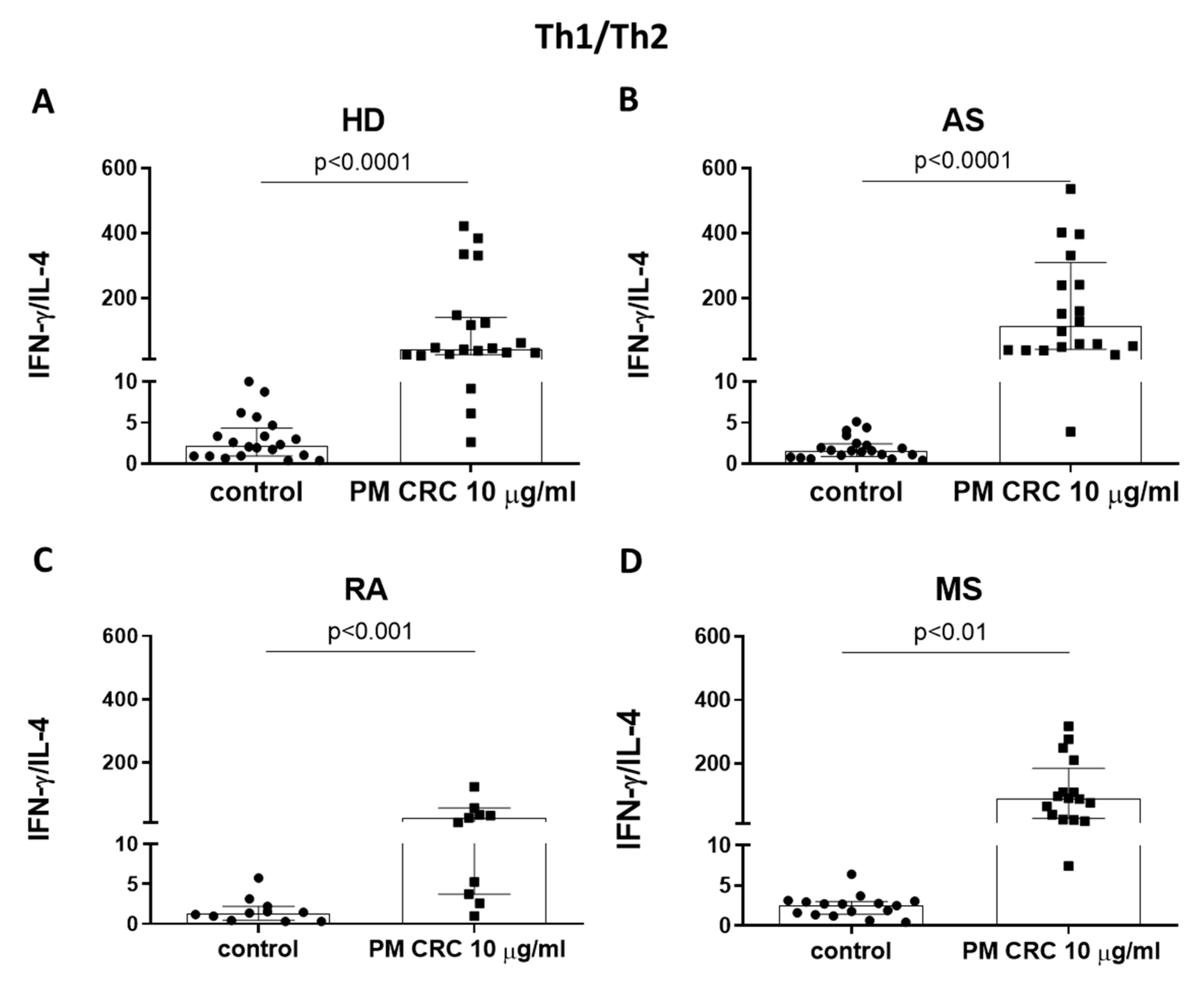
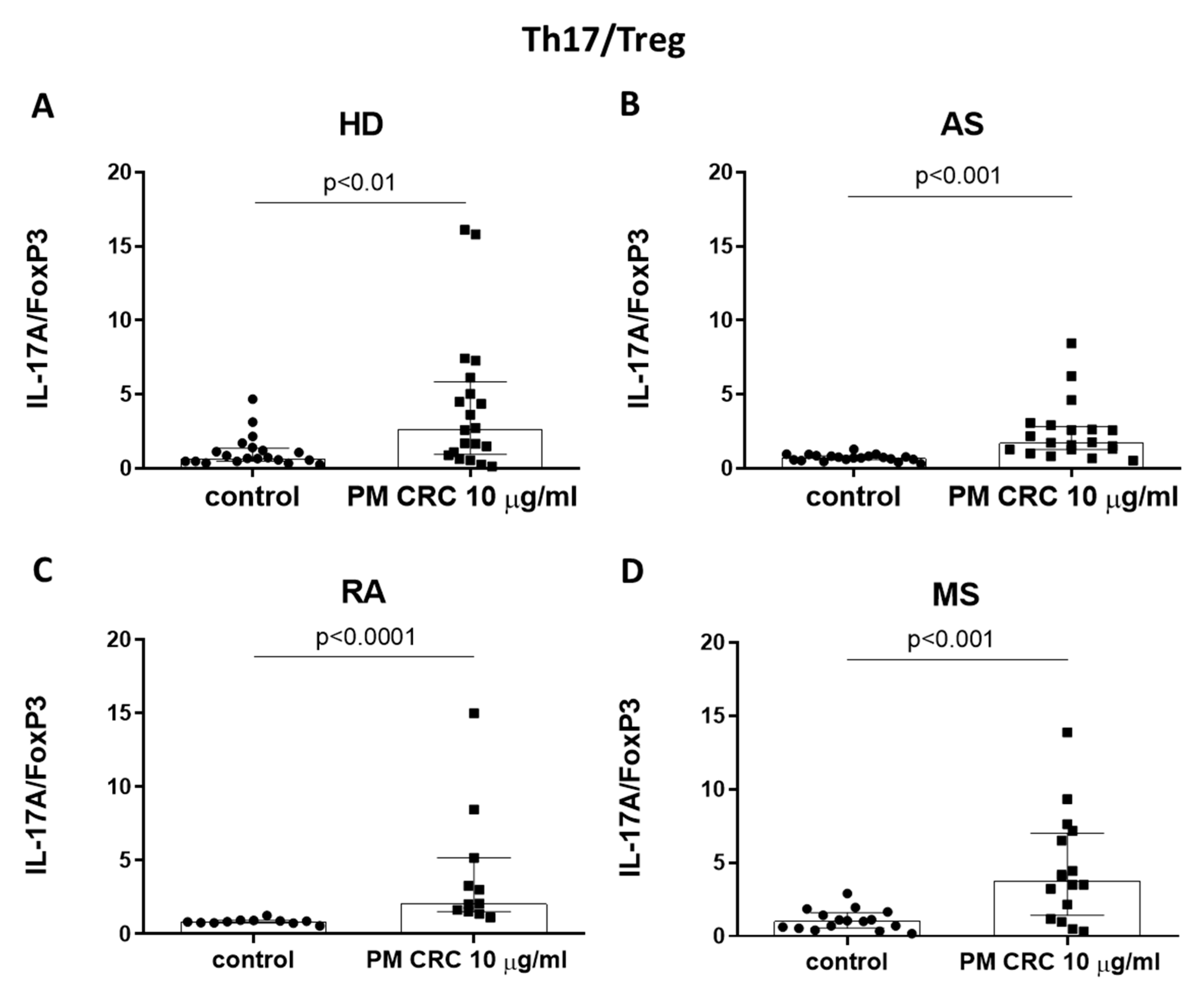
3.2. PM CRC Activate CD4+ T Lymphocytes, Skewing the Balance of Th1/Th2 and Th17/Treg Subsets in Patients and Healthy Donors

The effect of local air pollution on the polarization of CD4+ T lymphocytes was evaluated after a 3 h exposure of patients’ and HD PBMCs to PM CRC. The concentration of PM CRC (10 µg/mL) was established experimentally as non-cytotoxic (Figure S1). The population of CD4+ T cells is highly heterogenic, being composed of several subsets, mainly Th1, Th2, Th17, and Treg. With this in mind, we checked first if exposition of PBMCs to PM CRC cause CD4+ T cell subset polarization, as defined by the expression of intracellular proteins such as IFN-γ, IL-4, IL-17A, and FoxP3. The obtained data show that exposure of PBMCs to PM CRC caused a significant increase in the proportion of cells positive for IFN-γ and IL-17A, similarly as was described previously for NIST and LAP particles [21].
As the ratio of Th1/Th2 and Th17/Treg subsets correlates well with the functional status of the immune system [22,38,39], we further compared these parameters in HD and patients, after exposure of PBMC to PM CRC (Figure 1 and Figure 2). Cells stimulated with PMA + Ionomycin were used as a positive control (Figure S2).
Figure 1 shows a significant increase in the Th1/Th2 ratio in all studied groups after cell exposure to PM CRC, being highly skewed to the Th1 type. The most significant difference between stimulated and unstimulated samples was detected for patients with AS—a 71.4 fold increase (median value of the Th1/Th2 ratio 112.8 vs. 1.58, respectively) and for patients with MS—a 34.2 fold increase (89.09 vs. 2.6) (Figure 1B,D). The lowest increase in the Th1/Th2 ratio was detected in patients with RA—a 18.3 fold increase (median ratio 25.11 vs. 1.37), and HD—a 19.5 fold increase (median ratio 42.81 vs. 2.19) (Figure 1A,C).
Additionally, PBMCs exposed to PM CRC showed an increase in Th17/Treg ratio when compared to the unexposed control group (Figure 2). This was most significant for HD and patients with MS, where the Th17/Treg ratio increased by 3.8 (median ratio 2.66 vs. 0.69) and by 3.7 fold (median ratio 3.78 vs. 1.03), respectively (Figure 2A,D). In the case of patients with AS and RA—a 2.4-fold increase (median ratio 1.74 vs. 0.72), and a 2.5-fold increase (median ratio 2.07 vs. 0.84) was observed, respectively (Figure 2B,C).

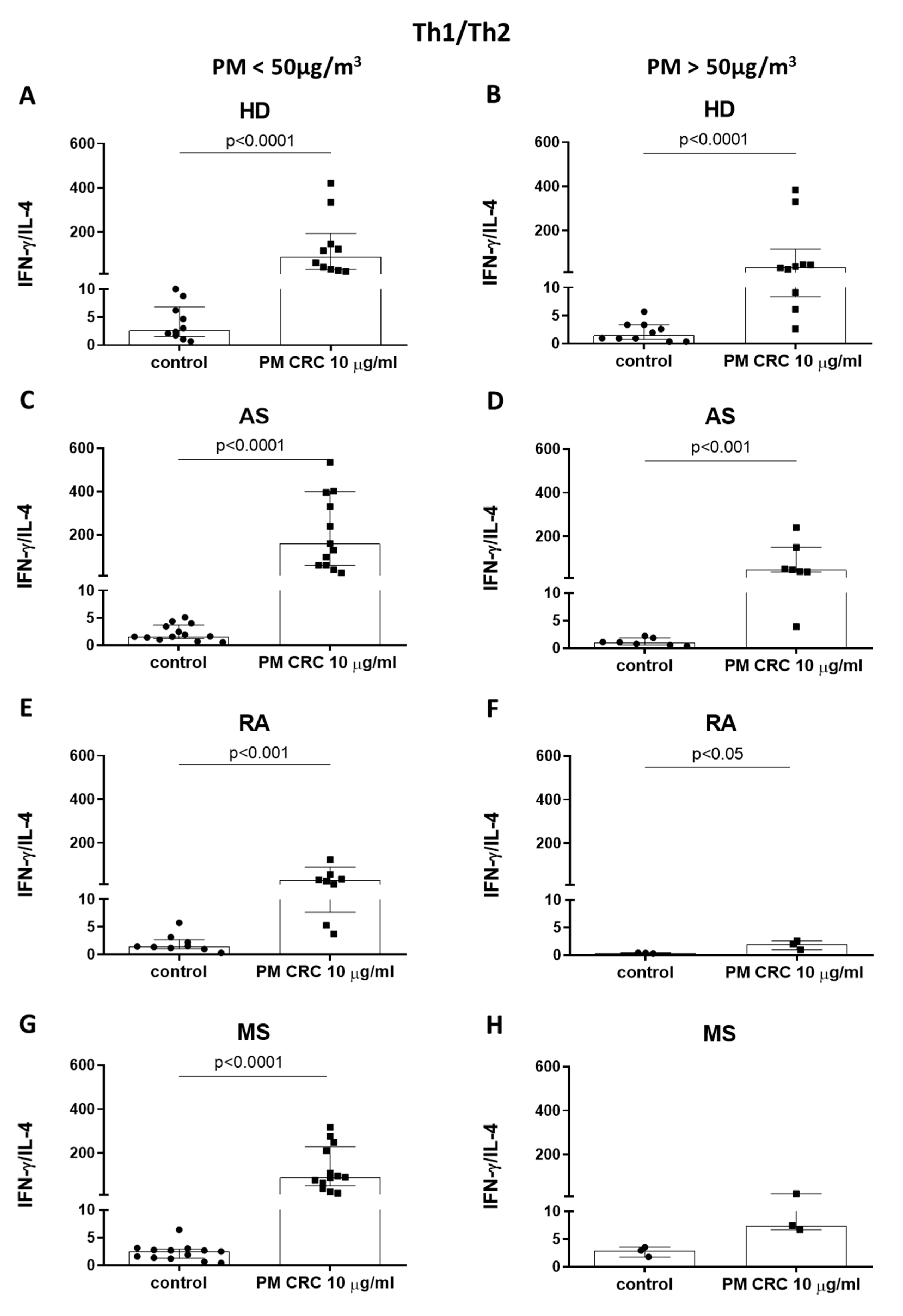
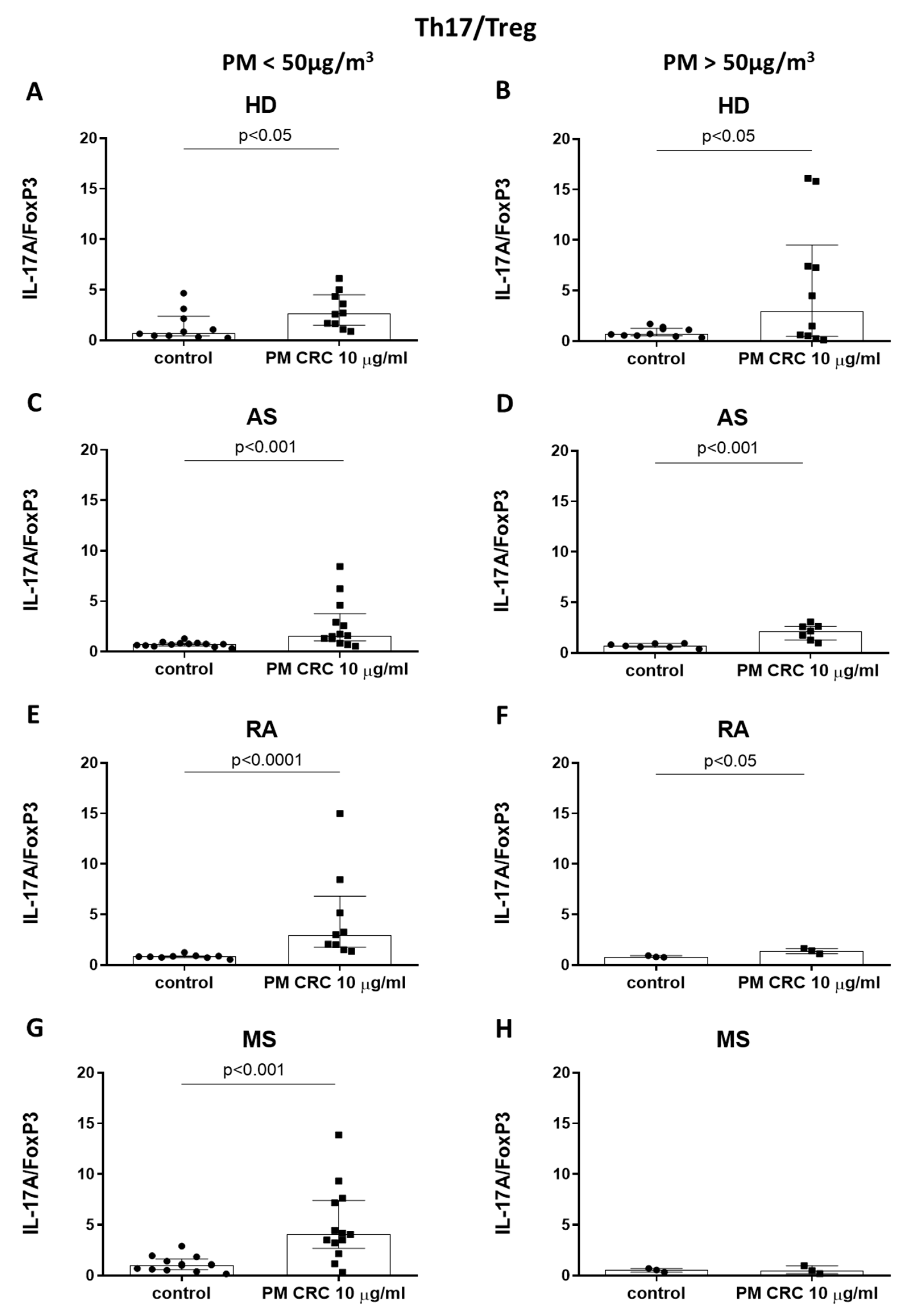
3.3. Polarization of CD4+ T Cells in Patients with Inflammatory or Autoimmune Disorders Depends on the Seasonal Changes in the Concentration of PM in the Air of Cracow

When referring to the air quality in Cracow, one must keep in mind that there is a significant difference in the daily concentration of PM during winter and summertime [7,40]. This is mainly due to smog covering the city in the winter. To answer whether a seasonal variation in the concentration of PM in the air of Cracow may affect the polarization of CD4+ T cells, blood samples were collected in two seasons when the concentration of PM in the air was lower (summer) and higher (winter) than the daily limit (50 µg/m3) (Table 2). To this end, PBMCs were exposed to PM CRC for 3 h (as above). Next, the CD4+ T cells positive for the cytoplasmic expression of IFN-γ; IL-4; IL-17A; FoxP3 were analysed and the corresponding Th1/Th2 and Th17/Treg ratios were calculated.
The obtained results show an increase in Th1/Th2 and Th17/Treg ratios after the exposure of PBMCs to PM CRC, being highly skewed into Th1 and Th17 cells in all groups in summer when the concentration of PM in the air was lower than the daily acceptable limit (Figure 3 and Figure 4). In respect to the Th1/Th2 ratio, the most pronounced effect of PM CRC was detected in the group of AS—a 97.5-fold increase (median ratio 159.0 vs. 1.63), MS—a 36-fold increase (median ratio 90.2 vs. 2.5) and HD—a 33.3-fold increase (median ratio 88.61 vs. 2.66) (Figure 3A,C,G). The lowest effect of PM CRC—an increase of 21.8-fold (median ratio 31.56 vs. 1.45)—was observed in patients with RA (Figure 3E).
Moreover, in the season with high PM concentration (above a daily limit), stimulation of patients’ PBMCs with PM CRC was less effective than in the summer.
This effect was more pronounced for patients with RA and AS, where the increase in the Th1/Th2 ratio by 5.0-fold (median ratio 2.0 vs. 0.4) and 44.0-fold (median ratio 48.4 vs. 1.1), respectively (Figure 3D,F), was much lower compared to the corresponding 21.8 and 97.5-fold increase in the summer. In the case of RA patients, however, the observed stimulatory effect in the wintertime reached statistical significance at the lowest level compared to other groups. No statistically significant stimulatory effect of PM CRC was observed in the group of MS patients (Figure 3H). In respect to the Th17/Treg ratio, in the season with low PM concentration, this parameter was significantly increased in all groups after PBMCs stimulation, reaching the highest value for MS—a 3.8-fold increase (median ratio 4.06 vs. 1.06) and RA patients—a 3.6-fold increase (median ratio 3.0 vs. 0.84), and HD—a 3.5-fold increase (median ratio 2.66 vs. 0.76). In the case of AS, the level of increase was the lowest—2.1-fold (median ratio 1.57 vs. 0.74) (Figure 4). On the other hand, in the season with high PM concentration in the air (winter), the Th17 response of patients with AS increased after stimulation with PM CRC by 3.0-fold (median ratio 2.17 vs. 0.71), while the response of patients with RA and HD increased by 1.75-fold (median ratio 1.4 vs. 0.8) and 4.3 (median ratio 3.0 vs. 0.7), respectively and was lower than in the summer. In the case of patients with MS, no difference between the stimulated and unstimulated group (median ratio 0.5 vs. 0.6) was observed (Figure 4H). The cell response in HD was at a similar level in both periods. No role of seasonal changes in the concentration of PM in the air was shown for Th2 and Treg cells, as their levels were undetectable in PBMCs of the studied groups despite the stimulation with PM CRC.
Taken together, the exposure of PBMCs from patients and HD to PM CRC caused the upregulation of Th1 and Th17 cells, indicating changes in the polarization of CD4+ T cell subsets towards proinflammatory status. This effect was dependent on the seasonal changes in the concentration of PM in the air of Cracow, being more pronounced in the season with the air pollution lower than a daily limit.

4. Discussion

A growing list of evidence suggests that environmental pollution and increasing concentration of PM in the air have an impact on the initiation, development, and exacerbation of allergies, inflammatory, cardiovascular, and autoimmune disorders [9,10,11,12,13,14]. Nowadays, air pollution is recognized as a major public health problem, with a strong influence on people’s life quality [42]. So far, our and other studies have documented a strong polarization of CD4+ T cells into Th1 and Th17 subsets in PBMCs of HD after the exposure to NIST and LAP particles, which differed in the content of organic components [21,43]. In this report, we assessed the effect of local air pollution in Cracow on the polarization of human CD4+ T cell subsets after in vitro exposure of PBMCs from patients with AS, RA, and MS. The diseases were selected based on evidence suggesting a role of the urban pollution on their prevalence in society [44,45,46,47,48,49,50]. This effect was determined by flow cytometry analysis of IFN-γ, IL-4, IL-17A, and Foxp3, characteristic for Th1, Th2, Th17, and Treg cells, respectively. Furthermore, Th1/Th2 and Th17/Treg ratios were assessed to estimate changes in the balance of Th1 to Th2 and Th17 to Treg subsets, caused by the exposure of PBMCs to PM CRC. These ratios were analysed seasonally (winter and summer), where differences in the concentration of PM10 in the air of Cracow (lower and higher than a daily limit of 50 µg/m3).
The obtained data revealed that the exposure of PBMCs to PM CRC shifted the balance of T cell subsets towards proinflammatory Th1 and Th17 cells, both in patients and HD. This was documented by elevated proportion of IFN-γ and IL-17A producing cells, with no concomitant changes in the level of Th2 and Treg cells in the studied groups. Simultaneously, an increase in the ratio of Th1/Th2 cells was detected in all groups, being most evident in AS (a 71.4-fold increase) and MS patients (a 34.2-fold increase), while cells from HD and patients with RA presented similar but less pronounced responses (a 18.3- and a 19.5-fold increase, respectively). For the Th17/Treg ratio, the stimulatory effect of PM CRC was much less indicated, with the most robust response observed in the group of patients with MS (a 3.7-fold increase), and HD (a 3.8-fold increase), while for patients with AS and MS, it was lower (a 2.4- and a 2.5-fold increase, respectively). It seems, that despite the health status, the exposition of PBMCs to PM CRC leads to the induction of inflammatory reaction, albeit with a different magnitude. In patients and HD, the difference in the Th1 polarization between unstimulated and PM CRC-stimulated cells was more significant than for the Th17 type. This suggests that Th1 cytokines might be more relevant in some of the studied diseases. In keeping, in the case of AS, the majority of pathogenic T cells are of the Th1 profile producing high levels of IFN-γ [51]. IFN-γ activates monocytes/macrophages and DCs, leading to the perpetuation of the pathogenic Th1 response. Although postulated, the role of Th17 cytokines in the pathogenesis of AS has not been unequivocally confirmed [52]. Additionally, in MS, much evidence has been obtained for a role of IFN-γ in the pathogenesis of the disease [53,54,55], however more recent studies have also suggested a role of Th17 cells in MS pathogenesis, involving the aberrations of IL-17 and IL-23 production in the disease [56]. In this context, it was shown that the exposure of innate immune cells to PM10 induces production of Th17 cytokines, causing progression of MS [50]. Currently, it is postulated that Th17 cells might play a role in the initial phases of MS, while Th1 cells might be important in later phases of the inflammation in the CNS [57].
On the other hand, RA is considered a Th1-associated disease [58] and it was documented that the frequency of IFN-γ producing CD4+ T cells is significantly elevated in the synovial fluid compared to the peripheral blood [59,60]. However, the frequencies of IFN-γ producing PBMCs in early arthritis correlated with disease activity, supporting the role of Th1 cells in the initiation of RA [61].
The present results also imply the crucial role of seasonal variation in the PM CRC concentration in the air on the polarization of T cell subsets in all investigated groups. In the area of the city of Cracow, the concentration of PM is 4-times higher in winter than during summer [62]. The obtained data show that the most pronounced effect of PM CRC on the upregulation of the Th1/Th2 and Th17/Treg ratios was observed in patients in the season with the concentration of PM10 lower than a daily limit of 50 µg/m3. This was not the case for HD, where the seasonal changes in the PM concentration in the air did not affect the magnitude of cell response. In the season with a concentration of PM10 higher than the daily limit, the effect of the PM CRC on the Th1/Th2 ratio in the patients’ groups was much lower compared to the summertime; the most significant reduction of the response level was detected for RA (c.a. 4 times less) and AS (c.a. 2 times less) patients.
The high concentration of air pollution also affected patients’ Th17/Treg responses, and this was either higher (AS—fold increase 3.0) or lower (RA—fold increase 1.75) than observed in the season with low air pollution (fold increase 2.1 vs. 3.6, respectively). Similarly to the Th1/Th2 response, no stimulatory effect of PM CRC in this season was observed in the group of patients with MS.
Our data may suggest that in the case of patients, especially with MS, lasting exposure to the air pollution with a daily concentration of PM10 higher than 50 µg/m3 makes T cells less sensitive to stimulation of PBMCs with PM CRC in vitro. These results indirectly corroborate the data, showing that a higher concentration of air pollution during winter is associated with the increased manifestation of MS [25,63,64].
It is worth mentioning that the activation of CD4+ T cells in response to PM requires monocytes and their accessory function. This was already confirmed by our previous report, where the exposition of T lymphocytes to the reference PM material resulted in their activation, only in the presence of monocytes. This effect was largely dependent on the organic compounds content in PM preparations [21].
In comparison to those data, PM CRC contain much more organic compounds than standard reference urban particulate matter; the carbon content in NIST SRM 1648a is certified as 12.7%, while PM CRC samples collected in Cracow (UrbanB) contain more than 40% of carbon [61]. One of the major organic components of PM is bacterial endotoxin (Lipopolysaccharide—LPS) [21]. Although we have no formal proof, we cannot exclude that the lower frequency of cytokine producing CD4+ T cells after stimulation with PM CRC in the season with high concentration of PM10 in the air (winter) is a result of T cell exhaustion due to the permanent signals driven by natural exposure to PM, containing LPS and/or other antigens [65]. The LPS-responding human blood T cells, occurring with a frequency less than 1:1000, were described by Ulmer et al., and the feature of this activation pathway is the MHC-unrestricted accessory cell activity of monocytes, providing costimulatory signals via direct cell-to-cell contact and release of soluble cytokines [66]. Another possible explanation of this observation is monocyte tolerance, a phenomenon described also in respect to LPS and TLR4 binding [67,68]. The tolerance and cross-tolerance phenomena might be induced via Pattern Recognition Receptors (PRRs), including Toll-like receptors (TLRs) TLR2, TLR4, and TLR9 [69], which recognize many, not only microbial products. Shoenfelt et al., suggested that PM might use distinct receptors and pro-inflammatory signalling pathways based on particle composition. For example, exposure of murine peritoneal macrophages to PM2.5, which had high levels of redox-active metals and low levels of endotoxin, induced cytokine secretion in a TLR2-dependent mechanism. Conversely, PM10, which contains high level of endotoxin, induced cytokine secretion in a TLR4-dependent mechanism [70].
The data suggest that PM induces the release of proinflammatory cytokines, including TNF-α, IL-1, IL-6, and IL-8 [21,71,72]. Here, we also observed increased TNF-α and IL-6 release after 3 h exposure of PBMCs to PM CRC, being the highest in patients with AS and MS (Figure S3). In the population of PBMCs, the main producers of TNF-α and IL-6 are monocytes [73]. Our unpublished data indicate that monocytes exposed to high concentrations of PM produce these cytokines and rapidly die through pyroptosis. Dying cells release damage-associated molecular patterns (DAMPs) that can protect the neighbouring, still alive monocytes, allowing for rechallenge with PM, and activate them for further production of the inflammatory mediators relevant for the activation of CD4+ Th cells (manuscript in preparation).
In the case of MS, TNF-α is a major mediator of the inflammatory response and is important in the pathogenesis and progression of MS [74]. Although TNF-α and IL-6 are also main pathogenic cytokines in RA, having a destructive effect on bones [75], and the “anti-cytokine therapy”, such as the anti-TNF-α or the anti-IL-6 therapy has revolutionized current RA treatment [76], we did not observe any significant increase in the secretion of these cytokines to the culture medium after stimulation of PBMCs with PM CRC, indicating that cells in the local environment in synovial and bone tissues in RA might respond differently to certain stimuli than those in vitro.
In summary, our results indicate that treatment of human PBMCs with PM CRC affects the balance of Th1/Th2 and Th17/Treg cells, promoting activity of Th1 and Th17 subsets both in patients with AS, RA and MS, and HD. Although the cells from HD and patients responded to stimulation with PM CRC in a similar fashion, the magnitude of Th1 and Th17 response in patients was different as compared to HD, suggesting the disease-related specificity. This effect was dependent on the seasonal concentration of PM in the air of Cracow (variations in PM10 and PM2.5 content).
However, due to some limitations, our results should be treated tentatively. An important factor that might have an impact on the obtained data is the gender of patients and HD. It is well documented that autoimmunity positively correlates with females, while inflammatory disorders, including AS, are correlated with male patients. This was reflected also in our study by the composition of RA, MS, and AS groups, with the two formers contained in the majority women, while the latter was dominated by men. Recently Kim et al. reported gender-related differences in the effects of air pollution on cognitive functions in elderly persons in South Korea [77]. In their succinct study, they showed that women were at a higher risk for decreased cognitive function associated with increased exposure to PM10 and PM2.5−10. In our study, we did not find a similar correlation (Figures S4 and S5). This may be due to the relatively low number of patients in our study groups and/or due to a lack of exclusion of potential confounding factors, e.g., age, geographical location, smoking status, drinking habits, body mass index, blood pressure, co-morbidities, etc. from the analysis. Another important aspect is that CD4+ T cells do not directly respond to PM; their activity requires monocytes, and their cytokines as accessory signals. However, a precise mechanism of monocyte interactions with ambient air pollution needs to be elucidated.

5. Conclusions

In vitro exposure of PBMCs to PM CRC promotes the Th1 and Th17-type response in HD and patients with AS, RA, and MS. In the case of patients, polarization of CD4+ T cells was also dependent on the seasonal variation in the concentration of PM (PM10 and PM2.5) in the air of Cracow, being more pronounced in the season with low air pollution. These findings further support the observations that air pollution induces a complex pro-inflammatory response, which may contribute to the development and/or exacerbation of many pathologies, including inflammatory and autoimmune disorders.

Supplementary Materials

The following supporting information can be downloaded at: https://0-www-mdpi-com.brum.beds.ac.uk/article/10.3390/atmos13040529/s1, Figure S1: Flow cytometry analysis of viability of cells after exposure of PBMCs to PM CRC; Figure S2: Flow cytometry analysis of intracellular proteins characteristic for specific Th subsets after exposure of PBMC to PM CRC; Figure S3: Effect of the PM CRC treatment on PBMCs production of TNFα and IL-6; Figure S4: Effect of the exposure of PBMCs to PM CRC on the ratio of Th1/Th2 cells by gender in patients and HD; Figure S5: Effect of the exposure of PBMCs to PM CRC on the ratio of Th17/Treg cells by gender in patients and HD.

Author Contributions

Conceptualization, methodology, writing—original draft A.G.-B.; investigation and data analysis A.G.-B. and K.W.; recruitment of patients J.P., A.S., P.L., K.J.-C. and M.K.; review of manuscript J.P., A.S., P.L., K.J.-C., M.K. and M.S.; conceptualization, formal analysis, supervision, and writing—review and editing J.B. All authors have read and agreed to the published version of the manuscript.

Funding

This study was supported by a grant from the National Science Centre (NCN) in Poland (project APARIC, GA no. 2015/16/W/ST5/00005); and by the European Commission H2020-MSCA-RISE-2016 program (project “CHARMED”, GA no. 734684).

Institutional Review Board Statement

The study was conducted following the Declaration of Helsinki and approved by the Bioethical Commission of Jagiellonian University (protocol code 122.6120.261.2015).

Informed Consent Statement

Informed consent was obtained from all subjects involved in the study.

Data Availability Statement

Not applicable.

Acknowledgments

The authors appreciate the kind help of Janusz Oszajca with the collection of PM samples from the air of Cracow.

Conflicts of Interest

The authors declare no conflict of interest.

References

  1. The European Parliament; The Council of The European Union. Directive 2008/50/EC of the European Parliament and the Council of 21 May 2008 on ambient air quality and cleaner air for Europe. Off. J. Eur. Union 2008, 152, 1–44. [Google Scholar]
  2. Polish Minister of the Environment. Regulation of the Minister of the Environment of 24 August 2012 on the levels of certain substances in the air. J. Laws 2012, 1031, 1–9. (In Polish). Available online: https://isap.sejm.gov.pl/isap.nsf/download.xsp/WDU20120001031/O/D20121031.pdf (accessed on 28 February 2022).
  3. De Brito Beirao, C.A.; Ortiz, G.; De Leeuw, F.; Wiana, M. Air Quality in Europe—2018 Report; European Environment Agency: Copenhagen, Denmark, 2018.
  4. World Bank Group. In the Spotlight. Air Quality in Poland, What Are the Issues and What Can Be Done? World Bank Group: Washington, DC, USA, 2019. [Google Scholar]
  5. Khomenko, S.; Cirach, M.; Pereira-Barboza, E.; Mueller, N.; Barrera-Gómez, J.; Rojas-Rueda, D.; de Hoogh, K.; Hoek, G.; Nieuwenhuijsen, M. Premature mortality due to air pollution in European cities: A health impact assessment. Lancet Planet. Health 2021, 5, 121–134. [Google Scholar] [CrossRef]
  6. Nazar, W.; Plata-Nazar, K. Changes in Air Pollution-Related Behaviour Measured by Google Trends Search Volume Index in Response to Reported Air Quality in Poland. Int. J. Environ. Res. Public Health 2021, 18, 11709. [Google Scholar] [CrossRef] [PubMed]
  7. Traczyk, P.; Gruszecka-Kosowska, A. The Condition of Air Pollution in Kraków, Poland, in 2005–2020, with Health Risk Assessment. Int. J. Environ. Res. Public Health 2020, 17, 6063. [Google Scholar] [CrossRef] [PubMed]
  8. Bokwa, A. The climate of the city and air pollution. Aura 2016, 9, 8–13. (In Polish) [Google Scholar]
  9. Brook, R.D.; Rajagopalan, S.; Pope, C.A.; Brook, J.R.; Bhatnagar, A.; Diez-Roux, A.V.; Holguin, F.; Hong, Y.; Luepker, R.V.; Mittleman, M.A.; et al. American Heart Association Council on Epidemiology and Prevention, Council on the Kidney in Cardiovascular Disease, and Council on Nutrition, Physical Activity, and Metabolism. Particulate matter air pollution and cardiovascular disease: An update to the scientific statement from the American Heart Association. Circulation 2010, 121, 2331–2378. [Google Scholar]
  10. Al-Kindi, S.G.; Brook, R.D.; Biswal, S.; Rajagopalan, S. Environmental determinants of cardiovascular disease: Lessons learned from air pollution. Nat. Rev. Cardiol. 2020, 17, 656–672. [Google Scholar] [CrossRef]
  11. Marchini, T.; Zirlik, A.; Wolf, D. Pathogenic Role of Air Pollution Particulate Matter in Cardiometabolic Disease: Evidence from Mice and Humans. Antioxid. Redox Signal. 2020, 33, 263–279. [Google Scholar] [CrossRef]
  12. Gawda, A.; Majka, G.; Nowak, B.; Marcinkiewicz, J. Air pollution, oxidative stress, and exacerbation of autoimmune diseases. Cent. Eur. J. Immunol. 2017, 42, 305–312. [Google Scholar] [CrossRef]
  13. Ji, H.; Hershey, G.K.K. Genetic and epigenetic influence on the response to environmental particulate matter. J. Allergy Clin. Immunol. 2013, 129, 33–41. [Google Scholar] [CrossRef] [Green Version]
  14. Ritz, S.A. Air pollution as a potential contributor to the “epidemic” of autoimmune disease. Med. Hypotheses 2010, 74, 110–117. [Google Scholar] [CrossRef] [PubMed]
  15. Doria, A.; Zen, M.; Bettio, S.; Gatto, M.; Bassi, N.; Nalotto, L.; Ghirardello, A.; Iaccarino, L.; Punzi, L. Autoinflammation and autoimmunity: Bridging the divide. Autoimmun. Rev. 2012, 12, 22–30. [Google Scholar] [CrossRef] [PubMed]
  16. Meng, Q.; Richmond-Bryant, J.; Lu, S.E.; Buckley, B.; Welsh, W.J.; Whitsel, E.A.; Hanna, A.; Yeatts, K.B.; Warren, J.; Herring, A.H.; et al. Cardiovascular outcomes and the physical and chemical properties of metal ions found in particulate matter air pollution: A QICAR study. Environ. Health Perspect. 2013, 121, 558–564. [Google Scholar] [CrossRef] [PubMed] [Green Version]
  17. Wright, R.O.; Baccarelli, A. Metals and neurotoxicology. J. Nutr. 2007, 137, 2809–2813. [Google Scholar] [CrossRef] [Green Version]
  18. Sawyer, K.; Mundandhara, S.; Ghio, A.J.; Madden, M.C. The effects of ambient particulate matter on human alveolar macrophage oxidative and inflammatory responses. J. Toxicol. Environ. Health A 2009, 73, 41–57. [Google Scholar] [CrossRef]
  19. Zelikoff, J.T.; Schermerhorn, K.R.; Fang, K.; Cohen, M.D.; Schlesinger, R.B. A role for associated transition metals in the immunotoxicity of inhaled ambient particulate matter. Environ. Health Perspect. 2002, 110, 871–875. [Google Scholar] [CrossRef] [Green Version]
  20. Nemmar, A.; Hoet, P.H.M.; Vanquickenborne, B.; Dinsdale, D.; Thomeer, M.; Hoylaerts, M.F.; Vanbilloen, H.; Mortelmans, L.; Nemery, B. Passage of inhaled particles into the blood circulation in humans. Circulation 2002, 105, 411–414. [Google Scholar] [CrossRef] [Green Version]
  21. Gałuszka, A.; Stec, M.; Węglarczyk, K.; Kluczewska, A.; Siedlar, M.; Baran, J. Transition metal containing particulate matter promotes Th1 and Th17 inflammatory response by monocyte activation in organic and inorganic compounds dependent manner. Int. J. Environ. Res. Public Health 2020, 17, 1227. [Google Scholar] [CrossRef] [Green Version]
  22. Crane, I.J.; Forrester, J.V. Th1 and Th2 lymphocytes in autoimmune disease. Crit. Rev. Immunol. 2005, 25, 75–102. [Google Scholar] [CrossRef]
  23. Lee, F.E.; Georas, S.N.; Beck, L.A. IL-17: Important for host defense, autoimmunity, and allergy? J. Investig. Dermatol. 2010, 130, 2540–2542. [Google Scholar]
  24. Araujo, J.A.; Barajas, B.; Kleinman, M.; Wang, X.; Bennett, B.J.; Gong, K.W.; Navab, M.; Harkema, J.; Sioutas, C.; Lusis, A.J.; et al. Ambient particulate pollutants in the ultrafine range promote early atherosclerosis and systemic oxidative stress. Circ. Res. 2008, 102, 589–596. [Google Scholar] [CrossRef] [PubMed] [Green Version]
  25. Oikonen, M.; Laaksonen, M.; Laippala, P.; Oksaranta, O.; Lilius, E.M.; Lindgren, S.; Rantio-Lehtimäki, A.; Anttinen, A.; Koski, K.; Erälinna, J.P. Ambient air quality and occurrence of multiple sclerosis relapse. Neuroepidemiology 2003, 22, 95–99. [Google Scholar] [CrossRef] [PubMed]
  26. Hou, T.; Liao, J.; Zhang, C.; Sun, C.; Li, X.; Wang, G. Elevated expression of miR-146, miR-139 and miR-340 involved in regulating Th1/Th2 balance with acute exposure of fine particulate matter in mice. Int. Immunopharmacol. 2018, 54, 68–77. [Google Scholar] [CrossRef] [PubMed]
  27. Li, Y.; Dong, T.; Jiang, X.; Wang, C.; Zhang, Y.; Li, Y.; Zheng, G.; Li, X.; Bai, J.; Li, H. Chronic and low-level particulate matter exposure can sustainably mediate lung damage and alter CD4 T cells during acute lung injury. Mol. Immunol. 2019, 112, 51–58. [Google Scholar] [CrossRef]
  28. Polman, C.H.; Reingold, S.C.; Banwell, B.; Clanet, M.; Cohen, J.A.; Filippi, M.; Fujihara, K.; Havrdova, E.; Hutchinson, M.; Kappos, L.; et al. Diagnostic criteria for multiple sclerosis: 2010 revisions to the McDonald criteria. Ann. Neurol. 2011, 69, 292–302. [Google Scholar] [CrossRef] [Green Version]
  29. Adams, H.P., Jr.; Bendixen, B.H.; Kappelle, L.J.; Biller, J.; Love, B.B.; Gordon, D.L.; Marsh, E.E., III. Classification of subtype of acute ischemic stroke. Definitions for use in a multicenter clinical trial. TOAST. Trial of Org 10172 in Acute Stroke Treatment. Stroke 1993, 24, 35–41. [Google Scholar] [CrossRef] [Green Version]
  30. Mikrut, M.; Mazuryk, O.; Macyk, W.; van Eldik, R.; Stochel, G. Generation and photogeneration of hydroxyl radicals and singlet oxygen by particulate matter and its inorganic components. J. Environ. Chem. Eng. 2021, 9, 106478. [Google Scholar] [CrossRef]
  31. Baran, J.; Kowalczyk, D.; Ożóg, M.; Zembala, M. Three-color flow cytometry detection of intracellular cytokines in peripheral blood mononuclear cells: Comparative analysis of phorbol myristate acetate-ionomycin and phytohemagglutinin stimulation. Clin. Diagn. Lab. Immunol. 2001, 8, 303–313. [Google Scholar] [CrossRef] [Green Version]
  32. Wnuk, M.; Maluchnik, M.; Perwieniec, J.; Podwojcic, K.; Szelag, M.; Walkiewicz, D.; Zakrzewski, M.; Kulakowska, A.; Brola, W.; Rejdak, K.; et al. Multiple sclerosis incidence and prevalence in Poland: Data from administrative health claims. Mult. Scler. Relat. Disord. 2021, 55, 103162. [Google Scholar] [CrossRef]
  33. Iltchev, P.; Śliwczyński, A.; Czeleko, T.; Sierocka, A.; Tłustochowicz, M.; Tłustochowicz, W.; Timler, D.; Brzozowska, M.; Szatko, F.; Marczak, M. Epidemiology of Rheumatoid Arthritis (RA) in rural and urban areas of Poland—2008–2012. Ann. Agric. Environ. Med. 2016, 23, 350–356. [Google Scholar] [CrossRef] [Green Version]
  34. Kruger, P.C.; Guzik, T.J.; Eikelboom, J.W. How can the results of the COMPASS trial benefit patients with coronary or peripheral artery disease in Poland? Kardiol. Pol. 2019, 77, 661–669. [Google Scholar] [CrossRef] [PubMed] [Green Version]
  35. Kvien, T.K.; Uhlig, T.; Ødegård, S.; Heiberg, M.S. Epidemiological aspects of rheumatoid arthritis: The sex ratio. Ann. N. Y. Acad. Sci. 2006, 1069, 212–222. [Google Scholar] [CrossRef] [PubMed]
  36. Sellner, J.; Kraus, J.; Awad, A.; Milo, R.; Hemmer, B.; Stüve, O. The increasing incidence and prevalence of female multiple sclerosis—A critical analysis of potential environmental factors. Autoimmun. Rev. 2011, 10, 495–502. [Google Scholar] [CrossRef] [PubMed]
  37. Fairweather, D. Sex differences in inflammation during atherosclerosis. Clin. Med. Insights Cardiol. 2015, 8, 49–59. [Google Scholar] [CrossRef] [Green Version]
  38. Druet, P.; Sheela, R.; Pelletier, L. Th1 and Th2 cells in autoimmunity. Clin. Exp. Immunol. 1995, 101, 9–12. [Google Scholar] [CrossRef]
  39. Pryjma, J.; Baran, J.; Ernst, M.; Woloszyn, M.; Flad, H.D. Altered antigen-presenting capacity of human monocytes after phagocytosis of bacteria. Infect. Immun. 1994, 62, 1961–1967. [Google Scholar] [CrossRef] [Green Version]
  40. Samek, L.; Styszko, K.; Stegowski, Z.; Zimnoch, M.; Skiba, A.; Turek-Fijak, A.; Gorczyca, Z.; Furman, P.; Kasper-Giebl, A.; Rozanski, K. Comparison of PM10 Sources at Traffic and Urban Background Sites Based on Elemental, Chemical and Isotopic Composition: Case Study from Krakow, Southern Poland. Atmosphere 2021, 12, 1364. [Google Scholar] [CrossRef]
  41. Measurement data bank of the Chief Inspectorate of Environmental Protection. (In Polish). Available online: https://powietrze.gios.gov.pl/pjp/archives (accessed on 15 February 2021).
  42. Cohen, A.J.; Brauer, M.; Burnett, R.; Anderson, H.R.; Frostad, J.; Estep, K.; Balakrishnan, K.; Brunekreef, B.; Dandona, L.; Dandona, R.; et al. Estimates and 25-year trends of the global burden of disease attributable to ambient air pollution: An analysis of data from the Global Burden of Diseases Study 2015. Lancet 2017, 389, 1907–1918. [Google Scholar] [CrossRef] [Green Version]
  43. Ma, Q.Y.; Huang, D.Y.; Zhang, H.J.; Wang, S.; Chen, X.F. Exposure to particulate matter 2.5 (PM2.5) induced macrophage-dependent inflammation, characterized by increased Th1/Th17 cytokine secretion and cytotoxicity. Int. Immunopharmacol. 2017, 50, 139–145. [Google Scholar] [CrossRef]
  44. Angelici, L.; Piola, M.; Cavalleri, T.; Randi, G.; Cortini, F.; Bergamaschi, R.; Baccarelli, A.A.; Bertazzi, P.A.; Pesatori, A.C.; Bollati, V. Effects of particulate matter exposure on multiple sclerosis hospital admission in Lombardy region, Italy. Environ. Res. 2016, 145, 68–73. [Google Scholar] [CrossRef] [Green Version]
  45. Wu, Q.; Xu, Z.; Dan, Y.L.; Cheng, J.; Zhao, C.N.; Mao, Y.M.; Xiang, K.; Hu, Y.Q.; He, Y.S.; Pan, H.F. Association between traffic-related air pollution and hospital readmissions for rheumatoid arthritis in Hefei, China: A time-series study. Environ. Pollut. 2021, 268, 115628. [Google Scholar] [CrossRef] [PubMed]
  46. Hart, J.E.; Källberg, H.; Laden, F.; Bellander, T.; Costenbader, K.H.; Holmqvist, M.; Klareskog, L.; Alfredsson, L.; Karlson, E.W. Ambient air pollution exposures and risk of rheumatoid arthritis: Results from the Swedish EIRA case-control study. Ann. Rheum. Dis. 2013, 72, 888–894. [Google Scholar] [CrossRef] [PubMed]
  47. Hennig, F.; Geisel, M.H.; Kälsch, H.; Lucht, S.; Mahabadi, A.A.; Moebus, S.; Erbel, R.; Lehmann, N.; Jöckel, K.H.; Scherag, A.; et al. Air Pollution and Progression of Atherosclerosis in Different Vessel Beds-Results from a Prospective Cohort Study in the Ruhr Area, Germany. Environ. Health Perspect. 2020, 128, 107003. [Google Scholar] [CrossRef] [PubMed]
  48. Hasslöf, H.; Molnár, P.; Andersson, E.M.; Spanne, M.; Gustafsson, S.; Stroh, E.; Engström, G.; Stockfelt, L. Long-term exposure to air pollution and atherosclerosis in the carotid arteries in the Malmö diet and cancer cohort. Environ. Res. 2020, 191, 110095. [Google Scholar] [CrossRef] [PubMed]
  49. Kaufman, J.D.; Adar, S.D.; Barr, R.G.; Budoff, M.; Burke, G.L.; Curl, C.L.; Daviglus, M.L.; Diez Roux, A.V.; Gassett, A.J.; Jacobs, D.R., Jr.; et al. Association between air pollution and coronary artery calcification within six metropolitan areas in the USA (the Multi-Ethnic Study of Atherosclerosis and Air Pollution): A longitudinal cohort study. Lancet 2016, 388, 696–704. [Google Scholar] [CrossRef] [Green Version]
  50. Cortese, A.; Lova, L.; Comoli, P.; Volpe, E.; Villa, S.; Mallucci, G.; La Salvia, S.; Romani, A.; Franciotta, D.; Bollati, V.; et al. Air pollution as a contributor to the inflammatory activity of multiple sclerosis. J. Neuroinflamm. 2020, 17, 334. [Google Scholar] [CrossRef]
  51. Tedgui, A.; Mallat, Z. Cytokines in atherosclerosis: Pathogenic and regulatory pathways. Physiol. Rev. 2006, 86, 515–581. [Google Scholar] [CrossRef] [Green Version]
  52. Taleb, S.; Tedgui, A.; Mallat, Z. IL-17 and Th17 cells in atherosclerosis: Subtle and contextual roles. Arterioscler. Thromb. Vasc. Biol. 2015, 35, 258–264. [Google Scholar] [CrossRef] [Green Version]
  53. Noronha, A.; Toscas, A.; Jensen, M.A. Interferon beta decreases T cell activation and interferon gamma production in multiple sclerosis. J. Neuroimmunol. 1993, 46, 145–153. [Google Scholar] [CrossRef]
  54. Panitch, H.S.; Hirsch, R.L.; Haley, A.S.; Johnson, K.P. Exacerbations of multiple sclerosis in patients treated with gamma interferon. Lancet 1987, 1, 893–895. [Google Scholar] [CrossRef]
  55. Steinman, L. A brief history of T(H)17, the first major revision in the T(H)1/T(H)2 hypothesis of T cell-mediated tissue damage. Nat. Med. 2007, 13, 139–145. [Google Scholar] [CrossRef] [PubMed]
  56. Luchtman, D.W.; Ellwardt, E.; Larochelle, C.; Zipp, F. IL-17 and related cytokines involved in the pathology and immunotherapy of multiple sclerosis: Current and future developments. Cytokine Growth Factor Rev. 2014, 25, 403–413. [Google Scholar] [CrossRef]
  57. Rostami, A.; Ciric, B. Role of Th17 cells in the pathogenesis of CNS inflammatory demyelination. J. Neurol. Sci. 2013, 333, 76–87. [Google Scholar] [CrossRef] [PubMed] [Green Version]
  58. Firestein, G.S. Evolving concepts of rheumatoid arthritis. Nature 2003, 423, 356–361. [Google Scholar] [CrossRef] [PubMed]
  59. Davis, L.S.; Cush, J.J.; Schulze-Koops, H.; Lipsky, P.E. Rheumatoid synovial CD4+ T cells exhibit a reduced capacity to differentiate into IL-4-producing T-helper-2 effector cells. Arthritis Res. 2001, 3, 54–64. [Google Scholar] [CrossRef] [Green Version]
  60. van der Graaff, W.L.; Prins, A.P.; Niers, T.M.; Dijkmans, B.A.; van Lier, R.A. Quantitation of interferon gamma- and interleukin-4-producing T cells in synovial fluid and peripheral blood of arthritis patients. Rheumatology 1999, 38, 214–220. [Google Scholar] [CrossRef] [Green Version]
  61. Kanik, K.S.; Hagiwara, E.; Yarboro, C.H.; Schumacher, H.R.; Wilder, R.L.; Klinman, D.M. Distinct patterns of cytokine secretion characterize new onset synovitis versus chronic rheumatoid arthritis. J. Rheumatol. 1998, 25, 16–22. [Google Scholar]
  62. Mikrut, M.; Macyk, W.; van Eldik, R.; Stochel, G. Physicochemical Analysis of Water Extracts of Particulate Matter from Polluted Air in the Area of Kraków, Poland. Atmosphere 2021, 12, 565. [Google Scholar] [CrossRef]
  63. Jeanjean, M.; Bind, M.A.; Roux, J.; Ongagna, J.C.; de Sèze, J.; Bard, D.; Leray, E. Ozone, NO2 and PM10 are associated with the occurrence of multiple sclerosis relapses. Evidence from seasonal multi-pollutant analyses. Environ. Res. 2018, 163, 43–52. [Google Scholar] [CrossRef] [Green Version]
  64. Roux, J.; Bard, D.; Le Pabic, E.; Segala, C.; Reis, J.; Ongagna, J.C.; de Sèze, J.; Leray, E. Air pollution by particulate matter PM10 may trigger multiple sclerosis relapses. Environ. Res. 2017, 156, 404–410. [Google Scholar] [CrossRef]
  65. Deiuliis, J.A.; Kampfrath, T.; Zhong, J.; Oghumu, S.; Maiseyeu, A.; Chen, L.C.; Sun, Q.; Satoskar, A.R.; Rajagopalan, S. Pulmonary T cell activation in response to chronic particulate air pollution. Am. J. Physiol. Lung Cell. Mol. Physiol. 2012, 302, 399–409. [Google Scholar] [CrossRef] [PubMed]
  66. Ulmer, A.J.; Flad, H.; Rietschel, T.; Mattern, T. Induction of proliferation and cytokine production in human T lymphocytes by lipopolysaccharide (LPS). Toxicology 2000, 152, 37–45. [Google Scholar] [CrossRef]
  67. Ifrim, D.C.; Quintin, J.; Joosten, L.A.; Jacobs, C.; Jansen, T.; Jacobs, L.; Gow, N.A.; Williams, D.L.; van der Meer, J.W.; Netea, M.G. Trained immunity or tolerance: Opposing functional programs induced in human monocytes after engagement of various pattern recognition receptors. Clin. Vaccine Immunol. 2014, 21, 534–545. [Google Scholar] [CrossRef] [PubMed] [Green Version]
  68. Biswas, S.K.; Lopez-Collazo, E. Endotoxin tolerance: New mechanisms, molecules and clinical significance. Trends Immunol. 2009, 30, 475–487. [Google Scholar] [CrossRef] [PubMed]
  69. Dalpke, A.H.; Lehner, M.D.; Hartung, T.; Heeg, K. Differential effects of CpG-DNA in Toll-like receptor-2/-4/-9 tolerance and cross-tolerance. Immunology 2005, 116, 203–212. [Google Scholar] [CrossRef] [PubMed]
  70. Shoenfelt, J.; Mitkus, R.J.; Zeisler, R.; Spatz, R.O.; Powell, J.; Fenton, M.J.; Squibb, K.A.; Medvedev, A.E. Involvement of TLR2 and TLR4 in inflammatory immune responses induced by fine and coarse ambient air particulate matter. J. Leukoc. Biol. 2009, 86, 303–312. [Google Scholar] [CrossRef] [Green Version]
  71. Aalapati, S.; Ganapathy, S.; Manapuram, S.; Anumolu, G.; Prakya, B.M. Toxicity and bioaccumulation of inhaled cerium oxide nanoparticles in CD1 mice. Nanotoxicology 2014, 8, 786–798. [Google Scholar]
  72. Steenhof, M.; Gosens, I.; Strak, M.; Godri, K.J.; Hoek, G.; Cassee, F.R.; Mudway, I.S.; Kelly, F.J.; Harrison, R.M.; Lebret, E.; et al. In vitro toxicity of particulate matter (PM) collected at different sites in the Netherlands is associated with PM composition, size fraction and oxidative potential–the RAPTES project. Part. Fibre Toxicol. 2011, 8, 26. [Google Scholar] [CrossRef] [Green Version]
  73. Tousoulis, D.; Oikonomou, E.; Economou, E.K.; Crea, F.; Kaski, J.C. Inflammatory cytokines in atherosclerosis: Current therapeutic approaches. Eur. Heart J. 2016, 37, 1723–1732. [Google Scholar] [CrossRef] [Green Version]
  74. Sharief, M.K.; Hentges, R. Association between tumor necrosis factor-alpha and disease progression in patients with multiple sclerosis. N. Engl. J. Med. 1991, 325, 467–472. [Google Scholar] [CrossRef]
  75. Lubberts, E. IL-17/Th17 targeting: On the road to prevent chronic destructive arthritis? Cytokine 2008, 41, 84–91. [Google Scholar] [CrossRef] [PubMed] [Green Version]
  76. Feldmann, M.; Brennan, F.M.; Maini, R.N. Role of cytokines in rheumatoid arthritis. Annu. Rev. Immunol. 1996, 14, 397–440. [Google Scholar] [CrossRef] [PubMed]
  77. Kim, H.; Noh, J.; Noh, Y.; Oh, S.S.; Koh, S.B.; Kim, C. Gender Difference in the Effects of Outdoor Air Pollution on Cognitive Function among Elderly in Korea. Front. Public Health 2019, 7, 375. [Google Scholar] [CrossRef] [PubMed]
Figure 1. Effect of the exposure of PBMCs to PM CRC on the ratio of Th1/Th2 cells in patients and HD. After PBMCs stimulation, the proportion of CD4+ cells positive for IFN-γ (Th1) and IL-4 (Th2), and the corresponding Th1/Th2 ratio were determined in HD (A) and patients with AS (B), RA (C), and MS (D). Data are presented as median ± interquartile range from 20 independent experiments for HD (A) and AS (B), 11 for RA (C), and 16 for MS (D). Statistically significant difference was estimated at p < 0.05.
Atmosphere 13 00529 g001
Figure 2. Effect of the exposure of PBMCs to PM CRC on the ratio of Th17/Treg cells in patients and HD. After PBMCs stimulation, the proportion of CD4+ cells positive for IL-17 (Th17) and FoxP3 (Treg), and the corresponding Th17/Treg ratio were determined in HD (A) and patients with AS (B), RA (C), and MS (D). Data are presented as median ± interquartile range from 20 independent experiments for HD (A) and AS (B), 11 for RA (C), and 16 for MS (D). Differences between the groups were considered statistically significant at p < 0.05.
Atmosphere 13 00529 g002
Figure 3. Effect of the exposure of PBMCs to PM CRC on the ratio of Th1/Th2 cells in patients and HD in relation to the seasonal changes in the concentration of PM in the air. After PBMCs stimulation, the proportion of CD4+ T cells positive for IFN-γ (Th1) and IL-4 (Th2), and the corresponding Th1/Th2 ratio were determined in HD (A,B) and patients with AS (C,D), RA (E,F), and MS (G,H). Results are presented concerning seasonal changes in PM10 concentration in the air, lower (A,C,E,G) or higher than the daily limit (50 µg/m3) (B,D,F,H). Data are presented as median ± interquartile range from 10 independent experiments for HD (A,B), 13 (C) and 7 (D) for AS, 8 (E) and 3 (F) for RA, 13 (G) and 3 (H) for MS. Statistically significant difference was estimated at p < 0.05.
Atmosphere 13 00529 g003
Figure 4. Effect of the exposure of PBMCs to PM CRC on the ratio of Th17/Treg cells in patients and HD with the seasonal changes in PM concentration in the air. After PBMCs stimulation, the frequency of CD4+ cells positive for IL-17A (Th17) and FoxP3 (Treg) and the corresponding Th17/Treg ratio were determined in HD (A,B) and patients with AS (C,D), RA (E,F), and MS (G,H). Results are presented concerning seasonal changes in PM10 concentration in the air, lower (A,C,E,G) or higher than the daily limit (50 µg/m3) (B,D,F,H). Data are presented as median ± interquartile range from 10 independent experiments for HD (A,B), 13 (C) and 7 (D) for AS, 8 (E) and 3 (F) for RA, 13 (G) and 3 (H) for MS. Statistically significant difference was estimated at p < 0.05.
Atmosphere 13 00529 g004
Table 1. Basic clinical characteristics of the groups of patients and healthy donors participated in the study.
GroupFrequency of Disease in the Population [%]Number Female to Male Ratio
[%]
Age
[Mean Years ± SD]
OverallFemaleMaleOverallFemaleMale
MS0.13 *161061.67030.88 ± 7.3631.00 ± 7.9730.5 ± 7.78
RA0.44 *11741.75043.55 ± 10.5140.50 ± 9.7545.29 ± 11.27
AS3.00 **207130.53869.90 ± 11.0275.14 ± 10.3067.08 ± 10.70
HD 207130.53838.90 ± 11.1739.57 ± 11.8738.54 ± 11.25
MS—multiple sclerosis; RA—rheumatoid arthritis; AS—atherosclerosis; HD—healthy donors. * frequency of the disease in the population of Lesser Poland [23,24]. ** frequency of the disease in the population of Poland [25].
Table 2. Mean concentration of PM10 and PM2.5 (mean in µg/m3 ± SD) in the air of Cracow in the summer and wintertime between 2019–2021.
Season
SummerWinter
PM10PM2.5PM10PM2.5
17.53 ± 9.279.98 ± 5.4676.43 ± 36.7750.46 ± 27.02
The daily concentration of PM10 and PM2.5 was recorded each time when blood sampling from patients and healthy donors occurred. Calculated based on the daily reports from the measurement data archives of the Chief Inspectorate of Environmental Protection in Poland [41].
Publisher’s Note: MDPI stays neutral with regard to jurisdictional claims in published maps and institutional affiliations.

