Enhancement of Light Output in ScxY1−xPO4:Eu3+ Solid Solutions
by
 ,
,
Dmitry Spassky
1,* ,
,
Viktoriia Voznyak-Levushkina
2,
Anastasiia Arapova
2,
Boris Zadneprovski
3,
Kirill Chernenko
4 and
Vitali Nagirnyi
5 1
Skobeltsyn Institute of Nuclear Physics, Moscow State University, Leninskiye Gory 1-2, 11991 Moscow, Russia
2
Physics Faculty, Moscow State University, Leninskiye Gory 1-2, 11991 Moscow, Russia
3
All-Russian Research Institute for Synthesis of Materials, Institutskaya str. 1, 601600 Alexandrov, Russia
4
MAX IV Laboratory, Lund University, PO BOX 118, SE-221 00 Lund, Sweden
5
Institute of Physics, University of Tartu, W. Ostwald str. 1, 50411 Tartu, Estonia
*
Author to whom correspondence should be addressed.
Symmetry 2020, 12(6), 946; https://0-doi-org.brum.beds.ac.uk/10.3390/sym12060946
Submission received: 20 April 2020
/
Revised: 9 May 2020
/
Accepted: 15 May 2020
/
Published: 4 June 2020
(This article belongs to the Special Issue Advances in Synchrotron and Undulator Radiation Studies)
Abstract
:The luminescence properties of ScxY1−xPO4:Eu3+ solid solutions have been studied under excitation by synchrotron radiation in the energy range of 4.5–50 eV. The luminescence originating from three different types of emission centers was observed, and the origin of the emission centers was determined. The light output of ScxY1−xPO4:Eu3+ was shown to depend non-linearly on the ratio of Sc and Y cations, whereas it is maximal in compounds with their equal content. The branching of the energy relaxation process between different emission centers is analyzed for the brightest Sc0.5Y0.5PO4:Eu3+ solid solution.
1. Introduction
Complex phosphates with tetragonal structures, REPO4 (RE = Tb − Lu, Y, Sc), are well-known due to their excellent luminescent properties, high chemical and thermal stability and radiation resistance [1,2,3,4]. One of the most studied phosphates, YPO4, has been proposed as an active material for a variety of applications, e.g., as a phosphor for X-ray imaging or plasma display panels, a scintillator for radiation detectors, and a host material for radioactive waste storage [2,5,6]. Recently, special attention has been paid to solid solutions of inorganic phosphors, in particular, the systems whose features are varied by cation substitution [7,8,9]. The main reason for this interest is connected with the possibility of the fine tailoring of the material properties such as the positions of the emission and excitation bands, the energy bandgap width, the depth of the charge carrier traps, etc. Solid solutions based on YPO4 have been extensively studied in the last decade. The tuning of the position of electron and hole traps has been shown for Y1−xLuxPO4 solid solutions doped with various rare-earth (RE) ions [10,11]. Such tuning of the trap depths in orthophosphates is supposed to allow the development of novel optical storage and persistent phosphors. A random distribution of two cations in Y1−xLuxPO4:Eu3+ solid solutions results in a broadening and shift of the Eu3+ luminescence peaks, thus also allowing a fine tuning of the luminescence characteristics [12]. The same study also demonstrated that 4f-4f Eu3+ transitions can serve as a sensitive probe for the perturbation of the local symmetry of the sites occupied by RE ions.
Another feature, which is frequently observed in solid solutions, is the non-linear dependence of luminescence intensity on the relative concentration of substitutional atoms. The maximum intensity is usually observed when the relative concentrations of substitutional atoms are more or less equal, so the dependence is represented by an arched near-symmetric curve [13,14,15,16]. The highest emission intensity under vacuum ultraviolet excitation was observed for Gd0.5Y0.5PO4:Tb3+, which exhibited a two-fold increase in luminescence intensity compared to YPO4:Tb3+ [17]. The effect has been ascribed to the tuning of the morphology and phase composition in the solid solutions. The increase in the luminescence emission intensity connected with a phase transition has been also reported for GdxLu1−xPO4:Ce solid solutions [18]. In contrast to the increase in luminescence intensity in the solid solutions of the abovementioned phosphates, a gradual decrease in light output with an increase in x under vacuum ultraviolet excitation has been reported for LuxY1−xPO4:Eu [12]. The effect has been ascribed to a higher mobility of low-energy conduction electrons in lutetium compounds.
RE ion doped ScPO4 phosphors possess luminescence properties rather similar to those of YPO4, but they are less studied. ScPO4 can potentially be used as a scintillator [19], a phosphor for pcLEDs [20] or a persistent phosphor [21]. The RE doped ScxY1−xPO4 solid solutions, possessing superior properties compared to their constituents, may appear promising to obtain an efficient phosphor material; however, they have been insufficiently studied so far. An emission band peaking at 680 nm has been reported for the ScxY1−xPO4:Eu3+ solid solutions, and its intensity has been shown to increase with x [22]. The excitation wavelength corresponds to the transparency region of phosphates, which implies a defect origin for the observed emission. YPO4 and ScPO4 are wide bandgap compounds with Eg > 7 eV [23], and the studies of their intrinsic luminescence properties and energy transfer processes from the host to RE ions require excitation in the fundamental absorption region, implying the use of vacuum ultraviolet spectroscopy techniques. Synchrotron radiation beamlines are the most convenient setups for such a type of experiments. Here, we present the results of the study of the influence of cation composition on the luminescence properties of ScxY1−xPO4:Eu3+ solid solutions excited by synchrotron radiation. The enhancement of material light output under interband excitation is demonstrated.
2. Materials and Methods
ScxY1−xPO4 solid solutions with different Sc/Y ratios (x = 0, 0.2, 0.4, 0.5, 0.6, 0.8 or 1.0), doped with 0.5 mol % Eu3+, were synthesized by the sol-gel method. The purity of the starting materials was 99.9%. The synthesis of the polycrystalline powders of scandium-yttrium orthophosphate was conducted in two stages. First, the amorphous precipitates of the mixture of RE hydroxides and hydrogen phosphates were obtained from the water solution by methods of chemical coprecipitation. Next, the precipitates were subjected to solid-phase crystallization by high-temperature annealing, and the final products were obtained. Water solutions of specially purified nitrates of scandium, yttrium and phosphorous acid in concentrations of 0.2 M were used for the synthesis. The nitrate solutions were mixed in proportions corresponding to the stoichiometry of the orthophosphate solid solution, and europium nitrate was added for doping by Eu3+ ions. The resulting mixture was added dropwise into the acid solution with continuous stirring. The obtained transparent solution was stirred for 1 h. The precipitation of the reaction products was realized by adding ammonia water solution to a pH value of 7.0–7.1. The solution with the precipitated hydroxyphosphates was stirred for 2 h. Then, the precipitate was separated from the solvent by vacuum filtration, washed repeatedly with distilled water and dried at 150 °C. The final annealing of the dried product was performed in a corundum crucible for 2 h at a temperature of 1100 °C in ambient atmosphere. XRD analysis showed that all samples were of a single phase and crystallized in the tetragonal system, I41/amd space group (no. 141).
Luminescence excitation spectra in the region 4.5–45 eV and emission spectra in the region 190–850 nm were obtained at T = 7 K using the photoluminescence endstation of the FinEstBeAMS undulator beamline at the 1.5 GeV storage ring of MAX IV synchrotron facility (Lund, Sweden) [24,25]. The higher orders of excitation were suppressed using the set of optical (fused silica, MgF2) and thin-film (In, Sn, Mg, Al) filters during the recording of the excitation spectra. The samples were placed into a closed-cycle helium cryostat equipped with a LakesShore 325 temperature controller. A fiber-coupled Andor Shamrock SR-303i (Andor Technology Ltd., Belfast, UK) spectrometer equipped with a photon counting head Hamamatsu H8259-01 was used for the detection of luminescence signals. An excitation flux curve obtained by means of a factory calibrated AXUV-100G diode (OptoDiode Corp, USA) was used for the correction of the excitation spectra. The presented luminescence spectra were not corrected for the spectral sensitivity of the registration channel.
Luminescence and excitation spectra in the ultraviolet spectral region were measured under excitation by a 150 W Xe lamp combined with a monochromator MDR-206. The sample was placed into a vacuum optical cryostat Cryotrade LN-120. The luminescence was detected using an Oriel MS257 spectrograph (L.O.T.-Oriel, USA) equipped with a Marconi 30-11 CCD detector.
3. Results and Discussion
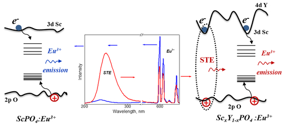
The luminescence spectra of all the samples except YPO4:Eu consist of two broad emission bands peaking at 240–250 nm and 340–370 nm as well as a set of narrow emission lines in the range of 580–720 nm. The characteristic luminescence spectra are presented for the Y0.5Sc0.5PO4:Eu3+ solid solution in Figure 1. The broad emission bands were also observed in undoped samples, and therefore, they cannot be related to the dopant emission. A broad band peaking at 240–250 nm belongs to an intrinsic emission previously observed in ScPO4 and attributed to the radiative annihilation of self-trapped excitons (STEs), whereas the electron and hole components of STEs are supposed to be localized on Sc ions and PO4 complexes, respectively [26]. The participation of Sc in the formation of STEs is further confirmed by the band structure calculations, which demonstrate that the bottom of the conduction band of ScPO4 is formed by the 3d Sc states [27,28] and by the absence of the corresponding emission band in YPO4:Eu3+ (see discussion below). The other broad band peaking around 340–370 nm is ascribed to the emission from crystal structure defects, probably from oxygen deficient oxyanionic complexes.
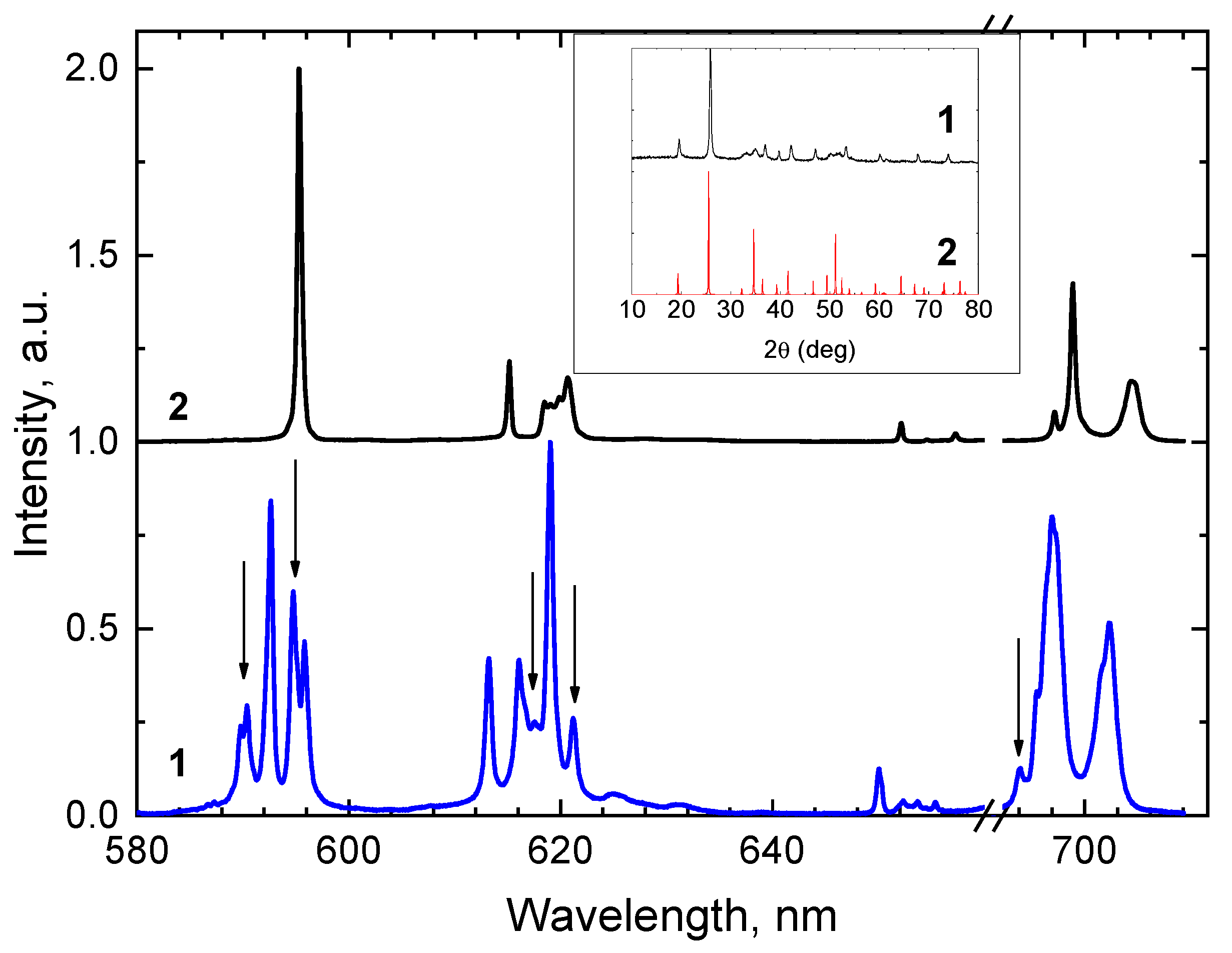
A set of narrow emission lines in the range of 580–720 nm is attributed to the 5D0-7Fj (j = 0, 1, 2, 3, 4) transitions of Eu3+ ions. The fine structure of these emission lines is presented for YPO4:Eu3+ and ScPO4:Eu3+ in Figure 2. For ScPO4:Eu3+, the spectrum is in good agreement with the previously obtained data [21]. For YPO4:Eu3+, several additional emission lines, which are not inherent for this compound [29], are observed. In particular, in the region of the magnetic dipole transition 5D0-7F1, (590–600 nm), the site symmetry, D2d, accepts the presence of two intense lines [30], which are observed in YPO4:Eu3+ at 592.7 and 596.1 nm. In the emission spectrum of the studied YPO4:Eu3+, an additional doublet at 589.8 and 590.4 nm and a line at 594.7 nm can be found in this region. Additional lines are detected also at 621.2 and 692.1 nm (all indicated by arrows in Figure 2). All these lines were also observed in the solid solutions. The presence of an additional phase originating from the yttrium based raw material is expected to be responsible for these extra lines. However, the presence of the additional phase is not manifested in the XRD pattern for YPO4:Eu3+ (see the inset to Figure 2), suggesting that its concentration is rather low. By the integration of the corresponding lines over the spectral region of interest, the contribution of the emission from this phase into the Eu3+ emission in the 580–720 nm spectral range is estimated to ~20%.
The dependences of the emission intensities of the ScxY1−xPO4:Eu3+ solid solutions on x under the interband excitation (Eex = 10 eV) are presented in Figure 3. The intensity of the emission integrated over the entire recorded region demonstrates a maximum value in a solid solution with x = 0.5. The effect is connected with the enhancement of the energy transfer efficiency from the host to emission centers and the decrease in non-radiative energy losses, resulting in the increase in light yield in solid-solution based phosphors and scintillators [31,32]. The enhancement is caused by the limitation of the free path length of charge carriers.
The dependences are different for the STEs and Eu3+ emissions. A defect related band is very weak at the 10 eV excitation and is not taken into account here. The intensity of the STEs emission band increases by a factor of 17 with the decrease in x from 1 to 0.4, then slightly decreases for x = 0.2, and finally, the band completely disappears in YPO4:Eu3+. On the one hand, this circumstance indicates that the STEs are created with the participation of Sc states. On the other hand, such a drastic increase in intensity with the decrease in Sc content implies that solid solutions with rather low content of Sc (x < 0.5) are more beneficial for the exciton creation. The distinctive difference between the constituents of the solid solution (ScPO4 and YPO4) is the energy of the conduction band bottom, which is, by ~1.5 eV, higher for YPO4 [23]. The lower content of Sc results in the creation of deep energy wells at the bottom of the conduction band, which prevent the separation of charge carriers due to migration and facilitate the creation of excitons [31]. The conditions beneficial for the STE creation suppress, in turn, the competitive process of energy transfer to the Eu3+ emission center by charge carriers at low concentrations of Sc. As a result, the maximum of Eu3+ emission intensity is shifted to solid solutions with high Sc content and is observed at x = 0.8. The reason is that for higher concentrations of Sc, at which the bottom of the conduction band becomes flat and predominantly formed by Sc states, the probability of exciton creation from e-h pairs decreases, leading to the enhancement of energy transfer to the Eu3+ emission centers. However, for the integrated intensity of both STE and Eu3+ emissions, the maximum is observed at an intermediate value of x = 0.5. It is worth noting that the additional phase does not influence the dependence of Eu3+ emission intensity on x. The dependence measured in the region of the 5D0-7F4 transitions, where the emission spectrum is not perturbed by the presence of an additional phase, is similar to that observed for the integrated emission, which is presented in Figure 3.
The excitation spectra for the STEs, defect-related and Eu3+ emissions are presented for a Sc0.5Y0.5PO4:Eu3+ solid solution in Figure 4. The low-energy onset observed at 7.1 eV in the excitation spectrum of the STEs emission (Figure 4a, curve 2) corresponds to the fundamental absorption edge of the material. The first peak at 7.4 eV is related to the direct exciton creation due the charge transfer transitions from oxygen to scandium. The intensity rise at the excitation energies above 17 eV is caused by the multiplication of electronic excitations, i.e., the number of electron-hole pairs created by one exciting photon (Figure 4b). Some peculiarities related to the excitation of the upper core levels are revealed in the excitation spectra at Eex > 20 eV. Such excitations cause usually peaks in reflection spectra and minima in excitation spectra. The minima arise due to the effects of near-surface losses as well as the emission quenching in the local regions with high densities of excitations [33]. The pronounced dips are observed in the excitation spectrum of the STEs emission at 34 eV and 39 eV and are ascribed to the manifestation of core cation excitons. The reflectivity spectra of various oxides with yttrium cation (Y3Al5O12, YAlO3, Y2O3) demonstrate a peak in the region of 34 eV as well, which corresponds to the excitation of the 4p Y3+ core level, namely to the 4p6-4p54d electron transitions in Y3+ [34,35,36]. We suppose this transition to be also responsible for the minimum at 34 eV in the excitation spectra of Sc0.5Y0.5PO4:Eu3+. The ionization energy of the upper core 3p Sc level is 28.3 eV, which is ~4.5 eV higher than that of the 4p upper core level of Y (23.1–24.4 eV) [37]. Therefore, the dip at 39 eV can be ascribed to the core exciton with the hole component at the 3p core level of Sc and electron at the 3d Sc states contributing to the conduction band bottom.
In the excitation spectrum of the Eu3+ emission, the narrow low-intensity lines at E < 4.5 eV (inset in Figure 4a) correspond to the intracenter 4f - 4f excitation within Eu3+, while a broad band peaking at 5.75 eV is attributed to the charge transfer 2p O–4f Eu transitions. The position of the first excitonic peak for the Eu3+ emission is slightly (by 0.15 eV) shifted to low energies in comparison to its position for the STEs emission. The shift is connected with the localization of excitons at the sites perturbed by Eu3+ ions substituting yttrium or scandium cations. Both Eu3+ and STEs emissions are efficiently excited in the region of the fundamental absorption, which serves as an additional confirmation that the energy transfer to the Eu3+ centers is a process competitive to the STEs creation. The onset of the intensity growth observed for the Eu3+ emission at 12 eV is, by 5 eV, lower than the threshold for the multiplication of electronic excitations. This rise can be ascribed to the impact excitation of Eu3+ ions in the process of the inelastic scattering of primary electrons [38,39,40].
The intensity decrease in the region of cation core exciton creation is much more pronounced in the excitation spectrum of the Eu3+ emission in comparison to that of the STEs emission. The modification of the energy distribution of hot (or non-thermalized) secondary e-h pairs occurs when the excitation energy is high enough to cause electron transitions from core levels. The simulation of the electron energy distribution showed that such transitions increase the part of low-energy electrons in the conduction band [41]. A similar conclusion was reached upon the analysis of the scintillation properties of several binary compounds in [42]. We suppose that a similar modification of the energy distribution of secondary excitations also takes place in the solid solutions studied. The decrease in the kinetic energy of the secondary e-h pairs results in the increased probability of exciton creation as it follows from the excitation spectra. In particular, it reduces the part of excitations, which can participate in the impact ionization of the emission centers, leading to a noticeable decrease in the efficiency of energy transfer to the Eu3+ centers.
The most pronounced feature of the excitation spectrum of the defect-related emission is a broad peak at 6.8 eV. The peak is situated below the fundamental absorption edge and is connected with the direct excitation of a defect emission center (Figure 4a, curve 3). The intensity of the excitation spectrum at the fundamental absorption edge is very low, suggesting a low probability of energy transfer to the defect emission center by excitons. The rise in the intensity is observed at Eex > 12 eV, demonstrating that a more efficient energy transfer from separated electrons and holes to the defect emission centers becomes possible starting from these energies.
4. Conclusions
The influence of cation composition on the luminescence properties of ScxY1−xPO4:Eu3+ solid solutions excited by synchrotron radiation has been studied. Two broad luminescence bands, one peaking at 235–250 nm and related to the intrinsic emission of self-trapped excitons and another peaking at 340–370 nm and connected with the extrinsic emission of crystal structure defects, as well as a set of narrow emission lines at 580–720 nm related to Eu3+, have been detected. The analysis of the fine structure of Eu3+ emission revealed the presence of an additional phase in yttrium-containing samples. The intensity of the emission integrated over the entire recorded region demonstrates a maximum value in a solid solution with x = 0.5, but the dependences are different for the STEs and Eu3+ emissions. The drastic intensity increase of the STEs emission band by a factor of 17 with a decrease in x from 1 to 0.4 is related to the creation of deep energy wells at the bottom of the conduction band of solid solutions with a lower content of Sc, which prevent the separation of charge carriers due to migration and facilitate the creation of excitons. The maximum of Eu3+ emission intensity is shifted to solid solutions with high Sc content (x = 0.8) due to the competition of energy transfer processes with the STEs emission. The peculiarities of the energy transfer to competitive emission centers are analyzed on the basis of the excitation spectra of the brightest solid solution Sc0.5Y0.5PO4:Eu3+.
Author Contributions
Conceptualization, D.S.; methodology, K.C., B.Z.; investigation, D.S., V.V.-L., A.A. and V.N.; writing—original draft preparation, D.S., V.V.-L. and V.N.; writing—review and editing, D.S., and V.N.; visualization, A.A. All authors have read and agreed to the published version of the manuscript.
Funding
The research leading to this result has been supported by the project CALIPSOplus under the Grant Agreement 730872 from the EU Framework Programme for Research and Innovation HORIZON 2020 as well as by the Estonian Research Council, project PUT PRG111 and the ERDF funding in Estonia granted to the Centre of Excellence TK141, Project No. 2014-2020.4.01.15-0011.
Acknowledgments
We are grateful to A.N. Vasil’ev for a fruitful discussion of the results. The measurements at MAX-lab were performed within the proposal 20190244.
Conflicts of Interest
The authors declare no conflict of interest.
References
- Moses, W.W.; Weber, M.J.; Derenzo, S.E.; Perry, D.; Berdahl, P.; Boatner, L.A. Prospects for dense, infrared emitting scintillators. IEEE Trans. Nucl. Sci. 1988, 45, 462–466. [Google Scholar] [CrossRef]
- Nakazawa, E. The lowest 4f-to-5d and charge-transfer transitions of rare earth ions in YPO4 hosts. J. Lumin. 2002, 100, 89–96. [Google Scholar] [CrossRef]
- Jüstel, T.; Huppertz, P.; Mayr, W.; Wiechert, D.U. Temperature-dependent spectra of YPO4:Me (Me = Ce, Pr, Nd, Bi). J. Lumin. 2004, 106, 225–233. [Google Scholar] [CrossRef]
- Boatner, L.A. Synthesis, structure, and properties of monazite, pretulite, and xenotime. Rev. Mineral. Geochem. 2002, 48, 87–121. [Google Scholar] [CrossRef]
- Makhov, V.N.; Kirikova, N.Y.; Kirm, M.; Krupa, J.C.; Liblik, P.; Lushchik, A.; Lushchik, C.; Negodin, E.; Zimmerer, G. Luminescence properties of YPO4:Nd3+: A promising VUV scintillator material. Nucl. Instrum. Meth. A 2002, 486, 437–442. [Google Scholar] [CrossRef]
- Boatner, L.A.; Beall, G.W.; Abraham, M.M.; Finch, C.B.; Huray, P.G.; Rappaz, M. Monazite and other lanthanide orthophosphates as alternate actinide waste forms. In Book Scientific Basis for Nuclear Waste Management; Northrup, C.J., Ed.; Plenum Publishing Corporation: New York, NY, USA, 1980; Volume 2, pp. 289–296. [Google Scholar] [CrossRef]
- Chen, L.; Chen, X.; Liu, F.; Chen, H.; Wang, H.; Zhao, E.; Jiang, Y.; Chan, T.; Wang, C.; Zhang, W.; et al. Charge deformation and orbital hybridization: Intrinsic mechanisms on tunable chromaticity of Y3Al5O12:Ce3+ luminescence by doping Gd3+ for warm white LEDs. Sci. Rep. 2015, 5, 11514. [Google Scholar] [CrossRef] [Green Version]
- Kamada, K.; Endo, T.; Tsutumi, K.; Yanagida, T.; Fujimoto, Y.; Fukabori, A.; Yoshikawa, A.; Pejchal, J.; Nikl, M. Composition engineering in cerium-doped (Lu,Gd)3(Ga, Al)5O12 single-crystal scintillators. Cryst. Growth Des. 2011, 11, 4484–4490. [Google Scholar] [CrossRef]
- Levushkina, V.; Spassky, D.; Brik, M.G.; Zych, E.; Madej, A.; Belsky, A.N.; Bartosiewicz, A.K.; Nikl, M. Mixed vanadates: Optimization of optical properties by varying chemical composition. J. Lumin. 2017, 189, 140–147. [Google Scholar] [CrossRef]
- Levushkina, V.S.; Spassky, D.A.; Aleksanyan, E.M.; Brik, M.G.; Tretyakova, M.S.; Zadneprovski, B.I.; Belsky, A.N. Bandgap engineering of the LuxY1−xPO4 mixed crystals. J. Lumin. 2016, 171, 33–39. [Google Scholar] [CrossRef]
- Lyu, T.; Dorenbos, P. Charge carrier trapping processes in lanthanide doped LaPO4, GdPO4, YPO4, and LuPO4. J. Mater. Chem. C 2018, 6, 369–379. [Google Scholar] [CrossRef] [Green Version]
- Levushkina, V.S.; Spassky, D.A.; Tretyakova, M.S.; Zadneprovski, B.I.; Kamenskikh, I.A.; Vasil’ev, A.N.; Belsky, A. Luminescence properties of solid solutions LuxY1-xPO4:Eu3+. Opt. Mater. 2018, 75, 607–611. [Google Scholar] [CrossRef]
- Gektin, A.V.; Belsky, A.N.; Vasil’ev, A.N. Scintillation efficiency improvement by mixed crystal use. IEEE Trans. Nucl. Sci. 2013, 61, 262–270. [Google Scholar] [CrossRef]
- Sidletskiy, O.; Gektin, A.; Belsky, A. Light yield improvement trends in mixed scintillation crystals. Phys. Status Solidi A 2014, 211, 2384–2387. [Google Scholar] [CrossRef]
- Belsky, A.N.; Auffray, E.; Lecoq, P.; Dujardin, C.; Garnier, N.; Canibano, H.; Pedrini, C.; Petrosyan, A.G. Progress in the development of LuAlO3-based scintillators. IEEE Trans. Nucl. Sci. 2001, 48, 1095–1100. [Google Scholar] [CrossRef]
- Spassky, D.; Vasil’ev, A.; Vielhauer, S.; Sidletskiy, O.; Voloshyna, O.; Belsky, A. Composition effect in luminescence properties of Y(NbxTa1-x)O4 mixed crystals. Opt. Mater. 2018, 80, 247–252. [Google Scholar] [CrossRef]
- Hernández, A.G.; Boyer, D.; Potdevin, A.; Chadeyron, G.; Murillo, A.G.; Romo, F.D.J.C.; Mahiou, R. Tuning the morphology of GdxY1-xPO4:Tb3+ powders and their emission intensity upon VUV excitation. Opt. Mater. 2017, 73, 350–357. [Google Scholar] [CrossRef]
- Sun, C.; Xue, D. The synergy effect of rare earth cations on local structure and PL emission in a Ce3+:REPO4 (RE = La, Gd, Lu, Y) system. Dalton Trans. 2017, 46, 7888–7896. [Google Scholar] [CrossRef]
- Makowski, M.; Witkowski, M.E.; Drozdowski, W.; Wojtowicz, A.J.; Wisniewski, K.; Boatner, L.A. Luminescence and scintillation properties of XPO4:Nd3+ (X = Y, Lu, Sc, La) crystals. Opt. Mater. 2018, 79, 273–278. [Google Scholar] [CrossRef] [Green Version]
- Mi, R.; Chen, J.; Liu, Y.G.; Fang, M.; Mei, L.; Huang, Z.; Wang, B.; Zhaob, C. Luminescence and energy transfer of a color tunable phosphor: Tb3+ and Eu3+ co-doped ScPO4. RSC Adv. 2016, 6, 28887–28894. [Google Scholar] [CrossRef]
- Jedoń, J.; Zeler, J.; Zych, E. On the orange-red persistent luminescence of ScPO4:Eu3+. J. Alloy. Compd. 2020, 816, 152–603. [Google Scholar] [CrossRef]
- Melnikov, P.; Massabni, A.M.; Malta, O. Luminescent properties of some rare earth phosphates. Phosphorus Sulfur 1996, 111, 1. [Google Scholar] [CrossRef]
- Dorenbos, P. The electronic level structure of lanthanide impurities in REPO4, REBO3, REAlO3, and RE2O3 (RE = La, Gd, Y, Lu, Sc) compounds. J. Phys. Condens. Mat. 2013, 25, 225–501. [Google Scholar] [CrossRef]
- Pankratov, V.; Pärna, R.; Kirm, M.; Nagirnyi, V.; Nõmmiste, E.; Omelkov, S.; Vielhauer, S.; Chernenko, K.; Reisberg, L.; Turunen, P.; et al. Progress in development of a new luminescence setup at the FinEstBeAMS beamline of the MAX IV laboratory. Radiat. Meas. 2019, 121, 91–98. [Google Scholar] [CrossRef]
- Pärna, R.; Sankari, R.; Kukk, E.; Nõmmiste, E.; Valden, M.; Lastusaari, M.; Kooser, K.; Kokko, K.; Hirsimäki, M.; Urpelainen, S.; et al. FinEstBeaMS—A wide-range Finnish-Estonian Beamline for Materials Science at the 1.5 GeV storage ring at the MAX IV Laboratory. Nucl. Instrum. Meth. A 2017, 859, 83–89. [Google Scholar] [CrossRef] [Green Version]
- Trukhin, A.N.; Shmits, K.; Jansons, J.L.; Boatner, L.A. Ultraviolet luminescence of ScPO4, AlPO4 and GaPO4 crystals. J. Phys. Condens. Mat. 2013, 25, 385–502. [Google Scholar] [CrossRef]
- Han, L.; Guo, C.; Ci, Z.; Wang, C.; Wang, Y.; Huang, Y. Morphology-controllable synthesis, electronic structure and multicolor-tunable luminescence properties of multifunctional ScPO4 based nano/micro-phosphor. Chem. Eng. J. 2017, 312, 204–219. [Google Scholar] [CrossRef]
- Xu, H.; Xu, B.; Liu, R.; Li, X.; Zhang, S.; Ouyang, C.; Zhong, S. Facile microwave synthesis of ScPO4·2H2O flowerlike superstructures: Morphology control, electronic structure and multicolor tunable luminescent properties. CrystEngComm 2017, 19, 5787–5796. [Google Scholar] [CrossRef]
- Brecher, C.; Samelson, H.; Riley, R.; Lempicki, A. Polarized spectra and crystal field parameters of Eu3+ in YPO4. J. Chem. Phys. 1968, 49, 3303. [Google Scholar] [CrossRef]
- Binnemans, K. Interpretation of europium (III) spectra. Coord. Chem. Rev. 2015, 295, 1–45. [Google Scholar] [CrossRef] [Green Version]
- Belsky, A.; Gektin, A.; Vasil’ev, A.N. Influence of Disorder in Scintillating Solid Solutions on Thermalization and Recombination of Electronic Excitations. Phys. Status Solidi B 2019. [Google Scholar] [CrossRef]
- Belsky, A.; Gektin, A.; Gridin, S.; Vasil’ev, A.N. Electronic and optical properties of scintillators based on mixed ionic crystals. In Engineering of Scintillation Materials and Radiation Technologies: Proceedings of ISMART 2016; Korzhik, M., Gektin, A., Eds.; Springer: Berlin/Heidelberg, Germany, 2017; Volume 200, pp. 63–82. [Google Scholar] [CrossRef]
- Belsky, A.N.; Chevallier, P.; Dhez, P.; Martin, P.; Pedrini, C.; Vasil’ev, A.N. X-ray excitation of luminescence of scintillator materials in the 7–22 keV region. Nucl. Instrum. Meth. A 1995, 361, 384–387. [Google Scholar] [CrossRef]
- Tomiki, T.; Shikenbaru, T.; Ganaha, Y.; Futemma, T.; Kato, H.; Yuri, M.; Fukutani, H.; Miyahara, T.; Shin, S.; Ishigame, M.; et al. Optical spectra of Y2O3 single crystals in the vacuum ultraviolet region. II. J. Phys. Soc. Jpn. 1992, 61, 2951–2963. [Google Scholar] [CrossRef]
- Tomiki, T.; Fukudome, F.; Kaminao, M.; Fujisawa, M.; Tanahara, Y.; Futemma, T. Optical spectra of Y3Al5O12 (YAG) single crystals in the vacuum ultraviolet region. J. Phys. Soc. Jpn. 1989, 58, 1801–1810. [Google Scholar] [CrossRef]
- Tomiki, T.; Ganaha, Y.; Shikenbaru, T.; Futemma, T.; Yuri, M.; Aiura, Y.; Fukutani, H.; Kato, H.; Miyahara, T.; Yonesu, A.; et al. Anisotropic optical spectra of YAlO3 (YAP) single crystals in the vacuum ultraviolet region. II. Spectra of Reflectivity. J. Phys. Soc. Jpn. 1994, 63, 1976–1985. [Google Scholar] [CrossRef]
- Cardona, M.; Ley, L. Photoemission in solids. Vol. 1: General principles; Vol. 2: Case studies. In Topics in Applied Physics; Cardona, M., Ley, L., Eds.; Springer Proceedings in Physics: Berlin/Heidelberg, Germany, 1978; p. 290. [Google Scholar]
- Feldbach, E.; Kamada, M.; Kirm, M.; Lushchik, A.; Lushchik, C.; Martinson, I. Direct excitation of Tl+ impurity ions by hot photoelectrons in wide-gap crystals. Phys. Rev. B 1997, 56, 13908. [Google Scholar] [CrossRef]
- Belsky, A.N.; Krupa, J.C. Luminescence excitation mechanisms of rare earth doped phosphors in the VUV range. Displays 1999, 19, 185–196. [Google Scholar] [CrossRef]
- Lushchik, A.; Lushchik, C.; Popov, A.I.; Schwartz, K.; Shablonin, E.; Vasil’chenko, E. Influence of complex impurity centres on radiation damage in wide-gap metal oxides. Nucl. Instrum. Meth. B 2016, 374, 90–96. [Google Scholar] [CrossRef] [Green Version]
- Belskiy, A.N.; Kamenskikh, I.A.; Mikhailin, V.V.; Shpinkov, I.N.; Vasil’ev, A.N. Electronic excitations in crystals with complex oxyanions. Phys. Scr. 1990, 41, 530–536. [Google Scholar] [CrossRef]
- Vasil’ev, A.N.; Gektin, A.V. Multiscale approach to estimation of scintillation characteristics. IEEE Trans. Nucl. Sci. 2013, 61, 235–245. [Google Scholar] [CrossRef]
Figure 1.
Luminescence spectra of Sc0.5Y0.5PO4:Eu3+ under excitation Eex = 10 eV (1) and Eex = 6.5 eV (2) at T = 7 K.
Figure 1.
Luminescence spectra of Sc0.5Y0.5PO4:Eu3+ under excitation Eex = 10 eV (1) and Eex = 6.5 eV (2) at T = 7 K.

Figure 2.
The luminescence spectra of YPO4:Eu3+ (1) and ScPO4:Eu3+ (2) under excitation Eex = 3.14 eV (395 nm) at T = 300 K. The arrows indicate the emission lines, which are not inherent to YPO4:Eu3+. In the inset: XRD pattern for YPO4 (1) and literature data for YPO4 according to ICSD#184543 (2).
Figure 2.
The luminescence spectra of YPO4:Eu3+ (1) and ScPO4:Eu3+ (2) under excitation Eex = 3.14 eV (395 nm) at T = 300 K. The arrows indicate the emission lines, which are not inherent to YPO4:Eu3+. In the inset: XRD pattern for YPO4 (1) and literature data for YPO4 according to ICSD#184543 (2).

Figure 3.
(a) Luminescence spectra of ScxY1−xPO4:Eu3+ under excitation Eex = 10 eV at T = 7 K, and (b) intensity dependences on Sc concentration for the STEs emission (signal is integrated over the range of 200–320 nm, curve 1), Eu3+ emission (signal is integrated over the range of 580–720 nm, curve 2) and the emission integrated over the entire range of 190–850 nm (curve 3). Lines are used to guide the eye.

Figure 4.
Luminescence excitation spectra of Sc0.5Y0.5PO4:Eu3+ measured at 240 nm (1), 595 nm (2) and 360 nm (3) at T = 7 K in the energy range 4.5–10 eV (a) and 4.5–45 eV (b). In the inset: excitation spectrum of Sc0.5Y0.5PO4:Eu3+ measured at 595 nm in the region of f-f Eu3+ transitions.
Figure 4.
Luminescence excitation spectra of Sc0.5Y0.5PO4:Eu3+ measured at 240 nm (1), 595 nm (2) and 360 nm (3) at T = 7 K in the energy range 4.5–10 eV (a) and 4.5–45 eV (b). In the inset: excitation spectrum of Sc0.5Y0.5PO4:Eu3+ measured at 595 nm in the region of f-f Eu3+ transitions.

© 2020 by the authors. Licensee MDPI, Basel, Switzerland. This article is an open access article distributed under the terms and conditions of the Creative Commons Attribution (CC BY) license (http://creativecommons.org/licenses/by/4.0/).
Share and Cite
MDPI and ACS Style
Spassky, D.; Voznyak-Levushkina, V.; Arapova, A.; Zadneprovski, B.; Chernenko, K.; Nagirnyi, V. Enhancement of Light Output in ScxY1−xPO4:Eu3+ Solid Solutions. Symmetry 2020, 12, 946. https://0-doi-org.brum.beds.ac.uk/10.3390/sym12060946
AMA Style
Spassky D, Voznyak-Levushkina V, Arapova A, Zadneprovski B, Chernenko K, Nagirnyi V. Enhancement of Light Output in ScxY1−xPO4:Eu3+ Solid Solutions. Symmetry. 2020; 12(6):946. https://0-doi-org.brum.beds.ac.uk/10.3390/sym12060946
Chicago/Turabian StyleSpassky, Dmitry, Viktoriia Voznyak-Levushkina, Anastasiia Arapova, Boris Zadneprovski, Kirill Chernenko, and Vitali Nagirnyi. 2020. "Enhancement of Light Output in ScxY1−xPO4:Eu3+ Solid Solutions" Symmetry 12, no. 6: 946. https://0-doi-org.brum.beds.ac.uk/10.3390/sym12060946
Note that from the first issue of 2016, this journal uses article numbers instead of page numbers. See further details here.