Article

Mineral Composition and Structural Characterization of the Clinoptilolite Powders Obtained from Zeolite-Rich Tuffs

by
Ewelina Pabiś-Mazgaj
1,*,
Tomasz Gawenda
2,
Paweł Pichniarczyk
1 and
Agata Stempkowska
2
1
Glass and Building Materials Division, Łukasiewicz Research Network Institute of Ceramics and Building Materials, 31-983 Cracow, Poland
2
Department of Environmental Engineering, Faculty of Civil Engineering and Resource Management, AGH University of Science and Technology, 30-059 Cracow, Poland
*
Author to whom correspondence should be addressed.
Submission received: 5 August 2021 / Revised: 16 September 2021 / Accepted: 18 September 2021 / Published: 23 September 2021

Abstract

:
Clinoptilolite is a precious zeolite mineral that has the most comprehensive physicochemical properties among all the zeolite group minerals. Due to these unique properties, clinoptilolite has a wide range of applications in many different industries. In Poland, the clinoptilolite occurs only as an accompanying mineral in the sedimentary rocks nearby Rzeszów. In Europe, the abundant clinoptilolite-bearing deposits are located in Slovakia and Ukraine, where clinoptilolite mineralization occurs in the volcanic tuffs. Due to clinoptilolite’s rare performance, it is extremely crucial to manage its deposits in a complementary manner. In this paper, the mineralogical and structural characterization of the clinoptilolite powders obtained by mineral processing of the clinoptilolite-rich tuffs from Slovakia and Ukraine deposits were discussed. The scope of research covered determination of the mineral composition of the tuffs, structural analysis of the clinoptilolite crystals, as well as textural and physical properties of the powders obtained by mineral processing of the tuffs. In addition, this paper includes the comparative study of the most significant zeolite deposits in the world and investigated clinoptilolite-rich tuffs. A wide spectrum of methods was used: X-ray powder diffraction (XRD), thermal analysis (DSC, TG), X-ray fluorescence (XRF), optical microscopy, Scanning Electron Microscopy (SEM-EDS), the laser diffraction technique, and low-temperature nitrogen adsorption/desorption. The test results indicated that the major component of the tuffs is clinoptilolite, which crystallized in the form of very fine-crystalline thin plates. The clinoptilolite mineralization in the Ukrainian and Slovakian tuffs exhibited a strong resemblance to the clinoptilolite crystals in Yemeni and Turkish tuffs. With respect to the mineral composition, the investigated tuffs showed excellent conformity with the Miocene white tuffs from Romania. The Ukrainian and Slovakian tuffs do not reveal the presence of the clay minerals, which is quite common for naturally occurring zeolite-rich rocks in various deposits in the world. The textural features together with mineral composition of the investigated samples incline that they are potentially suitable raw materials for the sorbent of petroleum compounds. Moreover, the obtained results can be useful indicators with respect to the crushing and compaction susceptibility of the Ukrainian and Slovakian clinoptilolite-rich tuffs.

1. Introduction

Zeolites are microporous aluminosilicates with a three-dimensional network composed of SiO4 and AlO4 tetrahedra joined by oxygen atoms. Depending on a certain zeolite type, the Si/Al ratio in its framework is changeable, but the Si content is always higher than the Al content. In the crystalline framework of zeolites, the specific system comprised of the channels and chambers with certain shape and size is observed. In normal condition, the channels and chambers are occupied by some cations and easily removable H2O molecules. The size distribution of the channel system in the zeolite framework is significantly important when it comes to its sorption and catalytic properties [1,2,3].
Clinoptilolite is the one of the most widespread zeolites, which belongs to the heulandite group [2,3,4,5]. The general formula of clinoptilolite is as follows:
(K, Na, Ca)6[(Si, Al)36O72] nH2O (n = 20–24).
The water content in the clinoptilolite elementary cell is changeable (from 20 to 24 H2O molecules) [6]. Clinoptilolite may occur in high-silica or low-silica form, which is determined as Ca-clinoptilolite [3]. The interest in clinoptilolite is not only associated with its abundance in nature but mainly with the most comprehensive physicochemical properties among all of the zeolite-group minerals [7,8].
The most significant clinoptilolite properties are high sorption and ion exchange capacity, ion exchange selectivity, molecular sieving properties, catalytic activity, acid resistance, and thermal stability up to 750 °C [7]. The latter one distinguished clinoptilolite to the isostructural heulandite, which shows thermal stability up to 400 °C [5,8,9,10]. Due to the very similar crystallographic structure of the clinoptilolite and heulandite, they were misidentified. Finally, in 1960, the clinoptilolite was marked out as an individual mineral [3,9]. Clinoptilolite has found particular application in environmental protection, industry, and agriculture. However, even now, new areas of the clinoptilolite management are being discovered. Due to this ongoing expansion of the applications, zeolites often are determined as the raw material of the 21st century [7,11]. It is worth highlighting that clinoptilolite was used on the large scale as the cesium and strontium absorbent to mitigate the detrimental effects of the Chernobyl disaster [12].
The location of clinoptilolite-rich deposits is mostly related to the areas of previous volcanic activity and hydrothermal processes. Moreover, in some regions, the zeolite crystallization occurs these days as a result of the “zeolitization” of the feldspathic rocks. Due to the “zeolitization” process, zeolite-bearing rocks are widespread throughout the earth’s crust and stratigraphically belong to the different geological eras [13]. According to the U.S. Geological Survey [14], the most abundant deposits in the world are in China, the Republic of Korea, Indonesia, New Zealand, the United States, and Cuba. Moreover, new deposits of the zeolite-rich rocks are still being discovered. A good example is the recently discovered the mordenite–clinoptilolite deposit in eastern Cuba [15]. It proved that even the areas well known for occurrences of zeolite still have undiscovered deposits. Based on the data provided by KMI Zeolite Inc. [16], the most valuable mineral commodity because of the high-purity clinoptilolite zeolite is in the U.S. state of Nevada. KMI reported [16] that so far, their deposit has exhibited the highest clinoptilolite concentration in Northern America, maybe even in the world. However, many countries are not willing to distribute the data on their mine reserves. There are also assumptions that reserves of some deposits are overestimated because of possible miscalculation. It should be emphasized that what makes deposits valuable is not the host rock but the zeolite mineralization concentration in it [14]. In Europe, the clinoptilolite-rich tuffs deposits are manly located in Slovakia, Ukraine, Turkey, Italy, and Romania [7,13,14,17,18]. However, the first one is reported as the economically most crucial natural zeolite occurrence in Europe [14,19].
In general, zeolites are not the rock-forming minerals and rarely occur in a pure form. The zeolite mineral type and its content in the rock depend on the host rock petrography (chemical and mineralogical composition of volcanic glass as well as the texture and structure of host rock) and forming conditions such as the pH and temperature of the water [12].
Usually, the various zeolite minerals co-occur in the particular rock. In addition, the zeolite mineralization is associated with some minor and accessory minerals such as feldspar, mica group minerals, chlorite group minerals, opal, cristobalite, zircon, tourmaline, iron, and sulfur compounds as well as clay minerals. The mentioned constituents are treated as unwanted impurities in zeolite mineralization [12,20]. The occurrences of secondary and accessory minerals in zeolite-rich rocks induce many constrains in their applications [20]. The high demand for zeolite minerals brings about development in synthetic zeolites [11]. For the first time, zeolites were obtained under the laboratory conditions in the 1950s [21]. Today, many methods for the synthesis of zeolites are well known and broadly discussed in the literature [11,22,23,24,25].
It should be emphasized that natural zeolites subjected to the appropriate activation processes can compete with the properties of the synthetic zeolites or create an even better tailored product for the specific industrial applications. One of the common performance-enhancement processes for natural zeolites is the mechanical activation [26,27,28]. The milling technology applied in mechanical activation has a significant impact on the processing properties of natural zeolites, and it is recognized as an up-and-coming and eco-friendly process in the production of the highly demanding nanozeolites [26,29,30]. In the interest of nanoscale zeolite production, many studies regarding the zeolite grinding optimization were carried out [26,27]. Bohács et al. [26] established the optimal conditions for the zeolite wet milling in a stirred media mill, whereas Sivashankari et al. [27] determined the most advantageous parameters for the zeolite dry milling process in a planetary ball mill. In addition, Şükrü et al. [28] stated that using a planetary ball mill compared to a conventional ball mill in the zeolite grinding process significantly enhanced the zeolite absorption capacity for the removal of heavy metals from wastewater solution. Moreover, Zolzaya et al. [31] reported that the mechanical activation in a vibration mill remarkably improved the cation exchange capacity of natural zeolites. In [32], the authors optimized the key parameters associated with the vibratory and planetary centrifugal milling processes for enhancement of the Sr2+ and Cu2+ selectivity of natural zeolites. In recent years, there has been considerable interest in alternative processes of the natural zeolites’ activation [33,34,35,36]. Silva et al. [33] proved that graphene-oxide activation by the spin-coating method improves the adsorption properties of natural zeolites. Whereas Kang et al. [34] reported that the fluorinated natural zeolite exhibits significantly better efficiency in the radon sorption compared to untreated natural zeolite. Other researchers demonstrated that the aluminum-coated natural zeolites [35] and surfactant-modified zeolites [37] are promising materials for the defluorination of drinking water. Many studies have been published on surfactant-modified zeolites as organic [38,39] and inorganic [37,40,41] toxicants adsorbents. Several studies [40,41,42,43] suggested that among the many surfactants, the hexadecyl-trimethylammonium bromide (HDTMA) is the most widely applied as an agent that can enhance the ability of zeolites to adsorb the organic pollutants by increasing zeolite hydrophobicity. In more recently published studies, Fuss et al. [44] investigated the chemical (acid, basic) and thermal methods for zeolite activation regarding the capacity of natural zeolites to adsorb the odors and toxic compounds. Researchers [44] proved that the thermally activated zeolites exhibit a significantly better performance in terms of humidity sorption, whereas the acid-activated zeolites are an efficient polycyclic aromatic hydrocarbons (PAHs) adsorbent.
Although it may seem that zeolite minerals are already well known, the interest in this group of minerals is not decreasing, and there is still need to investigate them [11]. Moreover, due to the versatility of the zeolite minerals, every particular zeolite commodity demands individual detailed research [45].
The aim of this work was to investigate and compare the features of clinoptilolite powders obtained by the mineral processing of Zakarpacki clinoptilolite-rich tuffs from Ukrainian and Slovakian deposits. In addition, a detailed comparative study of the most significant zeolite deposits in the world and investigation of the clinoptilolite-rich tuffs was conducted. The fundamental importance of possessing access to the natural zeolites is the long-term economic development of Ukraine and Slovakia. Moreover, the naturally occurring zeolites in the studied countries can be useful in tackling one of the biggest environmental challenges facing the world, which is climate change. The mineralogical and structural characterization of Ukrainian and Slovakian clinoptilolite-rich tuffs were examined by X-ray powder diffraction (XRD), thermal analysis (DSC, TG), X-ray fluorescence (XRF), optical microscopy, Scanning Electron Microscopy (SEM-EDS), the laser diffraction technique, and low-temperature nitrogen adsorption/desorption. Despite huge interest in zeolite-bearing deposits, no one as far as we know has provided such comprehensive petrographic characterization and comparative study of Ukrainian and Slovakian zeolite deposits, in respect to some of the global representative sources of zeolites.

2. Materials and Methods

Studies were carried out on the clinoptilolite powders obtained by mineral processing of the Miocene volcanic tuffs from two deposits located in Slovakia (Nižný Hrabovec Mine) and Ukraine (Sokyrnytsya Deposit). In order to carry out a profound analysis of the fine-grained clinoptilolite, the host rocks from Slovakian and Ukrainian mines were also examined. Petrogenesis of the studied tuffs is associated with a volcanic ash sedimentation in a marine environment, whereas occurrence of the clinoptilolite mineralization in tuffs is the result of the latter hydrothermal processes [46].

2.1. Macroscopic Analysis

At the beginning of the studies, the basic lithological characteristics such as the color and coherence structure of the clinoptilolite-rich tuffs used for making the zeolite powders were determined. The rocks’ lithology was defined using the Nikon SMZ1000 stereo zoom microscope equipped with the zoom range of 0.8–8×. Further mineralogical and structural characterization of the materials has required conducting different analytical methods, which are discussed below. All of the equipment except for the ASAP 2020 Micromeritic instrument (ASAP 2020, Micromeritics, Cracow, Poland) used in tests is in possession of the Łukasiewicz Research Network ICiMB Cracow.
The nitrogen adsorption/desorption tests with ASAP 2020 from Micromeritic were conducted in the Organic Technology Laboratory Faculty of Chemistry Jagiellonian University in Cracow.

2.2. X-ray Analysis

Mineralogical composition was determined, mainly, via X-ray powder diffraction analysis using a Philips APD PANalytical X’pert diffractometer with PW 3020 goniometer, Cu Kα radiation (radiation at 40 kV power and 35 mA current), and graphite monochromator. The measurements were done in the 5° to 55° 2θ range with step size of 0.02° 2θ. Diffraction data were processed by X’Pert HighScore Plus, PANalytical B.V software (version 2.2.5) combined with JCPDS-ICDD database. The Rietveld method was applied to estimate the clinoptilolite mineral content. Before analysis, the samples were ground to the particle size of 0.125 mm.

2.3. X-ray Fluorescence

The chemical analysis of the clinoptilolite-rich tuffs was determined by X-ray fluorescence spectroscopy with wavelength dispersive (WDXRF) using a Panalytical Epsilon 3X instrument. The analysis was conducted on the grounded to 0.125 mm whole rocks. The bulk oxides composition allowed assessing the total alkalis (K2O + Na2O) versus silica (SiO2) ratio. Based on that, the classification according to the TAS (Total Alkali Silica) of volcanic rocks chemical classification was carried out [47].

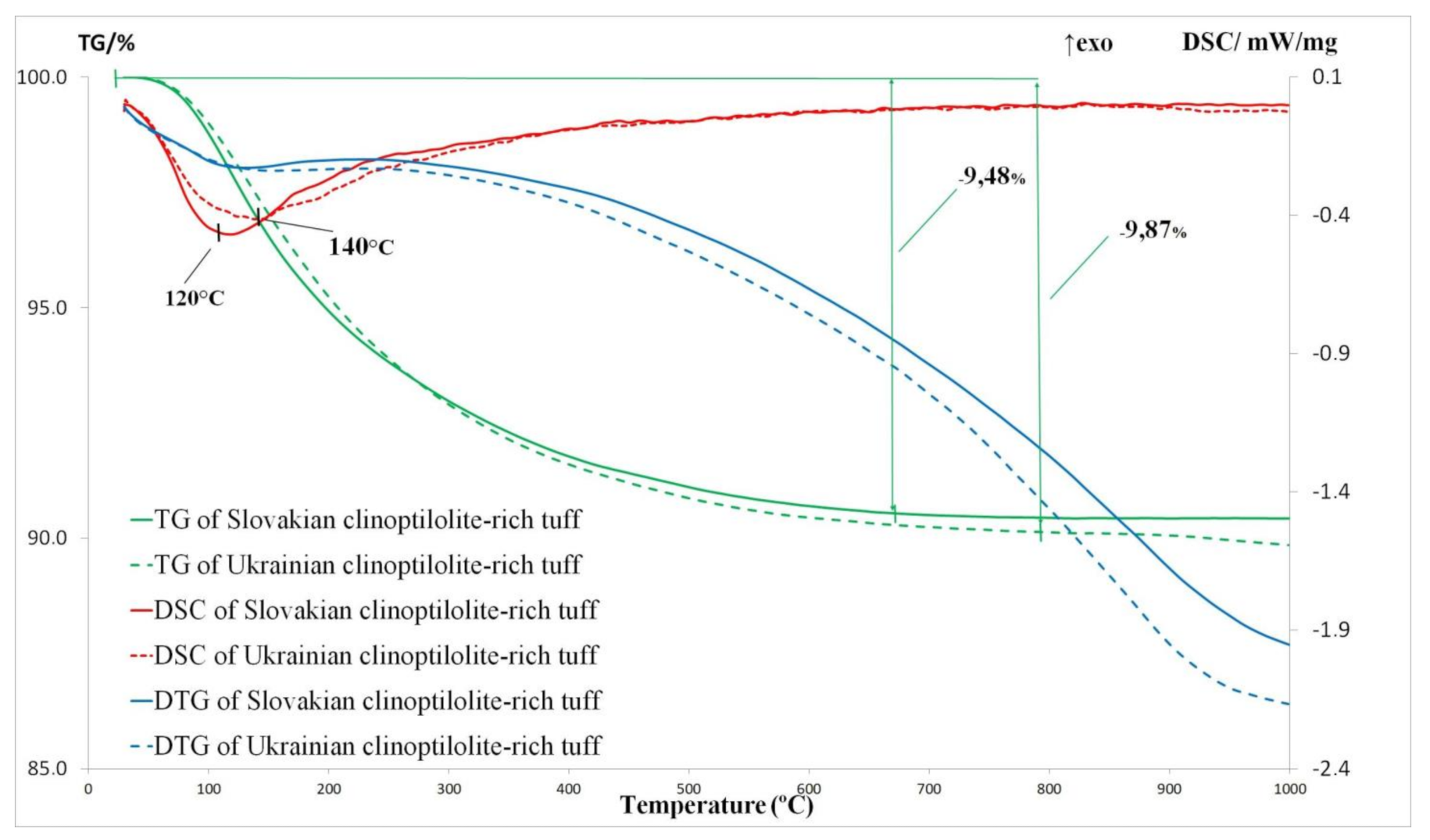
2.4. Thermal Analysis
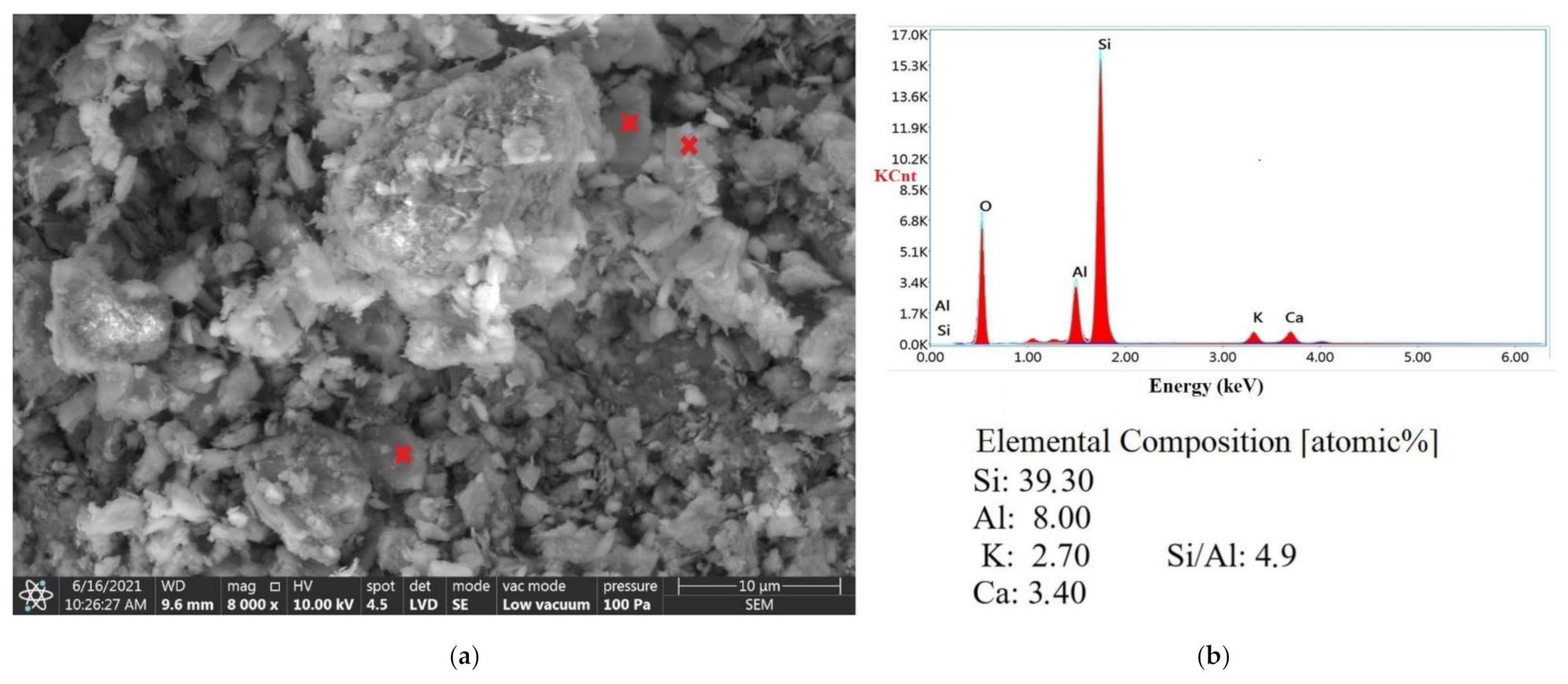
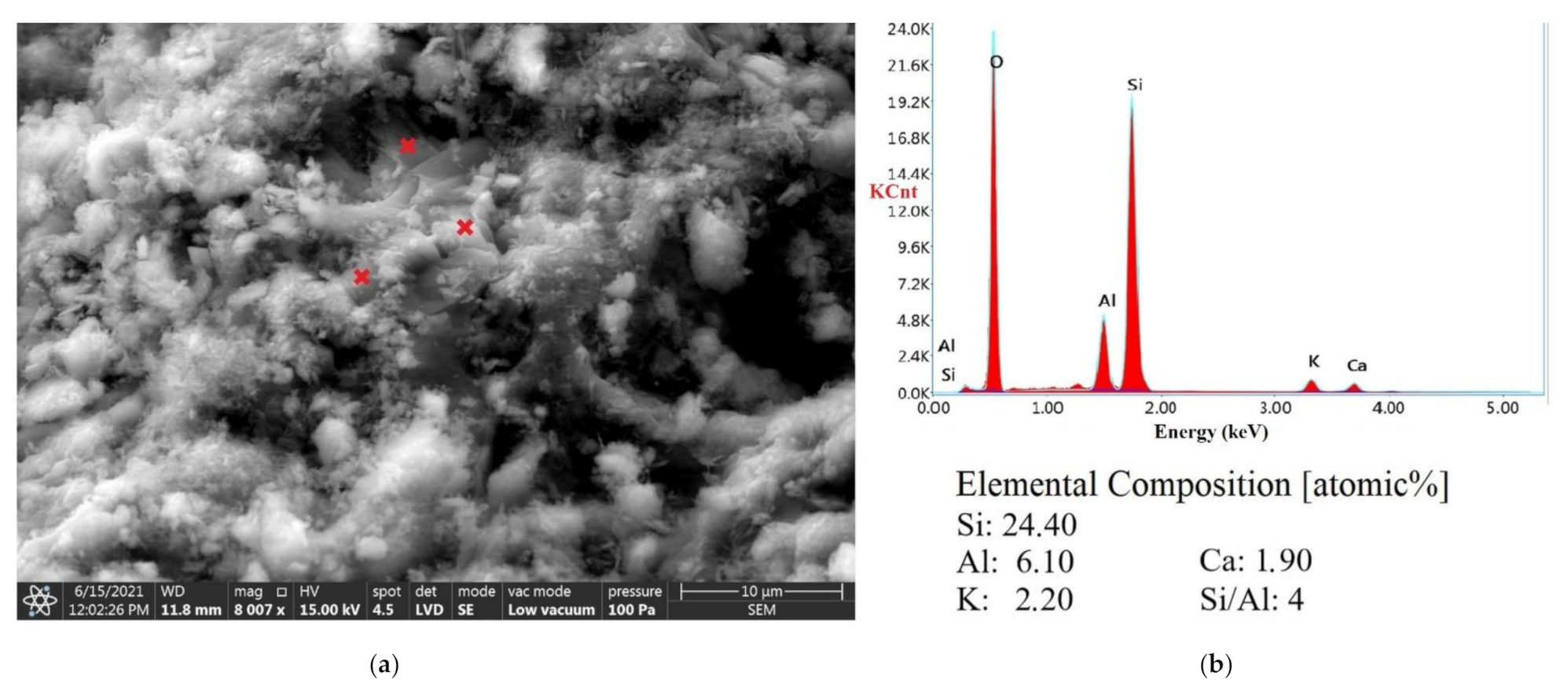
Thermal analysis has included the thermogravimetric (TG) and differential scanning calorimetry (DSC) measurements by using a Netzsch Jupiter STA 449 F3 instrument in the temperature range 30 to 1000 °C with a heating rate of 10 °C/min in oxide atmosphere (flow rate 80 mL/min). The thermal analysis enables determining the thermal properties of the test materials.

2.5. SEM Analysis

The data obtained from XRD analysis were supported by optical microscopy and SEM observations. The optical microscopy on a thin section was performed using a Nikon Eclipse LV 100 POL microscope combined with the digital camera Nikon Digital Sight DS –Fi1 runs by NIS–Elements BV 2.3. software. The microscope observations were carried out in polarized transmitted light on the standard thin sections polished with fine abrasives to a thickness of 0.30 µm. Optical microscopy was useful in the identification of some minerals in the tuffs and in the determination of structural features of the clinoptilolite-rich rocks. In order to determine the clinoptilolite texture and its Si/Al ratio as well as the grain morphology of the clinoptilolite powders, the scanning electron microscopy (SEM) with an energy-dispersive spectrometry was performed. The observations were conducted using a Quattro ESEM scanning electron microscope equipped with a field emission gun (FEG) and EDS detector. The observations were performed on the fragments of clinoptilolite-rich tuff fresh fractures. The fragments were attached onto aluminum stubs with double-stick carbon tape. The powder forms of the samples were treated in the same manner.

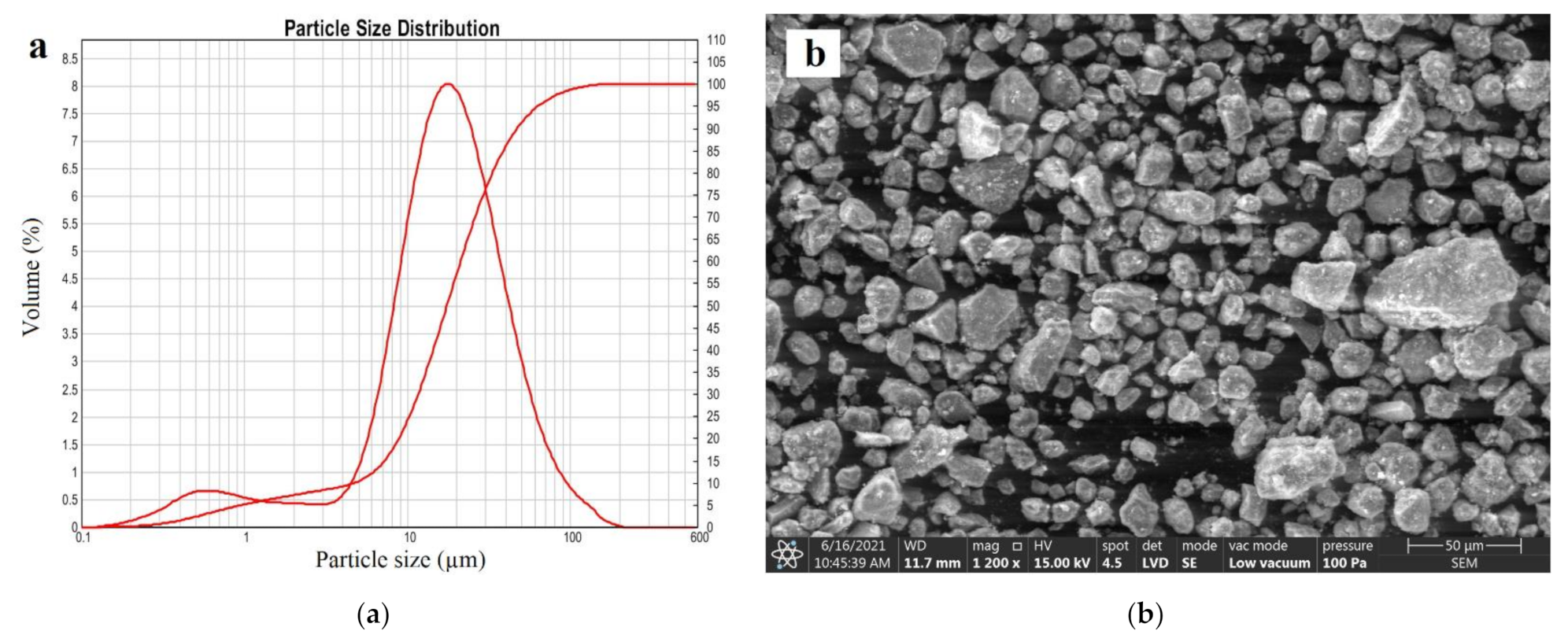
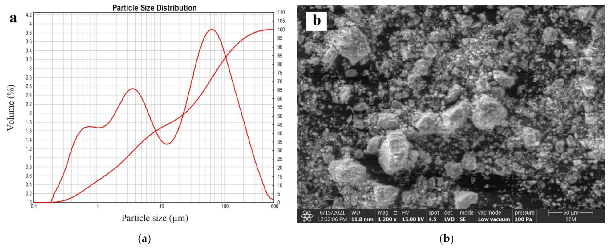
2.6. Particle Size Distribution

The particle size distribution of the powders was performed using the laser diffraction technique by the dry dispersion method and grains measurement according to ISO 13320 [48]. Tests were conducted by a Mastersizer 2000 Malvern device.

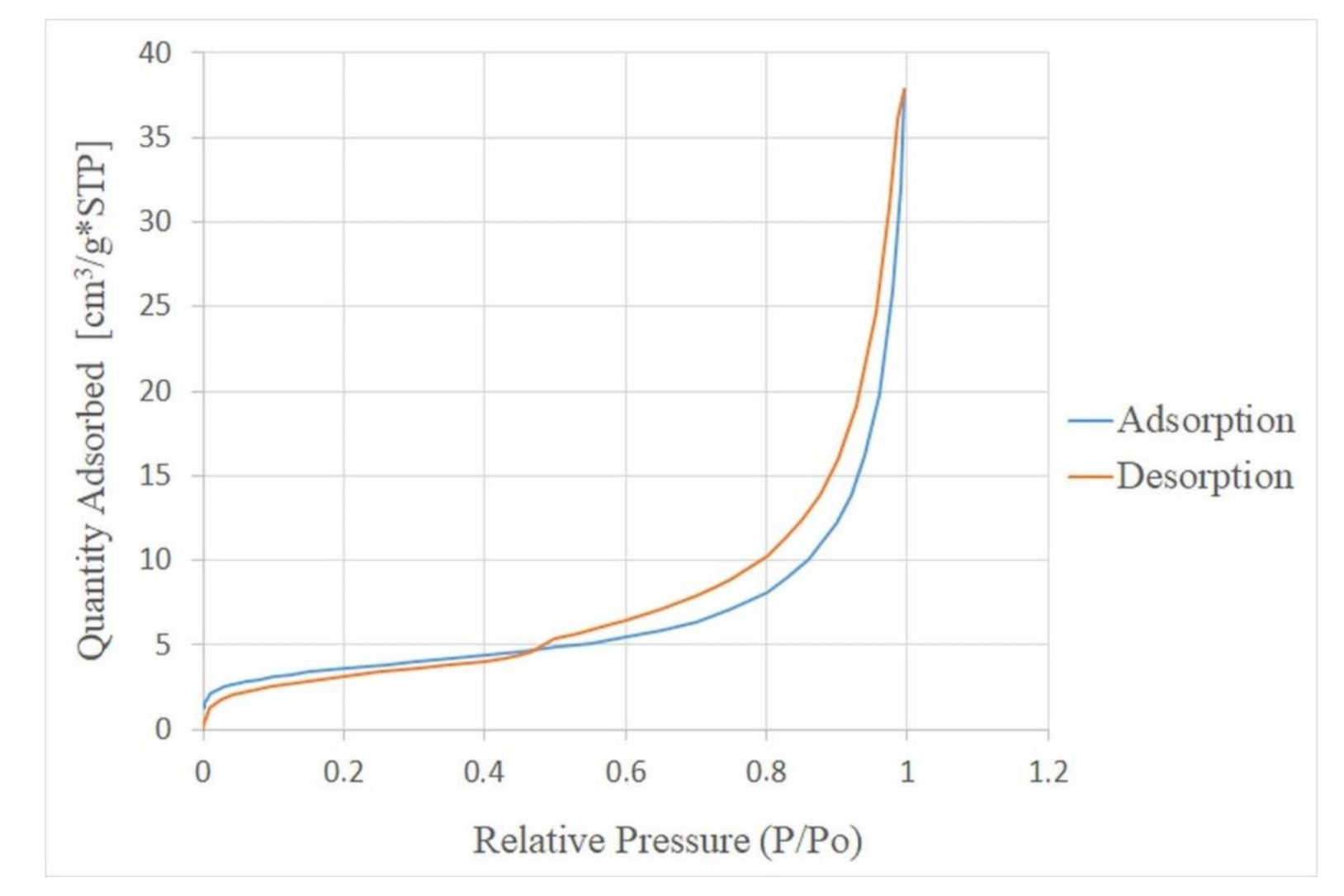
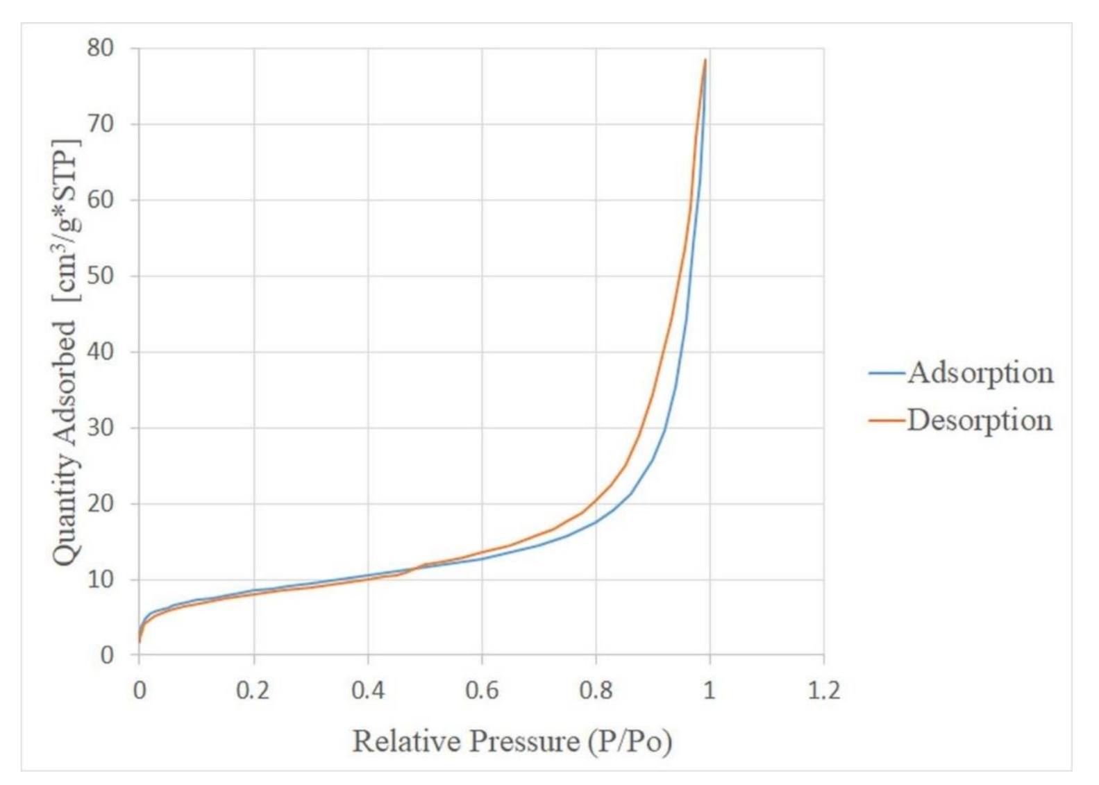
2.7. Textural and Physical Properties

The textural parameters of the clinoptilolite powders were determined by low-temperature nitrogen adsorption/desorption using an ASAP 2020 Micromeritic instrument at a relative vapor pressure from ≈10−3 to ≈0.99. The obtained adsorption data allowed determining such parameters as the specific surface area and porosity of the materials. Before performing the test, the samples were outgassed at the temperature of 200 °C to achieve a constant mass. The specific surface area (SBET) was calculated according to the Brunauer–Emmett–Teller (BET) method. The total pore volume (VTOT0.99) was evaluated using the t-plot method and Barretta–Joynera–Halendy (BJH). The area of micropores (Smic) was determined by using the t-plot method and the average diameter of pores (Ravg) on the basis of the BJH findings. Finally, in order to determine the macropore volume (Vmac), the following equation was adopted:
Vmac = Vtot0.99 − (VmicT + VmesBJH).

3. Results and Discussion

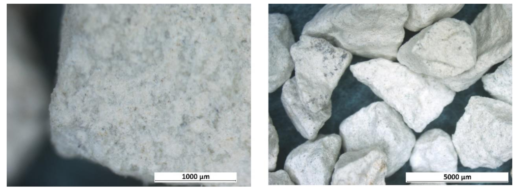
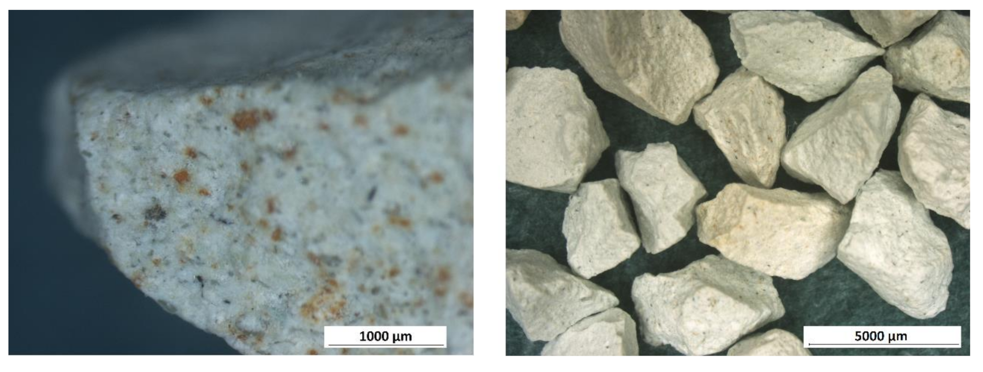
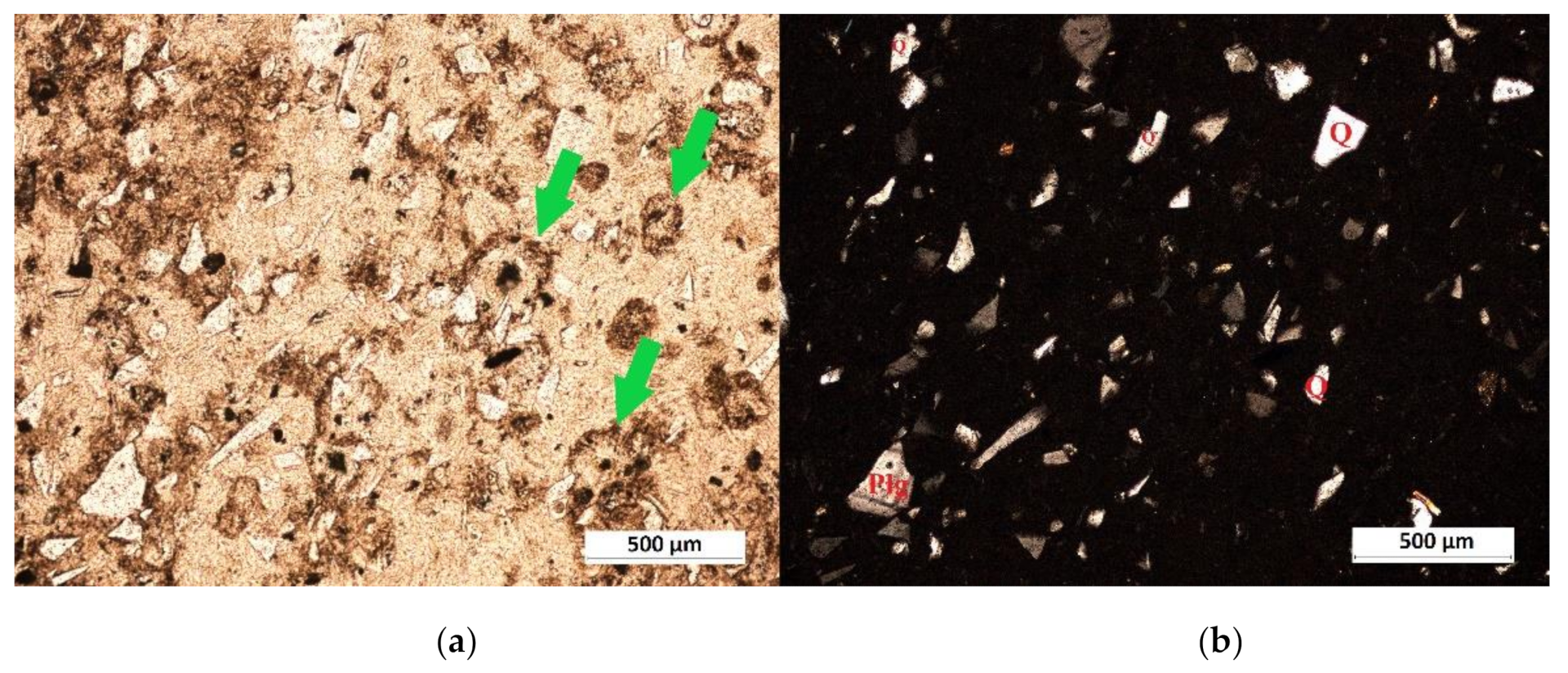
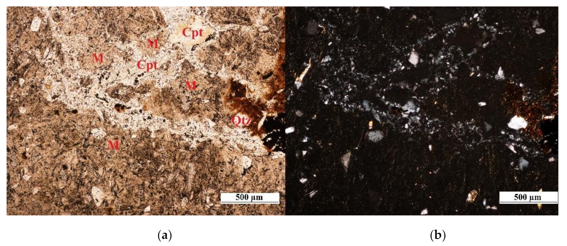
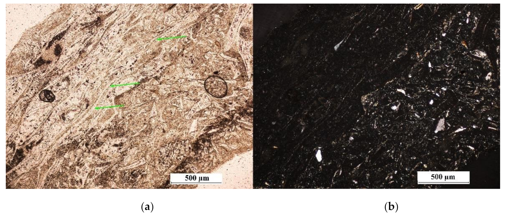
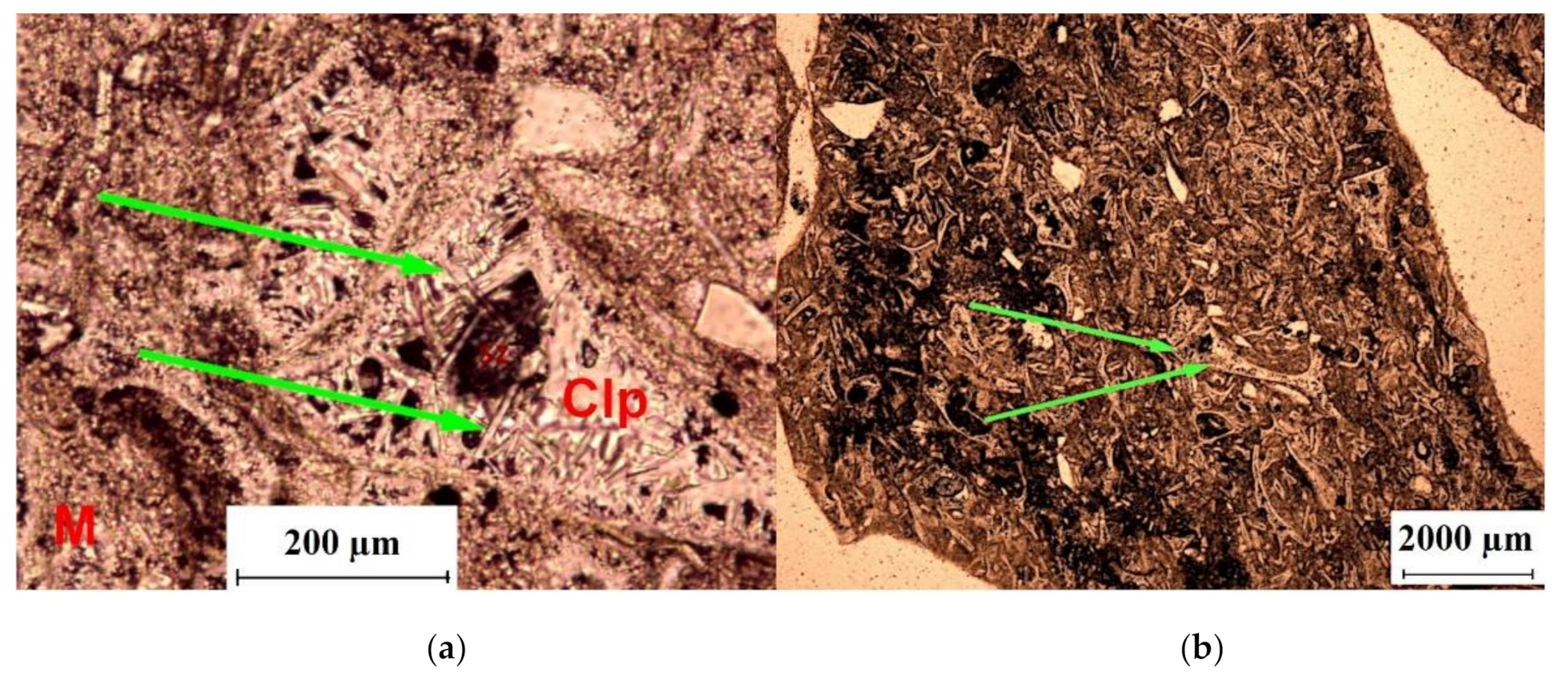
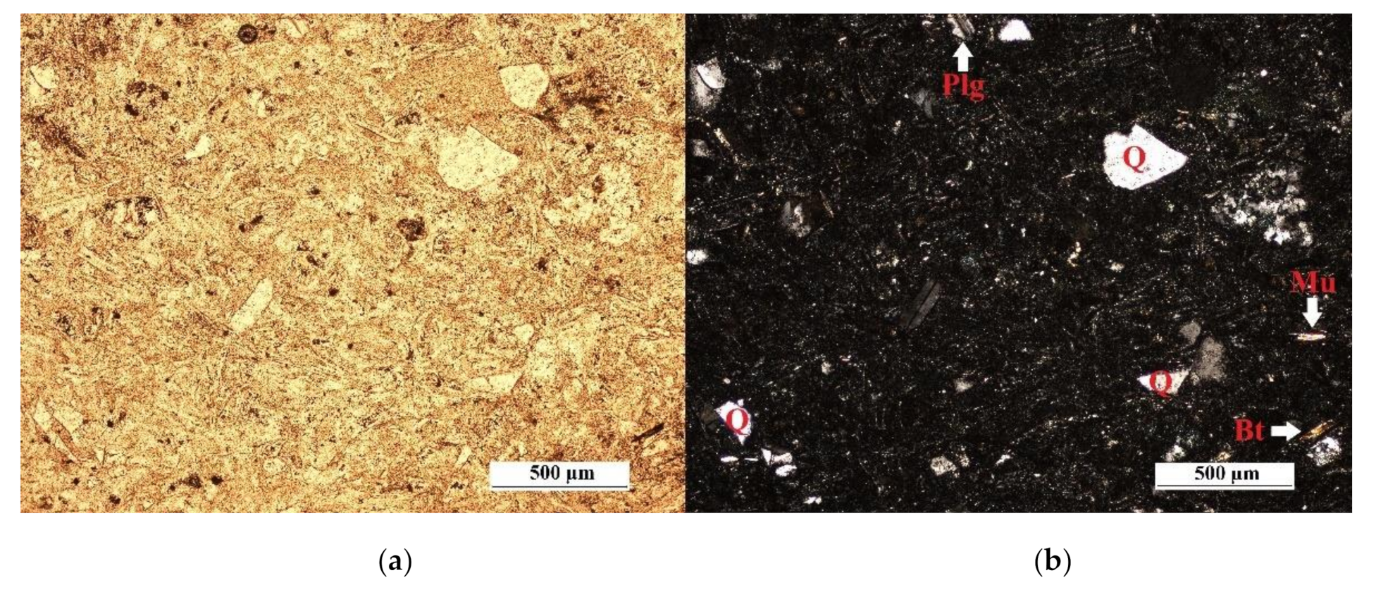
3.1. Macroscopic and Microscopic Analysis
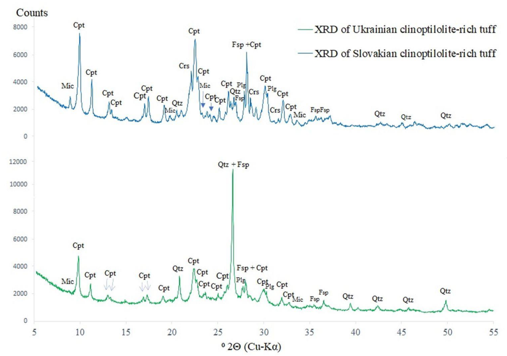
The investigated clinoptilolite-rich tuffs are light-colored with the shade of white for the Ukrainian tuff and green for the Slovakian (Figure 1 and Figure 2). The rocks are fine-grain with the massive and compact structure (Figure 1 and Figure 2). The Slovakian tuff can be marked out by the larger mineral assemblages disseminated through the rock (Figure 2). The macroscopic examinations have indicated that the most noticeable minerals in the Ukrainian tuff are quartz (with resinous luster) and iron oxides mineralization (fine orange spots) followed by feldspars, biotite (fine dark spots), and muscovite flakes (silver flakes).
By contrast, in the Slovakian tuff, the most noticeable mineral components are irone compounds and dark biotite flakes, which sometimes form the relatively large assemblages. The quartz mineralization in the Slovakian sample is scattered through the rock and far less abundant than in the Ukrainian tuff.
Petrographic examination under the polarizing microscope indicated that both studied tuffs are crystallo-vitroclastic rocks with the compact and slightly oriented structure. Primarily the major component of the clinoptilolite-rich tuffs was the volcanic glass, which has undergone a devitrification process. The range of the glass devitrification is distinct for the tuffs from different deposits. The Slovakian tuff is marked by significant glass devitrification. The bulk matrix of the rock comprised the mixture of the fine-crystalline clinoptilolite and feldspars. In the matrix, the globular vacuoles, which are the leftovers after the lava blister filled partially with ash and partially with fine-crystalline clinoptilolite, were identified (Figure 3). In addition, clinoptilolite in the Slovakian tuff is observed as the pseudomorphs after the glass shards. A bit larger plate-shaped clinoptilolite crystals with size up to 0.25 mm can be seen as an impregnation of the rock fractures (Figure 4). The mineral composition of Slovakian tuff based on the thin section examinations indicated that except for clinoptilolite (major mineral), feldspars, and plagioclases, the secondary minerals such as sharp-edged quartz, biotite laths, and iron mineralization assemblages can be found (Figure 3). The plagioclase crystalloclasts are up to 0.300 mm in size, and some of them show the zonal and polysynthetic twinnings. The quartz crystals are mainly fine crystalline below 0.100 mm in size, occasionally about a size of 0.200 mm. All of the quartz minerals are of pyroclastic origin. The crystalloclasts in the Slovakian tuff are well-maintained without any alternation features.
The thin section examinations of the Ukrainian tuff indicated that the range of the glass devitrification in the rock is significantly lower in contrast to the Slovakian tuff. The existing volcanic glass remains mainly are observed as the diversiform grains. In some parts of the rock, glass is shaping some kind of the undulatory laminars in the matrix, which is attributed to the glassy structure of the tuff (Figure 5). Moreover, in the Ukrainian tuff, the faunal remains filled partially with opal and partially with fine clinoptilolite plates in the range of 0.030–0.080 mm, and volcanic glass was identified as well (Figure 6). The remaining part of the Ukrainian tuff matrix includes very fine-crystalline clinoptilolite and feldspars with widespread pyroclastic edged-shape quartz crystalloclasts and polysynthetic plagioclases. The secondary mineral components are muscovite and biotite laths (Figure 7). The quartz crystalloclasts are fine crystalline and widespread in the rock matrix. Some of the quartz crystals can reach up to 0.300 mm. The plagioclases minerals are up to 0.200 mm in size, and some of them express polysynthetic twinnings. The mica laths can reach up to 0.150 mm. All of the crystalloclasts in the rock are well-maintained without any alteration features. The clinoptilolite mineralization in the tuffs is very fine crystalline, and it forms the rock matrix and the pseudomorphs after glass shards.
The obtained results correspond to the previous research of the Slovakian [19] and Ukrainian [49,50] clinoptilolite-rich tuffs. Moreover, the investigated tuffs exhibit strong similarity to the Miocene white tuffs from Romania [18] in respect to the macroscopic features and mineralogical composition, but they differ slightly in SiO2 and Al2O3 concentrations (Table 2). All of that can be attributed to the comparable formation conditions. In addition, macroscopically and geochemically, the Ukrainian and Slovakian tuffs show similarity to the Cenozoic tuffs from Central Sardinia, which were investigated by Mormone and Piochi [17] and the Neogenic tuffs from Western Turkey [51]. However, the petrographic examination under a polarizing microscope provided by researchers [17,51] indicated quite sharp distinctions in their mineral composition. In the Sardinian tuff, the main zeolite phase is mordenite accompanied by some smectite and glauconite, while in Turkish tuffs, the clinoptilolite mineralization is followed by some trace of the mordenite and smectite [17,51]. The above data proved that Slovakian and Ukrainian clinoptilolite-rich tuffs appear quite pure, contrary to the Sardinian and Turkish tuffs.
It is important to highlight that the comparative studies provided above apply to zeolite-rich tuffs belonging to the same stratigraphic period, which is Neogene. By contrast, the latest discovery of the new zeolitic deposit in Cuba derived from the Cretaceous and contains two zeolitic phases: mordenite and clinoptilolite [15].

3.2. XRD Analysis

The XRD analysis (Figure 8) indicated that in both studied tuffs, the main mineral phase is well-crystalized clinoptilolite identified based on the first major peak at 9.85° 2Θ and second major peak at 22.45° 2Θ. However, there is a quite sharp distinction in the clinoptilolite peaks intensities between Slovakian and Ukrainian rocks. The estimated clinoptilolite phase content for the Slovakian tuff was about 85 wt %, whereas for the Ukrainian one, it was about 70 wt %. It is in good agreement with the previous XRD analysis of the Slovakian tuff provided by Chmielewská et al. [52] and other researchers [21], whereas the XRD pattern of the sample from Ukraine correlates quite well with results demonstrated by Muir et al. [53]. Comparatively, the estimation of clinoptilolite phase content in investigated tuffs is close to some of the high-value global clinoptilolite-rich tuff deposits, as shown in Table 1. The results reported in Table 1 indicate that the U.S. state Nevada zeolite deposit contains the highest concentration of the clinoptilolite. However, according to the XRD analysis provided by KMI zeolite Inc. [16], the U.S. clinoptilolite is accompanied by trace amounts of montmorillonite, phlogopite, and Ca-stilbite, but it still it is of the highest level of purity in comparison to other naturally occurring zeolites (Table 1). In the case of Slovakian tuff, the clinoptilolite occurs in association with the trace amount of the cristobalite identified based on a major peak at 21.87° 2Θ and the K-feldspars with a major peak at 27.64° 2Θ. Other minor phases recognized in the Slovakian rock are mica group minerals (main peak at 8.82° 2Θ), quartz (main peak at 26.58° 2Θ), and plagioclases (main peak at 27.7° 2Θ).
Determination of the particular mica minerals based on XRD data is really difficult or even impossible because of the overlapping peaks. In order to provide the proper identification of the particular mica minerals, microscopy analysis is required. The XRD data and microscope observations enabled determining the presence of biotite in the Slovakian sample.
The XRD pattern of the Ukrainian tuff proved that except for the main phase, which is clinoptilolite, the sample includes minor phases such as quartz, K-feldspars, plagioclases, and mica group minerals. The microscopy observations indicated that minerals among the mica group are muscovite and biotite. The recorded data are in excellent conformity with mineralogical composition (XRD) of the Romanian white tuff [56], which implies the similar conditions of the clinoptilolite-rich tuffs crystallization.
The XRD patterns of Slovakian and Ukrainian clinoptilolite-rich tuffs (Figure 8) showed that the clinoptilolite content in Slovakian sample is about 15 wt % higher than in the Ukrainian one. In addition, there is no XRD evidence for cristobalite in the Ukrainian tuff compared to the Slovakian one. There is also the huge gap between Slovakian and Ukrainian tuffs with respect to the quartz content. It is clearly noticeable on the XRD patterns (Figure 8) that the quartz reflection (main peak at 26.58° 2Θ) is far more intense for the Ukrainian tuff compared to the quartz peak on the XRD spectrum of the Slovakian one. The slight enhancement of the background signal between 20° and 30° 2Θ in both Slovakian and Ukrainian tuffs indicates a trace of the amorphous phases associated with the remains of volcanic glass in the samples. The microscopy examination proved that phenomenon.

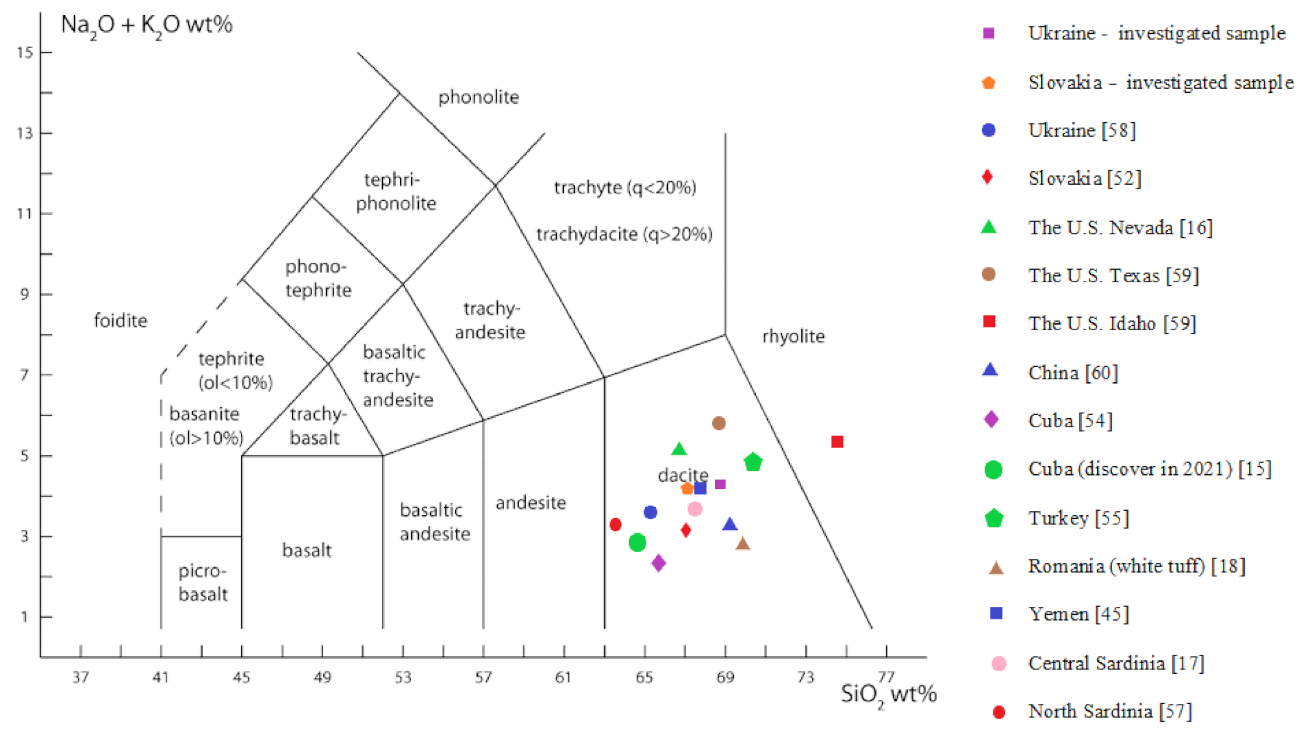
3.3. X-ray Fluorescence

Table 2 includes the chemical composition (XRF) of the investigated clinoptilolite-rich tuffs from Ukraine and Slovakia and, for the sake of comparison, an analysis of the various clinoptilolite-rich tuff deposits in the world. The Si/Al ratios listed in Table 2 were calculated on the basis of the bulk rocks XRF analysis. Due to this, the Si/Al ratios included Si and Al derived from many other rock minerals than clinoptilolite. Other Si and Al-bearing minerals are quartz, feldspars, plagioclases, mica group minerals, and clay minerals. The accurate data of the Si/Al ratio in the clinoptilolite structure can be provided by the EDS microanalysis, and it will be discussed later in the paper.
The obtained results show that the oxide composition of the Slovakian and Ukrainian samples is comparable. The SiO2 and Al2O3 are the main components, which is characteristic for zeolites. The exchangeable cations are Ca+2, Mg+2, Na+, and K+ with a prevalence of the last one. The Ukrainian clinoptilolite-rich tuff exhibits a bit higher Si/Al ratio in comparison to Slovakian clinoptilolite-rich tuff, which can impact the catalytic performance of zeolites. Taking into account that higher alumina content provides higher adsorption capacity [52], the Slovakian clinoptilolite-rich tuff will exhibit better adsorption properties than the Ukrainian one. Compared to the Ukrainian clinoptilolite-rich tuff studied by Ciężkowska et al. [58], the investigated Ukrainian one has a higher concentration of SiO2 and lower concentration of Al2O3. In addition, a decrease in the LOI value of the studied sample in comparison to the previous results [58] can be observed. Whereas the chemical data of the investigated Slovakian tuff are in coherence with the results provided by Chmielewská et al. [52].
Based on the collected data (Table 2), it can be concluded that there is no significant distinction in the chemical composition between various clinoptilolite-rich tuff deposits in the world and the studied samples. However, the clinoptilolite-rich tuff from the Idaho deposit has the highest SiO2 and quite high Al2O3 concentrations, which is in line with the highest Si/Al ratio in comparison to other reported clinoptilolite-rich tuff deposits (Table 2).
Figure 9 shows the chemical classification of the volcanic rocks (TAS diagram) with the clinoptilolite-rich tuffs from the most representative deposits in the world and investigated tuffs as well. According to the TAS diagram (Figure 9), all of the zeolitic tuffs except the tuff from the Idaho deposit are assigned to the dacite plot. The clinoptilolite-rich tuff from Idaho belongs to the rhyolitic one, displaying the most SiO2-enriched tuff.

3.4. Thermal Analysis

In Figure 10, the TG-DSC curves of the clinoptilolite-rich tuffs from the Ukrainian and Slovakian deposits are showed. The thermal analysis indicated in both cases the presence of a single, small but broad endothermal peak with the maximum at 120 °C and 140 °C for Slovakian and Ukrainian sample, respectively. The endothermal effects ended at 680 °C and 800 °C with mass loss of 9.48% and 9.87%. The fastest mass loss in samples can be observed from 65 to 300 °C, and it is associated with mass losses of about 6.84 wt % for the Slovakian and 6.97 wt % for the Ukrainian tuff. The endothermal effects visible for both tuffs are attributed to the dehydration of the zeolite minerals. It is compelling that the DSC curves for both studied zeolitic tuffs have not revealed any drastic transition in heat flow up to 1000 °C. It implies that the total structural decomposition of the examined clinoptilolite zeolites occurs at temperature over 1000 °C. However, it needs to be verified by the thermal analysis at temperature higher than 1000 °C and requires XRD analysis to detect any structural changes in clinoptilolite as the result of an amorphization.
The shape of the DSC curves indicates that the zeolite water in the studied samples is released step by step in the wide range of the temperature, which corresponds to the literature [59]. According to Franus et al. [61], that phenomenon can be associated with the mesoporous volume in the framework of the zeolite minerals. Korkuna et al. [62] state that the bond between the zeolitic water and the mineral framework is weaker. Due to the weaker zeolitic water bonding in the mineral framework, the water release temperature is relatively low: about 100 °C and from 100 to 200 °C. In addition, an endothermal effect with the maxima at 140 °C is visible on the Ukrainian tuff DSC curve, which corresponds to the literature data [63]. Breck [2] has proved that above 360 °C, the zeolite minerals undergo dihydroxylation. The dihydroxylation process in zeolite minerals is related to releasing the water, which exhibits a stronger bond in the zeolitic crystal framework.
The thermal behavior of the clinoptilolite-rich tuff from the Ukrainian deposit correlates quite well when compared to the previous study of the zeolitic tuff from the same source [50]. Chmielewská et al. [64] proved that the Slovakian clinoptilolite-rich tuff from the same deposit as the studied Slovakian sample begins to undergo a structural phase transition at the temperature around 900 °C.
Table 3 comprises data on the previous studies on the thermal stability of the clinoptilolite minerals from various deposits in the world as well as properties of the investigated samples. The thermal behavior of the zeolite minerals can be generalized, but the data presented in Table 3 have shown that depending on the source locations, a distinction in the temperature of the profound destabilization of the clinoptilolite crystal structure can appear. Based on the reported data (Table 3), the clinoptilolite structural decomposition temperature ranges from 450 to above 1000 °C. However, the collected data are just indicative because the evaluation methods used for determining the clinoptilolite structural decomposition in reported studies are different. The results in Table 3 show that the clinoptilolite-rich tuffs from Ukraine and Slovakia have a high and comparable to Cuban and Italian clinoptilolites thermal stability properties.

3.5. SEM Analysis

The SEM observations and EDS microanalysis enable determining a morphology of the clinoptilolite minerals in the studied samples and allow precisely defining the Si/Al ratios in the clinoptilolite minerals.
The clinoptilolite in both tuffs occurs in the form of thin plate-shaped (flaky) crystals, of sizes usually not exceeding 0.010 mm, which formed a tight mass (Figure 11 and Figure 12). The thinness of the clinoptilolite crystals is significantly noticeable in the Ukrainian tuff. The clinoptilolites thin plates create the close fit of the lamellar aggregates; rarely, they occur as individual plates. Moreover, the appearance of clinoptilolite crystals splitting is in agreement with the previous studies [50]. Sometimes, the clinoptilolite crystals in the Ukrainian tuff exhibit the distinct hexagonal shape (Figure 11a), which is consistent with the observation provided by Woszuk et al. [68]. The SEM images confirmed the previous statement reported by Sprynsky [50] on the hydrothermal origin of the clinoptilolite crystals in Ukrainian tuffs. The SEM observations of the Ukrainian and Slovakian clinoptilolite-rich tuffs imply a strong resemblance to the clinoptilolite crystals in Yemeni [45] and in Turkish tuffs [69].
The average of the Si/Al ratios determined from the particular microareas by means of EDS microanalysis indicated that the Si/Al ratio is 4.9 for the Ukrainian clinoptilolite and 4.0 for the Slovakian one. The obtained results are not compatible with the Si/Al ratios evaluated based on the bulk rocks XRF analysis (see Table 2). Moreover, Holub et al. [70] reported that the average Si/Al ratio for Slovakian clinoptilolite from the same deposit is 5.0. This can be explained by the natural variability in the clinoptilolite crystals within the deposit.
Taking into account that the maximum Si/Al ratio in natural zeolites is between 5 and 6, it can be pointed out that the clinoptilolite in the Ukrainian tuff shows relatively high value [3]. Chen et al. [60] state that the adsorption capacity of natural zeolites mainly depends on the Si/Al ratio and the specific surface area (SSA). Due to this fact, it is expected that Ukrainian zeolitic tuff will exhibit higher adsorption capacity than the Slovakian one.
For comparison, in Table 4, the previous Si/Al ratios (EDS analysis) of clinoptilolite crystals from various deposits in the world are presented. The data given in Table 4 prove that the Si/Al ratio for clinoptilolite crystals can vary considerably with respect to the deposit or even within it. The variability in Si/Al ratios is closely related to the different crystallization and hydrothermal conditions during crystals’ growth.

3.6. Particle Size Distribution

The analysis of particle size distribution (Figure 13 and Figure 14) of the studied samples showed that the clinoptilolite powder obtained from the Slovakian tuff has a grain size from about 0.2 to about 400 µm, while the clinoptilolite powder from the Ukrainian tuff is from about 0.2 to about 150 µm. However, the Slovakian clinoptilolite powder is significantly finer and more homogeneous in comparison to the Ukrainian one. The greater degree of the fragmentation of the Slovakian powder is indicated by the content of the grains size below 5 µm, which is about 35%, while in the Ukrainian clinoptilolite powder, the content of this fraction is just about 10%.
The Slovakian clinoptilolite particles are distributed tri-modally with the following peaks: from 0.6 to 1 µm, from 2 to 6 µm, and from 40 to 100 µm. The Ukrainian clinoptilolite powder shows the monomodal population curve with peaks from 10 to 20 µm; however, the grains from 10 to 30 µm dominated. The SEM observations of the powders obtained by mineral processing of the Slovakian and Ukrainian clinoptilolite tuffs confirmed the results of the grain distribution analysis by the laser method (Figure 13b and Figure 14b).
Moreover, the microarea observations of the Ukrainian clinoptilolite powder showed that regular particles with sharp edges dominated (Figure 13b), while in the Slovakian powder, the rounded particles are abundant (Figure 14b). In addition, the size distribution of the Slovakian powder is conducive to agglomerate, as seen in Figure 14b.
The mineral composition of the Ukrainian and Slovakian volcanic tuffs has a huge impact on the morphology of the studied clinoptilolite powders. The analysis of the mineral composition of the investigated tuffs indicated a significantly higher quartz content in the Ukrainian tuff compared to the Slovakian one. The silica impregnation in the Ukrainian tuff aggravates the disintegration during the mineral processing of the raw materials, causing greater wear of the working elements of the machines.
It is worth underlining that dusty materials are problematic when it comes to their management, and in most of the cases, such raw materials need to be compacted into larger grains. This also concerns the clinoptilolite powder, which is especially problematic when it comes to consolidation. Difficulty in the zeolite powder compaction is mainly related to its unique physicochemical properties, such as high alkaline pH, moisture swelling, and decreasing of sorption properties due to compaction processes with the binder application [71]. One of the factors influencing the material susceptibility to the compaction processes is its grain size [72,73,74]. The conducted studies of the morphology and particle size distribution of powders by means of laser diffraction indicated that Slovakian clinoptilolite powder will probably exhibit better susceptibility to consolidation processes due to the higher fine fractions content [73].

3.7. Textural and Physical Properties

The analysis of textural parameters (obtained by N2 adsorption/desorption analysis—Table 5) revealed that clinoptilolite powder obtained by mineral processing of the Slovakian clinoptilolite tuff has a better developed specific surface area and larger porosity in contrary to the clinoptilolite powder from Ukraine. The specific surface area (SBET) and total pore volume (Vtot0.99) are about two times higher than for the Slovakian clinoptilolite powder. The average pores size for the samples is quite similar, and it is about 10 nm.
With respect to the pore size distribution, both zeolitic powders can be classified as meso- and microporous, which make them the prospective raw materials for the organic and petroleum compounds adsorption [75]. The textural analysis showed relatively low micropore volume in the studied samples. It is about 3.4% and 2.5% for the Ukrainian and Slovakian materials, respectively. The samples exhibit a similar type of isotherm (Figure 15 and Figure 16), which is type II with some minimal features of type IV, and the hysteresis loop of type H3 according to the IUPAC classification [76]. The observed hysteresis loop corresponds to disordered slit-shaped pores, and it is typical for the aluminosilicates.

4. Conclusions

The XRD and petrographic analysis indicate that clinoptilolite is the only zeolite mineral in the studied rocks. Clinoptilolite crystallized mainly in the form of very fine-crystalline thin plates up to 0.010 mm in size; occasionally in the material from Slovakia, slightly larger crystals about 0.250 mm were observed.
The clinoptilolite tuffs have an oxide composition corresponding to the dacite plot, according to the TAS diagram. The Slovakian tuff is more abundant in clinoptilolite mineralization (about 15% higher clinoptilolite concentration in contrary to the Ukrainian tuff) and shows a higher degree of the volcanic glass devitrification and contains less impurities such as quartz minerals and no evidence of the faunal remains compared to the Ukrainian tuff. The above-mentioned differences in the mineral composition indicate different conditions of the petrogenetic processes of these rocks. The Ukrainian and Slovakian clinoptilolite-rich tuffs differ in terms of the Si/Al ratio and specific surface area. Compared the Slovakian one, Ukrainian clinoptilolite exhibits a higher value of the Si/Al. This implies the better adsorption capacity of Ukrainian zeolite. In contrast, the specific surface area obtained for both clinoptilolite-rich tuffs indicates the opposite.
The textural analysis of the clinoptilolite powders obtained by mineral processing of the studied tuffs showed that they are the meso-macropore materials. The morphology analysis together with the particle size distribution indicate that the Slovakian clinoptilolite powder probably will show a better susceptibility to compaction processes, due to its higher fine particle fractions content.
Based on the conducted comparative study of the most significant zeolite deposits in the world and examined clinoptilolite-rich tuffs, we can conclude that Ukrainian and Slovakian zeolites are comparable with many European zeolitic deposits, especially with the Romanian white tuff and the Turkish one. In addition, the estimation of clinoptilolite phase content in investigated tuffs is close to some high-value world clinoptilolite-rich tuff deposits such as the Cuban zeolitic commodity. Moreover, the thermal analysis results indicated that the clinoptilolite-rich tuffs from Ukraine and Slovakia have high thermal stability properties that are comparable to the Cuban and Italian clinoptilolites, which is crucial in many industrial applications.
We can see then that conducting the detailed mineralogical and structural characterization of zeolitic-bearing rocks is significantly important in order to assess the material behavior in the future technological processes. The results obtained in this study can be a useful indicator with respect to the crushing and compaction susceptibility of the Ukrainian and Slovakian clinoptilolite-rich tuffs.

Author Contributions

Conceptualization, E.P.-M. and T.G.; methodology, E.P.-M.; investigation, E.P.-M.; T.G. writing—original draft preparation, E.P.-M.; writing—review and editing, A.S.; visualization, A.S.; supervision, P.P. All authors have read and agreed to the published version of the manuscript.

Funding

This research is the result of the Implementation Doctorate Program IV edition for the years 2020–2024 financed by MEiN on the basis of the collaboration agreement no. DWD/4/28/2020 between AGH University of Science and Technology and Łukasiewicz Research Network Institute of Ceramics and Building Materials.

Data Availability Statement

The data presented in this study are available on request from the corresponding author.

Conflicts of Interest

The authors declare no conflict of interest.

References

  1. Auerbach, S.M.; Carrado, K.A.; Dutta, P.K. Handbook of Zeolite Science and Technology Copyright Year 2003; CRC Press: Boca Raton, FL, USA, 2003; ISBN 9780824740207. [Google Scholar]
  2. Breck, D.W. Zeolite Molecular Sieves: Structure, Chemistry and Use; John Wiley & Sons Inc.: New York, NY, USA, 1974. [Google Scholar]
  3. Ciciszwili, G.W.; Andronikaszwili, T.G.; Kirow, G.N.; Filizowa, D.Ł. Zeolity Naturalne; Wydawnictwa Naukowo-Techniczne: Warszawa, Poland, 1990. [Google Scholar]
  4. Mozgawa, W. Spektroskopia Oscylacyjna Zeolitów; Uczelniane Wydawnictwa Naukowo-Dydaktyczne AGH: Kraków, Poland, 2007. [Google Scholar]
  5. Mansouri, N.; Rikhtegar, N.; Panahi, S.H.; Atabi, F.; Shahraki, B.K. Porosity, characterization and structural properties of natural zeolite—Clinoptilolite—As a sorbent. Environ. Prot. Eng. 2013, 39, 139–152. [Google Scholar]
  6. Földvári, M. Handbook of Thermogravimetric System of Minerals and Its Use in Geological Practice; Geological Institute of Hungary: Budapest, Hungary, 2011; ISBN 978-963-671-288-4. [Google Scholar]
  7. Wieser, T. Zeolity—Kopaliny XXI wieku. Przegląd Geol. 1994, 9, 477–482. [Google Scholar]
  8. De Souza, V.C.; Villarroel-Rocha, J.; De Araújo, M.J.G.; Sapag, K.; Pergher, S.B.C. Basic Treatment in Natural Clinoptilolite for Improvement of Physicochemical Properties. Minerals 2018, 8, 595. [Google Scholar] [CrossRef] [Green Version]
  9. Boles, J.R. Composition, optical properties, cell dimensions and thermal Stability of some heulandite group zeolites. Am. Mineral. 1972, 57, 1463–1493. [Google Scholar]
  10. Koyama, K.; Takeuchi, Y. Clinoptilolite: The distribution of potassium atoms and its role in thermal stability. Z. Kristall. 1977, 145, 216–239. [Google Scholar] [CrossRef]
  11. Król, M. Natural vs. Synthetic zeolites. Crystals 2020, 10, 622. [Google Scholar] [CrossRef]
  12. Manecki, A.; Muszyński, M. Przewodnik do Petrografii; Uczelniane Wydawnictwa Naukowo-Dydaktyczne AGH: Kraków, Poland, 2008. [Google Scholar]
  13. Mastinu, A.; Kumar, A.; Maccarinelli, G.; Bonini, S.A.; Premoli, M.; Aria, F.; Gianoncelli, A.; Memo, M. Zeolite Clinoptilolite: Therapeutic Virtues of an Ancient Mineral. Molecules 2019, 24, 1517. [Google Scholar] [CrossRef] [Green Version]
  14. Flanagan, D.M. Zeolites (Natural). Miner. Commod. Summ. 2020, 188–189. [Google Scholar]
  15. Costafreda, J.L.; Martín, D.A. New Deposit of Mordenite–Clinoptilolite in the Eastern Region of Cuba: Uses as Pozzolans. Molecules 2021, 26, 4676. [Google Scholar] [CrossRef] [PubMed]
  16. Product Data Sheet; KMI Zeolite Inc.: Amargosa Valley, NV, USA; Available online: https://www.kmizeolite.com/technical-data/ (accessed on 5 August 2021).
  17. Mormone, A.; Piochi, M. Mineralogy, Geochemistry and Genesis of Zeolites in Cenozoic Pyroclastic Flows from the Asuni Area (Central Sardinia, Italy). Minerals 2020, 10, 268. [Google Scholar] [CrossRef] [Green Version]
  18. Bojar, A.-V.; Barbu, V.; Wojtowicz, A.; Bojar, H.-P.; Hałas, S.; Duliu, O.G. Miocene Slănic Tuff, Eastern Carpathians, Romania, in the Context of Badenian Salinity Crisis. Geosciences 2018, 8, 73. [Google Scholar] [CrossRef] [Green Version]
  19. Tschegg, C.; Rice, A.H.N.; Grasemann, B.; Matiasek, E.; Kobulej, P.; Dzivák, M.; Berger, T. Petrogenesis of a Large-Scale Miocene Zeolite Tuff in the Eastern Slovak Republic: The Nižný Hrabovec Open-Pit Clinoptilolite Mine. Econ. Geol. 2019, 114, 1177–1194. [Google Scholar] [CrossRef] [Green Version]
  20. Mondale, K.D.; Carland, R.M.; Aplan, F.F. The comparative ion exchange capacities of natural sedimentary and synthetic zeolites. Miner. Eng. 1995, 8, 535–548. [Google Scholar] [CrossRef]
  21. Barrer, R.M.; White, E.A.D. The hydrothermal chemistry of silicates. Part II. Synthetic crystalline sodium aluminosilicate. J. Chem. Soc. 1952, 286, 1561–1571. [Google Scholar] [CrossRef]
  22. Takeda, H.; Hashimoto, S.; Yokoyama, H.; Iwamoto, Y.; Iwamoto, Y. Characterization of zeolite in zeolite-geopolymer hybrid bulk materials derived from kaolinitic clays. Materials 2013, 6, 1767–1778. [Google Scholar] [CrossRef] [PubMed] [Green Version]
  23. Bandura, L.; Franus, M.; Józefaciuk, G.; Franus, W. Synthetic zeolites from fly ash as effective mineral sorbents for land-based petroleum spills cleanup. Fuel 2015, 147, 100–107. [Google Scholar] [CrossRef]
  24. Rożek, P.; Król, M.; Mozgawa, W. Geopolymer-zeolite composites: A review. J. Clean. Prod. 2019, 230, 557–579. [Google Scholar] [CrossRef]
  25. Wojciechowska, K.M.; Król, M.; Bajda, T.; Mozgawa, W. Sorption of heavy metal cations on mesoporous ZSM-5 and mordenite zeolites. Materials 2019, 12, 3271. [Google Scholar] [CrossRef] [Green Version]
  26. Bohács, K.; Faitli, J.; Bokányi, L.; Mucsi, G. Control of Natural Zeolite Properties by Mechanical Activation in Stirred Media Mill. Arch. Metall. Mater. 2017, 62, 1399–1406. [Google Scholar] [CrossRef] [Green Version]
  27. Sivashankari, L.; Rajkishore, S.K.; Lakshmanan, A.; Subramanian, K.S. Optimization of Dry Milling Process for Synthesizing Nano Zeolites. Int. J. Chem. Stud. 2019, 7, 328–333. [Google Scholar]
  28. Şükrü, U.; Musa, S.; Soner, T.; İrfan, T. Removal of heavy metals from wastewater solution using a mechanically activated novel zeolitic material. J. Min. Sci. 2020, 56, 1010–1023. [Google Scholar]
  29. Wakihara, T.; Ichikawa, R.; Tatami, J.; Endo, A.; Yoshida, K.; Sasaki, Y. Bead-Milling and Postmilling Recrystallization: An Organic Template-free Methodology for the Production of Nano-zeolites. Cryst. Growth Des. 2011, 11, 955–958. [Google Scholar] [CrossRef]
  30. Ejhieh, A.N.; Khorsandi, S. Photocatalytic degradation of 4-nitrophenol with ZnO supported nano-clinoptilolite zeolite. J. Ind. Eng. Chem. 2014, 20, 937–946. [Google Scholar] [CrossRef]
  31. Zolzaya, T.; Davaabal, B.; Ochirbat, Z.; Oyun-Erdene, G.; Min-jigmaa, A.; Temuujin, J. The mechanochemical activation study of Tsagaan-tsav zeolite. Mong. J. Chem. 2011, 12, 98–101. [Google Scholar] [CrossRef] [Green Version]
  32. Nikashina, V.; Streletskii, A.; Kolbanev, I.; Meshkova, I.; Grinev, V.; Serova, I.; Yusupov, T.; Shumskaya, L. Effect of mechanical activation on the properties of natural zeolites. Inorg. Mater. 2011, 47, 1341–1346. [Google Scholar] [CrossRef]
  33. Silva, M.R.; Lecus, A.; Gajdardziska-Josifovska, M.; Schofield, M.; Virnoche, M.; Chang, J.; Chen, J.; Garman, D. Graphene-oxide loading on natural zeolite particles for enhancement of adsorption properties. RSC Adv. 2020, 10, 4589–4597. [Google Scholar] [CrossRef] [Green Version]
  34. Kang, J.; Singh, B.K.; Um, W. Efficient radon removal using fluorine-functionalized natural zeolite. J. Environ. Radioact. 2021, 233, 106607. [Google Scholar] [CrossRef]
  35. Wirtu, Y.D.; Melak, F.; Yitbarek, M.; Astatkie, H. Aluminum Coated Natural Zeolite for Water Defluoridation: A Mechanistic Insight; Elsevier: Amsterdam, The Netherlands, 2021; Volume 12. [Google Scholar]
  36. Reeve, P.J.; Fallowfield, H.J. Natural and surfactant modified zeolites: A review of their applications for water remediation with a focus on surfactant desorption and toxicity towards microorganisms. J. Environ. Manag. 2018, 205, 253–261. [Google Scholar] [CrossRef]
  37. Donkor, E.A.; Buamah, R.; Kwakye-Awuah, B. Defluorination of drinking water using surfactant modified zeolites. J. Sci. Technol. 2016, 36, 15–21. [Google Scholar] [CrossRef] [Green Version]
  38. Bowman, R.S. Applications of surfactant-modified zeolites to environmental remediation. Microporous Mesoporous Mater. 2003, 61, 43–56. [Google Scholar] [CrossRef]
  39. Xie, Q.; Xie, J.; Wang, Z.; Wu, D.; Zhang, Z.; Kong, H. Adsorption of organic pollutants by surfactant modified zeolite as controlled by surfactant chain length. Microporous Mesoporous Mater. 2013, 179, 144–150. [Google Scholar] [CrossRef]
  40. Naghash, A.; Nezamzadeh-Ejhieh, A. Comparison of the efficiency of modified clinoptilolite with HDTMA and HDP surfactants for the removal of phosphate in aqueous solutions. J. Ind. Eng. Chem. 2015, 31, 185–191. [Google Scholar] [CrossRef]
  41. Tran, H.N.; Viet, P.V.; Chao, H.P. Surfactant modified zeolite as amphiphilic and dual-electronic adsorbent for removal of cationic and oxyanionic metal ions and organic compounds. Ecotoxicol. Environ. Saf. 2018, 147, 55–63. [Google Scholar] [CrossRef] [PubMed]
  42. Sullivan, E.J.; Hunter, D.B.; Bowman, R.S. Fourier transform Raman spectroscopy of sorbed HDTMA and the mechanism of chromate sorption to surfactant-modified clinoptilolite. Environ. Sci. Technol. 1998, 32, 1948–1955. [Google Scholar] [CrossRef]
  43. Al-Mashaqbeh, O.A.; Alsafadi, D.A.; Alsalhi, L.Z.; Bartelt-Hunt, S.L.; Snow, D.D. Removal of Carbamazepine onto Modified Zeolitic Tuff in Different Water Matrices: Batch and Continuous Flow Experiments. Water 2021, 13, 1084. [Google Scholar] [CrossRef]
  44. Fuss, V.L.B.; Bruj, G.; Dordai, L.; Roman, M.; Cadar, O.; Becze, A. Evaluation of the Impact of Different Natural Zeolite Treatments on the Capacity of Eliminating/Reducing Odors and Toxic Compounds. Materials 2021, 14, 3724. [Google Scholar] [CrossRef]
  45. Alshameri, A.; Xinghu, W.; Dawood, A.S.; Xin, C.; Yan, C.; Assabri, A.M. Characterization of Yemeni Natural Zeolite (Al-Ahyuq Area) and its Environment Applications: A Review. J. Ecol. Eng. 2019, 20, 157–166. [Google Scholar] [CrossRef]
  46. Tschegg, C.; Hou, Z.; Rice, A.H.N.; Fendrych, J.; Matiasek, E.; Berger, T.; Grasemann, B. Fault zone structures and strain localization in clinoptilolite-tuff (Nižný Hrabovec, Slovak Republic). J. Struct. Geol. 2020, 138, 104090. [Google Scholar] [CrossRef]
  47. Le Maitre, R.W.; Bateman, P.; Dudek, A.; Keller, J.; Lameyre, J.; Le Bas, M.J.; Sabine, P.A.; Schmid, R.; Sorensen, H.; Strekeisen, A.; et al. A Classification of Igneous Rocks and Glossary of Terms: Recommendations of the InternationalUnion of Geological Sciences, Subcommission on the Systematics of Igneous Rocks. Int. Union Geol. Sci. 1989, 552, 193. [Google Scholar]
  48. ISO 13320 Particle Size Analysis—Laser Diffraction Methods; The International Organization for Standarization: Geneva, Switzerland, 2020.
  49. Vasylechko, V.O.; Gryshchouk, G.V.; Kuz’ma, Y.B.; Zakordonskiy, V.P.; Vasylechko, L.O.; Lebedynets, L.O.; Kalytovs’ka, M.B. Adsorption of cadmium on acid-modified Transcarpathian clinoptilolite. Microporous Mesoporous Mater. 2003, 60, 183–196. [Google Scholar] [CrossRef]
  50. Sprynsky, M.; Golembiewski, R.; Trykowski, G.; Buszewski, B. Heterogeneity and hierarchy of clinoptilolite porosity. J. Phys. Chem. Solids 2010, 71, 1269–1277. [Google Scholar] [CrossRef]
  51. Kaçmaz, H. Mineralogical and petrographic study of zeolitic tuffs from Yenice-Saraycık (Demirci, Manisa), Turkey. Muhendis. Fak. Muhendis. 2018, 20, 647–653. [Google Scholar] [CrossRef]
  52. Chmielewská, E.; Tylus, W.; Drábik, M.; Majzlan, J.; Kravčak, J.; Williams, C.; Čaplovičová, M.; Čaplovič, L. Structure investigation of nano-FeO(OH) modified clinoptilolite tuff for antimony removal. Microporous Mesoporous Mater. 2017, 248, 222. [Google Scholar] [CrossRef]
  53. Muir, B.; Matusik, J.; Bajda, T. New insights into alkylammonium-functionalized clinoptilolite and Na-P1 zeolite: Structural and textural features. Appl. Surf. Sci. 2016, 361, 242–250. [Google Scholar] [CrossRef]
  54. Cervini-Silva, J.; Nieto-Camacho, A.; Kaufhold, S.; Ufer, K.; Palacios, E.; Montoya, A.; Dathe, W. Antiphlogistic effect by zeolite as determined by a murine inflammation model. Microporous Mesoporous Mater. 2016, 228, 207–214. [Google Scholar] [CrossRef]
  55. de Gennaro, B.; Aprea, P.; Liguori, B.; Galzerano, B.; Peluso, A.; Caputo, D. Zeolite-Rich Composite Materials for Environmental Remediation: Arsenic Removal from Water. Appl. Sci. 2020, 10, 6939. [Google Scholar] [CrossRef]
  56. Kalebić, B.; Pavlović, J.; Dikić, J.; Rečnik, A.; Gyergyek, S.; Škoro, N.; Rajić, N. Use of Natural Clinoptilolite in the Preparation of an Efficient Adsorbent for Ciprofloxacin Removal from Aqueous Media. Minerals 2021, 11, 518. [Google Scholar] [CrossRef]
  57. Castaldi, P.; Santona, L.; Cozza, C.; Giuliano, V.; Abbruzzese, C.; Nastro, V.; Melisa, P. Thermal and spectroscopic studies of zeolites exchanged with metal cations. J. Mol. Struct. 2005, 734, 99–105. [Google Scholar] [CrossRef]
  58. Ciezkowska, M.; Bajda, T.; Decewicz, P.; Dziewit, L.; Drewniak, L. Effect of Clinoptilolite and Halloysite Addition on Biogas Production and Microbial Community Structure during Anaerobic Digestion. Materials 2020, 13, 4127. [Google Scholar] [CrossRef]
  59. Burris, L.E.; Juenger, M.C.G. Effect of calcination on the reactivity of natural clinoptilolite zeolites used as supplementary cementitious materials. Constr. Build. Mater. 2020, 258, 119988. [Google Scholar] [CrossRef]
  60. Chen, H.-F.; Lin, Y.-J.; Chen, B.-H.; Yoshiyuki, I.; Liou, S.Y.-H.; Huang, R.-T. A Further Investigation of NH4+ Removal Mechanisms by Using Natural and Synthetic Zeolites in Different Concentrations and Temperatures. Minerals 2018, 8, 499. [Google Scholar] [CrossRef] [Green Version]
  61. Franus, W.; Woszuk, A.; Bandura, L.; Zofka, A. Właściwości zeolitu naturalnego i syntetycznego oraz ich wpływ na efekt spienienia asfaltu. Mater. Bud. 2017, 8, 60–64. [Google Scholar] [CrossRef]
  62. Korkuna, O.; Leboda, R.; Skubiszewska-Ziemba, J.; Vrublevska, T.; Gunko, V.M.; Ryczkowski, J. Structural and physicochemical properties of natural zeolites: Clinoptilolite and mordenite. Microporous Mesoporous Mater. 2006, 87, 243–254. [Google Scholar] [CrossRef]
  63. Moreno, P.V.; Arellano, J.J.C.; Ramırez, H.B. Characterization and preparation of porous membranes with a natural Mexican zeolite. J. Phys. Condens. Matter 2004, 16, 2345–2352. [Google Scholar] [CrossRef]
  64. Chmielewská, E.; Tylus, W.; Bujdoš, M. Study of Mono- and Bimetallic Fe and Mn Oxide-Supported Clinoptilolite for Improved Pb(II) Removal. Molecules 2021, 26, 4143. [Google Scholar] [CrossRef]
  65. Duvarcı, Ö.Ç.; Akdeniz, Y.; Özmıhçı, F.; Ülkü, S.; Balköse, D.; Çiftçioğlu, M. Thermal behaviour of a zeolitic tuff. Ceram. Int. 2007, 33, 795–801. [Google Scholar] [CrossRef] [Green Version]
  66. Stocker, K.; Ellersdorfer, M.; Lehner, M.; Raith, J.G. Characterization and Utilization of Natural Zeolites in Technical Applications. Berg. Huettenmaenn Monatsh 2017, 162, 142–147. [Google Scholar] [CrossRef]
  67. Donati, E.; Polcaro, C.M.; Ciccioli, P.; Galli, E. The comparative study of a laccase-natural clinoptilolite-based catalyst activity and free laccase activity on model compounds. J. Hazard. Mater. 2015, 289, 83–90. [Google Scholar] [CrossRef] [PubMed]
  68. Woszuk, A.; Wróbel, M.; Franus, W. Influence of Waste Engine Oil Addition on the Properties of Zeolite-Foamed Asphalt. Materials 2019, 12, 2265. [Google Scholar] [CrossRef] [PubMed] [Green Version]
  69. Güngör, D.; Özen, S. Development and characterization of clinoptilolite-, mordenite-, and analcime-based geopolymers: A comparative study. Case Stud. Constr. Mater. 2021, 15, e00576. [Google Scholar] [CrossRef]
  70. Holub, M.; Balintova, M.; Demcak, S.; Hurakova, M. Characterization of Natural Zeolite and Determination Its Adsorption Properties. Czas. Inżynierii Lądowej Sr. Archit. 2016, 63, 113–122. [Google Scholar] [CrossRef] [Green Version]
  71. Czuma, N.; Panek, R.; Baran, P.; Zarębska, K. The influence of binders for the pelletization of fly ash zeolites on sulfur dioxide sorption properties. Clay Miner. 2020, 55, 40–47. [Google Scholar] [CrossRef]
  72. Bembenek, M.; Hryniewicz, M. Badania i Opracowanie Metody Doboru Układu Zagęszczania Prasy Walcowej; Wydawnictwo AGH: Kraków, Poland, 2010. [Google Scholar]
  73. Kłassiena, P.W.; Griszajew, I.G. Podstawy Techniki Granulacji; Wydawnictwa Naukowo-Techniczne: Warszawa, Poland, 1989. [Google Scholar]
  74. Izak, P. Reologia w Ceramice, Wydanie Drugie Uzupełnione; Wydawnictwo AGH: Kraków, Poland, 2017. [Google Scholar]
  75. Muir, B.; Bajda, T. Organically modified zeolites in petroleum compounds spill cleanup—Production, efficiency, utilization. Fuel Process. Technol. 2016, 149, 153–162. [Google Scholar] [CrossRef]
  76. Thommes, M.; Kaneko, K.; Neimark, A.V.; Olivier, J.P.; Rodriguez-Reinoso, F.; Rouquerol, J.; Sing, K.S.W. Sing Physisorption of gases, with special reference to the evaluation of surface area and pore size distribution (IUPAC Technical Report). Pure Appl. Chem. 2015, 87, 1051–1069. [Google Scholar] [CrossRef] [Green Version]
Figure 1. Ukrainian clinoptilolite-rich tuff (stereoscopic microscope image).
Minerals 11 01030 g001
Figure 2. Slovakian clinoptilolite-rich tuff (stereoscopic microscope image).
Minerals 11 01030 g002
Figure 3. Slovakian clinoptilolite-rich tuff. Spherical vacuoles (green arrows) and scattered crystalloclasts (Q—quartz, Plg—plagioclase) in the rock matrix. Optical microscope, polarized transmitted light–PPL image (a). Optical microscope, polarized transmitted light–XPL image (b).
Minerals 11 01030 g003
Figure 4. Slovakian clinoptilolite-rich tuff. The larger clinoptilolite (Cpt) crystals in fine-crystalline rock matrix (M), quartz (Qtz). Optical microscope, polarized transmitted light–PPL image (a). Optical microscope, polarized transmitted light–XPL image (b).
Minerals 11 01030 g004
Figure 5. Ukrainian clinoptilolite-rich tuff. The remains of the volcanic glass in the form of a wavy laminated matrix (green arrows). Optical microscope, polarized transmitted light–PPL image (a). Optical microscope, polarized transmitted light–XPL image (b).
Minerals 11 01030 g005
Figure 6. Ukrainian clinoptilolite-rich tuff. Faunal remains (green arrows) filled partially with clinoptilolite mineralization (Clp) and partially with volcanic glass (sz); in the background, the rock matrix (M). Optical microscope, polarized transmitted light–PPL images (a,b).
Minerals 11 01030 g006
Figure 7. Ukrainian clinoptilolite-rich tuff. Crystalloclasts (Q—quartz, Plg—plagioclase, Mu—muscovite, Bt—biotite) scattered in the fine-crystalline rock matrix. Optical microscope, polarized transmitted light–PPL image (a). Optical microscope, polarized transmitted light–XPL image (b).
Minerals 11 01030 g007
Figure 8. XRD patterns of clinoptilolite-rich tuffs from Slovakia and Ukraine. (Cpt—clinoptilolite, Mic—mica group minerals, Qtz—quartz, Fsp—K-feldspar, Plg—plagioclase, Crs—cristobalite).
Minerals 11 01030 g008
Figure 9. The chemical classification of the clinoptilolite-rich tuffs from the most representative deposits in the world and the investigated tuffs according to the TAS diagram [47].
Minerals 11 01030 g009
Figure 10. Thermal analysis (TG-DSC) of the clinoptilolite-rich tuffs from Ukraine and Slovakia.
Minerals 11 01030 g010
Figure 11. SEM image of the clinoptilolite in Ukrainian tuff (EDS spots—red forks) (a) its EDS microanalysis (b).
Minerals 11 01030 g011
Figure 12. SEM image of the clinoptilolite in Slovakian tuff (EDS spots—red forks) (a) and its EDS microanalysis (b).
Minerals 11 01030 g012
Figure 13. Particle size distribution of the clinoptilolite powder obtained by mineral processing of the Ukrainian tuff (a); SEM images of the particles morphology (b).
Minerals 11 01030 g013
Figure 14. Particle size distribution of the clinoptilolite powder obtained by mineral processing of the Slovakian tuff (a); SEM images of the particles morphology (b).
Minerals 11 01030 g014
Figure 15. N2 adsorption/desorption isotherms for the Ukrainian clinoptilolite powder.
Minerals 11 01030 g015
Figure 16. N2 adsorption/desorption isotherms for Slovakian clinoptilolite powder.
Minerals 11 01030 g016
Table 1. The clinoptilolite content based on the Rietveld estimation in various zeolite deposits in the world.
DepositType of Clinoptilolite-Bearing RockClinoptilolite Content (wt %)
Ukraine-investigated sampleVolcanic tuff≈70
Slovakia-investigated sampleVolcanic tuff≈85
Slovakia [52]Volcanic tuff70 to 85
Ukraine [53]Volcanic tuff≈75
The U.S. Nevada [16]Volcanic tuff97
Cuba [54]Volcanic tuff65
Turkey [55]Volcanic tuff79
Yemen [45]Volcanic tuff68 to 72
Serbia [56]Volcanic tuff>80
North Sardinia [57]Volcanic tuff63
Table 2. The chemical composition of the various clinoptilolite-rich tuff deposits in the world.
DepositChemical Composition (wt %)Si/Al
Ratio 2
SiO2Al2O3Fe2O3CaOMgOK2ONa2OTiO2LOI 1
Ukraine-investigated sample68.5711.771.952.520.743.321.160.159.675.10
Slovakia-investigated sample67.1512.391.433.360.563.630.950.1710.274.90
Ukraine [58]65.2412.581.853.240.782.880.640.1812.23- 3
Slovakia [52]67.1612.302.302.911.102.280.660.1710.90- 3
The U.S. Nevada [16]66.7011.480.901.330.273.421.800.1313.955.80
The U.S Texas [59]68.0713.592.437.971.122.693.30- 3- 35.01
The U.S. Idaho [59]75.0412.852.383.480.804.860.50- 3- 35.84
China [60]69.1412.82- 33.700.051.941.58- 3- 34.58
Cuba [54]65.3011.201.602.601.101.061.500.2014.70- 3
Cuba (Discover in 2021) [15]64.6912.611.622.590.552.010.82- 313.164.62
Turkey [55]69.7111.741.212.300.314.410.76- 312.80- 3
Romania (white tuff) [18]70.7210.17- 34.260.672.140.820.1811.47- 3
Yemen [45]67.2011.692.772.120.413.670.90- 310.23- 3
Central Sardinia [17]67.2711.661.972.820.972.910.930.3910.90- 3
North Sardinia [57]63.3612.90- 33.101.292.161.390- 315.714.17
1 LOI: loss of ignition; 2 Si/Al ratio is the molar ratio of Si to Al based on the bulk rocks XRF analysis; 3 data not available.
Table 3. The thermal behavior of the clinoptilolite minerals from various deposits around the world.
Clinoptilolite Source LocationsEstimated *
Clinoptilolite Structural Decomposition Temperature (°C)
Ukraine-investigated sample>1000
Slovakia-investigated sample>1000
The U.S. Nevada [16]700
The U.S. Texas [59]965
The U.S. Idaho [59]965
Cuba [8]>1000
The U.K. [59]>920
Turkey [65]800
Austria [66]720
North Sardinia [57]450–500
Italy (Modena) [67]>1100
* Based on the different evaluation methods.
Table 4. The Si/Al ratios (EDS analysis) of clinoptilolite crystals from different deposits in the world.
DepositMolar Si/Al Ratio
Ukraine-investigated sample4.90 *
Slovakia-investigated sample4.00 *
Slovakia [70]
The U.S. Nevada [16]
5.00 *
5.80–6.40
China [60]2.19–5.73
Turkey [69]5.25–6.08
Central Sardinia [17]4.13–5.42
Serbia [56]5.03 *
* average ratio.
Table 5. Textural parameters of the clinoptilolite powders from Ukraine and Slovakia.
SampleTextural Properties
SBET
(m2/g)
VTOT0.99
(cm3/g)
VmicT
(cm3/g)
Smic
(m2/g)
VmesBJH
(cm3/g)
Smes
(m2/g)
Vmac
(cm3/g)
Ravg
(μm)
Ukrainian powder12.580.0590.0023.180.0299.9390.0290.012
Slovakian powder29.910.1220.0035.680.06423.8150.0550.011
Publisher’s Note: MDPI stays neutral with regard to jurisdictional claims in published maps and institutional affiliations.

