Triple Metachronous Malignancies with Thyroid Involvement: A Brief Overview of Five Case Reports over 20 Years of Institutional Experience


Abstract
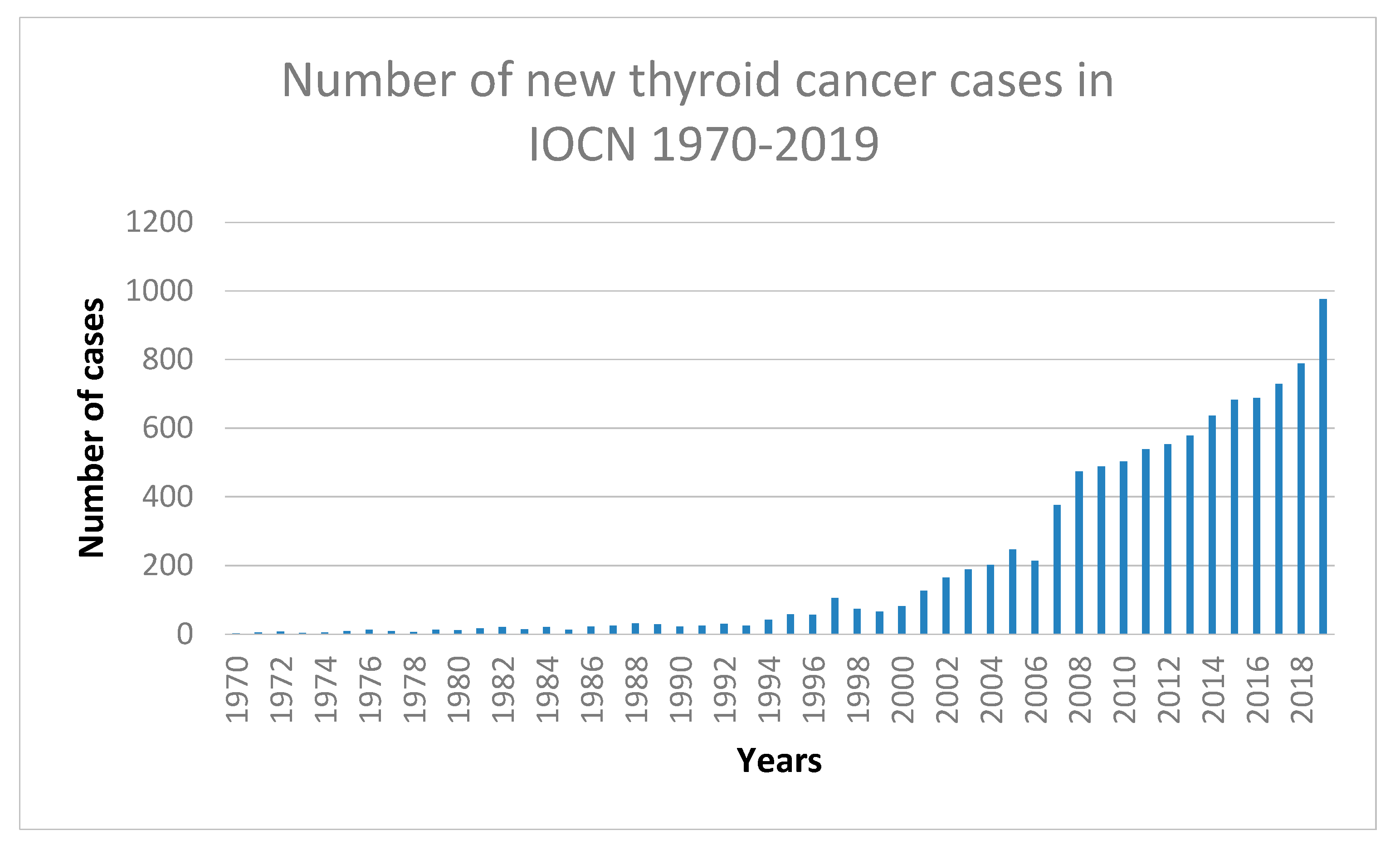
:1. Introduction
2. Methods
3. Case Presentations
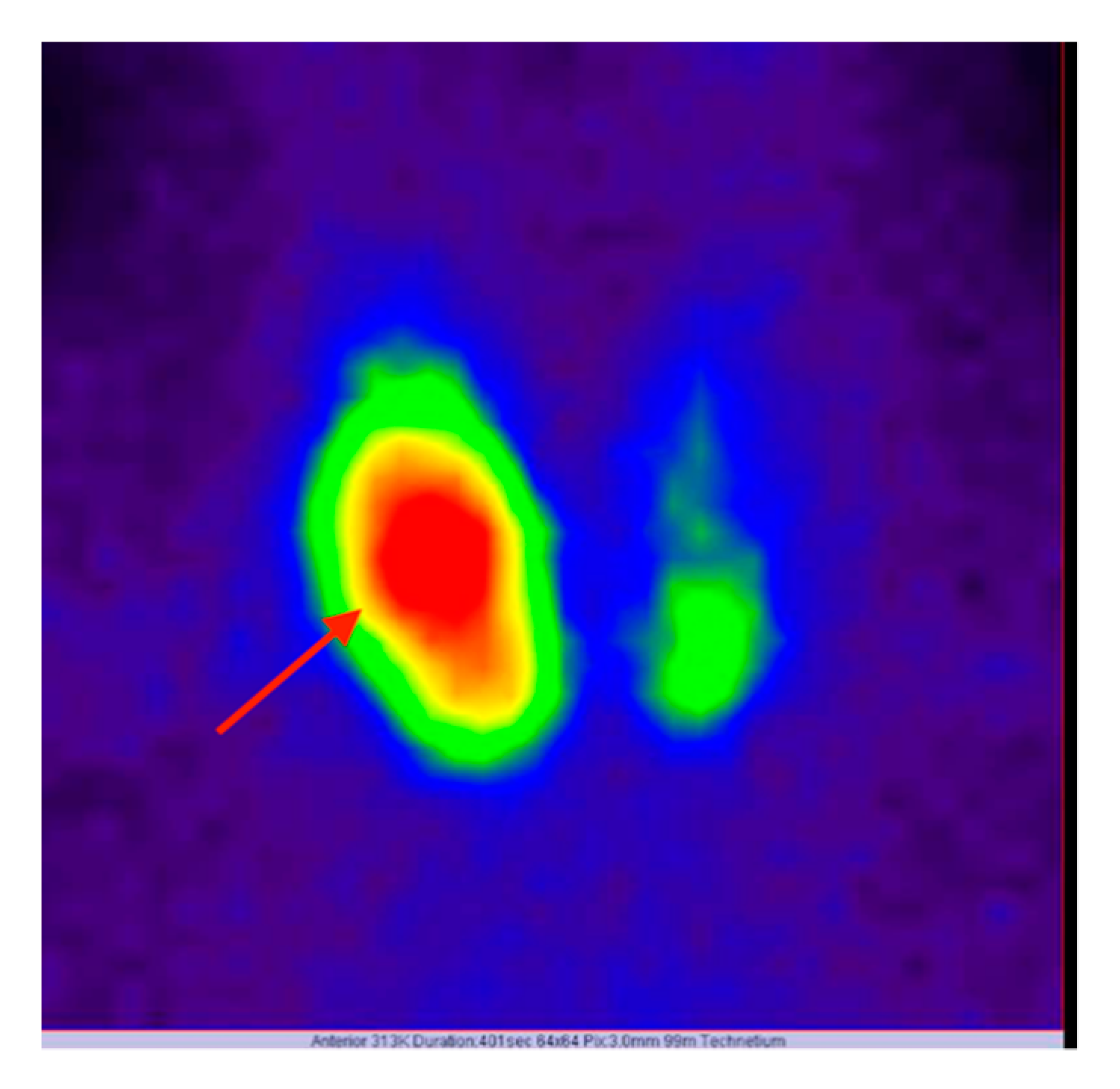
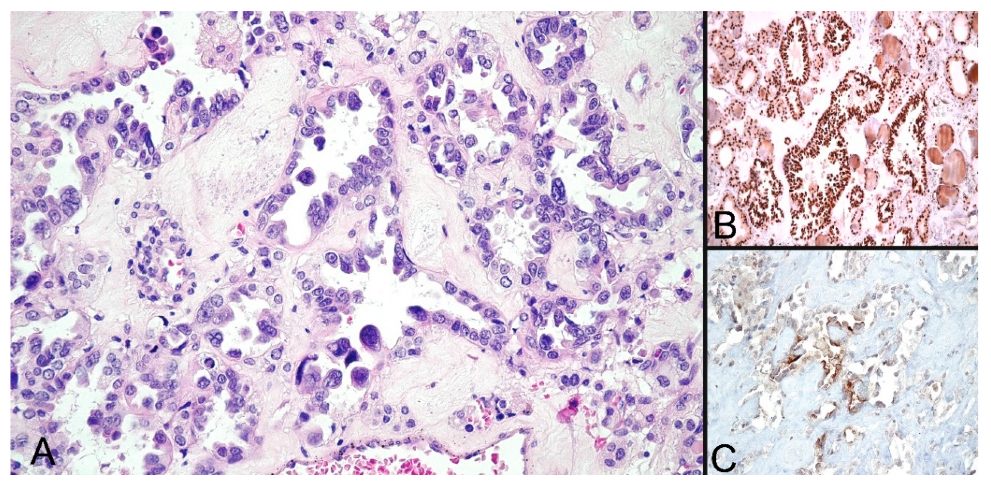
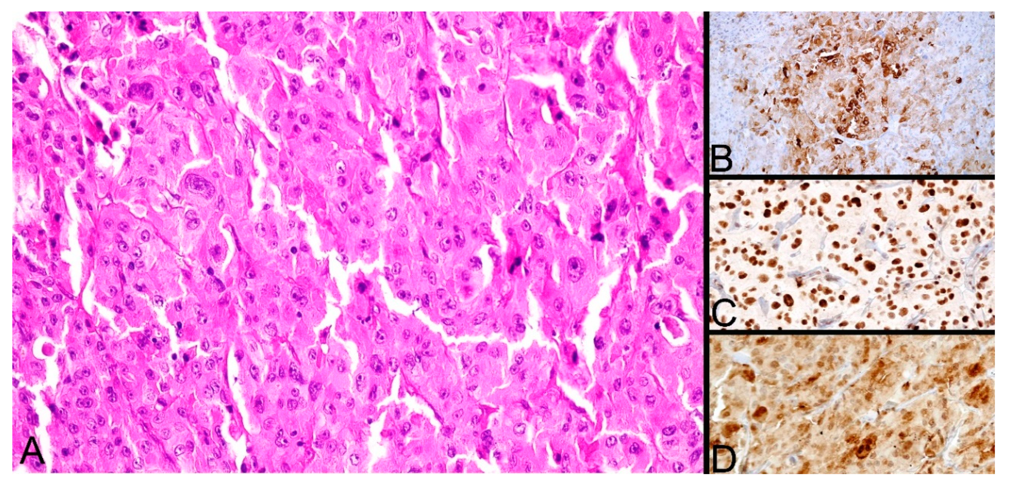
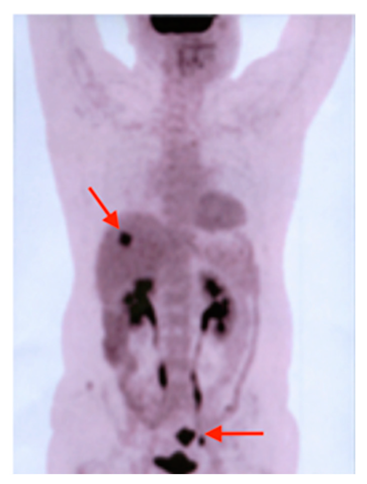
3.1. Case Presentation Nr. 1
3.2. Case Presentation Nr. 2
3.3. Case Presentation Nr. 3
3.4. Case Presentation Nr. 4
3.5. Case Presentation Nr. 5
3.6. Overview
4. Discussions
Author Contributions
Funding
Acknowledgments
Conflicts of Interest
References
- Billroth, T. Die Allgemeine Chirurgische Pathologie und Therapie in 51 Vorlesungen-Ein Handbuch fur Studierende und Arzte; Reimer, G., Ed.; Auflage: Berlin, Germany, 1889. [Google Scholar]
- Warren, S.; Gates, O. Multiple primary malignant tumors: A survey of the literature and a statistical study. Am. J. Cancer 1932, 16, 1358–1414. [Google Scholar]
- Spratt, J.S.; Hoag, M.G. Incidence of multiple primary cancers per man-year of follow up: 20-year review from the Ellis Fischel State Cancer Hospital. Ann. Surg. 1966, 164, 775–784. [Google Scholar] [CrossRef] [PubMed]
- Demandante, C.G.N.; Troyer, D.A.; Miles, T.P. Multiple primary malignant neoplasms: Case report and a comprehensive review of the literature. Am. J. Clin. Oncol. 2003, 26, 79–83. [Google Scholar] [CrossRef] [PubMed]
- Suzuki, T.; Takahashi, H.; Yao, K.; Inagi, K.; Nakayama, M.; Makoshi, T.; Nagai, H.; Okamoto, M. Multiple primary malignancies in the head and neck: A clinical review of 121 patients. Acta Otolaryngol. Suppl. 2002, 122, 88–92. [Google Scholar] [CrossRef]
- Xu, L.L.; Gu, K.S. Clinical retrospective analysis of cases with multiple primary malignant neoplasms. Genet. Mol. Res. 2014, 13, 9271–9284. [Google Scholar] [CrossRef] [PubMed]
- Zhai, C.; Cai, Y.; Lou, F.; Liu, Z.; Xie, J.; Zhou, X.; Zhanggui, W.; Fang, Y.; Pan, H.; Han, W. Multiple Primary Malignant Tumors—A Clinical Analysis of 15,321 Patients with Malignancies at a Single Center in China. J. Cancer 2018, 9, 2795–2801. [Google Scholar] [CrossRef] [PubMed]
- Li, F.; Zhong, W.-Z.; Niu, F.; Zhao, N.; Yang, J.-J.; Yan, H.-H.; Wu, Y.-L. Multiple primary malignancies involving lung cancer. BMC Cancer 2015, 15, 696. [Google Scholar] [CrossRef] [Green Version]
- Gokyer, A.; Kostek, O.; Hacioglu, M.; Erdogan, B.; Kodaz, H.; Turkmen, E.; Hacibekiroglu, I.; Uzunoglu, S.; Cicin, I.; Osman, K.; et al. Clinical features of the patient with multiple primary tumors: Single center experience. North. Clin. Istanbul 2017, 4, 43–51. [Google Scholar] [CrossRef] [PubMed] [Green Version]
- Lei, K.; He, X.; Yu, L.; Ni, C.; Chen, H.; Guan, D.; Sun, K.; Zou, H. Breast cancer prognosis is better in patients who develop subsequent metachronous thyroid cancer. PLoS ONE 2019, 14, e0215948. [Google Scholar] [CrossRef]
- Corso, G.; Veronesi, P.; Santomauro, G.I.; Maisonneuve, P.; Morigi, C.; Peruzzotti, G.; Intra, M.; Sacchini, V.; Galimberti, V. Multiple primary non-breast tumors in breast cancer survivors. J. Cancer Res. Clin. Oncol. 2018, 144, 979–986. [Google Scholar] [CrossRef]
- Yu, F.; Ma, J.; Huo, K.; Li, P. Association between breast cancer and thyroid cancer: A descriptive study. Transl. Cancer Res. 2017, 6, 393–401. [Google Scholar] [CrossRef]
- Kim, J.H.; Kang, I.; Nam, S.; Park, H.S.; Park, S.; Jeong, J.J.; Nam, K.-H.; Chung, W.Y.; Kim, S.I.; Cho, Y.U.; et al. Comparison of characteristics in patients with both thyroid and breast cancer: Based on order of incidence. Korean J. Clin. Oncol. 2017, 13, 1–9. [Google Scholar] [CrossRef]
- Piciu, D.; Pestean, C.; Barbus, E.; Larg, M.I.; Piciu, A. Second malignancies in patients with differentiated thyroid carcinoma treated with low and medium activities of radioactive I-131. Clujul Med. 2016, 89, 384–389. [Google Scholar] [CrossRef] [PubMed] [Green Version]
- Irimie, A.; Achimas-Cadariu, P.; Burz, C.; Puscas, E. Multiple Primary Malignancies—Epidemiological Analysis at a Single Tertiary Institution. J. Gastrointest. Liver Dis. 2010, 19, 69–73. [Google Scholar]
- Sharma, D.; Singh, G.; Kakkar, N.; Raj, S. Second primary malignancy: A retrospective analysis report from a tertiary cancer center of North India. Indian J. Cancer 2016, 53, 595. [Google Scholar] [CrossRef]
- Lee, J.S.; Moon, W.; Park, S.J.; Park, M.I.; Kim, K.J.; Jang, L.L.; Park, M.J.; Chun, B.K. Triple synchronous primary cancers of rectum, thyroid, and uterine cervix detected during the workup for hematochezia. Intern. Med. 2010, 49, 1745–1747. [Google Scholar] [CrossRef] [Green Version]
- Iqbal, F.R.W.; Sani, A.; Gendeh, B.S.; Aireen, I. Triple primary cancers of the larynx, lung and thyroid presenting in one patient. Med. J. Malays. 2008, 63, 417–418. [Google Scholar]
- Hamada, Y.; Takise, A.; Uno, D.; Itoh, H.; Ichikawa, H.; Morishta, Y. Synchronous primary triple cancers including the lung, stomach, and thyroid: A case report. Kyobu Geka 2000, 53, 101–105. [Google Scholar]
- Fukuoka, M.; Hagiwara, M.; Shimoshige, S.; Hirata, A.; Adachi, T.; Komura, H.; Shoji, T.; Kikuiri, T.; Ikeda, K.; Kimura, N.; et al. Primary leiomyosarcoma of the heart subsequent to double carcinomas of the thyroid and lung. Heart Vessels 2000, 15, 100–102. [Google Scholar] [CrossRef]
- Rai, R.S.; Deb, P.; Rai, R.; Gupta, E.; Panayach, J.S. Synchronous primary triple neoplasia (renal cell carcinoma and prostate cancer in combination with thyroid neoplasm). Report of an unusual case. Minerva Urol. Nefrol. 2007, 59, 451–454. [Google Scholar]
- Peng, C.; Li, Z.; Gao, H.; Zou, X.; Wang, X.; Zhou, C.; Niu, J. Synchronous primary sigmoid colon cancer and primary thyroid cancer followed by a malignant tumor of the kidney: Case report of multiple primary cancer and review of the literature. Oncol. Lett. 2019, 17, 2479–2484. [Google Scholar] [CrossRef] [PubMed] [Green Version]
- Peng, L.; Zeng, Z.; Teng, X.; Chen, Z.; Lin, L.; Bao, H.; Shao, Y.W.; Wang, Y.; Dong, Y.; Zhao, Q. Genomic profiling of synchronous triple primary tumors of the lung, thyroid and kidney in a young female patient: A case report. Oncol. Lett. 2018, 16, 6089–6094. [Google Scholar] [CrossRef]
- Kikuchi, N.; Ohashi, T.; Miura, T.; Nishibu, A.; Yamamoto, T. Triple cancers concurrently detected in a case of antitranscriptional intermediary factor-1γ antibody-positive dermatomyositis. Int. J. Dermatol. 2017, 56, 1516–1517. [Google Scholar] [CrossRef] [PubMed]
- Singh, N.J.; Tripathy, N.; Roy, P.; Manikantan, K.; Arun, P. Simultaneous Triple Primary Head and Neck Malignancies: A Rare Case Report. Head Neck Pathol. 2016, 10, 233–236. [Google Scholar] [CrossRef] [PubMed] [Green Version]
- Cohen, P.R. Segmental neurofibromatosis and cancer: Report of triple malignancy in a woman with mosaic Neurofibromatosis 1 and review of neoplasms in segmental neurofibromatosis. Dermatol. Online J. 2016, 22. pii: 13030/qt66k5j4wt. [Google Scholar]
- Oh, S.J.; Bae, D.S.; Suh, B.J. Synchronous triple primary cancers occurring in the stomach, kidney, and thyroid. Ann. Surg. Treat Res. 2015, 88, 345–348. [Google Scholar] [CrossRef]
- Adams, M.; Caffrey, R. Triple primary cancers of the head and neck: Case report and literature review. J. Laryngol. Otol. 2014, 128, 552–554. [Google Scholar] [CrossRef]
- Omür, O.; Ozcan, Z.; Yazici, B.; Akgün, A.; Oral, A.; Ozkiliç, H. Multiple primary tumors in differentiated thyroid carcinoma and relationship to thyroid cancer outcome. Endocr. J. 2008, 55, 365–372. [Google Scholar] [CrossRef] [Green Version]
- Shibutani, Y.; Inoue, D.; Sugawa, H.; Mori, T. A case report of primary triple cancers in the thyroid, stomach and rectum with evidence of variable oncoprotein expressions. Nihon Naibunpi Gakkai Zasshi 1994, 70, 951–956. [Google Scholar]
- Arimura, T.; Niwa, K.; Mitani, N.; Hagiwara, I.; Kawaida, T.; Shimazu, H. A resected case of triple cancer in the uterus, lung and thyroid. Nihon Kyobu Geka Gakkai Zasshi 1989, 37, 1233–1237. [Google Scholar]
- Ohi, H.; Kikuchi, K.; Futawatari, K.; Kowada, M. A histologically-verified triple cancer--report of a rare case involving a primary brain tumor. Gan No Rinsho 1988, 34, 1001–1005. [Google Scholar]
- Watanabe, S.; Kodama, T.; Shimosato, Y.; Arimoto, H.; Sugimura, T.; Suemasu, K.; Shiraishi, M. Multiple primary cancers in 5,456 autopsy cases in the National Cancer Center of Japan. J. Natl. Cancer Inst. 1984, 72, 1021–1027. [Google Scholar]
- Yamamoto, Y.; Moriwaki, S.; Takashima, S.; Yumoto, Y. Autopsy case of simultaneous triple malignancies--ovarian, chordoma and thyroid cancers. Gan No Rinsho 1983, 29, 1375–1378. [Google Scholar] [PubMed]
- Nemoto, R.; Shimizu, S.; Kato, T.; Tsuchida, S.; Takanashi, R.; Toda, T. Triple cancers consisting of renal cell carcinoma, epipharynx carcinoma, and thyroid carcinoma accompanied with primary hyperparathyroidism (author’s transl). Nippon Hinyokika Gakkai Zasshi 1977, 68, 288–293. [Google Scholar] [PubMed] [Green Version]
- Ishii, T.; Iri, H.; Yamamoto, S.; Sudoh, M. A triple cancer. Simultaneous occurrence of gastric carcinoid, adenocarcinoma and thyroid cancer. Am. J. Gastroenterol. 1977, 67, 171–176. [Google Scholar] [PubMed]
- Søndergaard, E.; Ebbehoj, A.; Poulsen, P.L.; Gormsen, L.C. Multiple Neoplasms Simultaneously Diagnosed by Complementary Triple-Tracer PET/CT and 123I-MIBG Scintigraphy. Clin. Nucl. Med. 2017, 42, e61–e66. [Google Scholar] [CrossRef]
- Ishimori, T.; Patel, P.V.; Wahl, R.L. Detection of unexpected additional primary malignancies with PET/CT. J. Nucl. Med. 2005, 46, 752–757. [Google Scholar]
- Israel, O.; Yefremov, N.; Bar-Shalom, R.; Kagana, O.; Frenkel, A.; Keidar, Z.; Fischer, D. PET/CT detection of unexpected gastrointestinal foci of 18F-FDG uptake: Incidence, localization patterns, and clinical significance. J. Nucl. Med. 2005, 46, 758–762. [Google Scholar]
- Even-Sapir, E.; Lerman, H.; Gutman, M.; Lievshitz, G.; Zuriel, L.; Polliack, A.; Inbar, M.; Metser, U. The presentation of malignant tumours and pre-malignant lesions incidentally found on PET-CT. Eur. J. Nucl. Med. Mol. Imaging 2006, 33, 541–552. [Google Scholar] [CrossRef]
- Jena, A.; Patnayak, R.; Lakshmi, A.Y.; Manilal, B.; Reddy, M.K. Multiple primary cancers: An enigma. South Asian J. Cancer 2016, 5, 29. [Google Scholar] [CrossRef]
- Chalstrey, L.J.; Benjamin, B. High incidence of breast cancer in thyroid cancer patients. Br. J. Cancer 1966, 20, 670–675. [Google Scholar] [CrossRef] [PubMed] [Green Version]
- Rahbari, R.; Zhang, L.; Kebebew, E. Thyroid cancer gender disparity. Future Oncol. 2010, 6, 1771–1779. [Google Scholar] [CrossRef] [PubMed] [Green Version]
- Piciu, D.; Irimie, A.; Piciu, A. Investigation of thyroid carcinoma over 40 years, using the database of the Ion Chiricuta Institute of Oncology Cluj-Napoca. J. BUON 2014, 19, 524–529. [Google Scholar] [PubMed]
- Kapoor, A.; Narayan, S.; Nirban, R.; Purohit, R.; Bfagri, P.; Kumar, H.; Singhal, M. A rare case report of triple malignancy: Carcinoma urinary bladder, larynx, and breast in a single patient. Clin. Cancer Investig. J. 2014, 3, 558. [Google Scholar] [CrossRef]
- Piciu, D.; Barbus, E.; Piciu, A.; Fetica, B. Mazabraud’s syndrome and thyroid cancer, a very rare and confusing association: A case report. BMC Endocr. Disord. 2015, 15, 39. [Google Scholar] [CrossRef] [Green Version]
- Ihle, M.A.; Huss, S.; Jeske, W.; Hartmann, W.; Merkelbach-Bruse, S.; Schildhaus, H.-U.; Büttner, R.; Sihto, H.; Sundby Hall, K.; Eriksson, M.; et al. Expression of cell cycle regulators and frequency of TP53 mutations in high risk gastrointestinal stromal tumors prior to adjuvant imatinib treatment. PLoS ONE 2018, 13, e0193048. [Google Scholar] [CrossRef] [Green Version]
- Namal, E. Medical Treatment with Two Targeted Therapies for an Oncology Patient with Double Primary Cancer: Case Report. Turk. J. Oncol. 2016, 31, 51–54. [Google Scholar] [CrossRef]
- Borda, A.; Zahan, A.-E.; Piciu, D.; Barbuș, E.; Berger, N.; Nechifor-Boilă, A. A 15 year institutional experience of well-differentiated follicular cell-derived thyroid carcinomas; impact of the new 2017 TNM and WHO Classifications of Tumors of Endocrine Organs on the epidemiological trends and pathological characteristics. Endocrine 2019, 67, 630–642. [Google Scholar] [CrossRef]


| Case No. | Location of Malignancy | Histology | Gender | Age at Thyroid Carcinoma Diagnosis | Initial Diagnosis (Year) | Disease-Free Interval (Years) |
|---|---|---|---|---|---|---|
| Case 1 | ovarian | serous adenocarcinoma | female | 53 years | 2017 | NA |
| thyroid | papillary microcarcinoma | 2018 | 1 | |||
| lung | acinar adenocarcinoma | 2019 | 1 | |||
| Case 2 | endometrial | endometrioid carcinoma | female | 71 years | 2005 | NA |
| ovarian | serous adenocarcinoma | 2005 | 0 | |||
| thyroid | Hürthle cell carcinoma | 2019 | 14 | |||
| Case 3 | thyroid | papillary carcinoma | male | 58 years | 2008 | NA |
| stomach | gastro-intestinal stromal tumor | 2014 | 6 | |||
| colon | adenocarcinoma | 2014 | 0 | |||
| Case 4 | vulva | keratinizing squamous cell carcinoma | female | 53 years | 1993 | NA |
| endometrial | endometrioid carcinoma | 2000 | 7 | |||
| thyroid | papillary carcinoma | 2002 | 2 | |||
| Case 5 | thyroid | papillary carcinoma | male | 46 years | 2005 | NA |
| colon | adenocarcinoma | 2007 | 2 | |||
| prostate | adenocarcinoma | 2013 | 6 |
| Investigators | Year of Publication | Location of Second and Third Malignancies besides the Thyroid Carcinoma |
|---|---|---|
| Peng et al. [22] | 2019 | Colon; Kidney |
| Peng et al. [23] | 2018 | Lung; Kidney |
| Kikuchi et al. [24] | 2017 | Stomach; Breast |
| Singh et al. [25] | 2016 | Larynx; Lymph nodes |
| Cohen et al. [26] | 2016 | Kidney; Skin |
| Oh et al. [27] | 2015 | Stomach; Kidney |
| Adams and Caffrey [28] | 2014 | Larynx; Thyroid (second) |
| Lee et al. [17] | 2010 | Rectum; Uterus |
| Omür et al. [29] | 2008 | Breast; Stomach |
| Iqbal et al. [18] | 2008 | Larynx; Lung |
| Rai et al. [21] | 2007 | Kidney; Prostate |
| Hamada et al. [19] | 2000 | Lung; Stomach |
| Fukuoka et al. [20] | 2000 | Heart; Lung |
| Shibutani et al. [30] | 1994 | Stomach; Rectum |
| Arimura et al. [31] | 1989 | Uterus; Lung |
| Ohi et al. [32] | 1988 | Skin; Brain |
| Watanabe et al. [33] | 1984 | not specified |
| Yamamoto et al. [34] | 1983 | Ovary; Brain |
| Nemoto et al. [35] | 1977 | Kidney; Nasopharynx |
| Ishii et al. [36] | 1977 | Stomach; Stomach |
© 2020 by the authors. Licensee MDPI, Basel, Switzerland. This article is an open access article distributed under the terms and conditions of the Creative Commons Attribution (CC BY) license (http://creativecommons.org/licenses/by/4.0/).
Share and Cite
Bădan, M.-I.; Piciu, D. Triple Metachronous Malignancies with Thyroid Involvement: A Brief Overview of Five Case Reports over 20 Years of Institutional Experience. Diagnostics 2020, 10, 168. https://0-doi-org.brum.beds.ac.uk/10.3390/diagnostics10030168
Bădan M-I, Piciu D. Triple Metachronous Malignancies with Thyroid Involvement: A Brief Overview of Five Case Reports over 20 Years of Institutional Experience. Diagnostics. 2020; 10(3):168. https://0-doi-org.brum.beds.ac.uk/10.3390/diagnostics10030168
Chicago/Turabian StyleBădan, Marius-Ioan, and Doina Piciu. 2020. "Triple Metachronous Malignancies with Thyroid Involvement: A Brief Overview of Five Case Reports over 20 Years of Institutional Experience" Diagnostics 10, no. 3: 168. https://0-doi-org.brum.beds.ac.uk/10.3390/diagnostics10030168