
Cytauxzoon sp. and Hepatozoon spp. in Domestic Cats: A Preliminary Study in North-Eastern Italy

 ,
,  , ,
, ,
Abstract
:1. Introduction
2. Results
2.1. Feline Population
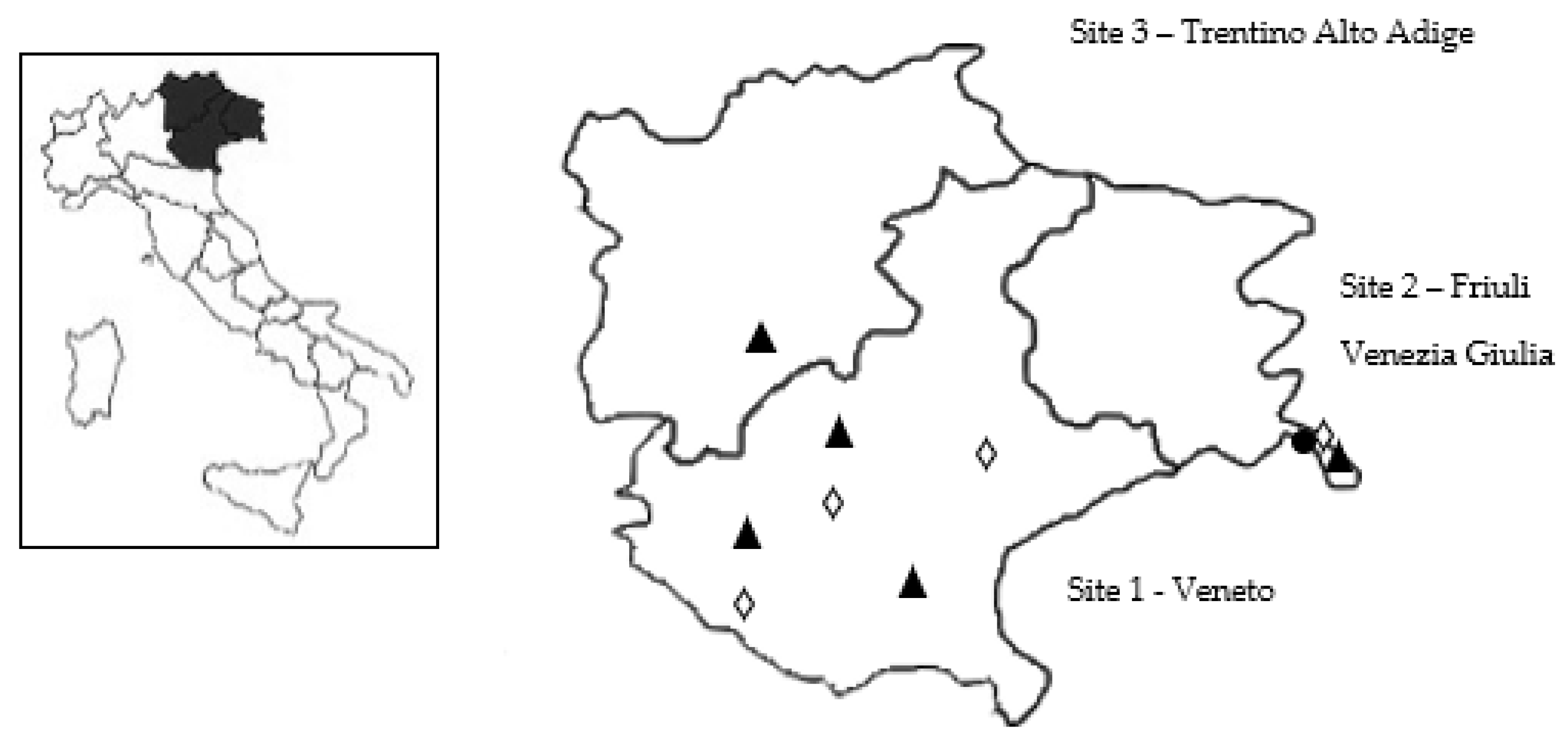
2.2. Laboratory Analysis and Geographical Distribution
2.3. Statistical Evaluation
3. Discussion
4. Materials and Methods
4.1. Blood Collection, Blood Analysis, and DNA Extraction
4.2. Molecular Analysis and Sequencing
4.3. Data Analysis
Author Contributions
Funding
Institutional Review Board Statement
Informed Consent Statement
Data Availability Statement
Acknowledgments
Conflicts of Interest
References
- Taylor, M.A.; Coop, R.L.; Wall, R.L. Veterinary Parasitology, 3rd ed.; Blackwell Publishing: Oxford, UK, 2007; p. 874. [Google Scholar]
- Wagner, J.E. A fatal cytauxzoonosis-like disease in cats. J. Am. Vet. Med. Assoc. 1976, 168, 585–588. [Google Scholar]
- Miller, J.; Davis, C.D. Increasing frequency of feline cytauxzoonosis cases diagnosed in western Kentucky from 2001 to 2011. Vet. Parasitol. 2013, 198, 205–208. [Google Scholar] [CrossRef] [Green Version]
- Tarigo, J.L.; Scholl, E.H.; McK Bird, D.; Brown, C.C.; Cohn, L.A.; Dean, G.A.; Levy, M.G.; Doolan, D.L.; Trieu, A.; Nordone, S.K.; et al. A novel candidate vaccine for cytauxzoonosis inferred from comparative apicomplexan genomics. PLoS ONE 2013, 8, e71233. [Google Scholar] [CrossRef]
- Criado-Fornelio, A.; Gónzalez-del-Río, M.A.; Buling-Saraña, A.; Barba-Carretero, J.C. The “expanding universe” of piroplasms. Vet. Parasitol. 2004, 119, 337–345. [Google Scholar] [CrossRef]
- Díaz-Regañón, D.; Villaescusa, A.; Ayllón, T.; Rodríguez-Franco, F.; Baneth, G.; Calleja-Bueno, L.; García-Sancho, M.; Agulla, B.; Sainz, Á. Molecular detection of Hepatozoon spp. and Cytauxzoon sp. in domestic and stray cats from Madrid, Spain. Parasites Vectors 2017, 10, 112. [Google Scholar] [CrossRef] [Green Version]
- Criado-Fornelio, A.; Buling, A.; Pingret, J.L.; Etievant, M.; Boucraut-Baralon, C.; Alongi, A.; Agnone, A.; Torina, A. Hemoprotozoa of domestic animals in France: Prevalence and molecular characterization. Vet. Parasitol. 2009, 159, 73–76. [Google Scholar] [CrossRef] [PubMed] [Green Version]
- Legroux, J.P.; Halos, L.; René-Martellet, M.; Servonnet, M.; Pingret, J.L.; Bourdoiseau, G.; Baneth, G.; Chabanne, L. First clinical case report of Cytauxzoon sp. infection in a domestic cat in France. BMC Vet. Res. 2017, 13, 81. [Google Scholar] [CrossRef] [Green Version]
- Alho, A.M.; Silva, J.; Fonseca, M.J.; Santos, F.; Nunes, C.; De Carvalho, L.M.; Rodrigues, M.; Cardoso, L. First report of Cytauxzoon sp. infection in a domestic cat from Portugal. Parasites Vectors 2016, 9, 220. [Google Scholar] [CrossRef] [PubMed] [Green Version]
- Nentwig, A.; Meli, M.L.; Schrack, J.; Reichler, I.M.; Riond, B.; Gloor, C.; Howard, J.; Hofmann-Lehmann, R.; Willi, B. First report of Cytauxzoon sp. infection in domestic cats in Switzerland: Natural and transfusion-transmitted infections. Parasites Vectors 2018, 11, 292. [Google Scholar] [CrossRef] [PubMed]
- Panait, L.C.; Stock, G.; Globokar, M.; Balzer, J.; Groth, B.; Mihalca, A.D.; Pantchev, N. First report of Cytauxzoon sp. infection in Germany: Organism description and molecular confirmation in a domestic cat. Parasitol. Res. 2020, 119, 3005–3011. [Google Scholar] [CrossRef]
- Carli, E.; Trotta, M.; Chinelli, R.; Drigo, M.; Sinigoi, L.; Tosolini, P.; Furlanello, T.; Millotti, A.; Caldin, M.; Solano-Gallego, L. Cytauxzoon sp. infection in the first endemic focus described in domestic cats in Europe. Vet. Parasitol. 2012, 183, 343–352. [Google Scholar] [CrossRef] [PubMed]
- Carli, E.; Trotta, M.; Bianchi, E.; Furlanello, T.; Caldin, M.; Pietrobelli, M.; Solano-Gallego, L. Cytauxzoon sp. infection in two free ranging young cats: Clinicopathological findings, therapy and follow up. Türkiye Parazitolojii Derg. 2014, 38, 185–189. [Google Scholar] [CrossRef] [PubMed]
- Jalovecka, M.; Sojka, D.; Ascencio, M.; Schnittger, L. Babesia life cycle—When phylogeny meets biology. Trends Parasitol. 2019, 35, 356–368. [Google Scholar] [CrossRef] [PubMed]
- Panait, L.C.; Mihalca, A.D.; Modrý, D.; Juránková, J.; Ionică, A.M.; Deak, G.; Gherman, C.M.; Heddergott, M.; Hodžić, A.; Veronesi, F.; et al. Three new species of Cytauxzoon in European wild felids. Vet. Parasitol. 2021, 290, 109344. [Google Scholar] [CrossRef]
- Patton, W.S. The haemogregarines of mammals and reptiles. Parasitology 1908, 1, 318–321. [Google Scholar] [CrossRef] [Green Version]
- Klopfer, U.; Nobel, T.A.; Neumann, F. Hepatozoon-like parasite (schizonts) in the myocardium of the domestic cat. Vet. Pathol. 1973, 10, 185–190. [Google Scholar] [CrossRef]
- Leeflang, P.; Ilemobade, A.A. Tick-borne disease of domestic animals in northern Nigeria. II. Research summary, 1966 to 1976. Trop. Anim. Health Prod. 1977, 9, 211–218. [Google Scholar] [CrossRef]
- Van Amstel, S. Hepatozoonose i’n kat. J. S. Afr. Vet. Med. Assoc. 1979, 50, 215–216. [Google Scholar]
- Pereira, C.; Maia, J.P.; Marcos, R.; Luzzago, C.; Puente-Payo, P.; Dall’Ara, P.; Faustino, A.; Lauzi, S. Molecular detection of Hepatozoon felis in cats from Maio Island, Republic of Cape Verde and global distribution of feline hepatozoonosis. Parasites Vectors 2019, 12, 294. [Google Scholar] [CrossRef]
- Ewing, G.O. Granulomatous cholangiohepatitis in a cat due to a protozoan parasite resembling Hepatozoon canis. Feline Pract. 1977, 7, 37–40. [Google Scholar]
- Perez, R.R.; Rubini, A.S.; O’Dwyer, L.H. The first report of Hepatozoon spp. (Apicomplexa, Hepatozoidae) in domestic cats from São Paulo state, Brazil. Parasitol. Res. 2004, 94, 83–85. [Google Scholar] [CrossRef]
- Beaufils, J.P.; Martin-Granel, J.; Jumelle, P. Hepatozoon spp. parasitemia and feline leukemia virus infection in two cats. Feline Pract. 1998, 26, 10–13. [Google Scholar]
- Vilhena, H.; Martinez-Díaz, V.L.; Cardoso, L.; Vieira, L.; Altet, L.; Francino, O.; Pastor, J.; Silvestre-Ferreira, A.C. Feline vector-borne pathogens in the north and center of Portugal. Parasites Vectors 2013, 6, 99. [Google Scholar] [CrossRef] [Green Version]
- Attipa, C.; Papasouliotis, K.; Solano-Gallego, L.; Baneth, G.; Nachum-Biala, Y.; Sarvani, E.; Knowles, T.G.; Mengi, S.; Morris, D.; Helps, C.; et al. Prevalence study and risk factor analysis of selected bacterial, protozoal and viral, including vector-borne, pathogens in cats from Cyprus. Parasites Vectors 2017, 10, 130. [Google Scholar] [CrossRef] [Green Version]
- Kegler, K.; Nufer, U.; Alic, A.; Posthaus, H.; Olias, P.; Basso, W. Fatal infection with emerging apicomplexan parasite Hepatozoon silvestris in a domestic cat. Parasites Vectors 2018, 11, 428. [Google Scholar] [CrossRef] [Green Version]
- Basso, W.; Görnerb, D.; Globokarc, M.; Keidelc, A.; Pantchevc, N. First autochthonous case of clinical Hepatozoon felis infection in a domestic cat in Central Europe. Parasitol. Int. 2019, 72, 101945. [Google Scholar] [CrossRef] [PubMed]
- Morelli, S.; Diakou, A.; Traversa, D.; Di Gennaro, E.; Simonato, G.; Colombo, M.; Dimzas, D.; Grillini, M.; Frangipane di Regalbono, A.; Beugnet, F.; et al. First record of Hepatozoon spp. in domestic cats in Greece. Ticks Tick Borne Dis. 2021, 12, 101580. [Google Scholar] [CrossRef] [PubMed]
- Ebani, V.V.; Guardone, L.; Marra, F.; Altomonte, I.; Nardoni, S.; Mancianti, F. Arthropod-borne pathogens in stray cats from Northern Italy: A serological and molecular survey. Animals 2020, 10, 2334. [Google Scholar] [CrossRef] [PubMed]
- Giannelli, A.; Latrofa, M.S.; Nachum-Biala, Y.; Hodžić, A.; Greco, G.; Attanasi, A.; Annoscia, G.; Otranto, D.; Baneth, G. Three different Hepatozoon species in domestic cats from southern Italy. Ticks Tick Borne Dis. 2017, 8, 721–724. [Google Scholar] [CrossRef] [PubMed]
- Otranto, D.; Napoli, E.; Latrofa, M.S.; Annoscia, G.; Tarallo, V.D.; Greco, G.; Lorusso, E.; Gulotta, L.; Falsone, L.; Basano, F.S.; et al. Feline and canine leishmaniosis and other vector-borne diseases in the Aeolian Islands: Pathogen and vector circulation in a confined environment. Vet. Parasitol. 2017, 236, 144–151. [Google Scholar] [CrossRef] [PubMed]
- Stevanović, O.; Diakou, A.; Morelli, S.; Paraš, S.; Trbojević, I.; Nedić, D.; Sladojević, Ž.; Kasagić, D.; Di Cesare, A. Severe verminous pneumonia caused by natural mixed infection with Aelurostrongylus abstrusus and Angiostrongylus chabaudi in a European wildcat from Western Balkan area. Acta Parasitol. 2019, 64, 411–417. [Google Scholar] [CrossRef] [PubMed]
- Diakou, A.; Dimzas, D.; Astaras, C.; Savvas, I.; Di Cesare, A.; Morelli, S.; Neofitos, Κ.; Migli, D.; Traversa, D. Clinical investigations and treatment outcome in a European wildcat (Felis silvestris silvestris) infected by cardio-pulmonary nematodes. Vet. Parasitol. Reg. Stud. Rep. 2020, 19, 100357. [Google Scholar] [CrossRef] [PubMed]
- Di Cesare, A.; Morelli, S.; Colombo, M.; Simonato, G.; Veronesi, F.; Marcer, F.; Diakou, A.; D’Angelosante, R.; Pantchev, N.; Psaralexi, E.; et al. Is angiostrongylosis a realistic threat for domestic cats? Front. Vet. Sci. 2020, 7, 195. [Google Scholar] [CrossRef]
- Traversa, D.; Morelli, S.; Di Cesare, A.; Diakou, A. Felid cardiopulmonary nematodes: Dilemmas solved and new questions posed. Pathogens 2021, 10, 30. [Google Scholar] [CrossRef]
- Kocan, A.A.; Blouin, E.F.; Glenn, B.L. Hematologic and serum chemical values for free-ranging bobcats, Felis rufus (Schreber), with reference to animals with natural infections of Cytauxzoon felis Kier, 1979. J. Wildl. Dis. 1985, 21, 190–192. [Google Scholar] [CrossRef] [PubMed]
- Mason, V.R.; Van Den Bussche, R.A.; Meinkoth, J.H.; Hoovert, J.P.; Kokan, A.A. A new species of Cytauxzoon from Pallas’ cats caught in Mongolia and comments on the systematics and taxonomy of piroplasmids. J. Parasitol. 2005, 91, 420–426. [Google Scholar]
- Millán, J.; Naranjo, V.; Rodríguez, A.; De la Lastra, J.M.; Mangold, A.J.; De la Fuente, J. Prevalence of infection and 18S rRNA gene sequences of Cytauxzoon species in Iberian lynx (Lynx pardinus) in Spain. Parasitology 2007, 134, 995–1001. [Google Scholar] [CrossRef] [Green Version]
- Gallusová, M.; Jirsová, D.; Mihalca, A.D.; Gherman, C.M.; D’Amico, G.; Qablan, M.A.; Modrý, D. Cytauxzoon infections in wild felids from Carpathian-Danubian-Pontic space: Further evidence for a different Cytauxzoon species in European felids. J. Parasitol. 2016, 102, 377–380. [Google Scholar] [CrossRef]
- Veronesi, F.; Ravagnan, S.; Cerquetella, M.; Carli, E.; Olivieri, E.; Santoro, A.; Pesaro, S.; Berardi, S.; Rossi, G.; Ragni, B.; et al. First detection of Cytauxzoon spp. infection in European wildcats (Felis silvestris silvestris) of Italy. Ticks Tick Borne Dis. 2016, 7, 853–858. [Google Scholar] [CrossRef]
- Hodžić, A.; Alić, A.; Prašović, S.; Otranto, D.; Baneth, G.; Duscher, G.G. Hepatozoon silvestris sp. nov.: Morphological and molecular characterization of a new species of Hepatozoon (Adeleorina: Hepatozoidae) from the European wild cat (Felis silvestris silvestris). Parasitology 2017, 144, 650–661. [Google Scholar] [CrossRef] [Green Version]
- Hodžić, A.; Alić, A.; Duscher, G.G. High diversity of blood-associated parasites and bacteria in European wild cats in Bosnia and Herzegovina: A molecular study. Ticks Tick Borne Dis. 2018, 9, 589–593. [Google Scholar] [CrossRef]
- Ortuño, M.; Nachum-Biala, Y.; García-Bocanegra, I.; Resa, M.; Berriatua, E.; Baneth, G. An epidemiological study in wild carnivores from Spanish Mediterranean ecosystems reveals association between Leishmania infantum, Babesia spp. and Hepatozoon spp. infection and new hosts for Hepatozoon martis, Hepatozoon canis and Sarcocystis spp. Transbound. Emerg. Dis. 2021, 1–16. [Google Scholar]
- Criado-Fornelio, A.; Ruas, J.L.; Casado, N.; Farias, N.A.; Soares, M.P.; Müller, G.; Brumt, J.G.; Berne, M.E.; Buling-Saraña, A.; Barba-Carretero, J.C. New molecular data on mammalian Hepatozoon species (Apicomplexa: Adeleorina) from Brazil and Spain. J. Parasitol. 2006, 92, 93–99. [Google Scholar] [CrossRef]
- Baneth, G.; Sheiner, A.; Eyal, O.; Hahn, S.; Beaufils, J.P.; Anug, Y.; Talmi-Frank, D. Redescription of Hepatozoon felis (Apicomplexa: Hepatozoidae) based on phylogenetic analysis, tissue and blood form morphology, and possible transplacental transmission. Parasites Vectors 2013, 6, 102. [Google Scholar] [CrossRef] [PubMed] [Green Version]
- Morganti, G.; Veronesi, F.; Stefanetti, V.; Di Muccio, T.; Fiorentino, E.; Diaferia, M.; Santoro, A.; Passamonti, F.; Gramiccia, M. Emerging feline vector-borne pathogens in Italy. Parasites Vectors 2019, 12, 193. [Google Scholar] [CrossRef]
- Spada, E.; Proverbio, D.; Galluzzo, P.; Perego, R.; Bagnagatti De Giorgi, G.; Roggero, N.; Caracappa, S. Frequency of piroplasms Babesia microti and Cytauxzoon felis in stray cats from northern Italy. Biomed. Res. Int. 2014, 2014, 943754. [Google Scholar] [CrossRef] [Green Version]
- Latrofa, M.S.; Iatta, R.; Toniolo, F.; Furlanello, T.; Ravagnan, S.; Capelli, G.; Schunack, B.; Chomel, B.; Zatelli, A.; Mendoza-Roldan, J.; et al. A molecular survey of vector-borne pathogens and haemoplasmas in owned cats across Italy. Parasites Vectors 2020, 13, 116. [Google Scholar] [CrossRef] [PubMed] [Green Version]
- Baneth, G. Perspectives on canine and feline hepatozoonosis. Vet. Parasitol. 2011, 181, 3–11. [Google Scholar] [CrossRef]
- Fattori, U.; Rucli, A.; Zanetti, M. Grandi Carnivori ed Ungulati Nell’area Confinaria Italo-Slovena. Stato di Conservazione, 2nd ed.; Regione Autonoma Friuli Venezia Giulia: Udine, Italy, 2010; pp. 1–80. [Google Scholar]
- Mattucci, F.; Oliveira, R.; Bizzarri, L.; Vercillo, F.; Anile, S.; Ragni, B.; Lapini, L.; Sforzi, A.; Alves, P.C.; Lyons, L.A.; et al. Genentic structure of wildcat (Felis silvestris) populations in Italy. Ecol. Evol. 2013, 3, 2443–2458. [Google Scholar] [CrossRef]
- Genovesi, P.; Angelini, P.; Bianchi, E.; Dupré, E.; Ercole, S.; Giacanelli, V.; Ronchi, F.; Stoch, F. Specie e Habitat di Interesse Comunitario in Italia: Distribuzione, Stato di conservazione e Trend; ISPRA Serie Rapporti; ISPRA-Settore Editoria: Roma, Italy, 2014; p. 194. [Google Scholar]
- Anile, S.; Devillard, S.; Ragni, B.; Rovero, F.; Mattucci, F.; Lo Valvo, M. Habitat fragmentation and anthropogenic factors affect wildcat Felis silvestris silvestris occupancy and detectability on Mt Etna. Wildl. Biol. 2019, 1, 1–13. [Google Scholar] [CrossRef] [Green Version]
- Traversa, D.; Morelli, S.; Cassini, R.; Crisi, P.E.; Russi, I.; Grillotti, E.; Manzocchi, S.; Simonato, G.; Beraldo, P.; Viglietti, A.; et al. Occurrence of canine and feline extra-intestinal nematodes in key endemic regions of Italy. Acta Trop. 2019, 193, 227–235. [Google Scholar] [CrossRef] [PubMed]
- Tabar, M.D.; Altet, L.; Francino, O.; Sánchez, A.; Ferrer, L.; Roura, X. Vector-borne infections in cats: Molecular study in Barcelona area (Spain). Vet. Parasitol. 2008, 151, 332–336. [Google Scholar] [CrossRef] [PubMed]


| Site 1 n (%) | Site 2 n (%) | Site 3 n (%) | Total n (%) | ||
|---|---|---|---|---|---|
| Sex | M | 49 (49.5) | 13 (33.3) | 12 (60.0) | 74 (46.8) |
| F | 50 (50,5) | 26 (66.7) | 8 (40.0) | 84 (53.2) | |
| Age classes | <12 months | 38 (38.4) | 9 (23.1) | 5 (25.0) | 52 (32.9) |
| 12–35 months | 23 (23.2) | 15 (38.5) | 8 (40.0) | 46 (29.1) | |
| ≥36 months | 37 (37.4) | 13 (33.3) | 7 (35.0) | 57 (36.1) | |
| NR a | 1 (1.0) | 2 (5.1) | 0 (0.0) | 3 (1.9) | |
| Management | Owned cats | 64 (64.6) | 19 (48.7) | 20 (100.0) | 103 (65.2) |
| Stray cats | 35 (35.4) | 20 (51.3) | 0 (0.0) | 55 (34.8) | |
| Lifestyle | Indoor | 28 (28.3) | 11 (28.2) | 7 (35.0) | 46 (29.1) |
| Outdoor | 71 (71.7) | 28 (71.8) | 13 (65.0) | 112 (70.9) | |
| Immunosuppressive infections (FIV and/or FeLV) | Positive | 15 (15.2) | 9 (23.1) | 1 (5.0) | 25 (15.8) |
| Negative | 84 (84.8) | 30 (76.9) | 19 (95.0) | 133 (84.2) | |
| Clinical signs (gastro-intestinal and respiratory signs) | Presence | 10 (10.1) | 1 (2.6) | 1 (5.0) | 12 (7.6) |
| Absence | 89 (89.9) | 38 (97.4) | 19 (95.0) | 146 (92.4) | |
| Ectoparasites infestations | Presence | 15 (15.2) | 11 (28.2) | 3 (15.0) | 29 (18.4) |
| Absence | 84 (84.8) | 28 (71.8) | 17 (85.0) | 129 (81.6) | |
| Total | 99 | 39 | 20 | 158 |
| Haemoparasite | ||||||||
|---|---|---|---|---|---|---|---|---|
| Factors | Variables | Tested | Cytauxzoon sp. n (%) | Hepatozoon spp. n (%) | Hepatozoon felis n (%) | Hepatozoon silvestris n (%) | ||
| Sex | M | 74 | 1 (1.4) | 13 (17.6) | 5 (6.8) | 8 (10.8) | ||
| F | 84 | 5 (6.0) | 13 (15.5) | 5 (6.0) | 8 (9.5) | |||
| Age Class | <12 months | 52 | 0 (0.0) | 9 (17.3) | 5 (9.6) | 4 (7.7) | ||
| 12–35 months | 46 | 1 (2.2) | 7 (15.2) | 0 (0.0) | 7 (15.2) | |||
| ≥36 months | 57 | 4 (7.0) | 9 (15.8) | 5 (8.8) | 4 (7.0) | |||
| NR a | 3 | 1 (33.3) | 1 (33.3) | 0 (0.0) | 1 (33.3) | |||
| Region | Site 1 | 99 | 0 (0.0) | * | 12 (12.1) | 5 (5.1) | 7 (7.1) | * |
| Site 2 | 39 | 6 (15.4) | 11 (28.2) | 2 (5.1) | 9 (23.1) | |||
| Site 3 | 20 | 0 (0.0) | 3 (15.0) | 3 (15.0) | 0 (0.0) | |||
| Management | Owned cats | 103 | 0 (0.0) | * | 12 (11.7) | 9 (8.7) | 3 (2.9) | * |
| Stray cats | 55 | 6 (10.9) | 14 (25.5) | 1 (1.8) | 13 (23.6) | |||
| Lifestyle | Indoor | 46 | 0 (0.0) | 5 (10.9) | 4 (8.7) | 1 (2.2) | ||
| Outdoor | 112 | 6 (5.4) | 21 (18.8) | 6 (5.4) | 15 (13.4) | |||
| Immunosuppressive infections (FIV and/or FeLV) | Positive | 25 | 3 (12.0) | 5 (20.0) | 2 (8.0) | 3 (12.0) | ||
| Negative | 133 | 3 (2.3) | 21 (15.8) | 8 (6.0) | 13 (9.8) | |||
| Clinical signs (gastro-intestinal and respiratory signs) | Presence | 12 | 0 (0.0) | 1 (3.8) | 0 (0.0) | 1 (8.3) | ||
| Absence | 148 | 6 (4.1) | 25 (16.9) | 10 (6.8) | 15 (10.1) | |||
| Ectoparasites infestation | Presence | 29 | 2 (6.9) | 8 (27.6) | 2 (6.9) | 6 (20.7) | ||
| Absence | 129 | 4 (3.1) | 18 (14.0) | 8 (6.2) | 10 (7.8) | |||
| Total | 158 | 6 (3.8) | 26 (16.5) | 10 (6.3) | 16 (10.1) | |||
Publisher’s Note: MDPI stays neutral with regard to jurisdictional claims in published maps and institutional affiliations. |
© 2021 by the authors. Licensee MDPI, Basel, Switzerland. This article is an open access article distributed under the terms and conditions of the Creative Commons Attribution (CC BY) license (https://creativecommons.org/licenses/by/4.0/).
Share and Cite
Grillini, M.; Simonato, G.; Tessarin, C.; Dotto, G.; Traversa, D.; Cassini, R.; Marchiori, E.; Frangipane di Regalbono, A. Cytauxzoon sp. and Hepatozoon spp. in Domestic Cats: A Preliminary Study in North-Eastern Italy. Pathogens 2021, 10, 1214. https://0-doi-org.brum.beds.ac.uk/10.3390/pathogens10091214
Grillini M, Simonato G, Tessarin C, Dotto G, Traversa D, Cassini R, Marchiori E, Frangipane di Regalbono A. Cytauxzoon sp. and Hepatozoon spp. in Domestic Cats: A Preliminary Study in North-Eastern Italy. Pathogens. 2021; 10(9):1214. https://0-doi-org.brum.beds.ac.uk/10.3390/pathogens10091214
Chicago/Turabian StyleGrillini, Marika, Giulia Simonato, Cinzia Tessarin, Giorgia Dotto, Donato Traversa, Rudi Cassini, Erica Marchiori, and Antonio Frangipane di Regalbono. 2021. "Cytauxzoon sp. and Hepatozoon spp. in Domestic Cats: A Preliminary Study in North-Eastern Italy" Pathogens 10, no. 9: 1214. https://0-doi-org.brum.beds.ac.uk/10.3390/pathogens10091214