Article

A First Report of Molecular Typing, Virulence Traits, and Phenotypic and Genotypic Resistance Patterns of Newly Emerging XDR and MDR Aeromonas veronii in Mugil seheli

by
Abdelazeem M. Algammal
1,*,
Reham A. Ibrahim
2,
Khyreyah J. Alfifi
3,
Hanaa Ghabban
3,
Saad Alghamdi
4,
Ahmed Kabrah
4,
Ahmed R. Khafagy
1,
Gehan M. Abou-Elela
2,
Nermeen M. Abu-Elala
5,6,
Matthew Gavino Donadu
7,8,* and
Reham M. El-Tarabili
1
1
Department of Bacteriology, Immunology, and Mycology, Faculty of Veterinary Medicine, Suez Canal University, Ismailia 41522, Egypt
2
National Institute of Oceanography and Fisheries, Cairo 11516, Egypt
3
Department of Biology, Faculty of Science, University of Tabuk, Tabuk 71421, Saudi Arabia
4
Laboratory Medicine Department, Faculty of Applied Medical Sciences, Umm Al-Qura University, Makkah 21955, Saudi Arabia
5
Department of Aquatic Animal Medicine and Management, Faculty of Veterinary Medicine, Cairo University, Giza 12211, Egypt
6
Faculty of Veterinary Medicine, King Salman International University, El Tor 46612, Egypt
7
Department of Biomedical Sciences, University of Sassari, 07100 Sassari, Italy
8
Department of Medical, Surgical and Experimental Sciences, University of Sassari, 07100 Sassari, Italy
*
Authors to whom correspondence should be addressed.
Submission received: 20 September 2022 / Revised: 7 October 2022 / Accepted: 27 October 2022 / Published: 29 October 2022
(This article belongs to the Special Issue Emerging Infections in Aquatic Animals)

Abstract

:
Aeromonas veronii is associated with substantial economic losses in the fish industry and with food-borne illness in humans. This study aimed to determine the prevalence, antibiogram profiles, sequence analysis, virulence and antimicrobial resistance genes, and pathogenicity of A. veronii recovered from Mugil seheli. A total of 80 fish were randomly gathered from various private farms in Suez Province, Egypt. Subsequently, samples were subjected to clinical, post-mortem, and bacteriological examinations. The retrieved isolates were tested for sequence analysis, antibiogram profile, pathogenicity, and PCR detection of virulence and resistance genes. The prevalence of A. veronii in the examined M. seheli was 22.5 % (18/80). The phylogenetic analyses revealed that the tested A. veronii strains shared high genetic similarity with other A. veronii strains from India, UK, and China. Using PCR it was revealed that the retrieved A. veronii isolates harbored the aerA, alt, ser, ompAII, act, ahp, and nuc virulence genes with prevalence of 100%, 82.9%, 61.7%, 55.3%, 44.7%, 36.17%, and 29.8%, respectively. Our findings revealed that 29.8% (14/47) of the retrieved A. veronii strains were XDR to nine antimicrobial classes and carried blaTEM, blaCTX-M, blaSHV, tetA, aadA1, and sul1 resistance genes. Likewise, 19.1% (9/47) of the obtained A. veronii strains were MDR to eight classes and possessed blaTEM, blaCTX-M, blaSHV, tetA, aadA1, and sul1 genes. The pathogenicity testing indicated that the mortality rates positively correlated with the prevalence of virulence-determinant genes. To our knowledge, this is the first report to reveal the occurrence of XDR and MDR A. veronii in M. seheli, an emergence that represents a risk to public health. Emerging XDR and MDR A. veronii in M. seheli frequently harbored aerA, alt, ser, ompAII, and act virulence genes, and blaTEM, sul1, tetA, blaCTX-M, blaSHV, and aadA1 resistance genes.

1. Introduction

The fast-growing demand for seafood presents a significant challenge for the enhancement of fisheries and aquaculture production worldwide. In 2014, the contribution of aquaculture to the human food supply overtook the production of natural water resources for the first time [1]. Egypt is the leading African country in terms of aquaculture production, with about 1.8 million tons of aquatic animal production, including freshwater and marine fish, shellfish, and crustaceans [2]. Mullets belong to the Mugilidae family, consisting of more than 72 species from 17 fish genera; Mugil cephalus (gray mullet) and Mugil seheli (bluespot mullet) are frequently cultured in the Suez Canal region. Mullet species occupy third place in terms of fish production in Egypt [3,4]. M. seheli is a commercially important fish species in the Suez Bay and in Egypt, although it grows slowly. It has a larger market price in Egypt than other mullets, due to its highly regarded flavor [5].
Infectious bacterial diseases adversely affect the aquaculture industry through direct economic losses related to fish mortality, and indirectly due to costs associated with disease control and reduction of production [3,4,6]. Aeromonas infection was previously reported in Nile tilapia (Oreochromis niloticus) [6], Chinese longsnout catfish (Leiocassis longirostris günther) [7,8], freshwater goldfish (Carassius auratus) [9], and catfish (Ictalurus punctatus) [10], resulting in severe economic losses in the fish industry and threatening public health [11]. A. veronii is categorized into two subspecies, veronii and sobria [12]. A. veronii is frequently incriminated in marine fish hemorrhagic septicemia [10]. A. veronii infection causes food-borne illness in humans, characterized by diarrhea, gastroenteritis, and sepsis [11,13]. The morbidity rate of A. veronii infection in fish was observed to be higher in summer when the water temperature is over 18 °C, usually reaching its peak in July [12].
The identification of Aeromonas species by traditional techniques is difficult due to the lack of a specific biochemical scheme to differentiate between them. Hence, the use of molecular assays provides more reliable identification and limits the incongruities associated with the biochemical identification of these pathogens [8,13]. Polymerase chain reaction (PCR) is an essential laboratory test for accurate and prompt bacterial identification and the investigation of virulence genes. 16SrRNA and the housekeeping genes are the most common target genes for identification of Aeromonas species [14]. Aeromonads have various virulence determinants that empower them to overcome host defense mechanisms. Various toxins and enzymes are included among these virulence factors, including aerolysin (aer), cytotonic enterotoxins (act and alt), serine proteases (ser and aph), nuclease (nuc), and outer membrane proteins (ompAI and ompAII). These virulence determinants are encoded by specific virulence genes that regulate the potential pathogenicity of A. veronii [15,16,17]. A. veronii infection has been linked to widespread fish mortality in Egypt [18].
Antimicrobial resistance has increased worldwide due to the widespread improper use of antibiotics. Moreover, antibiotic residues in different fish products are considered a public health threat [19,20,21,22]. Antibiotic residues in aquaculture products may harm human health by suppressing or eliminating beneficial bacterial flora in the gastrointestinal tract (GIT) [23,24,25]. A. veronii has public health and epidemiological importance as a primary pathogen of fish, due to the emergence of MDR strains.
Although several previous studies have clarified the incidence of A. veronii among different fish species, to the best of our knowledge, no previous reports have revealed the emergence of A. veronii in M. seheli. The current study aimed to investigate the prevalence, antibiogram profile, sequence analysis, virulence and antimicrobial resistance genes, and pathogenicity of A. veronii recovered from M. seheli.

2. Materials and Methods

2.1. Animal Ethics

The handling of fish and all the experiments were approved by the Animal Ethics Review Committee of Suez Canal University (AERC-SCU), Egypt.

2.2. Sampling

A total of 80 moribund fish (M. seheli with an average body weight of 60 ± 5 g) were randomly collected from various private farms in Suez Governorate, Egypt, between July and September 2020. The collected fish were rapidly transported in aerated sealed plastic bags to the microbiology laboratory at the National Institute of Oceanography and Fisheries (NIOF), Suez, Egypt, for clinical and bacteriological examination.

2.3. Clinical and Postmortem Examinations

The naturally infected M. seheli were screened for detection of any abnormalities. The clinical inspection was carried out as previously described [26]. Necropsy was performed on moribund M. seheli [27].

2.4. Isolation and Identification of A. veronii

A loopful of the obtained samples (liver, kidney, and gills) was streaked directly onto tryptic soy agar (TSA) and Aeromonas isolation medium base (supplemented with ampicillin) (Oxoid, Hampshire, UK) and incubated at 28 °C for 18–24 h. [28,29]. The identification of A. veronii was performed using Gram’s staining, culture characteristics, hemolysis on blood agar, and biochemical characterization (oxidase, catalase, methyl red, Voges–Proskauer, citrate utilization, gelatin liquefaction, casein, starch liquefaction, sugar fermentation, H2S production, urea hydrolysis, bile esculin hydrolysis, and nitrate reduction tests). Additionally, the identification of A. veronii was confirmed by PCR detection of the 16srRNA gene as previously stated [30], and gene sequencing was carried out.

2.5. 16S rRNA Gene Sequencing and Phylogenetic Analyses

PCR amplification of the 16S rRNA gene was performed for all recovered A. veronii isolates. The retrieved A. veronii strains displayed congruence in their phenotypic features. Consequently, the PCR products of three isolates (chosen at random) were subjected to direct sequencing in both directions following purification using a PureLink PCR-Product purification kit (Thermo-Fisher Scientific, Bremen, Germany). The obtained sequences were placed in the GenBank with accession numbers MW831507, MW836109, and MW599727. Multiple alignments were performed on the obtained sequences. The phylogenetic tree was established according to the neighbor-joining approach with 1000 bootstrap resampling using MEGA X software [31,32].

2.6. Antibiogram of the Recovered A. veronii Isolates

The antimicrobial susceptibility of the obtained A. veronii isolates was examined using the disc diffusion method on Mueller–Hinton agar (Oxoid, UK). Thirteen antimicrobial agents were tested, including piperacillin/tazobactam (TZP, 100/10 μg), ampicillin (AMP, 10 μg), amoxicillin/clavulanic acid (AMC, 30 μg), ceftriaxone (CRO, 30 μg), cefotaxime (CXT, 30μg), sulfamethoxazole/trimethoprim (SXT, 25 μg), streptomycin (S, 10 μg), polymyxin B (PB, 300U), tetracycline (TE, 10 μg), chloramphenicol (C, 30 μg), rifamycin SV (RF, 30 μg), erythromycin (E, 15 μg), and ciprofloxacin (CIP 10 μg) (Oxoid, UK). The results were interpreted as previously described in CLSI guidelines [33]. All plates were incubated at 37 °C for 24 h. The E. coli-ATCC25922 strain was implemented as a reference. A. veronii isolates were classified as extensively drug-resistant (XDR: resistant to one or more antibiotics in all tested classes, except 1 or 2 classes) or multidrug-resistant (MDR: resistant to ≥ one agent in ≥ 3 antimicrobial classes), as previously described [34]. Furthermore, the multiple antibiotic-resistance index values (MAR index: number of antimicrobial agents to which the isolates are resistant/total number of tested antimicrobial agents) were estimated [35].

2.7. Determination of Virulence and Antimicrobial Resistance Genes in the Recovered A. veronii Strains

PCR was employed to determine the virulence-determinant genes (aerA, ser, act, alt, ahp, nuc, and ompAII) and antimicrobial-resistant genes (blaTEM, blaSHV, blaCTX-M, aadA1, sul1, and tetA) in the retrieved A. veronii strains. DNA extraction was carried out using the PureLink DNA extraction kit (Thermo-Fisher Scientific, Bremen, Germany/Cat. No. A29790). Negative controls (DNA-free) and positive control strains (provided by the AHRI, Dokki, Egypt) were included in the PCR assay. The obtained PCR products were separated on agar gel by electrophoresis. Afterwards, the gel was photographed. The primer sequences (Thermo-Fisher Scientific, Karlsruhe, Germany) and cycling conditions are presented in Table 1.

2.8. Pathogenicity Test

2.8.1. Fish Sampling and Accommodation Period

Approximately 180 apparently healthy Tilapia zillii (weighting 45 ± 10 g) were collected from private farms in Suez Governorate, Egypt and acclimatized in large fiberglass tanks of 1500 L capacity containing aerated sea water (supplied from the same source as the fish) for 15 days before testing began. T. zillii was selected as a typical model of marine fish due to its simplicity of handling, whereas the handling of M. seheli is difficult. The water parameters were maintained within the permissible limits for T. zillii. The tank was filled with aerated sea water. Dissolved oxygen was monitored at 5 ± 1 mg L−1 using automatic air suppliers (RINA, Genova, Italy), while the water temperature was maintained at 27 ± 0.52 °C. The tank pH was regulated at 7.5, and a cycle of 13 h light/11 h dark was adopted. Ammonia and nitrite levels were measured twice a week and never exceeded 0.05 or 0.25 mg L−1, respectively. Moreover, six fish were randomly sampled and subjected to parasitological examination (gills and body surface were microscopically examined for the presence of parasites) and bacteriological examination. Only apparently healthy fish with no signs of disease were collected for experimental challenge.

2.8.2. Experimental Setup

Approximately 120 acclimated T. zillii were assigned into 6 groups, each containing 2 subgroups (n = 10). Each subgroup contained 10 fish in 100 L capacity glass tanks. Five groups of fish (G1–G5) were experimentally challenged I/P with 0.2 mL sterile saline cotaining (3 × 10⁸ cfu/mL) virulent A. veronii. Each group was challenged with a corresponding A. veronii strain: Strain 1 harbored the aerA gene; Strain 2 harbored aerA and ser genes; Strain 3 harbored aerA, ahp, ser, and nuc genes; Strain 4 harbored aerA, ompAII, alt, ahp, and act genes; Strain 5ed harbored aerA, alt, ahp, act, ser, nuc, and ompAII genes. Another group (C: negative control) was I/P injected with sterile saline solution (0.85% NaCl). Five strains of A. veronii were selected and cultured on tryptic soy broth (Oxoid, UK) for inoculum preparation at 28 °C for 24 h. Then, the bacterial suspension was modified to the final concentration (3 × 10⁸ vcfu/mL) using a 0.5 McFarland standard as previously described [44]. The clinical signs, post-mortem findings, and mortality rates were checked for 14 days post-challenge as previously described [45]. To establish Koch’s postulates, dead fish were bacteriologically examined for bacterial re-isolation.

2.9. Statistical Analyses

The Chi-square test was applied to analyze the data frequencies using SAS software (version 9.4, SAS Institute, Cary, NC, USA); the level of significance was p-value< 0.05. Moreover, the correlations between antimicrobial drugs and antimicrobial resistance genes were determined using R-software (version 4.0.2; https://www.r-project.org/) (accessed on 1 July 2022).

3. Results

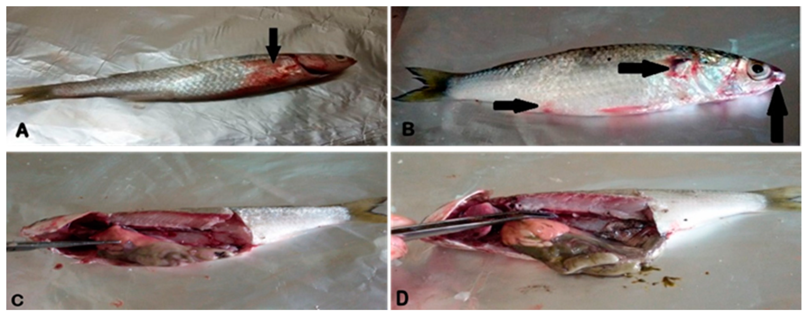
3.1. Clinical and Post-Mortem Findings

In the current study, the clinical inspection of naturally infected M. seheli revealed dark skin discoloration with detached scales (Figure 1A) and distinct hemorrhages at the base of the fins (Figure 1B). Moreover, the post-mortem findings of naturally infected M. seheli revealed hepatomegaly, friable liver with hemorrhagic patches (Figure 1C), and congested kidneys (Figure 1D).

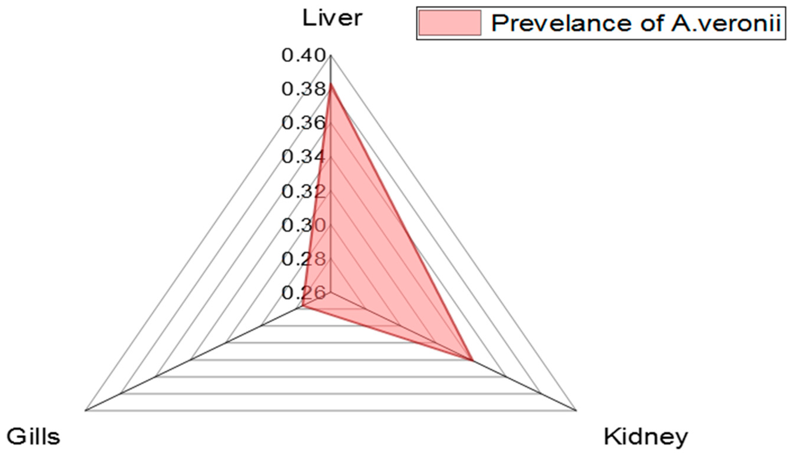
3.2. Phenotypic Features and the Prevalence of A. veronii in the Examined M. seheli

All the recovered A. veronii isolates were Gram-negative, motile, straight rods. After 24 h at 28 °C, the bacteria grew effectively on the TSA medium, giving characteristically creamy, round, convex, shiny colonies. Colonies subsequently appeared green with black centers on Aeromonas-selective agar media. Moreover, the recovered colonies were convex, round, and hemolytic on blood agar, turning dark green after prolonged incubation. Biochemically, the obtained A. veronii isolates tested positive for oxidase, catalase, Voges–Proskauer, gelatin liquefaction, methyl red, casein, starch liquefaction, citrate utilization, and fermentation of glucose and sucrose. Moreover, the recovered A. veronii isolates were negative for H2S production, urea hydrolysis, bile esculin hydrolysis, nitrate reduction, and mannose fermentation.
The prevalence of A. veronii among the examined M. seheli was 22.5% (18/80). To measure the intensity of A. veronii among various examined organs of M. seheli, three different organs (liver, kidney, and gills) from the same fish were examined, with the highest prevalence noticed in the liver (38.3%), then the kidneys (34.1%), and gills (27.6%), as revealed in Table 2 and Figure 2. Statistically, there was no significant difference in the distribution of A. veronii among the examined internal organs of naturally infected M. seheli (p> 0.05).

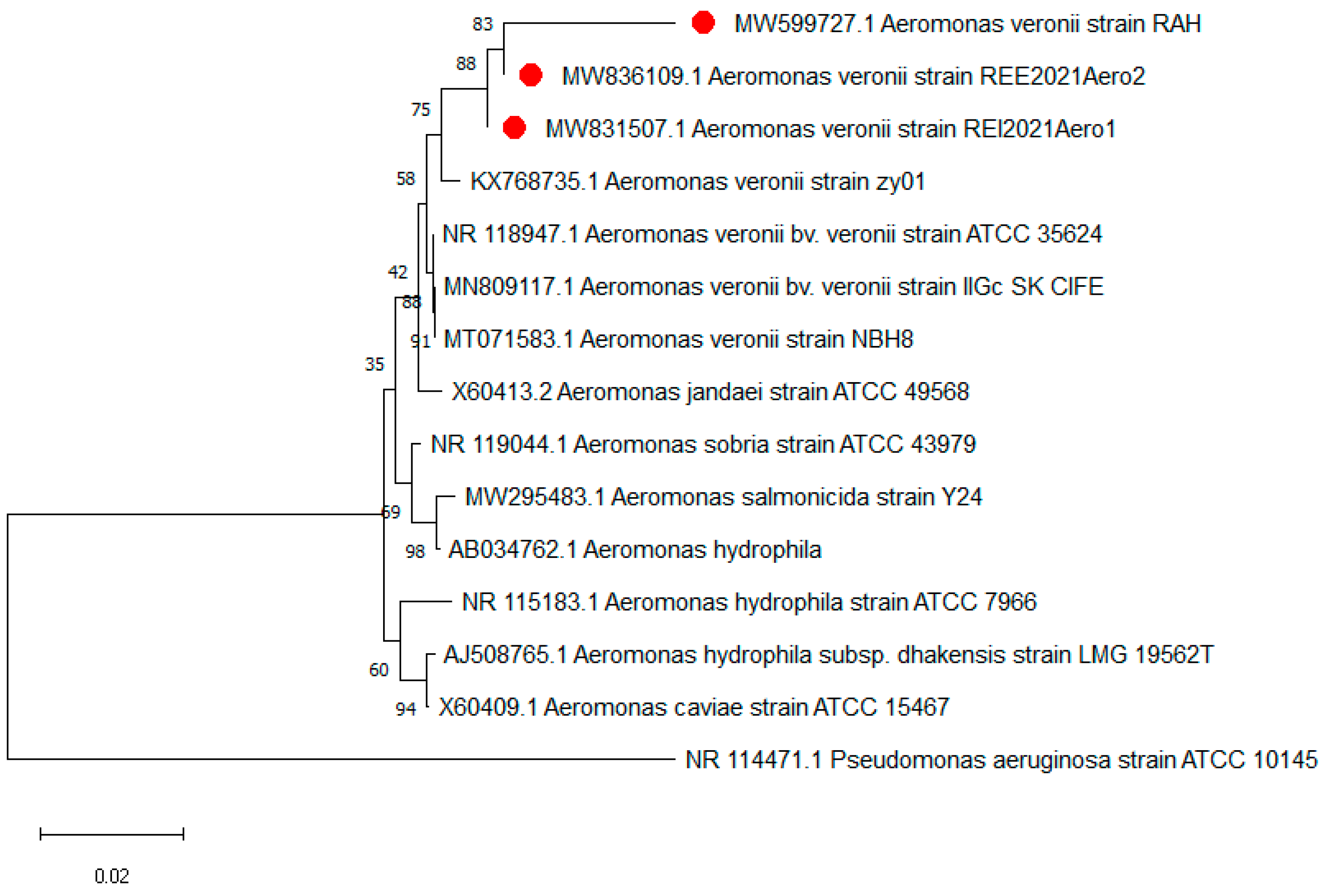
3.3. 16srRNA Gene Sequencing and Phylogenetic Analyses

All the isolated A. veronii strains were positive for the 16srRNA gene. The 16srRNA gene sequencing showed that the tested A. veronii strains (accession nos.: MW831507, MW836109, and MW599727) had a common ancestor. Likewise, the tested A. veronii strains exhibited high similarity of genetic identity compared with other A. veronii strains from different sources, such as A. veronii strain zy01 (accession no.: KX768735) from China (94.5–98.9%), A. veronii strain ATCC35624 (accession no.: NR_118947) from UK (94.8–98.8%), A. veronii strain IIGc_SK_CIFE (accession no.: MN809117) isolated from Nile tilapia in India (94.8–98.8%), and A. veronii strain NBH8 (accession no.: MT071583) from China (94.8–98.8%), as illustrated in Figure 3.

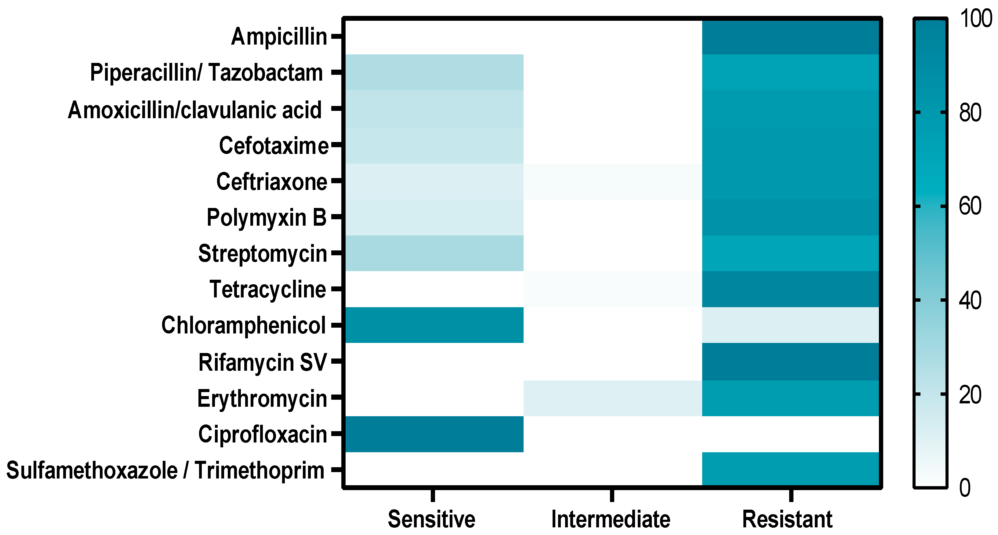
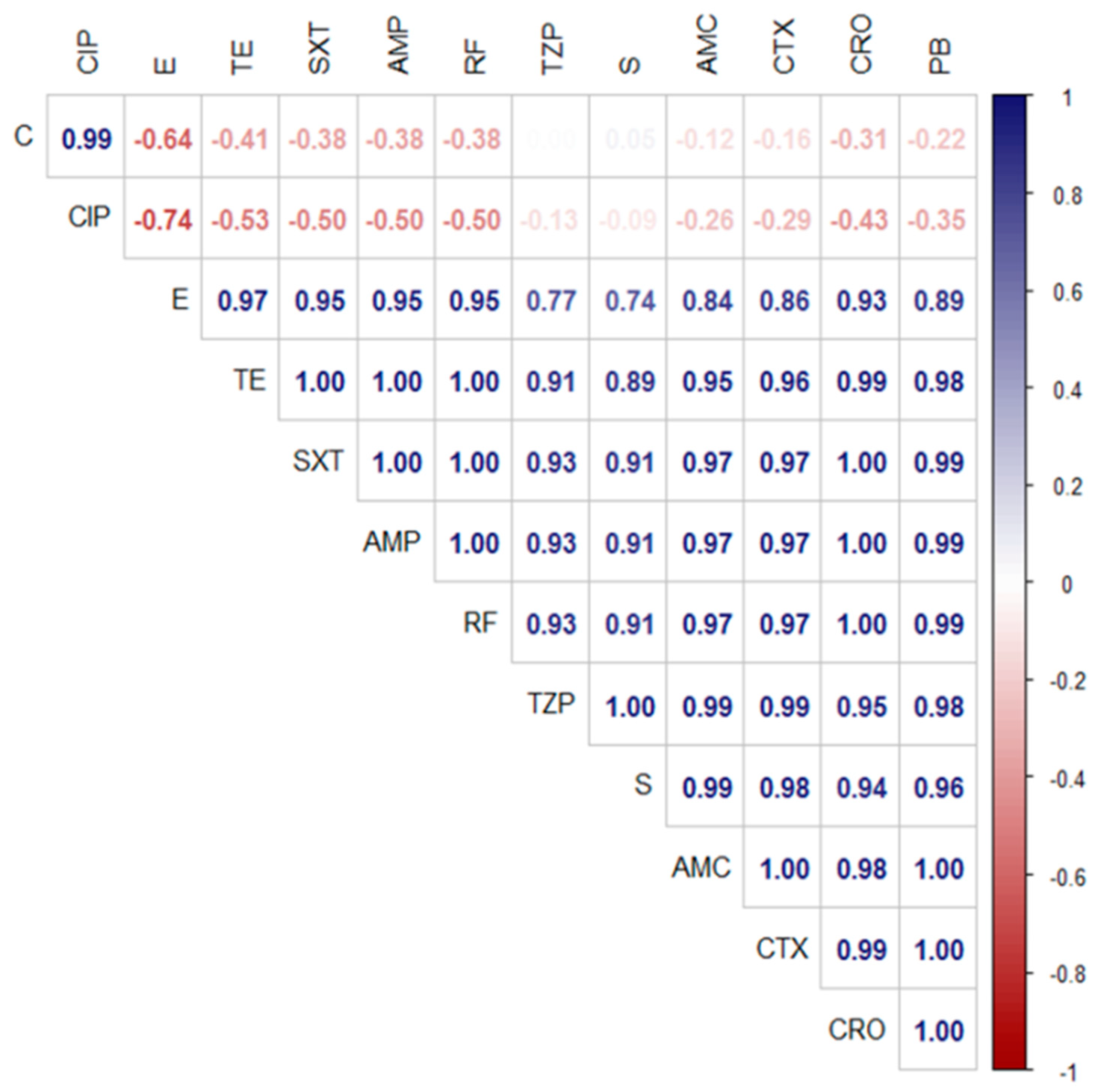
3.4. Antimicrobial Resistance Profiles of the Retrieved A. veronii Isolates

The antimicrobial susceptibility testing revealed that the retrieved A. veronii strains showed significant resistance to different antimicrobial agents including ampicillin, rifamycin SV, sulfamethoxazole/trimethoprim (100% for each), tetracycline (95.7%), polymyxin B (85.1%), cefotaxime, ceftriaxone (80.9% for each), amoxicillin/clavulanic acid (78.8%), erythromycin (76.5%), piperacillin/ tazobactam (72.3%), and streptomycin (70.2 %). Moreover, ciprofloxacin (100%) and chloramphenicol (87.3%) revealed a potent antimicrobial activity against the retrieved A. veronii strains from M. seheli, as indicated in Table 3 and Figure 4. Significant differences (p < 0.05) were observed in the sensitivity of A. veronii isolates to various antibiotics, and remarkable positive correlations were recorded, e.g., TZP, S, and AMC (r = 0.99), SXT, RF, and PB (r = 0.99), TZP and CTX (r = 0.99), TE and CRO (r = 0.99), S and CTX (r = 0.98), TE and PB (r = 0.98), CRO and AMC (r = 0.98), SXT, AMP, RF, and AMC (r = 0.97), E and TE (r = 0.97), SXT, AMP, RF, and CTX (r = 0.97), PB and S (r = 0.96), TE and CTX (r = 0.96), and TE and AMC (r = 0.95), as illustrated in Figure 5.

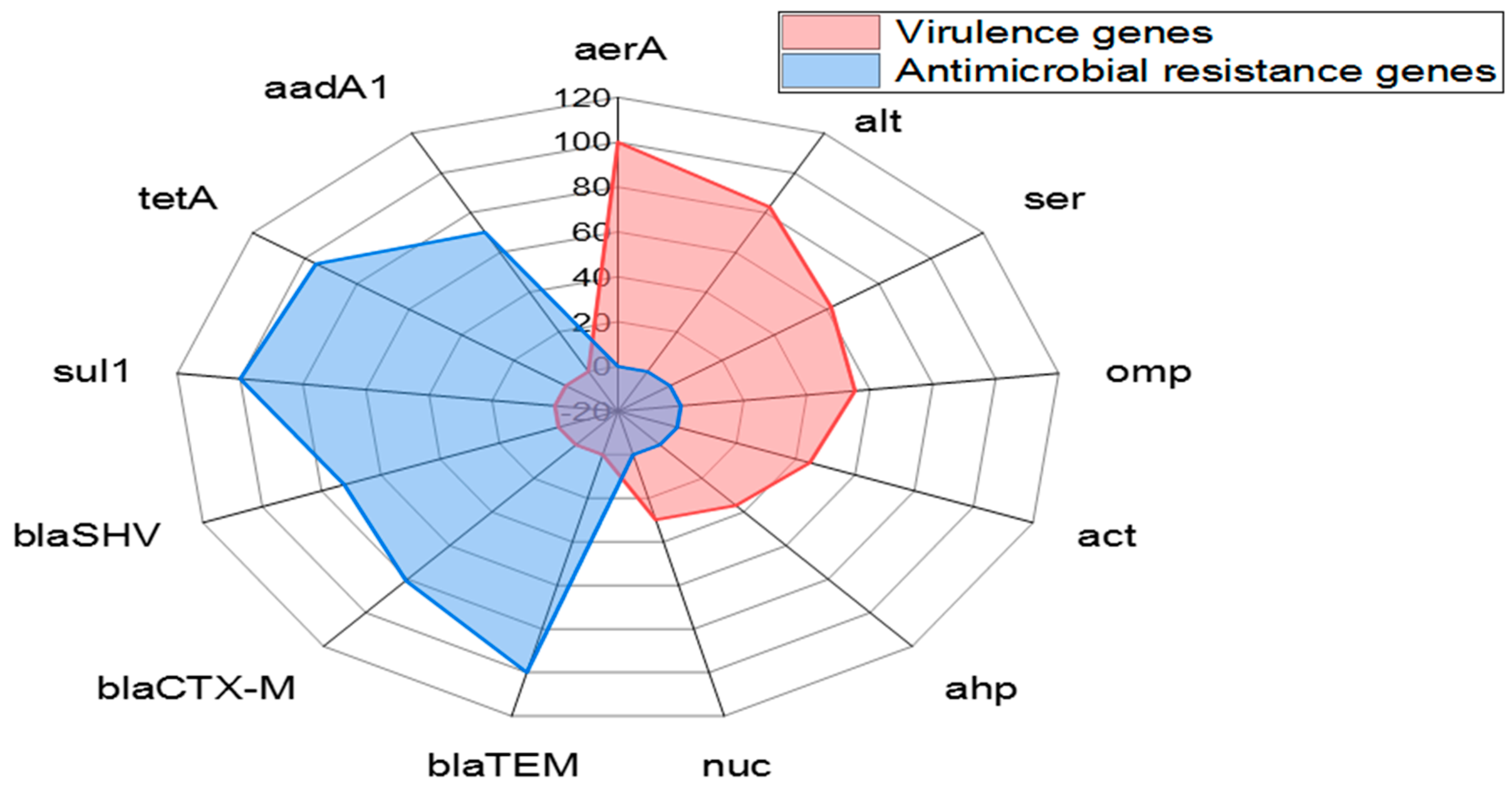
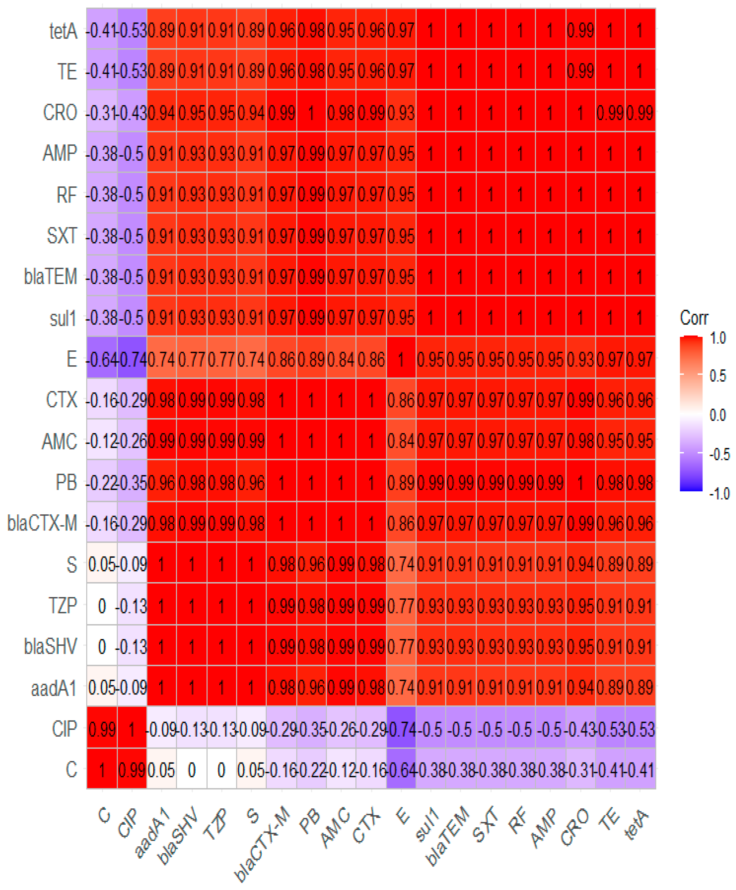
3.5. Dissemination of Virulence-Determinant and Antimicrobial Resistance Genes in the Emerging A. veronii Strains

The PCR indicated that the aerA gene (100%) was the principal virulence gene in A. veronii strains recovered from M. seheli, followed by alt (82.9%), ser (61.7%), ompAII (55.3%), act (44.7%), ahp (36.17 %), and nuc (29.8%) virulence genes. Likewise, the tested A. veronii strains carried the blaTEM, sul1, tetA, blaCTX-M, blaSHV, and aadA1 resistance genes with prevalence of 100%, 100%, 95.7%, 80.9%, 72.3%, and 70.2%, respectively, as revealed in Table 4 and Figure 6. A significant difference (p < 0.05) was noticed in the distribution of virulence-determinant genes in the obtained A. veronii strains. Conversely, there was no significant difference (p > 0.05) in the dissemination of resistance genes among the obtained A.veronii.

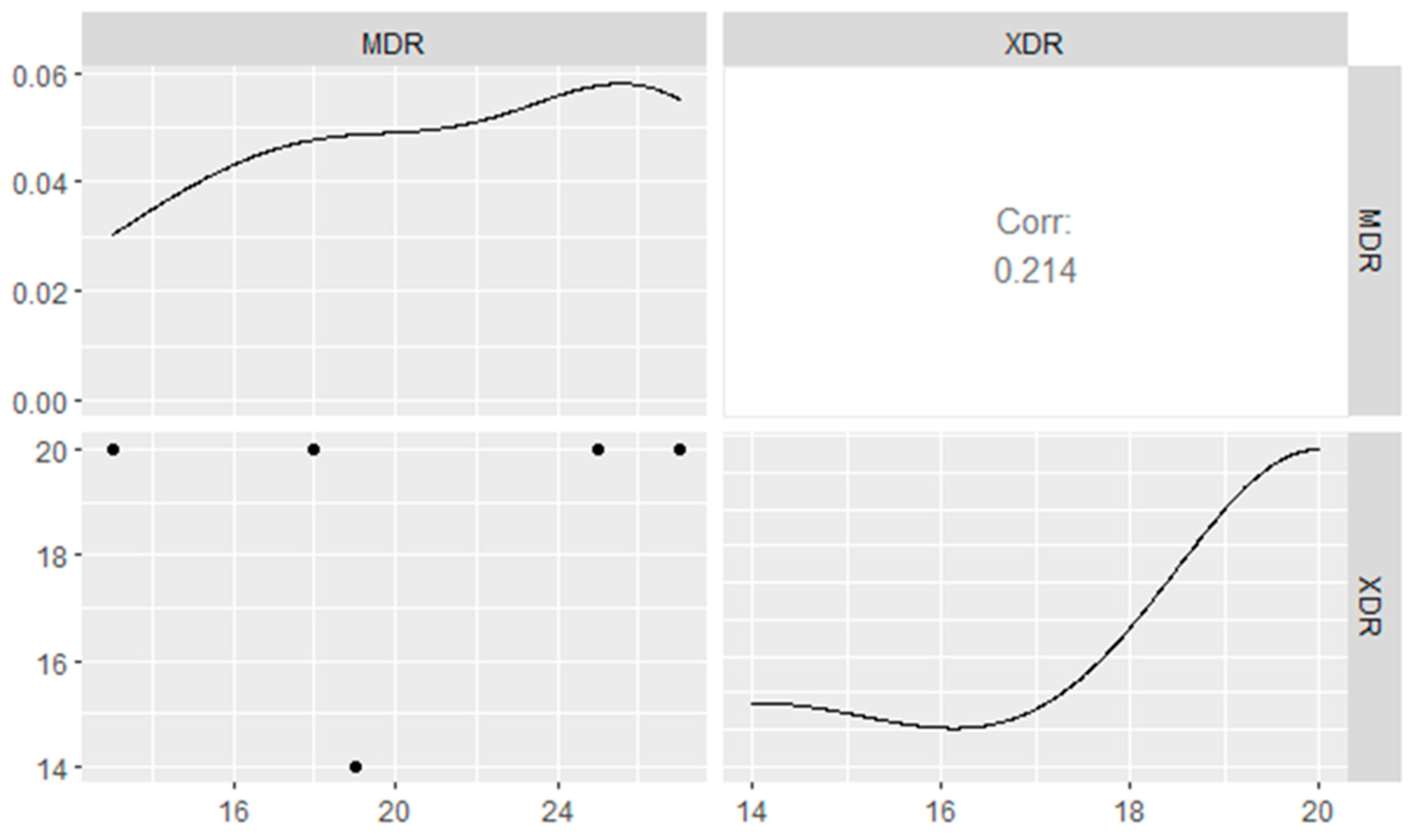
3.6. Genotypic and Phenotypic Multidrug-Resistance Patterns of the Emerging A. veronii Strains

Our findings revealed that 29.8% (14/47) of the recovered A. veronii strains were extensively drug-resistant (XDR) to nine classes and carried blaTEM, blaCTX-M, blaSHV, tetA, aadA1, and sul1 resistance genes. Likewise, 19.1% (9/47) of the obtained strains displayed multi-drug resistance (MDR) to eight antimicrobial classes and possessed blaTEM, blaCTX-M, blaSHV, tetA, aadA1, and sul1 resistance genes. Meanwhile, 14.9% (7/47) of the recovered A. veronii strains were MDR to seven classes and carried blaTEM, blaSHV, tetA, and sul1 genes. In addition, 12.8% (6/47) of the obtained A. veronii strains were XDR to nine different classes and possessed blaTEM, blaCTX-M, aadA1, tetA, and sul1 genes, as shown in Table 5 and Figure 7. Moreover, our findings revealed that the MAR index values were > 0.2, signifying that the A. veronii strains isolated from M. seheli originated from high-risk contamination. Furthermore, the correlation coefficient (r) between the resistance genes detected in A. veronii isolates and the tested antimicrobial agents was estimated. Remarkable positive correlations were recorded, including blaTEM gene and AMP (r = 1), blaCTX-M and CTX (r = 1), tetA gene and TE (r = 1), sul1 gene and STX (r = 1), aadA1 gene and S (r = 1), blaSHV and TZP (r = 1), blaCTX-M and CRO (r = 0.99), blaSHV gene, AMC, and CTX (r = 0.99), blaTEM gene and AMC (r = 0.97), blaSHV gene and CRO (r = 0.95), blaSHV and AMP (r = 0.93), and blaTEM gene and TZP (r = 0.93), as shown in Figure 8.

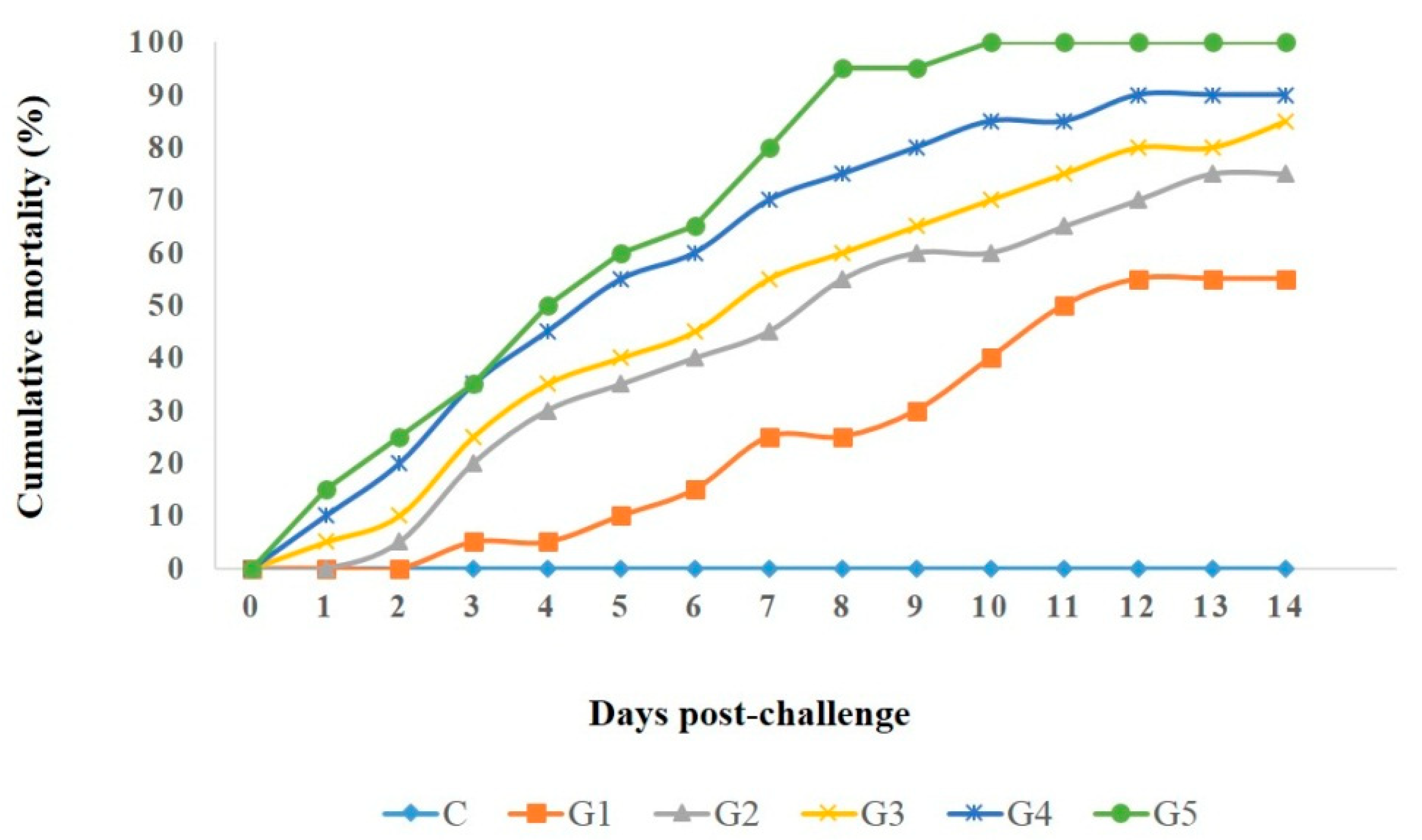
3.7. Pathogenicity Test

Five A. veronii strains (harboring one, two, four, five, and seven virulence genes, respectively) were selected for the pathogenicity test, as illustrated in Table 6. The clinical signs, pathological lesions, morbidity, and mortality rates in the different groups were monitored for 14 days after challenge. The results showed that fish in the control group had no deaths or pathologic lesions. In contrast, the other groups had substantial mortality rates and septicemic lesions, identical to those reported in naturally infected fish, including dark skin discoloration with detached scales, skin ulcers, and distinct hemorrhages at the base of the fins. Moreover, the mortality rate positively correlated with the virulence-determinant genes, and the highest mortality rate (100%) was recorded in the group (G5) inoculated with A. veronii Strain 5 which harbored seven virulence genes (as described in Figure 9). Clinically, the majority of infected fish showed detached scales, darkness of the skin, a hemorrhagic vent, slow movement, and hemorrhagic patches, mainly at the base of fins. Post-mortem inspection demonstrated that the tested fish exhibited characteristic septicemia, including enlarged kidneys, a congested liver, and accumulated bloody serous fluid in the abdominal cavity. Furthermore, A. veronii was re-isolated from various internal organs of the diseased and dead fish.

4. Discussion

Aeromonads are the most common septicemic bacterial fish pathogens, and are considered emerging food-borne pathogens associated with a significant threat to public health [46]. In the present work, the findings of clinical and post-mortem examinations were consistent with the results of [47,48,49], who observed congested gills, scattered hemorrhages on the skin, and detached scales, in addition to congested, friable and enlarged liver, and degenerative changes in the kidneys and spleen of fish naturally infected with Aeromonads. The degree of pathological alterations and the mortality rate are correlated with the severity of infection, fish immunity, and virulence determinants of Aeromonas species [50,51].
During the bacteriological examination, all retrieved isolates were recognized as A. veronii according to their morphological and biochemical features, and the recovered isolates revealed coordination of their phenotypic features. These results were consistent with those recorded by [6], who recovered A. veronii from O. niloticus in Egypt.
In the present study, A. veronii was recovered from moribund M. seheli with a prevalence of 22.5%, and the liver was the most predominant affected organ. The prevalence of A. veronii in this study was higher than that described by [6], who recorded only three isolates from diseased O. niloticus, and nearly similar to that reported by [11], who isolated 87 A. veronii strains from freshwater fish. A. veronii affects a variety of fish species and can live in environments where it may pose harm to the aquaculture industry and threaten food safety [52]. The prevalence of infection is attributed mainly to various predisposing variables, including stress resulting from fish density in intensive systems, poor management, poor hygienic conditions, poor water quality, insufficient oxygen, inappropriate pH, and temperature [14].
Aeromonas species are difficult to differentiate at the species level by conventional methods, due to the lack of a precise biochemical scheme to discriminate between them. Hence, molecular identification is essential for the differential diagnosis of Aeromonas species. The technique of 16S rRNA sequencing is one of the most reliable molecular methods for identifying A. veronii [52]. In this study, all recovered isolates of A. veronii tested positive for 16S rRNA using specific primers. Moreover, the 16SrRNA phylogenetic analysis highlighted that the tested A. veronii strains originated from a common ancestor (accession nos: MW831507, MW836109, and MW599727). Furthermore, the tested A. veronii strains revealed a remarkable genetic similarity with other A. veronii strains from different geographical regions, such as A. veronii strain zy01 from China, A. veronii strain IIGc_SK_CIFE from India, A. veronii strain NBH8 from China, and A. veronii strain ATCC35624 from UK [53]. These results emphasize the epidemiological map and underline the public health significance of A. veronii.
Regarding the antibiogram profiling, ciprofloxacin showed an optimistic antimicrobial activity against the retrieved A. veronii strains from M. seheli. Areomonads are generally susceptible to fluoroquinolones [12]. In contrast, the retrieved A. veronii strains were highly resistant to sulfonamides, penicillin, tetracycline, cephalosporin, β-Lactam-β-lactamase-inhibitor combination, polymyxin, aminoglycosides, and macrolides. Our findings were similar to those recorded by [9,47]. The resistance of A. veronii to various antibiotics affects the health of animals and humans. Inappropriate application of antibiotics in the aquaculture system and the capability of A. veronii to obtain resistance genes from other MDR pathogens are the key predisposing causes contributing to the emergence of multiple drug-resistant superbugs. Therefore, regular use of antimicrobial sensitivity tests and screening for the existence of MDR strains are essential for selecting suitable antibiotics. The emergence of multidrug resistance in bacterial pathogens is attributed mainly to the propagation of antimicrobial resistance genes by horizontal transfer mediated by plasmids [54,55].
The detection of virulence-determinant genes is vital for understanding their potential pathogenicity and the prevention of probable infectious disease [56]. In this study, PCR revealed that the tested A. veronii strains frequently carried the aerA gene, followed by alt, ser, ompAII, act, ahp, and nuc virulence genes. Our findings are consistent with the results of [7,14,56]. Screening of virulence-determinant genes is a vital tool for identifying the possible pathogenicity of Aeromonads [57]. The pathogenicity of A. veronii is related to the expression of certain virulence determinants. Its pathogenicity is attributed mainly to the aerolysin toxin, cytotonic enterotoxins, serine proteases, outer membrane protein, and nuclease enzymes that are encoded by aerA, alt, act, ser, ahp, ompAII, and nuc genes, respectively [6,58]. The aer gene encodes for aerolysin toxin, which plays a significant role in the occurrence of infection. Aerolysin toxin is the primary virulence-determinant factor in Aeromonads, contributing to disease pathogenesis [7]. Moreover, cytotoxic enterotoxins (encoded by alt and act genes) and aerolysin toxin are essential virulence determinants for Aeromonads, and are categorized as potent foodborne pathogens. Both of these virulence determinants exert a substantial effect on the pathogenesis of disease [58]. Protease enzymes (encoded by ser and ahp genes) are common in Areomonads; they play a significant role in the proliferation of bacteria. Furthermore, they endorse the destruction of the mucosa and discoloration of the scales in fish, facilitating the invasion of bacterial pathogens. Serine proteases are characterized by potent caseinolytic activity [59]. The outer membrane proteins (encoded by the ompA gene) are responsible for mucosal adhesion in A. veronii. They exert a significant role in the attachment of A. veronii to the intestinal mucosa of the host [60].
Concerning the multi-drug resistance patterns in the retrieved A. veronii strains, a high percentage of the recovered A. veronii was XDR to nine different classes and carried blaTEM, blaCTX-M, blaSHV, tetA, aadA1, and sul1 resistance genes. Furthermore, most of the isolated A. veronii were MDR to seven or eight different classes and possessed blaTEM, blaCTX-M, blaSHV, tetA, sul1, and aadA1 resistance genes. Multi-drug resistance is thought to be one of the major hazards to public health across the world. It occurs due to the misuse of antibiotics in the aquaculture sector and in medical practice, and may include acquisition of antimicrobial resistance genes via mobile genetic elements [55,61,62,63]. The blaTEM and blaSHV resistance genes mainly mediate resistance to penicillin. Interestingly, the blaTEM gene is the most predominant β-lactamase gene, commonly found in Aeromonads [14,64]. The resistance to sulfonamides and tetracycline is attributed mainly to the sul1and tetA resistance genes, respectively, which were the most predominant resistance genes found in this study. This was similar to the results of [64], who stated that tetracycline- and trimethoprim-resistance genes were demonstrated in all A. veronii genomes, an observation attributed mainly to the wide use of tetracycline and trimethoprim/sulfamethoxazole in the health sector and in veterinary settings. Moreover, the blaCTX-M gene is responsible for cephalosporin resistance as well as resistance to β-Lactam-β-lactamase-inhibitor combinations. Furthermore, aadA1 is one of the most common aminoglycoside-resistance genes. The development of genes encoding antibiotic resistance on either the bacterial chromosome or plasmid is commonly attributed to the widespread unregulated use of antibiotics. The remarkable increase in antimicrobial resistance represents a rising obstacle in the treatment of diseases caused by MDR pathogens in humans and fish, and is considered a public health threat [40,55,65].
In the results of the pathogenicity tests, fish challenged with A. veronii showed different mortality rates that positively correlated with the prevalence of virulence genes in the inoculated strain. They exhibited typical clinical signs observed in naturally infected fish. These findings are similar to the results reported by [66]. The pathogenicity testing highlighted the virulence and pathogenicity of the A. veronii strains recovered from M. seheli. The pathogenicity tests revealed that the more virulence genes carried by a strain, the higher was the mortality rate.

5. Conclusions

In summary, to the best of our knowledge, this is the first study to have revealed the occurrence of XDR and MDR A. veronii strains in M. seheli. The recovered A. veronii strains commonly harbored the aerA, alt, ser, ompAII, and act virulence genes. The emerging A. veronii strains were XDR or MDR to several antimicrobial classes (for example, sulfonamides, penicillin, tetracycline, cephalosporin, β-Lactam-β-lactamase-inhibitor combination, polymyxin, aminoglycosides, and macrolides) and frequently carried blaTEM, sul1, tetA, blaCTX-M, blaSHV, and aadA1 resistance genes. Ciprofloxacin revealed optimistic antimicrobial activity against the XDR and MDR A. veronii strains retrieved from M. seheli. Conventional isolation methods and molecular assays are reliable epidemiological tools for identifying A. veronii in fish. Distressingly, the occurrence of XDR and MDR A. veronii strains is currently recognized as a public health threat, which moreover adversely affects the fish industry. Accordingly, regular practice of antimicrobial sensitivity tests and the proper use of antibiotics are called for in the aquaculture and health sectors.

Author Contributions

A.M.A., R.A.I., K.J.A., H.G., S.A., A.K., A.R.K., G.M.A.-E., N.M.A.-E., M.G.D., and R.M.E.-T. made significant contributions to the work reported, whether in the conception, study design, execution, acquisition of data, analysis and interpretation, or in all these areas, took part in drafting, revising, or critically reviewing the article, gave final approval to the version to be published, agreed on the journal to which the article was submitted, and agreed to be accountable for all aspects of the work. All authors have read and agreed to the published version of the manuscript.

Funding

This research received no external funding.

Institutional Review Board Statement

The handling of fish and all the experiments were approved by the Animal Ethics Review Committee of Suez Canal University (AERC-SCU), Egypt (approval no.: 201859).

Informed Consent Statement

Not applicable.

Data Availability Statement

Not applicable.

Conflicts of Interest

The authors declare no conflict of interest.

References

  1. FAO. The State of World Fisheries and Aquaculture. Contributing to Food Security and Nutrition for All; FAO: Rome, Italy, 2016. [Google Scholar]
  2. Abouelmaatti, R.R.; Algammal, A.M.; LI, X.; MA, J.; Abdelnaby, E.A.; Elfeil, W.M. Cloning and analysis of Nile tilapia Toll-like receptors type-3 mRNA. Centr. Eur. J. Immunol. 2013, 38, 277–282. [Google Scholar] [CrossRef]
  3. Shaalan, M.; El-Mahdy, M.; Saleh, M.; El-Matbouli, M. Aquaculture in Egypt: Insights on the Current Trends and Future Perspectives for Sustainable Development. Rev. Fish. Sci. Aquac. 2018, 26, 99–110. [Google Scholar] [CrossRef]
  4. Mehanna, S.F.; El-Sherbeny, A.S.; El-Mor, M.; Eid, N.M. Age, Growth and Mortality of Liza Carinata Valenciennes, 1836 (Pisces: Mugilidae) in Bitter Lakes, Suez Canal, Egypt. Egypt. J. Aquat. Biol. Fish. 2019, 23, 283–290. [Google Scholar] [CrossRef] [Green Version]
  5. Nelson, J.S. Fishes of the World, 4th ed.; John Wiley & Sons: Hoboken, NJ, USA, 2006. [Google Scholar]
  6. Abd El Latif, A.M.A.; Elabd, H.; Amin, A.; Eldeen, A.I.N.; Shaheen, A.A. High Mortalities Caused by Aeromonas veronii: Identification, Pathogenicity, and Histopathological studies in Oreochromis niloticus. Aquac. Int. 2019, 27, 1725–1737. [Google Scholar] [CrossRef]
  7. Nawaz, M.; Khan, S.A.; Khan, A.A.; Sung, K.; Tran, Q.; Kerdahi, K.; Steele, R. Detection and Characterization of Virulence Genes and Integrons in Aeromonas veronii Isolated from Catfish. Food Microbiol. 2010, 27, 327–331. [Google Scholar] [CrossRef]
  8. Hoai, T.; Trang, T.; Van Tuyen, N.; Aquaculture, N.G. Aeromonas veronii Caused Disease and Mortality in Channel Catfish in Vietnam. Aquaculture 2019, 513, 734425. [Google Scholar] [CrossRef]
  9. Shameena, S.S.; Kumar, K.; Kumar, S.; Kumar, S.; Rathore, G. Virulence Characteristics of Aeromonas veronii Biovars Isolated from Infected Freshwater Goldfish (Carassius Auratus). Aquaculture 2020, 518, 734819. [Google Scholar] [CrossRef]
  10. Cai, S.; Wu, Z.; Jian, J.; Lu, Y.; Tang, J. Characterization of Pathogenic Aeromonas veronii Bv. Veronii Associated with Ulcerative Syndrome from Chinese Longsnout Catfish (Leiocassis Longirostris Günther). SciELO Bras. 2012, 43, 382–388. [Google Scholar] [CrossRef] [Green Version]
  11. Li, T.; Raza, S.H.A.; Yang, B.; Sun, Y.; Wang, G.; Sun, W.; Qian, A.; Wang, C.; Kang, Y.; Shan, X. Aeromonas veronii Infection in Commercial Freshwater Fish: A Potential Threat to Public Health. Animals 2020, 10, 608. [Google Scholar] [CrossRef] [Green Version]
  12. Janda, J.M.; Abbott, S.L. The Genus Aeromonas: Taxonomy, Pathogenicity, and Infection. Clin. Microbiol. Rev. 2010, 23, 35–73. [Google Scholar] [CrossRef]
  13. Fernandez-Bravo, A.; Fort-Gallifa, I.; Ballester, F.; Pujol, I.; Gomez-Bertomeu, F.; Dominguez, M.; Micó, M.; Alcoceba, E.; Simó-Sisó, J.M.; Figueras, M.J. A Case of Aeromonas Trota in an Immunocompromised Patient with Diarrhea. Microorganisms 2020, 8, 399. [Google Scholar] [CrossRef] [Green Version]
  14. Algammal, A.; Mohamed, M.; Tawfiek, B.; Pathogens, W.H. Molecular Typing, Antibiogram and PCR-RFLP Based Detection of Aeromonas Hydrophila Complex Isolated from Oreochromis niloticus. Pathogens 2020, 9, 238. [Google Scholar] [CrossRef] [Green Version]
  15. Sun, J.; Zhang, X.; Gao, X.; Jiang, Q.; Wen, Y.; Lin, L. Characterization of Virulence Properties of Aeromonas veronii Isolated from Diseased Gibel Carp (Carassius Gibelio). Int. J. Mol. Sci. 2016, 17, 496. [Google Scholar] [CrossRef]
  16. Hossain, S.; De-Silva, B.C.J.; Dahanayake, P.S.; Heo, G.J. Characterization of Virulence Properties and Multi-Drug Resistance Profiles in Motile Aeromonas Spp. Isolated from Zebrafish (Danio Rerio). Lett. Appl. Microbiol. 2018, 67, 598–605. [Google Scholar] [CrossRef]
  17. Zhang, D.X.; Kang, Y.H.; Song, M.F.; Shu, H.P.; Guo, S.N.; Jia, J.P.; Tao, L.T.; Zhao, Z.L.; Zhang, L.; Wang, C.F.; et al. Identity and Virulence Properties of Aeromonas Isolates from Healthy Northern Snakehead (Channa Argus) in China. Lett. Appl. Microbiol. 2019, 69, 100–109. [Google Scholar] [CrossRef]
  18. Abu-Elala, N.; Abdelsalam, M.; Marouf, S.; Setta, A.; Abu-Elala, N.; Abdel-Salam, M. Comparative Analysis of Virulence Genes, Antibiotic Resistance and GyrB-based Phylogeny of Motile Aeromonas Species Isolates from Nile Tilapia and Domestic Fowl. Lett. Appl. Microbiol. 2015, 61, 429–436. [Google Scholar] [CrossRef]
  19. Castro-Escarpulli, G.; Aguilera, G.; Castro-Escarpulli, G.; Figueras, M.J.; Aguilera-Arreola, G.; Soler, L.; Fernández-Rendón, E.; Aparicio, G.O.; Guarro, J.; Chacón, M.R. Characterisation of Aeromonas Spp. Isolated from Frozen Fish Intended for Human Consumption in Mexico. Int. J. Food Microbiol. 2003, 84, 41–49. [Google Scholar] [CrossRef]
  20. Algammal, A.M.; Hashem, H.R.; Al-Otaibi, A.S.; Alfifi, K.J.; El-Dawody, E.M.; Mahrous, E.; Hetta, H.F.; El-Kholy, A.W.; Ramadan, H.; El-Tarabili, R.M. Emerging MDR-Mycobacterium avium subsp. avium in house-reared domestic birds as the first report in Egypt. BMC Microbiol. 2021, 21, 237. [Google Scholar] [CrossRef]
  21. Kareem, S.M.; Al-kadmy, I.M.S.; Kazaal, S.S.; Mohammed Ali, A.N.; Aziz, S.N.; Makharita, R.R.; Algammal, A.M.; Al-Rejaie, S.; Behl, T.; Batiha, G.E.-S.; et al. Detection of gyrA and parC Mutations and Prevalence of Plasmid-Mediated Quinolone Resistance Genes in Klebsiella pneumoniae. Infect. Drug Resist. 2021, 14, 555–563. [Google Scholar] [CrossRef]
  22. Algammal, A.M.; Alfifi, K.J.; Mabrok, M.; Alatawy, M.; Abdel-moneam, D.A.; Alghamdi, S.; Azab, M.M.; Ibrahim, R.A.; Hetta, H.F.; El-Tarabili, R.M. Newly Emerging MDR B. cereus in Mugil Seheli as the First Report Commonly Harbor Nhe, Hbl, CytK, and Pc-Plc Virulence Genes and bla1, bla2, tetA, and ermA. Infect. Drug Resist. 2022, 15, 2167–2185. [Google Scholar] [CrossRef]
  23. Cabello, F.C. Heavy Use of Prophylactic Antibiotics in Aquaculture: A Growing Problem for Human and Animal Health and for the Environment. Environ. Microbiol. 2006, 8, 1137–1144. [Google Scholar] [CrossRef] [PubMed]
  24. WHO. Report of a Joint FAO/OIE/WHO Expert Consultation on Antimicrobial Use in Aquaculture and Antimicrobial Resistance; WHO: Seoul, Korea, 2006.
  25. Colquhoun, D.J.; Aarflot, L.; Melvold, C.F. GyrA and Par C Mutations and Associated Quinolone Resistance in Vibrio anguillarum Serotype O2b Strains Isolated from Farmed Atlantic Cod (Gadus Morhua) in Norway. Antimicrob. Agents Chemother. 2007, 51, 2597–2599. [Google Scholar] [CrossRef] [PubMed] [Green Version]
  26. Austin, B.; Austin, D. Characteristics of the pathogens: Gram-negative bacteria. In Bacterial Fish Pathogens; Springer: Berlin/Heidelberg, Germany, 2007; pp. 81–150. [Google Scholar]
  27. Noga, E.J. Fish Disease: Diagnosis and Treatment; John Wiley & Sons: Hoboken, NJ, USA, 2010. [Google Scholar]
  28. Monir, M.S.; Borty, S.C.; Bagum, N.; Rahman, M.K.; Islam, M.A.; Mahmud, Y. Identification of Pathogenic Bacteria Isolated from Diseased Stinging Catfish, Shing (Heteropneustes Fossilis) Cultured in Greater Mymensingh, Bangladesh. Asian Australas. J. Biosci. Biotechnol. 2016, 1, 116–124. [Google Scholar]
  29. Eissa, I.A.M.; Maather, E.; Mona, S.; Desuky, E.; Mona, Z.; Bakry, M. Aeromonas veronii biovar sobria a causative agent of mass mortalities in cultured Nile Tilapia in El-Sharkia governorate, Egypt. Life Sci. J. 2015, 12, 90–97. [Google Scholar]
  30. Gordon, L.; Giraud, E.; Ganiè, J.-P.; Armand, F.; Bouju-Albert, A.; De La Cotte, N.; Mangion, C.; Le Bris, H. Antimicrobial Resistance Survey in a River Receiving Effluents from Freshwater Fish Farms. J. Appl. Microbiol. 2007, 102, 1167–1176. [Google Scholar] [CrossRef]
  31. Tamura, K.; Nei, M.; Kumar, S. Prospects for Inferring Very Large Phylogenies by Using the Neighbor-Joining Method. Proc. Natl. Acad. Sci. USA 2004, 101, 11030–11035. [Google Scholar] [CrossRef] [Green Version]
  32. Kumar, S.; Stecher, G.; Li, M.; Knyaz, C.; Tamura, K. MEGA X: Molecular Evolutionary Genetics Analysis across Computing Platforms. Mol. Biol. Evol. 2018, 35, 1547–1549. [Google Scholar] [CrossRef]
  33. CLSI, Clinical and Laboratory Standards Institute. Performance Standards for Antimicrobial Susceptibility Testing for Bacteria Isolated from Animals, 4th ed.; CLSI Supplement VET08; CLSI: Wayne, PA, USA, 2018. [Google Scholar]
  34. Magiorakos, A.P.; Srinivasan, A.; Carey, R.; Carmeli, Y.; Falagas, M.E.; Giske, C.G.; Harbarth, S.; Hindler, J.F.; Kahlmeter, G.; Olsson-Liljequist, B.; et al. Multidrug-Resistant, Extensively Drug-Resistant and Pandrug-Resistant Bacteria: An International Expert Proposal for Interim Standard Definitions for Acquired Resistance. Clin. Microbiol. Infect. 2012, 18, 268–281. [Google Scholar] [CrossRef] [Green Version]
  35. Krumperman, P.H. Multiple Antibiotic Resistance Indexing of Escherichia Coli to Identify High-Risk Sources of Fecal Contamination of Foods. Appl. Environ. Microbiol. 1983, 46, 165–170. [Google Scholar] [CrossRef] [Green Version]
  36. Oliveira, S.T.L.; Veneroni-Gouveia, G.; Costa, M.M. Molecular Characterization of Virulence Factors in Aeromonas hydrophila Obtained from Fish. Pesqui. Vet. Bras. 2012, 32, 701–706. [Google Scholar] [CrossRef]
  37. Nam, I.; Joh, K. Rapid Detection of Virulence Factors of Aeromonas Isolated from a Trout Farm by Hexaplex-PCR. J. Microbiol. 2007, 45, 297–304. [Google Scholar]
  38. Hu, M.; Wang, N.; Pan, Z.H.; Lu, C.P.; Liu, Y.J. Identity and Virulence Properties of Aeromonas Isolates from Diseased Fish, Healthy Controls and Water Environment in China. Lett. Appl. Microbiol. 2012, 55, 224–233. [Google Scholar] [CrossRef]
  39. Wang, H. Comparative Study between Biological Characteristics of the Different Animal Species Aeromonas veronii and Four Virulence Genes. Master’s Thesis, Agricultural University, Jilin, China, 2014. [Google Scholar]
  40. Colom, K.; Pèrez, J.; Alonso, R.; Fernández-Aranguiz, A.; Lariňo, E.; Cisterna, R. Simple and reliable multiplex PCR assay for detection of blaTEM, blaSHV and blaOXA-1 genes in Enterobacteriaceae. FEMS Microbiol. Lett. 2003, 223, 147–151. [Google Scholar] [CrossRef]
  41. Archambault, M.; Petrov, P.; Hendriksen, R.S.; Asseva, G.; Bangtrakulnonth, A.; Hasman, H.; Aarestrup, F.M. Molecular Characterization and Occurrence of Extended-Spectrum β-Lactamase Resistance Genes among Salmonella Enterica Serovar Corvallis from Thailand, Bulgaria, and Denmark. Microb. Drug Resist. 2006, 12, 192–198. [Google Scholar] [CrossRef]
  42. Randall, L.P.; Cooles, S.W.; Osborn, M.K.; Piddock, L.J.V.; Woodward, M.J. Antibiotic Resistance Genes, Integrons and Multiple Antibiotic Resistance in Thirty-Five Serotypes of Salmonella Enterica isolated from Humans and Animals in the UK. J. Antimicrob. Chemother. 2004, 53, 208–216. [Google Scholar] [CrossRef] [Green Version]
  43. Ibekwe, A.M.; Murinda, S.E.; Graves, A.K. Genetic Diversity and Antimicrobial Resistance of Escherichia coli from Human and Animal Sources Uncovers Multiple Resistances from Human Sources. PLoS ONE 2011, 6, e20819. [Google Scholar] [CrossRef] [Green Version]
  44. El-Bahar, H.M.; Ali, N.G.; Aboyadak, I.M.; Khalil, S.A.E.S.; Ibrahim, M.S. Virulence Genes Contributing to Aeromonas hydrophila Pathogenicity in Oreochromis Niloticus. Int. Microbiol. 2019, 22, 479–490. [Google Scholar] [CrossRef]
  45. Azzam-Sayuti, M.; Ina-Salwany, M.Y.; Zamri-Saad, M.; Annas, S.; Yusof, M.T.; Monir, M.S.; Mohamad, A.; Muhamad-Sofie, M.H.N.; Lee, J.Y.; Chin, Y.K.; et al. Comparative Pathogenicity of Aeromonas spp. in Cultured Red Hybrid Tilapia (Oreochromis niloticus × O. mossambicus). Biology 2021, 10, 1192. [Google Scholar] [CrossRef]
  46. Igbinosa, I.H.; Igumbor, E.U.; Aghdasi, F.; Tom, M.; Okoh, A.I. Emerging Aeromonas Species Infections and Their Significance in Public Health. Sci. World J. 2012, 2012, 625023. [Google Scholar] [CrossRef] [Green Version]
  47. Jagoda, S.S.; Wijewardana, T.G.; Arulkanthan, A.; Igarashi, Y.; Tan, E.; Kinoshita, S.; Watabe, S.; Asakawa, S. Characterization and antimicrobial susceptibility of motile aeromonads isolated from freshwater ornamental fish showing signs of septicaemia. Dis. Aquat. Org. 2014, 109, 127–137. [Google Scholar] [CrossRef] [Green Version]
  48. Monir, M.S.; Bagum, N.; Kabir, S.L.; Borty, S.C.; Doulah, M.A. Isolation, Molecular Identification and Characterization of Aeromonas hydrophila from Infected Air-Breathing Catfish Magur (Clarias Batrachus) Cultured in Mymensingh, Bangladesh. Asian Australas. J. Food Saf. Secur. 2017, 1, 17–24. [Google Scholar] [CrossRef]
  49. Abdel-moneam, D.A.; Ibrahim, R.A.; Nashaat, M.; Shaalan, M. Multifactorial Causes of Mass Mortality in Oreochromis niloticus in Kafr El-Sheikh, Egypt, 198. Bull. Eur. Ass. Fish Pathol. 2021, 41, 2021. [Google Scholar]
  50. Ling, S.H.M.; Wang, X.H.; Lim, T.M.; Leung, K.Y. Green Fluorescent Protein -Tagged Edwardsiella tarda Reveals Portal of Entry in Fish. FEMS Microbiol. Lett. 2001, 194, 239–243. [Google Scholar] [CrossRef] [PubMed] [Green Version]
  51. Elsheshtawy, A.; Yehia, N.; Elkemary, M.; Soliman, H. Investigation of Nile tilapia Summer Mortality in Kafr El-Sheikh Governorate, Egypt. Genet. Aquat. Org. 2019, 3, 17–25. [Google Scholar] [CrossRef]
  52. Abbott, S.L.; Cheung, W.K.W.; Janda, J.M. The Genus Aeromonas: Biochemical Characteristics, Atypical Reactions, and Phenotypic Identification Schemes. J. Clin. Microbiol. 2003, 41, 2348–2357. [Google Scholar] [CrossRef] [Green Version]
  53. Martinez-Murcia, A.J.; Benlloch, S.; Collins, M.D. Phylogenetic interrelationships of members of the genera Aeromonas and Plesiomonas as determined by 16S ribosomal DNA sequencing: Lack of congruence with results of DNA-DNA hybridizations. Int J. Syst. Bacteriol. 1992, 42, 412–421. [Google Scholar] [CrossRef] [Green Version]
  54. Smyrli, M.; Prapas, A.; Rigos, G.; Kokkari, C.; Pavlidis, M.; Katharios, P. Aeromonas veronii infection associated with high morbidity and mortality in farmed European seabass Dicentrarchus labrax in the Aegean Sea, Greece. Fish Pathol. 2017, 52, 68–81. [Google Scholar] [CrossRef] [Green Version]
  55. Algammal, A.M.; Mabrok, M.; Ezzat, M.; Alfifi, K.J.; Aboelkheir, M.; Esawy, A.M.; Elmasry, N.; El-Tarabili, R.M. Prevalence, antimicrobial resistance (AMR) pattern, virulence determinant and AMR genes of emerging multi-drug resistant Edwardsiella tarda in Nile tilapia and African catfish. Aquaculture 2022, 548, 737643. [Google Scholar] [CrossRef]
  56. Zhu, M.; Wang, X.R.; Li, J.; Li, G.Y.; Liu, Z.P.; Mo, Z.L. Identification and Virulence Properties of Aeromonas veronii Bv. Sobria Isolates Causing an Ulcerative Syndrome of Loach Misgurnus Anguillicaudatus. J. Fish Dis. 2016, 39, 777–781. [Google Scholar]
  57. Yogananth, N.; Bhakyaraj, R.; Chanthuru, A.; Anbalagan, T.; Nila, K.M. Detection of Virulence Gene in Aeromonas hydrophila Isolated from Fish Samples Using PCR Technique. Glob. J. Biotechnol. Biochem. 2009, 4, 51–53. [Google Scholar]
  58. Sha, J.; Kozlova, E.V.; Chopra, A. Role of Various Enterotoxins in Aeromonas hydrophila-Induced Gastroenteritis: Generation of Enterotoxin Gene-Deficient Mutants and Evaluation of Their Enterotoxic Activity. Infect. Immun. 2002, 70, 1924–1935. [Google Scholar] [CrossRef] [Green Version]
  59. Beaz-Hidalgo, R.; Figueras, M.J. Aeromonas spp. whole genomes and virulence factors implicated in fish disease. J. Fish Dis. 2013, 36, 371–388. [Google Scholar] [CrossRef]
  60. Namba, A.; Mano, N.; Takano, H.; Beppu, T.; Ueda, K.; Hirose, H. OmpA Is an Adhesion Factor of Aeromonas veronii, an Optimistic Pathogen That Habituates in Carp Intestinal Tract. J. Appl. Microbiol. 2008, 105, 1441–1451. [Google Scholar] [CrossRef]
  61. Piotrowska, M.; Popowska, M. The Prevalence of Antibiotic Resistance Genes among Aeromonas Species in Aquatic Environments. Ann. Microbiol. 2014, 64, 921–934. [Google Scholar] [CrossRef]
  62. Elgendy, M.Y.; Shaalan, M.; Abdelsalam, M.; Eissa, A.E.; El-Adawy, M.M.; Seida, A.A. Antibacterial activity of silver nanoparticles against antibiotic-resistant Aeromonas veronii infections in Nile tilapia, Oreochromis niloticus (L.), in vitro and in vivo assay. Aquac. Res. 2022, 53, 901–920. [Google Scholar] [CrossRef]
  63. Ali, S.E.; Mahana, O.; Mohan, C.V.; Delamare-Deboutteville, J.; Elgendy, M.Y. Genetic characterization and antimicrobial profiling of bacterial isolates collected from Nile tilapia (Oreochromis niloticus) affected by summer mortality syndrome. J. Fish Dis. 2022, 00, 1–15. [Google Scholar] [CrossRef]
  64. Tekedar, H.C.; Arick, M.A.; Hsu, C.Y.; Thrash, A.; Blom, J.; Lawrence, M.L.; Abdelhamed, H. Identification of Antimicrobial Resistance Determinants in Aeromonas veronii Strain MS-17-88 Recovered from Channel Catfish (Ictalurus Punctatus). Front. Cell. Infect. Microbiol. 2020, 10, 348. [Google Scholar] [CrossRef]
  65. Fauzi, F.; Hamdan, R.H.; Mohamed, M.; Tan, L.P. Detection of Integron and Antibiotic Resistance Genes of Aeromonas Spp. Isolated from Freshwater Fish in Pahang, Malaysia. Charting Sustain. In Charting the Sustainable Future of ASEAN in Science and Technology; Springer: Berlin/Heidelberg, Germany, 2020; pp. 473–486. [Google Scholar] [CrossRef]
  66. Chen, F.; Sun, J.; Han, Z.; Yang, X.; Xian, J.A.A.; Lv, A.; Hu, X.; Shi, H. Isolation, Identification and Characteristics of Aeromonas veronii from Diseased Crucian Carp (Carassius Auratus Gibelio). Front. Microbiol. 2019, 10, 2742. [Google Scholar] [CrossRef]
Figure 1. M. seheli showing (A): Erosion of gill cover and skin hemorrhages, (B): Erosion and hemorrhages in the opercular region, anus, and base of the fins, (C): Friable pale liver and congested kidney, (D): Congested kidney.
Pathogens 11 01262 g001
Figure 2. The distribution of A. veronii among different internal organs of naturally infected M. seheli.
Pathogens 11 01262 g002
Figure 3. Phylogenetic tree based on 16S rRNA gene sequencing. The tree clarifies the genetic relatedness of the tested A. veronii strains and other strains deposited in the GenBank database. The tested strains in the present study are indicated with red circles.
Pathogens 11 01262 g003
Figure 4. Antimicrobial susceptibility patterns of A. veronii isolates retrieved from examined M. seheli samples.
Pathogens 11 01262 g004
Figure 5. The correlation coefficient (r) between various tested antibiotics in the disc diffusion test.
Pathogens 11 01262 g005
Figure 6. Distribution of virulence and antimicrobial resistance genes among A. veronii strains retrieved from the examined M. seheli.
Pathogens 11 01262 g006
Figure 7. Distribution of XDR and MDR patterns among A. veronni strains isolated from the examined M. seheli. The horizontal axis indicates MDR and XDR patterns, while the vertical axis indicates the antimicrobial resistance genes.
Pathogens 11 01262 g007
Figure 8. The correlation coefficient (r) between the tested antibiotics and the identified antimicrobial resistance genes.
Pathogens 11 01262 g008
Figure 9. Cumulative mortality of T. zillii experimentally challenged with different virulent strains of A. veronii (G1-G5: Fish groups received a bacterial dose of 3 × 10⁸ cfu/mL of various A. veronii strains S1–S5, respectively), data were analyzed 14 days after challenge.
Pathogens 11 01262 g009
Table 1. List of primer sequences and PCR cycling conditions.
GenePrimer SequenceCycling Conditions (35 Cycles)Amplified
Product
Reference
Denatur.AnnealingExtension
16SrRNA
Aeromonas species
F: CTACTTTTGCCGGCGAGCGG94 °C for 30 s50 °C for 40 s72 °C for 50 s953 bp[30]
R: TGATTCCCGAAGGCACTCCC
aerA
(Aerolysin)
F: CCTATGGCCTGAGCGAGAAG94 °C for 30 s55.5 °C for 30 s72 °C for 30 s431 bp[36]
R: CCAGTTCCAGTCCCACCACT
ser
(Serine protease)
F: ACGGAGTGCGTTCTTCCTACTCCAG94 °C for 1 min64 °C for 30 s72 °C for 45 s211 bp[37]
R: CCGTTCATCACACCGTTGTAGTCG
nuc
(Nuclease)
F: CAGGATCTGAACCGCCTCTATCAGG94 °C for 1 min64 °C for 30 s72 °C for 45 s504
R: GTCCCAAGCTTCGAACAGTTTACGC
act
Cytotoxic enterotoxin
F: GAGAAGGTGACCACCAAGAACA94 °C for 4 min59 °C for 30 s72 °C for 1 min232 bp[18]
R: AACTGACATCGGCCTTGAACTC
alt
(Heat-labile cytotonic enterotoxin)
F: TGACCCAGTCCTGGCACGGC94 °C for 4 min59 °C for 30 s72 °C for 1 min442 bp[38]
R: GGTGATCGATCACCACCAGC
ahp
(Serine protease)
F: ATTGGATCCCTGCCTA94 °C for 4 min59 °C for 30 s72 °C for 1 min911 bp
R: GCTAAGCTTGCATCCG
ompAII
(Outer membrane protein II)
F:GCTGAATTCATGAAACTCAAAATGGCTC94 °C for 1 min55 °C for 1 min72 °C for 1 min1001[39]
R: GCGAAGCTTTTACTGTTGTACTTGC
blaTEM
(Penicillin-resistance)
F: ATCAGCAATAAACCAGC94 °C
30 s
54 °C
40 s
72 °C
45 s
516[40]
R: CCCCGAAGAACGTTTTC
blaSHV
(Penicillin-resistance)
F: AGGATTGACTGCCTTTTTG94 °C
30 s
54 °C
40 s
72 °C
40 s
392
R: ATTTGCTGATTTCGCTCG
blaCTX-M
(Cephalosporines-resistance)
F: ATGTGCAGYACCAGTAARGTKATGGC94 °C
30 s
54 °C
40 s
72 °C
45 s
593[41]
R: TGGGTRAARTARGTSACCAGAAYC AGC GG
aadA1
(Aminoglycosides-resistance)
F: TATCAGAGGTAGTTGGCGTCAT94 °C
30 s
54 °C
40 s
72 °C
45 s
484[42]
R: GTTCCTAGCGTTAAGGTTTCATT
tetA
(Tetracycline-resistance)
F: GGTTCACTCGAACGACGTCA94 °C
30 s
50 °C
40 s
72 °C
45 s
576
R: CTGTCCGACAAGTTGCATGA
sul1
(sulfonamide-resistance)
F: CGGCGTGGGCTACCTGAACG94 °C
30 s
54 °C
40 s
72 °C
45 s
433[43]
R: GCCGATCGCGTGAAGTTCCG
Table 2. The intensity of A. veronii in different internal organs of naturally infected M. seheli.
OrganNo. of Positive IsolatesPercentage of Positive IsolatesChi Square
p Value
Liver1838.3%0.80851
0.6675 NS *
Kidneys1634.1%
Gills1327.6%
Total47100%
NS * = Non-significant.
Table 3. Antimicrobial resistance profiles of the recovered A. veronii strains (n = 47).
Antimicrobial ClassAntimicrobial
Agents
SensitiveIntermediateResistant
n%n%n%
PenicillinAmpicillin----47100
Piperacillin/ Tazobactam1327.7--3472.3
β-Lactam-β-lactamase-inhibitor combinationAmoxicillin/clavulanic acid1021.2--3778.8
CephalosporinCefotaxime919.1--3880.9
Ceftriaxone612.736.43880.9
PolymyxinPolymyxin B714.9--4085.1
AminoglycosidesStreptomycin1429.8--3370.2
TetracyclineTetracycline--24.34595.7
PhenicolsChloramphenicol4187.3--612.7
AnsamycinRifamycin SV----47100
MacrolidesErythromycin--1123.43676.5
FluroquinolonesCiprofloxacin47100----
SulfonamidesSulfamethoxazole/Trimethoprim----47100
Chi square
p value
252.82
<0.0001
92.875
<0.001
76.817
<0.0001
Table 4. Prevalence of virulence and antimicrobial resistance genes in the retrieved A. veronii strains (n = 47).
Type of GenesGenesNo of Positive Isolates%Chi-Square
p-Value
Virulence genesaerA4710030.891
<0.0001
alt3982.9
ser2961.7
ompAII2655.3
act2144.7
ahp1736.17
nuc1429.8
Antimicrobial resistance genesblaTEM471005.1475
0.3981 NS *
blaCTX-M3880.9
blaSHV3472.3
sul147100
tetA4595.7
aadA13370.2
NS * = Non-significant.
Table 5. Distribution of phenotypic multi-drug resistance patterns and antimicrobial resistance genes among the retrieved A. veronii isolates.
No. of Isolates%Type of ResistancePhenotypic ResistanceResistance GenesMAR
1429.8%XDRNine classes:
  • AMP, TZP
  • AMC
  • RF
  • SXT
  • TE
  • PB
  • CRO, CTX
  • S
  • E
blaTEM,blaCTX-M, blaSHV,tetA, aadA1, and sul10.84
919.1%MDREight classes:
  • AMP, TZP
  • AMC
  • RF
  • SXT
  • TE
  • PB
  • CRO, CTX
  • S
blaTEM, blaCTX-M, blaSHV, tetA, aadA1, and sul10.76
714.9%MDRSeven classes:
  • AMP, TZP
  • AMC
  • RF
  • SXT
  • TE
  • PB
  • E
blaTEM,blaSHV, tetA, and sul10.61
612.8%XDRNine classes:
  • AMP
  • RF
  • SXT
  • TE
  • PB
  • CRO, CTX
  • S
  • E
  • C
blaTEM, blaCTX-M,aadA1, tetA, and sul10.76
510.6%MDRSeven classes:
  • AMP
  • AMC
  • RF
  • SXT
  • TE
  • CRO, CTX
  • E
blaTEM, blaCTX-M, tetA, and sul10.61
48.5%MDREight classes:
  • AMP, TZP
  • RF
  • SXT
  • TE
  • PB
  • CRO, CTX
  • S
  • E
blaTEM blaCTX-M,blaSHV,aadA1, tetA, and sul10.76
24.2%MDRFour classes:
  • AMP
  • AMC
  • RF
  • SXT
blaTEM and sul10.31
Piperacillin/tazobactam (TZP), ampicillin (AMP), amoxicillin/clavulanic acid (AMC), ceftriaxone (CRO), cefotaxime (CTX), polymyxin B (PB), tetracycline (TE), rifamycin SV (RF), erythromycin (E), chloramphenicol (C), streptomycin (S), ciprofloxacin (CIP), and sulfmethoxazole/trimethoprim (SXT).
Table 6. The cumulative mortality rate in different groups.
A. veronii StrainsVirulence GenesCorresponding GroupCumulative Mortality %
Strain 5aerA, alt, ahp, act, ser, nuc, and ompAIIG5100
Strain 4aerA, ompAII, alt, ahp, and actG490
Strain 3aerA, ahp, ser, and nucG385
Strain 2aerA and serG275
Strain 1aerAG155
Publisher’s Note: MDPI stays neutral with regard to jurisdictional claims in published maps and institutional affiliations.

