Article

Molecular Detection of Non-O157 Shiga Toxin-Producing Escherichia coli (STEC) Directly from Stool Using Multiplex qPCR Assays

1
Department of Laboratory Medicine and Pathology, University of Alberta, Edmonton, AB T6G 2R3, Canada
2
Alberta Precision Laboratories-Public Health Laboratory (ProvLab), Edmonton, AB T6G 2J2, Canada
*
Author to whom correspondence should be addressed.
Submission received: 31 December 2021 / Revised: 18 January 2022 / Accepted: 28 January 2022 / Published: 31 January 2022

Abstract

:
Non-O157 Shiga toxin-producing E. coli (STEC) can cause outbreaks that have great economic and health impact. Since the implementation of STEC screening in Alberta in 2018, it is also essential to have a molecular serotyping method with faster turnaround time for cluster identification and surveillance purposes. This study sought to perform molecular serotyping of the top six non-O157 (O26, O45, O103, O111, O121 and O145) STEC serotypes directly from stools and enrichment broths compared to conventional methods on isolates. Multiplex, serotyping qPCR assays were used to determine sensitivity and specificity of the top six non-O157 STEC serotypes. Sensitivity and specificity were assessed for both singleplex and multiplex qPCR assays for comparison of the top six serotypes. Blinded stool specimens (n = 116) or broth samples (n = 482) submitted from frontline microbiology laboratories for STEC investigation were analyzed by qPCR. Both singleplex and multiplex assays were comparable, and we observed 100% specificity with a limit of detection of 100 colony-forming units per mL. Direct molecular serotyping from stool specimens mostly correlated (88%) with conventional serotyping of the cultured isolate. In cases of discordant serotypes, the top six non-O157 STEC mixed infections were identified and confirmed by culture and conventional serotyping. Detection of non-O157 STEC can be done directly from stool specimens using multiplex PCR assays with the ability to identify mixed infections, which would otherwise remain undetected by conventional serotyping of a single colony. This method can be easily implemented into a frontline diagnostic laboratory to enhance surveillance of non-O157 STEC, as more frontline microbiology laboratories move to culture independent assays.

1. Introduction

Shiga toxin-producing Escherichia coli (STEC) are one of the major causes of acute gastroenteritis (AGE) in Canada, which may result in complications and hospitalization [1]. E. coli O157, one of the most prominent STEC serotypes (synonymous with Verotoxigenic Escherichia coli, VTEC), has been implicated in various food-borne outbreaks world-wide [1]. AGE caused by STEC can lead to hemolytic uremic syndrome (HUS), a potentially fatal condition resulting in kidney failure. HUS is due to the presence of two Shiga toxin virulence factors, encoded stx1 and stx2 within the lambdoid prophages that inhibit protein synthesis [2]. The elderly and pediatric populations are most at risk of developing complications during infection. The Stx2 toxin has been shown to be more severe and associated with the development of HUS [3]. Furthermore, STEC require a very low infectious dose (fewer than 100 colony-forming units (CFU/mL)) and therefore have the potential to cause large numbers of outbreaks [4].
Ruminants, particularly cattle, have been identified as the primary reservoir for STEC where they do not cause the same disease but are rather shed into waterways or cause food-associated disease [5]. One of the most widely recognized cases of waterway contamination due to STEC was Walkerton, Ontario in 2000, where 2300 individuals were affected, causing 65 hospitalizations and 7 deaths [6]. Over 600 STEC serotypes have been identified (though not all are associated with human disease) from various sources, including the environment, food, humans and animals, with outbreak serotype predominance varying depending on the geographic region [7,8,9]. In 2011, E. coli O104:H4 was responsible for a major outbreak in Germany that resulted in 3816 cases, 845 HUS and 54 deaths over a period of 3 months [10], clearly demonstrating that STEC non-O157 infection can be catastrophic. In North America, the most commonly observed “top six” serotypes are O26, O45, O103, O111, O121 and O145 [11], which some have, on rare occasions, resulted in HUS (O26, O103, O111 and O121) [12].
Current diagnostic identification of O157 STEC infection relies on culture using MacConkey agar. We have recently shown that CHROMagar™ STEC agar plates were better able to identify non-O157 STEC isolates from stool samples as compared to the traditional Sorbitol–MacConkey agar used in STEC O157 detection [12]. We have also shown that non-O157 STEC isolates can be difficult to enrich in certain broth media, suggesting a potential for mixed infections that can be missed during an outbreak [13]. Culture-independent diagnostic tests (CIDT), which detect the presence of the stx gene(s), are becoming more readily available, allowing for rapid diagnosis, as well as increased surveillance [14,15]. CIDT are optimal for diagnostic identification, as they do not require culture prior to testing, which greatly decreases the turnaround time. However, conventional culture and STEC isolation are required for surveillance and serotype identification purposes using pulsed-field gel electrophoresis and whole genome sequencing [16,17]. In Alberta, STEC (both O157 and non-O157) screening began in 2019 alongside CIDT implementation in frontline microbiology laboratories. STEC identified by frontline laboratories were referred to Alberta Precision Laboratories ProvLab (APL-ProvLab) for isolation using chromogenic agar and colonies confirmed by PCR as part of the surveillance program. Since the implementation of this algorithm, over 60% of STEC detected were non-O157, causing diarrhea in this province. Non-O157 serotyping is performed at the Public Health Agency of the Canada National Microbiology Laboratory (PHAC-NML) located in Winnipeg, Manitoba, and thus the shipping of isolates for serotyping further increases the turnaround time for reporting. This study sought to assess the performance of a molecular serotyping qPCR assay directly from stools and enriched broth culture samples to predict the top six non-O157 (O26, O45, O103, O111, O121 and O145) serotypes as compared to the conventional isolate serotyping method.

2. Materials and Methods

2.1. Sensitivity Assay

One of each of the top six non-O157 STEC (O26, O45, O103, O111, O121 and O145) clinical human isolate stocks were retrieved from frozen skim milk stored at −80 °C and cultured on sheep blood agar plates (BAP) (Dalynn Biologicals, Calgary, AB, Canada). After overnight incubation at 37 °C, single colonies from each culture plate were then grown overnight at 37 °C in Tryptic Soy Broth (TSB) prepared by APL-ProvLab in Edmonton, Alberta, Canada. Each enriched culture broth was diluted to an optical density of 0.5 and performed ten-fold serial dilutions from 10−1 to 10−8. An aliquot of 100 μL was inoculated onto BAP (Dalynn Biologicals, Calgary, Canada) in triplicates and incubated overnight to determine the CFU/mL. Aliquots of cell suspensions containing 102–104 CFU/mL were centrifuged at 13,000× g for 5 min; supernatant was discarded, and DNA was extracted from the pellet by boiling in a rapid lysis buffer (100 mM NaCl, 10 mM Tris-HCL pH 8.3, 1 mM EDTA pH 9.0, 1% Triton X-100) as described previously [18]. An amount of 5 μL DNA was used as template in a total of 25 μL reaction volume for serotyping as singleplex or multiplex assays, as shown in Table 1. The amplification contained 12.5 μL of 1× PrimeTime® Gene Expression Master Mix (Integrated DNA Technology, Skokie, IL, USA), 0.33 μM of each primer, 0.22 μM probe, 5 μL DNA template and molecular biology grade water in a total of 25 μL reaction volume. A positive and no-template controls were included in each run, and the qPCR conditions consisted of 95 °C for 1 min followed by 40 cycles of 95 °C for 5 s and 58 °C for 45 s, performed on the 7500 FAST qPCR system (Applied Biosystems, Foster City, CA, USA).

2.2. Specificity Assay

Specificity was determined using a panel of non-STEC isolates consisting of the following isolates; Aeromonas hydrophila, Salmonella serovar Enteriditis, Salmonella serovar Typhimurium, Shigella boydii (Serotype 2), Shigella flexneri (Serotype 2), Shigella sonnei, Shigella dysenteriae (Serotype 2), Yersinia enterocolitica; and 13 reference strains consisted of Proteus mirabalis (ATCC12453), Campylobacter coli (ATCC33559), Campylobacter jejuni (ATCC33291), Citrobacter freundii (ATCC8090), Klebsiella pneumoniae (ATCC31488), Enterobacter cloacae (ATCC13047), Staphylococcus aureus (ATCC25923), Staphylococcus aureus (ATCC25913), Escherichia coli (ATCC25922), Bacillus cereus (ATCC14579) and Staphylococcus epidermis (ATCC12228). As for inclusivity panel, the following clinical STEC strains (n = 201) were included. These were comprised of O26 (n = 34), O45 (n = 3), O103 (n = 39), O111 (n = 35), O121 (n = 29), O145 (n = 17) in addition to non-top six isolates (n = 44), as shown in Table S1.
Each isolate was grown on BAP (Dalynn Biologicals, Calgary, Canada) under the respective growth conditions; single-colony DNA extraction was performed using a single colony suspended in rapid lysis and performed the procedure as described in previous section. PCR assays were performed as singleplex and multiplex assays with both sets of primers and probes, as described above.

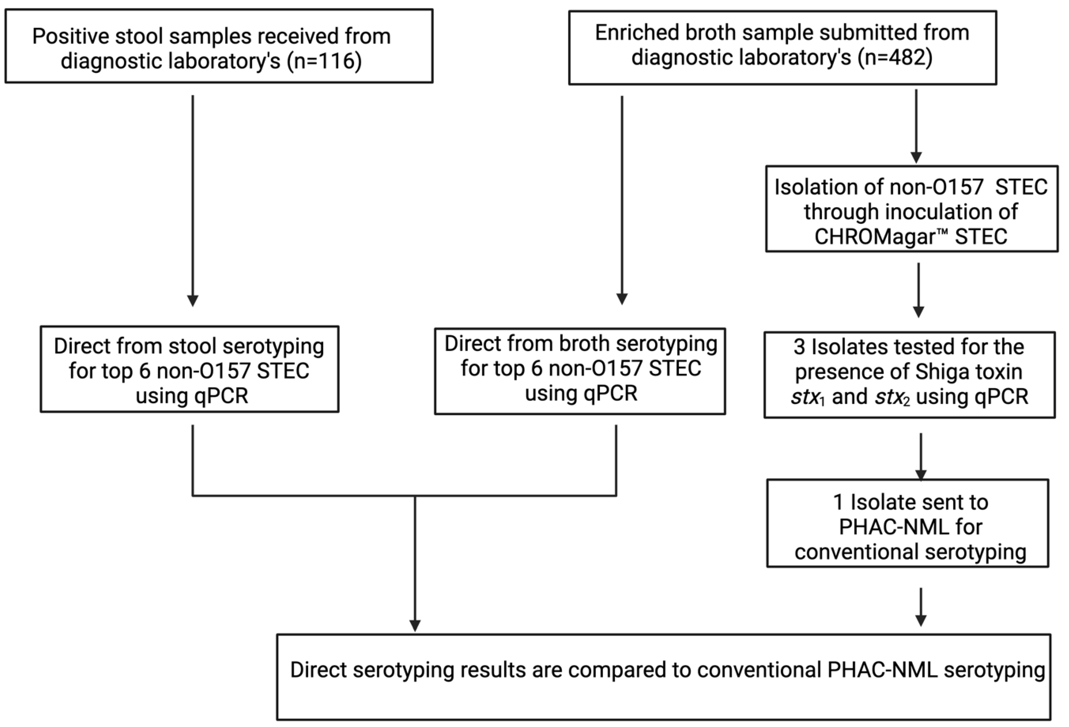
2.3. Patient Samples

All patient samples were either STEC-positive stool samples and/or enriched cultures from positive stool using either Gram Negative broth (GNB) (Dalynn Biologicals, Calgary, Canada) provided by Calgary Laboratory Services or Tryptic Soy Broth (TSB) (Dalynn Biologicals, Calgary, Canada) from DynaLIFE (Edmonton, AB, Canada). These samples were previously screened positive by SHIGA TOXIN QUIK CHEK™ immunoassay (TechLab Inc, Blacksburg, VA, USA) by the providing laboratories as non-O157 STEC. Workflow for all clinical samples is shown in Figure 1, with specific methods in the following Section 2.4 and Section 2.5.

2.4. Direct Molecular Serotyping from Clinical Samples

Enriched culture in TSB and GNB (n = 482) were extracted using rapid lysis buffer. An amount of 250 µL of enriched culture was centrifuged (13,000× g for 10 min), supernatant was discarded, and the cell pellet was resuspended in rapid lysis buffer and heated to 95 °C for 15 min using a heating block (Thermo Fisher, Mississauga, Canada). The samples were then briefly centrifuged (13,000× g for 10 min), and the supernatant stored at 4 °C until qPCR was performed. Cycle threshold above 0.1 Delta Rn and Ct value ≤ 36 were considered positive.
DNA from stool samples (n = 116) were extracted using the NucliSENS easyMag system (bioMerieux, Montreal, QC, Canada). Approximately 10 µL loop of stool was suspended in 1 mL of NucliSENS Lysis buffer in SK38 soil grinding lysis bead tubes (Luminex Corporation, Toronto, ON, Canada) and shaken on a vortex at max speed for 10 min; a sample was left at room temperature for 15 min and followed by centrifugation (15,871× g for 5 min). A total of 200 µL was extracted and with a final elution volume of 70 µL.
The primers’ and probes’ (Integrated DNA Technology, Coralville, IA, USA) design was based on reference sequences [19], as shown in Table 1. The total reaction contained 12.5 μL of 1× PrimeTime® Gene Expression Master Mix (Integrated DNA Technology, Coralville, USA), 0.33 μM of each primer, 0.22 μM probe, 5 μL DNA template and molecular biology grade water in a total of 25 μL reaction volume. A no-template control was included in each run. qPCR amplification conditions consisted of 95 °C for 1 min, followed by 40 cycles of 95 °C for 5 s and 58 °C for 45 s performed on the 7500 FAST qPCR system (Applied Biosystems, Mississauga, Canada).

2.5. Isolation of Non-O157 STEC

All enriched broth cultures (100 μL) originally from positive STEC clinical stool samples were inoculated onto CHROMagar™ STEC plates (Micronostyx, Ottawa, Canada) for strain isolation and further characterization. Individual mauve color colonies (n = 3) of the same morphotype were picked from each plate and suspended in rapid lysis buffer, heated, centrifuged, and 5 μL of the supernatant was used for STEC confirmation using our in-house qPCR. Once confirmed as STEC by stx identification, the isolate was forwarded to PHAC-NML for conventional serotyping. Serotyping results from stools, enriched cultures and isolates were compared, and if discordance was observed, then the samples were considered to contain >1 serotypes indicative of a mixed infection. Additional isolates were picked for qPCR serotyping to identify the remaining serotype and were subsequently sent to the PHAC-NML for confirmation (Figure 1).

2.6. Statistics

Sensitivity and specificity data were analyzed using Excel (Microsoft Office). Comparison between singleplex and multiplex was analyzed with a Wilcoxon matched pairs signed-rank test using Prism9 for Mac (Graph Pad, San Diego, CA, USA).

3. Results

3.1. Sensitivity and Specificity

The singleplex and multiplex assays using primers and probes originating from Perelle et al. [19] were found to be highly specific and sensitive, with a detection limit of 1 × 10² CFU/mL, as shown in Table 2. The results between the singleplex and multiplex of both assays do not show major differences (p > 0.05), as illustrated by the crossing point of the qPCR (Table 2). As for the specificity assay, no cross reactivity was observed between the isolates of the non-top six STEC or the exclusivity panel of different bacterial species. We observed 100% specificity, as well as a true positive rate of 100%, based on the 157 top six isolates, for the qPCR assays.

3.2. Clinical Samples Testing

We were able to determine the serotype from stool samples (n = 116) as well as enriched broths (n = 482) from stools using the direct molecular serotyping method. Isolates obtained from the enrichment broths were sent to the PHAC-NML for conventional serotyping and were compared to the direct-from-stool/broth serotyping assay. Concordance between direct serotyping from stool, broth and conventional isolate serotyping were found for 102 samples (88%) (Table 3); 1 sample was found to have discordance between direct serotyping from stool and broth to conventional serotyping (Table 3). The majority of the discordance was found to be between direct serotyping from stool (stool-only column) to direct serotyping from broth and conventional isolate serotyping, with one stool being positive for two top-six serotypes (n = 13 samples, 11.2%) (Table 3). Concordance was also measured with the remaining broth samples (n = 366 samples), which we did not receive a matching stool sample for (Table 4). We observed a 97.5% concordance between broths and isolates. There were eight serotypes that were identified through the enriched broths, and other serotypes were found when the isolates were picked from agar plates for traditional serotyping. The STEC isolates that failed to be typed by the top six molecular serotypes were classified as non-top six, and their serotypes were determined by the PHAC-NML, as shown in Supplemental Table S2. Next, we compared the stx characteristics between the isolates and found most of the infections were from stx1 (Table 5).

3.3. Mixed Infections between Broth and Stool Samples

Any discordant sample between direct-from-stool/broth qPCR and conventional serotyping were treated as if they were mixed infections and were further investigated. For discordant serotyping, results from one stool and eight additional broth samples, which were initially typed as top six, but the picked colony determined by conventional typing gave discordant results, further colonies from the CHROMagar™ STEC plates were subjected to qPCR to confirm as mixed infection. Colonies from six of these samples were subtyped by conventional serotyping, and the confirmed mixed infections are shown in Table 6. For the remaining three samples, we failed to isolate any additional colonies, although the broths tested positive by PCR. In addition, there were 13 stool samples with discordant results as compared to broth and conventional serotyping, which we were not able to isolate. There was no pattern with regard to serotypes involved in mixed infections, and most were discovered when the in-house qPCR detected a top-six serotype, but the PHAC-NML result from the single isolate was reported as a non-top-six serotype (Table S3).

4. Discussion

STEC remain one of the most prevalent food-borne associated infectious diseases in Canada, with Alberta having the highest burden/prevalence of the disease [20]. Currently, little data have been published on the incidence of non-O157 infections within Alberta prior to the implementation of routine STEC screening in 2018. Although the O157 serotype is the most recognized cause of E. coli-induced gastroenteritis, non-O157 outbreaks have been increasing in recent years. According to the EU One Health Zoonosis report in 2020, there was an overall trend for reporting STEC cases between 2015 to 2019 due to the awareness of the importance of STEC detection associated with outbreaks in the EU [21]. In Canada, there was a recommendation in 2018 from the Canadian Public Health Laboratories Network for all Canadian laboratories to detect all STEC, especially the non-O157, for better surveillance purposes [14]. An outbreak of O121 in 2016 linked to wheat flour was confirmed using pulsed-field gel electrophoresis and whole genome sequencing [22], of which both techniques require growth of the pathogen. Culture is essential for cluster detection and surveillance; however, direct stool serotyping will give the laboratory an early indication of the top six non-O157 STEC serotypes along with the stx gene status. Culture is still required after this initial step, but it will shorten the turnaround time for reporting. Our direct stool serotyping method is only limited to the top six serotypes, which consisted of the majority (72.4%) of samples tested, but any non-top-six samples still require conventional serotyping method at the PHAC-NML.
Sensitivity and specificity were comparable between both multiplex and singleplex assays, indicating that the multiplex assay is sufficient for implementation to support existing STEC surveillance programs. The primers also showed high specificity, indicating they could distinguish between the different serotypes, which is crucial in cases of mixed infections. When clinical samples were tested, O26 was the predominant serotype identified, followed by O111. Surprisingly, there were multiple non-top six STEC involved in diarrheal disease found in both stool and enriched broth samples. As for mixed infections, all the confirmed samples contained both top-six and non-top-six serotypes and were found through discordance between serotyping methods. This suggests that our current approach likely underestimates the number of non-top-six serotypes involved in infections and outbreaks.
One of the limitations of this study is the fact that we only analyzed the top six serotypes, and it is possible there are mixed infections with less common serotypes of STEC. However, it would not be economical for diagnostic laboratories to screen every sample for hundreds of potential mixed infections with very rare serotypes. As only a single colony was picked from a plate to be sent for conventional serotyping, it is possible that non-top-six STEC can be missed; therefore, more colonies could be picked and conventionally serotyped to increase surveillance of the non-top-six STEC. Although we determined the limit of detection was 100 cells using pure culture, it is still possible that the bacterial load in a stool sample is below the detectable limit, and therefore a serotype may false-negatively be assumed to be non-top six. We observed seven stool samples where the top-six serotype was not detectable until after enrichment in TSB broth, which is a limitation of relying solely on stool samples for any diagnostic test. However, enrichment into broth introduces the possibility that mixed infections may be missed due to the growth of the more abundant serotype. Another possibility of missing serotypes is the fact that stool samples are incredibly complex and may contain inhibitors that interfere with qPCR, particularly for those serotypes that have a low abundance, which is likely why we had a higher number of discordances with stool samples (Table S3). Interestingly, this seems to have been the case with O103 stool samples (five samples), as most of the mismatched isolates (only detected by one of the serotyping methods) were due to this serotype. This may indicate O103 primers as being less sensitive, particularly when dealing with stool samples, as they also had a higher Ct value for pure culture as compared to the other serotypes (Table S2).
In conclusion, we have shown that direct-from-stool serotyping can be performed for positive STEC stools using multiplex qPCR. Furthermore, we have identified the potential for mixed infections to occur. This assay can be implemented alongside routine diagnostic tools for rapid identification of the top six serotypes that cause the majority of STEC outbreaks in North America.

Supplementary Materials

The following supporting information can be downloaded at: https://0-www-mdpi-com.brum.beds.ac.uk/article/10.3390/microorganisms10020329/s1, Table S1: List of all non-top 6 isolates used for inclusivity panel; Table S2: List of all non-top 6 isolates determined by conventional serotyping; Table S3: List of discordant samples.

Author Contributions

Conceptualization, B.P. and L.C.; methodology, B.P.; formal analysis, M.B.-J.; investigation, M.B.-J., B.P., C.L. and J.S.; writing—original draft preparation, M.B.-J.; writing—review and editing, B.P., C.L., J.S. and L.C.; supervision, L.C.; funding acquisition, L.C. All authors have read and agreed to the published version of the manuscript.

Funding

This research was supported by Alberta Health Residual Funds.

Institutional Review Board Statement

Not applicable.

Informed Consent Statement

Not applicable.

Data Availability Statement

Not applicable.

Acknowledgments

The authors would like to thank Christina Ferrato, Joanne Callfas, the Bacterial Typing Unit at Alberta Precision Laboratories: Provincial Public Health Laboratory, AB and the Public Health Agency of Canada-National Microbiology Laboratory for help in providing serotyping. We would also like to thank Calgary Laboratory Services and DynaLIFE for providing stool and enrichment broth cultures.

Conflicts of Interest

The authors have no conflict of interest to declare.

References

  1. Thomas, M.K.; Murray, R.; Flockhart, L.; Pintar, K.; Fazil, A.; Nesbitt, A.; Marshall, B.; Tataryn, J.; Pollari, F. Estimates of foodborne illness-related hospitalizations and deaths in Canada for 30 specified pathogens and unspecified agents. Foodborne Pathog. Dis 2015, 12, 820–827. [Google Scholar] [CrossRef] [PubMed]
  2. Bryan, A.; Youngster, I.; McAdam, A.J. Shiga Toxin Producing Escherichia coli. Clin. Lab. Med. 2015, 35, 247–272. [Google Scholar] [CrossRef] [PubMed]
  3. Fuller, C.A.; Pellino, C.A.; Flagler, M.J.; Strasser, J.E.; Weiss, A.A. Shiga toxin subtypes display dramatic differences in potency. Infect. Immun. 2011, 79, 1329–1337. [Google Scholar] [CrossRef] [PubMed] [Green Version]
  4. Crockett, C.S.; Haas, C.N.; Fazil, A.; Rose, J.B.; Gerba, C.P. Prevalence of shigellosis in the U.S.: Consistency with dose-response information. Int. J. Food Microbiol. 1996, 30, 87–99. [Google Scholar] [CrossRef] [Green Version]
  5. Menge, C. The Role of Escherichia coli Shiga Toxins in STEC Colonization of Cattle. Toxins 2020, 12, 607. [Google Scholar] [CrossRef] [PubMed]
  6. Hrudey, S.E.; Payment, P.; Huck, P.M.; Gillham, R.W.; Hrudey, E.J. A fatal waterborne disease epidemic in Walkerton, Ontario: Comparison with other waterborne outbreaks in the developed world. Water Sci. Technol. 2003, 47, 7–14. [Google Scholar] [CrossRef] [PubMed]
  7. Karama, M.; Cenci-Goga, B.T.; Malahlela, M.; Smith, A.M.; Keddy, K.H.; El-Ashram, S.; Kabiru, L.M.; Kalake, A. Virulence Characteristics and Antimicrobial Resistance Profiles of Shiga Toxin-Producing Escherichia coli Isolates from Humans in South Africa: 2006–2013. Toxins 2019, 11, 424. [Google Scholar] [CrossRef] [PubMed] [Green Version]
  8. Peng, Z.; Liang, W.; Hu, Z.; Li, X.; Guo, R.; Hua, L.; Tang, X.; Tan, C.; Chen, H.; Wang, X.; et al. O-serogroups, virulence genes, antimicrobial susceptibility, and MLST genotypes of Shiga toxin-producing Escherichia coli from swine and cattle in Central China. BMC Vet. Res. 2019, 15, 427. [Google Scholar] [CrossRef]
  9. De Rauw, K.; Jacobs, S.; Pierard, D. Twenty-seven years of screening for Shiga toxin-producing Escherichia coli in a university hospital. Brussels, Belgium, 1987–2014. PLoS ONE 2018, 13, e0199968. [Google Scholar] [CrossRef]
  10. Beutin, L.; Martin, A. Outbreak of Shiga toxin-producing Escherichia coli (STEC) O104:H4 infection in Germany causes a paradigm shift with regard to human pathogenicity of STEC strains. J. Food Prot. 2012, 75, 408–418. [Google Scholar] [CrossRef]
  11. Fratamico, P.M.; Bagi, L.K.; Abdul-Wakeel, A. Detection and Isolation of the "Top Seven" Shiga Toxin-Producing Escherichia coli in Ground Beef: Comparison of RapidFinder Kits to the U.S. Department of Agriculture Microbiology Laboratory Guidebook Method. J. Food Prot. 2017, 80, 829–836. [Google Scholar] [CrossRef]
  12. Zelyas, N.; Poon, A.; Patterson-Fortin, L.; Johnson, R.P.; Lee, W.; Chui, L. Assessment of commercial chromogenic solid media for the detection of non-O157 Shiga toxin-producing Escherichia coli (STEC). Diagn. Microbiol. Infect. Dis. 2016, 85, 302–308. [Google Scholar] [CrossRef] [PubMed]
  13. Bording-Jorgensen, M.; Tyrrell, H.; Lloyd, C.; Chui, L. Comparison of Common Enrichment Broths Used in Diagnostic Laboratories for Shiga Toxin—Producing Escherichia coli. Microorganisms 2020, 9, 503. [Google Scholar] [CrossRef]
  14. Chui, L.; Christianson, S.; Alexander, D.C.; Arseneau, V.; Bekal, S.; Berenger, B.; Chen, Y.; Davidson, R.; Farrell, D.J.; German, G.J.; et al. CPHLN recommendations for the laboratory detection of Shiga toxin-producing Escherichia coli (O157 and non-O157). Can. Commun. Dis. Rep. 2018, 44, 304–307. [Google Scholar] [CrossRef]
  15. Marder, E.P.; Cieslak, P.R.; Cronquist, A.B.; Dunn, J.; Lathrop, S.; Rabatsky-Ehr, T.; Ryan, P.; Smith, K.; Tobin-D’Angelo, M.; Vugia, D.J.; et al. Incidence and Trends of Infections with Pathogens Transmitted Commonly Through Food and the Effect of Increasing Use of Culture-Independent Diagnostic Tests on Surveillance-Foodborne Diseases Active Surveillance Network, 10 U.S. Sites, 2013–2016. MMWR. Morb. Mortal. Wkly. Rep. 2017, 66, 397–403. [Google Scholar] [CrossRef] [Green Version]
  16. Jaros, P.; Dufour, M.; Gilpin, B.; Freeman, M.M.; Ribot, E.M. PFGE for Shiga toxin-producing Escherichia coli O157:H7 (STEC O157) and non-O157 STEC. Methods Mol. Biol. 2015, 1301, 171–189. [Google Scholar] [CrossRef]
  17. Bogaerts, B.; Nouws, S.; Verhaegen, B.; Denayer, S.; Van Braekel, J.; Winand, R.; Fu, Q.; Crombe, F.; Pierard, D.; Marchal, K.; et al. Validation strategy of a bioinformatics whole genome sequencing workflow for Shiga toxin-producing Escherichia coli using a reference collection extensively characterized with conventional methods. Microb. Genom. 2021, 7, 531. [Google Scholar] [CrossRef]
  18. Holland, J.L.; Louie, L.; Simor, A.E.; Louie, M. PCR detection of Escherichia coli O157:H7 directly from stools: Evaluation of commercial extraction methods for purifying fecal DNA. J. Clin. Microbiol. 2000, 38, 4108–4113. [Google Scholar] [CrossRef] [Green Version]
  19. Perelle, S.; Dilasser, F.; Grout, J.; Fach, P. Detection by 5’-nuclease PCR of Shiga-toxin producing Escherichia coli O26, O55, O91, O103, O111, O113, O145 and O157:H7, associated with the world’s most frequent clinical cases. Mol. Cell. Probes 2004, 18, 185–192. [Google Scholar] [CrossRef]
  20. Reynolds, C.; Checkley, S.; Chui, L.; Otto, S.; Neumann, N.F. Evaluating the risks associated with Shiga-toxin-producing Escherichia coli (STEC) in private well waters in Canada. Can. J. Microbiol. 2020, 66, 337–350. [Google Scholar] [CrossRef]
  21. European Food Safety Authority; European Centre for Disease Prevention and Control. The European Union One Health 2020 Zoonoses Report. EFSA J. 2021, 19, 324. [CrossRef]
  22. Gill, A.; Carrillo, C.; Hadley, M.; Kenwell, R.; Chui, L. Bacteriological analysis of wheat flour associated with an outbreak of Shiga toxin-producing Escherichia coli O121. Food Microbiol. 2019, 82, 474–481. [Google Scholar] [CrossRef] [PubMed]
Figure 1. STEC serotyping workflow comparing enriched broth culture and stool direct serotyping to conventional serotyping provided by the Public Health Agency of Canada—National Microbiology Laboratory (PHAC-NML), Winnipeg, Manitoba, Canada. Positive stool (n = 116) and broth (n = 482) samples were directly molecularly serotyped using qPCR. Additionally, broth cultures were plated onto CHROMagar™ STEC (Micronostyx. Ottawa, ON, Canada) where 3 colonies were picked and characterized for stx using qPCR. One of the isolates was then sent to the PHAC-NML for conventional serotyping and result generated was then compared to the in-house direct serotyping. Any discordant result between direct and conventional serotyping was treated as a mixed infection and an attempt was made to isolate the additional serotype for confirmation.
Microorganisms 10 00329 g001
Table 1. Primer and probe sequences used in this study.
Reference Gene, Primer/ProbeSequence 5′–3′ [19]
stx1-FTTT GTY ACT GTS ACA GCW GAA GCY TTA CG
stx1-RCCC CAG TTC ARW GTR AGR TCM ACR TC
stx1-PCTG GAT GAT CTC AGT GGG CGT TCT TAT GTA A
stx2-FTTT GTY ACT GTS ACA GCW GAA GCY TTA CG
stx2-RCCC CAG TTC ARW GTR AGR TCM ACR TC
stx2-PTCG TCA GGC ACT GTC TGA AAC TGC TCC
wzx O26-FCGC GAC GGC AGA GAA AAT T
wzx O26-RAGC AGG CTT TTA TAT TCT CCA ACT TT
wzx O26-PCCC CGT TAA ATC AAT ACT ATT TCA CGA GGT TGA
wzx O45-FTAC GTC TGG CTG CAG GG
wzx O45-RACT TGC AGC AAA AAA TCC CC
wzx O45-PTTC GTT GCG TTG TGC ATG GTG GC
wzx O103-FCAA GGT GAT TAC GAA AAT GCA TGT
wzx O103-RGAA AAA AGC ACC CCC GTA CTT AT
wzx O103-PCAT AGC CTG TTG TTT TAT
wbdl O111-FCGA GGC AAC ACA TTA TAT AGT GCT TT
wbdl O111-RTTT TTG AAT AGT TAT GAA CAT CTT GTT TAG C
wbdl O111-PTTG AAT CTC CCA GAT GAT CAA CAT CGT GAA
wzx O121-FTGG TCT CTT AGA CTT AGG GC
wzx O121-RTTA GCA ATT TTC TGT AGT CCA GC
wzx O121-PTCC AAC AAT TGG TCG TGA AAC AGC TCG
ihpl O145-FCGA TAA TAT TTA CCC CAC CAG TAC AG
ihpl O145-RCCG CCA TTC AGA ATG CAC ACA ATA TCG
ihpl O145-PACA GTG CCA GCA TTC GCT TGC GA
In the sequences: Y is (C, T), S is (C, G), W is (A, T), R is (A, G), M is (A, C).
Table 2. Singleplex and Multiplex qPCR of Non-O157 STEC Isolates.
Singleplex (Ct Values)
Primer/Probe SetO26O111O45O121O103O145
Target OrganismE. coli O26E. coli O111E. coli O45E. coli O121E. coli O103E. coli O145
104 (CFU/mL)25.627.526.325.229.925.5
103 (CFU/mL)29.330.830.628.833.528.7
102 (CFU/mL)3534.33333.83531.8
Multiplex (Ct Values)
Primer/Probe SetO26/O45O26/O45O121/O145O121/O145O103/O111O111/O103
Target OrganismE. coli O26E. coli O45E. coli O145E. coli O121E. coli O103E. coli O111
104 (CFU/mL)25.626.325.425.329.427.4
103 (CFU/mL)29.430.528.928.933.431.3
102 (CFU/mL)353231.5333534.6
Table 3. Serotypes identified through direct stool or broth serotyping and conventional isolate serotyping (n = 116) *.
SerotypeStools/Broths/IsolateStools/BrothsStools/IsolateBroths/IsolateBroth-OnlyIsolate-OnlyStool-Only
E. coli O2637101002 #
E. coli O11117000001 #
E. coli O1217001003
E. coli O10310008001
E. coli O1456000000
E. coli non-top six25003017
* Broth-Only, Isolate-Only and Stool-Only columns are discordant results that may indicate mixed infections. # Indicates stool sample in which two top five serotypes were detected.
Table 4. Serotypes identified through direct broth serotyping and conventional isolate serotyping (n = 366) *.
SerotypeBroths/IsolateBroth-OnlyIsolate Only
E. coli O2610610
E. coli O1116311
E. coli O1212501
E. coli O1035040
E. coli O1451110
E. coli non-top six10227
* Broth-Only and Isolate-Only columns are discordant results that may indicate mixed infections.
Table 5. stx Characteristics of the isolates (n = 488).
Serotypestx1stx2stx1 and stx2
E. coli O261171316
E. coli O11164107
E. coli O1213140
E. coli O1036424
E. coli O1451242
E. coli non-top six105267
Table 6. Mixed infections identified using in-house multiplex qPCR.
In-House Direct SerotypeConventional Serotype Based on Picked Colonies from CHROMagar™ STEC Plate
O26O69
O145O27
O111O: undetermined
O26O: undetermined
O103O187
O103O69
Publisher’s Note: MDPI stays neutral with regard to jurisdictional claims in published maps and institutional affiliations.

