Article

Genetic Diversity of Babesia canis Strains in Dogs in Lithuania

by
Jana Radzijevskaja
1,*,
Dalytė Mardosaitė-Busaitienė
1,
Asta Aleksandravičienė
1,
Birutė Karvelienė
2,
Miglė Razgūnaitė
1,
Inga Stadalienė
2 and
Algimantas Paulauskas
1
1
Faculty of Natural Sciences, Vytautas Magnus University, K. Donelaičio Str. 58, LT-44248 Kaunas, Lithuania
2
Faculty of Veterinary Medicine, Lithuanian University of Health Sciences, Tilžės Str. 18, LT-47181 Kaunas, Lithuania
*
Author to whom correspondence should be addressed.
Submission received: 1 July 2022 / Revised: 15 July 2022 / Accepted: 16 July 2022 / Published: 18 July 2022
(This article belongs to the Special Issue Vector-Borne Diseases in Temperate and Tropical Regions)

Abstract

:
Canine babesiosis is an emerging and rapidly expanding tick-borne disease in central and northeast Europe. In the last two decades, the endemic area of Babesia canis has expanded from central Europe to the Baltic region. This study aimed to investigate the genetic diversity of B. canis strains isolated from naturally infected dogs in different regions of Lithuania using PCR-RFLP and sequence analyses based on a partial region of 18S rRNA and Bc28.1 genes. Blood samples from 149 dogs suspected of having babesiosis were collected in Lithuania during 2016–2017. Based on PCR-RFLP profiles and two nucleotide substitutions observed in 18S rRNA gene sequences, three B. canis genotypes were identified in Lithuania—18S rRNA-A, 18S rRNA-B and 18S rRNA-A/B—with the A/B genotype predominating (83.9%). Based on the obtained PCR-RFLP profiles of the Bc28.1 gene, four B. canis genotypes were identified: Bc28.1-B (53.8%), Bc28.1-34 (20.8%), Bc28.1-A (17.9%), and Bc28.1-34/A or B (7.5%). Sequence analysis of the partial Bc28.1 gene revealed eighteen polymorphic sites and thirteen sequence variants among the Lithuanian samples. The B. canis genotypes obtained were detected with varying prevalences in different regions of Lithuania.

1. Introduction

Canine babesiosis is an emerging tick-borne disease in dogs found worldwide that is caused by haemoprotozoan Babesia parasites, which are divided into two groups (small and large babesias) based on the size and morphology of intraerythrocytic forms [1]. Members of both groups are known to cause disease in canines (large-sized Babesia canis, Babesia rossi and Babesia vogeli and small-sized Babesia gibsoni, Babesia conradae and Babesia microti-like) [2].
The vast majority of clinical babesiosis cases in European dogs are caused by B. canis [3]. The clinical signs associated with B. canis vary from mild transient illness to acute disease, potentially leading to mortality. Canine babesiosis can be classified as complicated (with a high mortality rate) and uncomplicated (with low mortality rates). Complicated forms with a high mortality rate have mostly been observed in central Europe [4].
Canine babesiosis is an emerging and rapidly expanding infectious disease in central and northeast Europe [5]. The endemic area of B. canis has expanded from central Europe to the Baltic region, and new endemic foci of canine babesiosis have been documented in Germany, Poland, Lithuania and Latvia [6,7,8,9]. The spread of B. canis to new areas is closely connected to the expansion of the range of its main vector, the Dermacentor reticulatus tick [10,11,12]. In the last decade, D. reticulatus has expanded its range in Baltic countries. In Lithuania, D. reticulatus has been detected in new localities in which this species had not been previously reported. In Latvia, the presence of D. reticulatus has been confirmed in some of the southern regions [13] and subsequently in western and central Latvia, including the Riga region, indicating that the northern border of D. reticulatus in Europe has moved northwards [14]. In Lithuania, Babesia spp. was detected in 1.2% (26 of 2255) of D. reticulatus, whose prevalence in different locations varied from 0% to 11% [15]. In Latvia, B. canis was detected in D. reticulatus ticks with a prevalence of 0.34% [14].
Canine babesiosis is a major veterinary problem in Lithuania. The first cases of the disease were registered in the central part of the country in 2003. An increasing number of cases with a wide variety of clinical signs have been recorded in different regions since 2010 and canine babesiosis has become endemic throughout the country. The State Food and Veterinary Service of Lithuania does not hold statistics on the annual incidence of complicated and uncomplicated clinical babesiosis cases or record mortality among the Lithuanian dog population. Between 2011 and 2021, veterinary practitioners in various small animal clinics in central (Kaunas), eastern (Vilnius) and western (Klaipėda) parts of Lithuania documented between 20 and 150 cases of babesiosis per year, with the highest number of cases detected in central regions of the country. The Small Animal Clinic (Faculty of Veterinary Medicine, Lithuanian University of Health Sciences) in Kaunas documented 188 autochthonous canine babesiosis cases in 2019–2021: the incidence of canine babesiosis in 2019 was 59 cases per year and 8.6/1000 dogs, in 2020 it was 74 cases per year and 10.5/1000 dogs, and in 2021 it was 55 cases per year and 7.1/1000 dogs. In the first half of 2022, 28 babesiosis cases were diagnosed in dogs brought to this veterinary clinic.
Over the past few years, with increasing information about the prevention of canine babesiosis, some veterinarians have observed a declining trend in the incidence of the disease, especially its complicated forms [16]. Microscopic evaluation continues to be the most accessible diagnostic test for most veterinarians in Lithuania. The first study on the detection and molecular characterisation of B. canis strains based on sequence analysis of a partial region of 18S rRNA gene was conducted in Lithuania in 2014 [7]. The results of the study confirmed that B. canis is the etiological agent of the disease in dogs in the central part of Lithuania, and demonstrated the necessity of using molecular analysis for an accurate diagnosis of canine babesiosis (in cases when symptoms are weak or unspecific and blood smears cannot provide sufficient diagnostic information to veterinarians).
Babesia canis strains are distinct based on their virulence, antigenic differences and chromosome size polymorphism [17]. The difference in the virulence of B. canis strains is associated with observed genetic heterogeneity among B. canis isolates. In the last few decades, Pirodog® (Merial, S.A.S., Lyon, France) and Nobivac® Piro vaccines have been developed and are commercially available in Europe. Although dogs can be vaccinated against canine babesiosis, the level of protection is highly variable, which may be due to the genetic diversity of B. canis strains.
Babesia canis strains have been classified based on polymorphism in 18S rRNA and Bc28.1 genes [17,18]. The 18S rRNA gene is relatively conserved and demonstrates very low genetic variation among B. canis strains, which limits the usefulness of this marker in studies of intraspecific diversity.
The Bc28.1 gene belongs to multigene families composed of polymorphic genes [19]. This gene encodes a major protein from the B. canis merozoite surface, a 28 kDa GPI (glycosyl-phosphatidylinositol) anchored protein, which is involved in initial attachment to canine erythrocytes. Members of the Bc28 gene family exhibit a high degree of intra-species antigenic variation and have been shown to be potential virulence factors. They have also recently attracted attention as candidates for innovative vaccine strategies and diagnostics [20,21]. Genetic variability and antigenic variation are considered to be important mechanisms in the survival of Babesia parasites in their vertebrate hosts [17]. The Bc28.1 gene is currently used as a genetic marker for the evaluation of the genetic diversity of B. canis strains and their distribution in different European regions.
The aim of this study was to investigate the genetic diversity of B. canis strains isolated from naturally infected dogs in Lithuania using PCR-RFLP and sequence analyses based on a partial region of 18S rRNA and Bc28.1 genes.

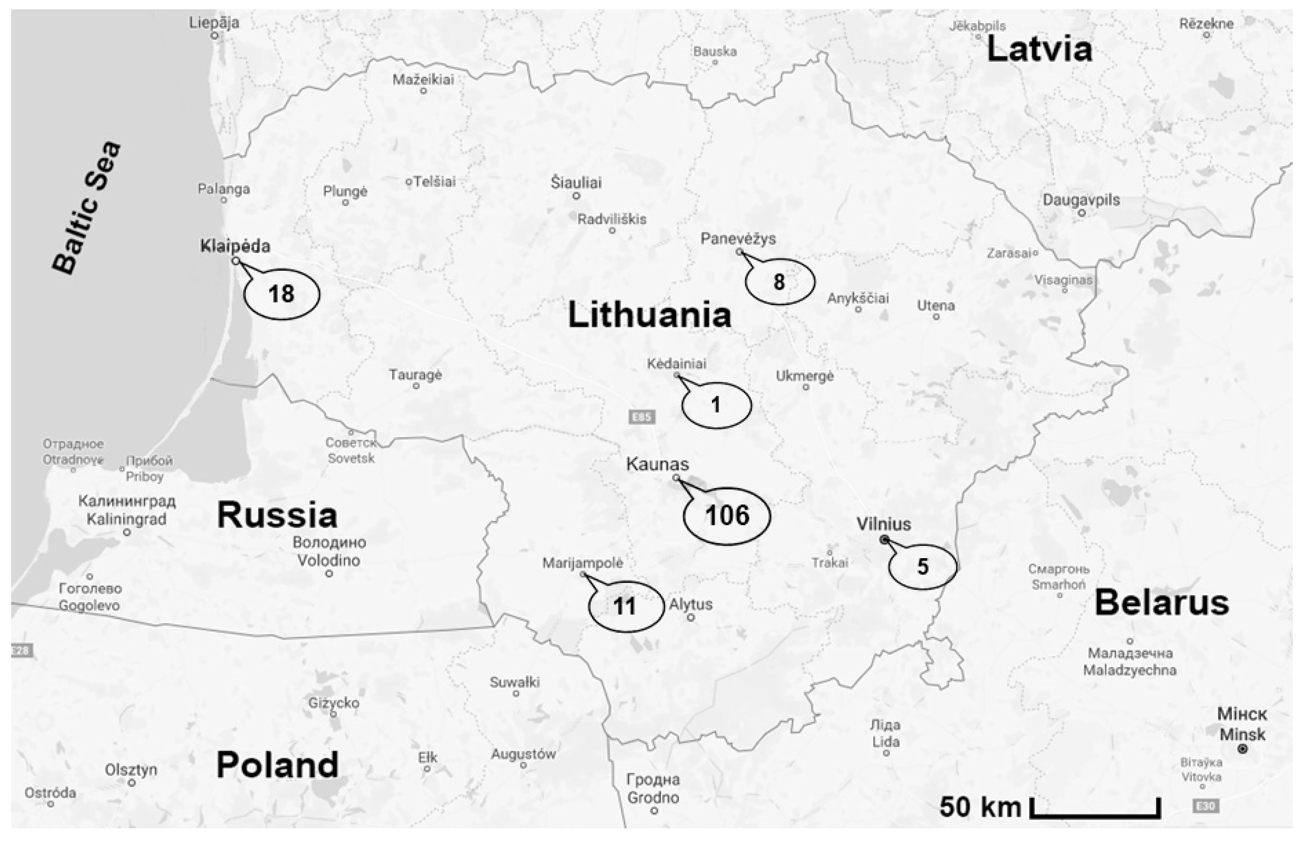
2. Materials and Methods

During the seasonal spring outbreak of babesiosis in 2016–2017, 149 blood samples were collected from dogs of various breeds and ages with a presumptive diagnosis of babesiosis. The disease was diagnosed based on clinical signs and the examination of blood smears. The samples were collected from different veterinary clinics in six regions of Lithuania (Figure 1). The clinicopathological findings in Babesia infections were slight to severe thrombocytopenia, severe immune-mediated haemolytic anaemia (IMHA), cerebral babesiosis and acute kidney injury (AKI).

2.1. DNA Extraction

Genomic DNA was extracted from 200 μL aliquots of EDTA blood (taken from the vena cephalica of the examined dog) using the GeneJet Whole Blood Genomic DNA Purification kit (Thermo Fisher Scientific, Vilnius, Lithuania), according to the manufacturer’s instructions.

2.2. PCR-RFLP Analysis

Partial sequences from 18S rRNA and Bc28.1 genes of B. canis were amplified. Conventional PCR was performed using primers BAB GF2 and primers BAB GR2, which amplify a 559 bp region of the 18S rRNA gene of B. canis [18]. A 710 bp fragment of the Bc28.1 gene was amplified with F281&2a and R281 primers, as described in Carcy et al. [17]. Both PCR reactions were carried out in a 25 µL final volume containing 5 X MyTaq DNA buffer, 10 pmol of each primer, 5 U Taq DNA polymerase (Thermo Fisher Scientific, Vilnius, Lithuania), double-distilled water and 5 µL of DNA template. Amplification reaction of partial 18S rRNA gene was performed as follows: initial denaturation at 92 °C for 2 min, 50 cycles of denaturation at 92 °C for 60 s, annealing at 52 °C for 60 s and extension at 72 °C for 90 s and a final extension step at 72 °C for 5 min. PCR conditions for Bc28.1 gene were initial denaturation at 94 °C for 3 min, followed by 35 cycles of denaturation at 94 °C for 30 s, annealing at 55 °C for 30 s and extension at 72 °C for 60 s. The final extension was at 72 °C for 7 min. In each PCR run, negative (double-distilled water) and positive (DNA of Babesia positive ticks, infection confirmed by sequencing) controls were used.
Positive 18S rRNA gene PCR products were digested with the HincII restriction enzyme [18] allowing the classification of B. canis isolates into two genotypes: 18S RNA-A, where the PCR fragment (559 bp) was cut into two fragments of 409 and 150 bp, and 18S RNA-B, where the 559 bp PCR fragment remained uncut (Figure S1).
PCR fragments of the Bc28.1 gene were digested with the combination of two restriction enzymes (MboI and AluI) allowing the classification of B. canis isolates into three genotypes: Bc28.1-A, Bc28.1-B and Bc28.1-34 [17]. Genotype Bc28.1-A was identified after MboI digestion when the 710 bp PCR fragment was cut into 300, 290, 70 or 50 bp fragments (Figure S2). AluI digestion enabled the identification of strains from genotype Bc28.1-B, resulting in 320, 240, 90 and 60 bp fragments and genotype Bc28.1-34 into 410, 240 and 60 bp fragments (Figure S3).
A 2% agarose gel was prepared by visualising the results after the PCR-RFLP analysis in the ultra-violet transilluminator GelDoc-It 310 and VisionWorks LS (Ultra-Violet Products Ltd., Cambridge, UK). DNA fragment sizes were assessed by comparison with the GeneRulerTM 50 bp DNA Ladder (Thermo Fisher Scientific, Vilnius, Lithuania).

2.3. Sequence Analysis

Representative PCR products of 62 samples were extracted from the agarose gel and purified using the GeneJET Gel Extraction Kit (Thermo Fisher Scientific, Vilnius, Lithuania) according to the manufacturer’s instructions and sent for sequencing (Macrogen, Amsterdam, The Netherlands).
The obtained sequences were edited using the Mega X software package, aligned with each other, and compared with the sequence data available from GenBank using the NCBI BLAST® software (http://blast.ncbi.nlm.nih.gov, accessed on 30 March 2022). A phylogenetic tree based on B. canis Bc28.1 gene sequences was created using the neighbour-joining method with bootstrap analysis of 1000 replicates. The most appropriate model of nucleotide substitution determined according to the Bayesian information criterion was the Jukes-Cantor model, with a gamma distribution of among-site variation (JC + G). Partial 18S rRNA and Bc28.1 gene sequences for representative samples were submitted to GenBank under accession numbers MN078319 to MN078323 for the 18S rRNA gene and MN078324 to MN078336 for the Bc28.1 gene.

3. Results

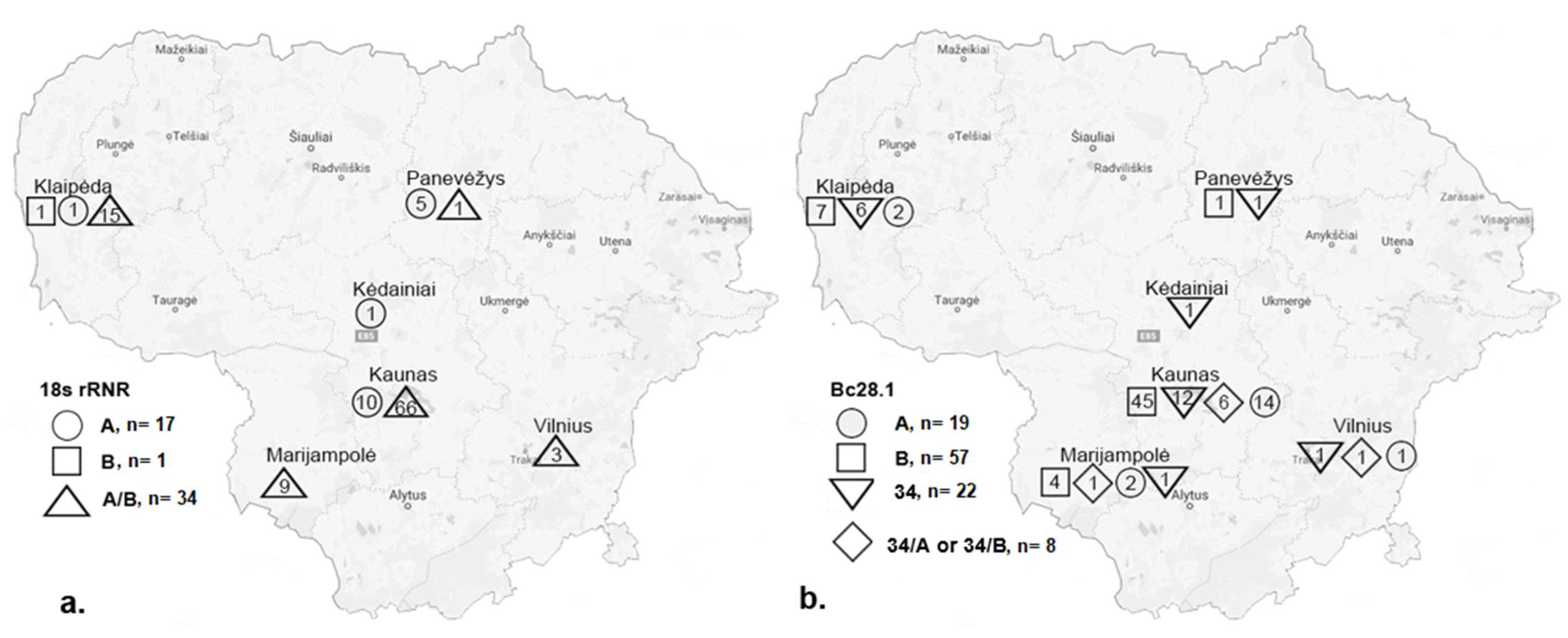
3.1. Babesia canis 18S rRNA Genotypes

After amplification of the partial 18S rRNA gene, B. canis DNA was detected in 112 (75.2%) samples. All these samples were subjected to digestion with the HincII restriction enzyme. A selected number of amplicons (n = 30) derived from samples collected in different Lithuanian regions were sequenced to identify the molecular heterogeneity of different isolates (Table 1).
Based on the RFLP profiles obtained and two nucleotide GA/AG substitutions observed in 18S rRNA gene sequences, three B. canis genotypes were identified. Of these genotypes, a “mixed” genotype predominated that showed PCR-RFLP profiles specific to both 18S rRNA-A and 18S rRNA-B genotypes (Figure S1) and the presence of both G and A nucleotides (R/R) at positions 92 and 93 (corresponding to 610 and 611 nucleotides in the whole-length ssrRNA gene) (Table 1). A total of three single nucleotide polymorphisms in the partial 18S rRNA gene of B. canis and five different sequence variants were identified by sequence analysis (Table 1).
The predominant 18S rRNA-A/B genotype was detected in 94 (83.9%) blood samples from dogs and in all investigated regions of Lithuania except Kėdainiai. The B. canis genotype 18S rRNA-A was detected in seventeen (15.2%) dogs from Kaunas, Klaipėda, Kėdainiai and Panevėžys, while the B. canis genotype 18S rRNA-B was detected in just one dog (0.9%) in Klaipėda (Figure 2a).

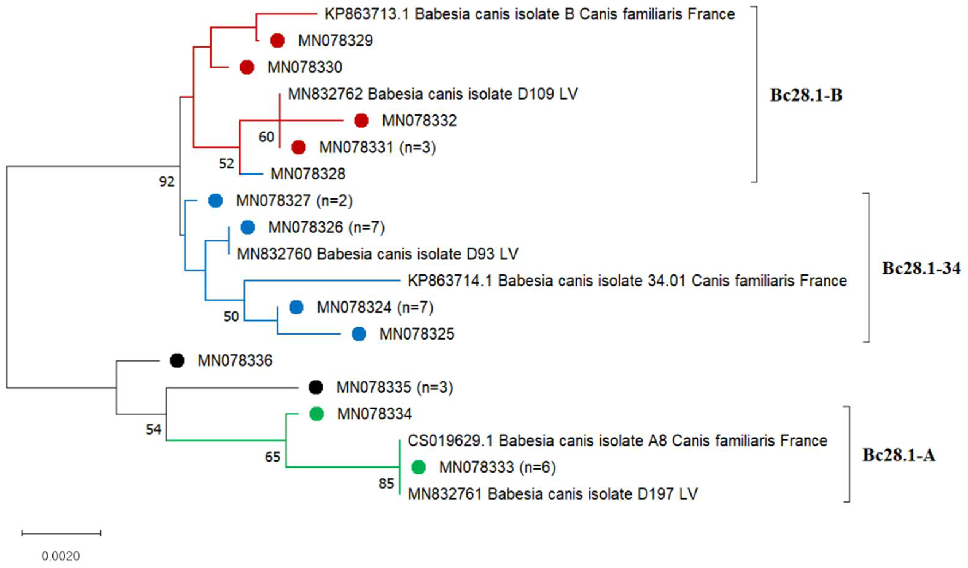
3.2. Babesia canis Bc28.1 Genotypes

After amplification of the partial region of Bc28.1 gene, B. canis DNA was detected in 106 (71.1%) samples. All these samples were subjected to digestion with the combination of two restriction enzymes (MboI and AluI). A selected number of amplicons (n = 35) derived from samples collected in different Lithuanian regions were sequenced (Table 2).
Based on the RFLP profiles obtained, three genotypes were identified: Bc28.1-B (57/106; 53.8%), Bc28.1-34 (22/106; 20.8%) and Bc28.1-A (19/106; 17.9%). Some analysed samples showed specific profiles of Bc28.1-34 and Bc28.1-A or Bc28.1-B (8/106; 7.5%) when the PCR fragment was cut into 410, 320, 240, 90 and 60 bp fragments (possibly co-infection) (Figures S2 and S3).
Sequence analysis of the 631 bp fragment of the Bc28.1 gene revealed 18 polymorphic sites (15 nucleotide substitutions and 3 deletions) in the analysed samples (Table 2).
Analysis of the 210 amino acids of the Bc28.1 protein sequences revealed amino acid substitutions in 10 positions: 26 (aspartic acid to asparagine), 28 (lysine to glutamine), 50 (glutamic acid to lysine), 109 (glutamic acid to aspartic acid), 110 (aspartic acid to asparagine), 113 (lysine to asparagine), 149 (leucine to valine), 156 (lysine to asparagine), 173 (threonine to asparagine) and 204 (serine to asparagine). All other nucleotide changes were silent (Table 2).
Based on two observed nucleotide substitutions, G/A (150 position) and C/T (477 position), four genotypes were distinguished in accordance with the genotypes obtained by PCR-RFPL analysis [17] (Table 2).
Bc28.1 gene sequences of B. canis strains obtained in Lithuania were 99–100% identical to the corresponding sequences from laboratory B. canis strains obtained in France [17]. Lithuanian B. canis strains identified as belonging to Bc28.1-A genotype were 100% identical to the B. canis laboratory strain A8 deposited in GenBank (Acc.No. CS019629) or differed from it by two nucleotides. Other Lithuanian B. canis Bc28.1 gene sequences that belonged to the Bc28.1-B and Bc28.1-34 genotypes differed from the corresponding sequences of the B. canis laboratory strain B (GenBank Acc.No. KP863713) by one to four nucleotides and three deletions and B. canis laboratory strain 34.01 (GenBank Acc.No. KP863714) by three to five nucleotides, respectively (Table 2).
Five Bc28.1 gene sequence variants within the B. canis Bc28.1-34 genotype, four sequence variants within the Bc28.1-B genotype and two sequence variants within the Bc28.1-A genotype were detected. In four samples, G nucleotide was observed at position 150, which is characteristic of the Bc28.1-34 genotype, and T nucleotide was observed at position 477, which is characteristic of the Bc28.1-A genotype. Therefore, these B. canis strains have been described as belonging to the “mixed” Bc28.1-34/A genotype. Two different sequence variants were detected within this genotype (Table 2).
The phylogenetic tree was created to include 35 sequences of the Bc28.1 gene obtained in this study and the 6 corresponding sequences of B. canis isolates obtained from dogs in France (Acc.No. KP863713, KP863714, CS019629) and Latvia (Acc.No. MN832760, MN832761, MN832762) (Figure 3). Most of the analysed sequences in the phylogenetic tree were separated into three major clusters according to the Bc28.1 genotype: Bc28.1-A, Bc28.1-B and Bc28.1-34. Sequence MN078328 (identified as belonging to the Bc28.1-34 genotype) clustered with the Bc28.1-B genotype sequences. Four sequences of the mixed genotype 34/A grouped with the Bc28.1-A genotype sequences and formed a single cluster on the phylogenetic tree (Figure 3).

4. Discussion

The molecular characterisation of B. canis 18S rRNA and Bc28.1 genes indicates the presence of genetically heterogenic B. canis strains in Lithuania. Molecular analysis of the 18S rRNA gene showed a separation of the sequences into three genotypes based on two single nucleotide polymorphisms. In Europe, four B. canis genotypes related to GA → AG nucleotides substitutions (GA, AG, AA, RR) are present [22,23]. In the present study, the vast majority of dogs (83.9%) were infected with the “mixed” 18S rRNA-A/B genotype (displaying the presence of both G and A nucleotides (R/R)), which was detected in all investigated regions of Lithuania except Kėdainiai (probably because there was only one sample analysed from this region) (Table 1; Figure 2a). In previous studies, the “mixed” genotype has been identified with a high prevalence in dogs in central Lithuania (65.8%) and in D. reticulatus ticks across the country (43.8%) [7,15]. A similar result on the dominance of the “mixed” 18S rRNA-A/B genotype in dogs has been reported in Latvia (91%; 39/43) [8] and Poland (87%) [24], and in ticks in Switzerland [23]. The presence of the ambiguous nucleotides detected in 18S rRNA sequences of B. canis isolates is explained by the genetic heterogeneity occurring among copies of the ss rRNA genes or by mixed infections with parasites of different 18S rRNA genotypes [22,23,24,25].
B. canis genotypes 18S rRNA-A (GA nucleotides) and 18S rRNA-B (AG nucleotides) occur in European countries at different rates of prevalence [17,26]. In the previous studies conducted in Lithuania, the 18S rRNA-A genotype was identified in 50% and 34.2% of B. canis sequences originating from D. reticulatus ticks and dogs, respectively [7,15]. The B. canis 18S rRNA-B genotype (AG nucleotides) is uncommon in Lithuania and has so far only been detected in one dog and one tick sample [15].
Babesia canis 18S rRNA-A and 18S rRNA-B genotypes were initially described in Poland by Adaszek and Winiarczyk [18]. Adaszek et al. [27] examined different strains of B. canis isolated from 76 dogs and found an association between the intensification of thrombocytopenia and particular 18S rRNA genotypes of the pathogen: the strains of B. canis genotype 18S rRNA-B were found to be more virulent in relation to thrombocytopenia (mean number of thrombocytes 61.11 × 10⁹/L) than genotype 18S rRNA-A (mean number of thrombocytes 27.47 × 10⁹/L). In this study, haematological data on thrombocytopenia was available for 40 dogs from Kaunas. Among these dogs, the B. canis 18S rRNA-A/B genotype was detected in 39 individuals and the 18S rRNA-A genotype in 1 dog. Thrombocyte counts below the lower reference range value (150 × 109/L) were detected in 28 (70%) dogs. Marked thrombocytopenia (thrombocyte counts lower than 27 × 109/L) has been found in 22 (55%) dogs (21 dogs with mixed A/B genotype and 1 with A genotype). Thrombocyte counts in the remaining dogs varied between 193 × 109/L and 488 × 109/L. As most infections were associated with the “mixed” 18S rRNA-A/B genotype, it was impossible to find a correlation between different 18S rRNA genotypes and the virulence of B. canis strains. It is believed that genotyping of B. canis strains based on the 18S rRNA gene is not suitable to determine the virulence of strains among 18S rRNA-A and B genotypes in regions where the “mixed” 18S rRNA-A/B genotype dominates.
Carcy et al. [17] first used the polymorphism of the Bc28.1 gene to study the genetic diversity of B. canis in Europe. Based on PCR-RFLP and sequence analysis applied to four B. canis laboratory strains (A8, B, 34.01, and G) originating from France, three genetic groups—Bc28.1-A, Bc28.1-B and Bc28.1-34/G—were identified, which is in agreement with the classification of strains according to differences in chromosome profiles, virulence and antigenic variation. The B. canis B strain is more virulent in dogs than the B. canis A strain, while 34.01 isolate is genetically distinct from the B. canis isolates A and B on the basis of chromosome size polymorphism [28]. In the present study, clinicopathological findings were compared with B. canis genotype data, and no relationship was observed between the Bc28.1 genotype and the severity of the disease. Data on complicated and uncomplicated forms of canine babesiosis were recorded from 45 dogs at one Kaunas veterinary clinic. Eight (17.8%) dogs developed a complicated form of the disease. In these dogs, three Bc28.1 genotypes were detected: B (n = 4; 50%), A (n = 3; 37.5%) and 34 (n = 1; 12.5%). Uncomplicated babesiosis was diagnosed in 37 dogs, with the B genotype detected in 23 samples (62%), A in 5 samples (13.5%), 34 in 5 samples (13.5%) and 34/A in 4 samples (10.8%).
Carcy et al. [17] analysed B. canis isolates collected from dogs in nine European countries between 2002 and 2010. The distribution and prevalence of B. canis Bc28.1 genotypes differed between four European regions. In France, genotype B dominated (62.4%), followed by genotypes A (37.1%) and 34 (11.8%). In south-west Europe, genotype B predominated, while in north-east Europe it was genotype A. In central Europe, both genotypes (A and B) were found with a similar prevalence. Carcy et al. [17] identified genotype 34 only in the south of France (mostly as co-infection with genotypes A or B). This genotype is now found in Lithuania and Latvia [29]. The expansion of D. reticulatus during the last decade in Europe could favour the spreading of certain B. canis genotypes in new areas. The results of the present study show that the distribution and prevalence of B. canis Bc28.1 genotypes in Lithuania are specific and similar to findings in France but differ from those obtained in north-east and central Europe [17].
Different patterns in the prevalence and geographical distribution of B. canis Bc28.1 genotypes have been found in Lithuania’s neighbour, Latvia. In a recent study conducted by Kivrane et al. [29], the presence of three B. canis Bc28.1 gene sequence types (Figure 2) with a non-uniform geographical distribution was detected. Based on the obtained results, the authors suggest two separate events in the establishment of B. canis foci in Latvia. Among the strains detected, predominant strains of the A genotype were mainly detected in Riga and the Riga region (Acc.No. MN832761). In the past few years, D. reticulatus had been detected in geographically separate new localities in the Riga region, indicating the development of new foci outside the major distribution area [14]. These recently established populations of D. reticulatus could favour the spread of B. canis strains of the A genotype in this region of Latvia. In Latvia, the B. canis strain of the B genotype (Acc.No. MN832762) was detected in just one sample obtained near the Lithuanian border [29], whereas, in Lithuania, this genotype was the most frequent. B. canis strains closely related to the 34.01 (Acc.No. MN832760) were detected in different regions of Latvia, mostly in the region of Daugavpils, which is relatively close to Lithuania and where the first populations of D. reticulatus ticks were detected [13]. No genetic variation was observed among sequences of A and 34 genotypes in the Latvian samples [29], which is in contrast to the findings of the present study. In Lithuania, five different sequence variants were detected within the B. canis 34 genotype, and two within the A genotype. It is noteworthy that all Bc28.1 gene sequence variants obtained in Latvia were detected in the present study. These findings could indicate the recent expansion of D. reticulatus ticks from Lithuania and the establishment of particular B. canis strains in Latvia. However, other transmission routes from Belarus and Russia (where the B. canis Bc28.1-A genotype dominates [17]) are also possible.
Local ecological factors may play a crucial role in the survival of particular B. canis genotypes [17]. The emergence of B. canis is mainly associated with the expanding range of D. reticulatus, environmental changes, and migrations of humans and animals. In the past two decades, changes in climatic and ecological conditions, as well as the anthropogenic impact on the formation of the landscape, have made many areas in Baltic countries suitable for the expansion of animal hosts for all active stages of ticks and for the establishment of D. reticulatus, which has high ecological plasticity [13]. The geographical range of D. reticulatus in Europe is fragmented and divided into two main zones: western Europe and eastern Europe. A recent study investigated the D. reticulatus population structure through its distribution range in western and eastern Europe using microsatellite markers and demonstrated that this tick forms two genetically distinct groups across Europe (located in western and eastern Europe, with an overlap in central Europe) [30]. Genetically different D. reticulatus populations have been detected in the Czech Republic, Slovakia, western and eastern Poland, and Lithuania. Analysis of two mtDNA 12S rRNA and 16S rRNA markers has demonstrated that there are different haplotypes of D. reticulatus present in Europe ([31], Paulauskas et al., unpublished). This suggests that genetically different D. reticulatus populations could harbour different B. canis genotypes. Further studies are needed to analyse the relationship between B. canis strains, their geographical distribution and genotypes of D. reticulatus.
Co-infections of B. canis with other agents of canine vector-borne diseases could occur. Co-infected cases are of major clinical importance because they often lead to atypical clinical signs and abnormal laboratory findings, and may cause misdiagnosis and failures in treatment and prognosis [32]. Co-infections with B. canis and other vector-borne pathogens may result in greater pathogenicity and complications in the infected dogs. Blood samples from 70 dogs infected with B. canis (which were examined in this study) were analysed by real-time PCR for the presence of the other vector-borne pathogens transmitted by mosquitoes (Dirofilaria repens) and I. ricinus ticks (Anaplasma phagocytophilum and Borrelia spp.) [33], and double, triple or even quadruple co-infections in dogs were detected. These findings demonstrate that co-infections of B. canis with other vector-borne pathogens in dogs are to be expected in Lithuania and that simultaneous detection of multiple vector-borne pathogens in dogs is needed for accurate diagnosis of canine vector-borne diseases in the country.

5. Conclusions

The molecular characterisation of B. canis 18S rRNR and Bc28.1 genes based on PCR-RFLP and sequence analyses indicated the presence of genetically heterogenic B. canis strains in Lithuania. The B. canis genotypes obtained were detected with different rates of prevalence in different regions of Lithuania. These findings provide a better understanding of the epidemiology of canine babesiosis and demonstrate the need for further investigation of the relationship between the genetic structure of B. canis parasites, their geographical distribution and strain virulence. Determination of the genetic diversity of the Bc28.1 gene may be helpful for vaccine development and the assessment of its potential use in Baltic countries.

Supplementary Materials

The following supporting information can be downloaded at: https://0-www-mdpi-com.brum.beds.ac.uk/article/10.3390/microorganisms10071446/s1, Figure S1: Identification of B. canis genotypes using the HincII restriction enzyme. Lines M—Gene RulerTM 50 bp DNA ladder (Thermo Fisher Scientific Baltics, Lithuania); Line K−—negative control; Line K+—positive control; Line 1—genotype B; Lines 10, 12—genotype A; Lines 2–9, 11, 13–15—genotype A/B; Figure S2: Identification of B. canis genotypes using the MboI restriction enzyme. Lines M—Gene RulerTM 50 bp DNA ladder (Thermo Fisher Scientific Baltics, Lithuania); Line K−—negative control; Line K+—positive control; Lines 1–4, 6–10, 12–15—genotype B/34.01; Lines 5,11—genotype A; Figure S3: Identification of B. canis genotypes using the AluI restriction enzyme. Lines M—Gene RulerTM 50 bp DNA ladder (Thermo Fisher Scientific Baltics, Lithuania); Lines 1–8, 10–14—genotype Bc28.1-B; Line 16—genotype Bc28.1-34.01; Line 9—“mixed” genotype.

Author Contributions

Conceptualization, J.R., A.P. and B.K.; methodology, J.R., A.A. and D.M.-B.; software, J.R., D.M.-B. and A.A.; formal analysis, D.M.-B., A.A., M.R. and I.S.; investigation, J.R., D.M.-B., A.A. and M.R.; resources, A.P., B.K. and I.S.; funding acquisition, J.R. and A.P.; supervision, A.P.; writing—original draft preparation, J.R., D.M.-B. and A.A.; writing—review and editing, A.P., B.K. and J.R. All authors have read and agreed to the published version of the manuscript.

Funding

Part of this research is funded by the European Social Fund under the No. 09.3.3-LMT-K-712-03-0081 “Development of Competences of Scientists, other Researchers and Students through Practical Research Activities” measure.

Data Availability Statement

Data are contained within the article.

Acknowledgments

The authors are grateful to Vytautas Sabūnas for his help in collecting dog blood samples.

Conflicts of Interest

The authors declare no conflict of interest.

References

  1. Schnittger, L.; Rodriguez, A.E.; Florin-Christensen, M.; Morrison, D.A. Babesia: A world emerging. Infect. Genet. Evol. 2012, 12, 1788–1809. [Google Scholar] [CrossRef] [PubMed]
  2. Irwin, P.J. Canine babesiosis: From molecular taxonomy to control. Parasit. Vectors 2009, 2 (Suppl. S1), S4. [Google Scholar] [CrossRef] [PubMed]
  3. Solano-Gallego, L.; Sainz, Á.; Roura, X.; Estrada-Peña, A.; Miró, G. A review of canine babesiosis: The European perspective. Parasit. Vectors 2016, 9, 336. [Google Scholar] [CrossRef] [PubMed]
  4. Matijatko, V.; Torti, M.; Schetters, T.P. Canine babesiosis in Europe: How many diseases? Trends Parasitol. 2012, 28, 99–105. [Google Scholar] [CrossRef] [PubMed]
  5. Bajer, A.; Beck, A.; Beck, R.; Behnke, J.M.; Dwużnik-Szarek, D.; Eichenberger, R.M.; Farkas, R.; Fuehrer, H.P.; Heddergott, M.; Jokelainen, P.; et al. Babesiosis in Southeastern, Central and Northeastern Europe: An Emerging and Re-Emerging Tick-Borne Disease of Humans and Animals. Microorganisms 2022, 10, 945. [Google Scholar] [CrossRef]
  6. Berzina, I.; Capligina, V.; Baumanis, V.; Ranka, R.; Cirule, D.; Matise, I. Autochthonous canine babesiosis caused by Babesia canis canis in Latvia. Vet. Parasitol. 2013, 196, 515–518. [Google Scholar] [CrossRef]
  7. Paulauskas, A.; Radzijevskaja, J.; Karvelienė, B.; Grigonis, A.; Aleksandravičienė, A.; Zamokas, G.; Babickaitė, L.; Sabūnas, V.; Petkevičius, S. Detection and molecular characterization of canine babesiosis causative agent Babesia canis in naturally infected dog in Lithuania. Vet. Parasitol. 2014, 205, 702–706. [Google Scholar] [CrossRef]
  8. Seleznova, M.; Kivrane, A.; Namina, A.; Krumins, R.; Aleinikova, D.; Lazovska, M.; Akopjana, S.; Capligina, V.; Ranka, R. Babesiosis in Latvian domestic dogs, 2016–2019. Ticks Tick Borne Dis. 2020, 11, 101459. [Google Scholar] [CrossRef]
  9. Schäfer, I.; Helm, C.; Marsboom, C.; Hendrickx, G.; Kohn, B.; Krücken, J.; Samson-Himmelstjerna, G.; Müller, E. Infections with Babesia spp. in dogs living in Germany (2007–2020). J. Vet. Intern. Med. 2021, 35, 3199. [Google Scholar]
  10. Rubel, F.; Brugger, K.; Pfeffer, M.; Chitimia-Dobler, L.; Didyk, Y.M.; Leverenz, S.; Dautel, H.; Kahl, O. Geographical distribution of Dermacentor marginatus and Dermacentor reticulatus in Europe. Ticks Tick Borne Dis. 2016, 7, 224–233. [Google Scholar] [CrossRef]
  11. Drehmann, M.; Springer, A.; Lindau, A.; Fachet, K.; Mai, S.; Thoma, D.; Schneider, C.R.; Chitimia-Dobler, L.; Bröker, M.; Dobler, G.; et al. The spatial distribution of Dermacentor ticks (Ixodidae) in Germany—Evidence of a continuing spread of Dermacentor reticulatus. Front. Vet. Sci. 2020, 7, 578220. [Google Scholar] [CrossRef] [PubMed]
  12. Dwużnik-Szarek, D.; Mierzejewska, E.J.; Rodo, A.; Goździk, K.; Behnke-Borowczyk, J.; Kiewra, D.; Kartawik, N.; Bajer, A. Monitoring the expansion of Dermacentor reticulatus and occurrence of canine babesiosis in Poland in 2016–2018. Parasit. Vectors 2021, 14, 267. [Google Scholar] [CrossRef]
  13. Paulauskas, A.; Radzijevskaja, J.; Mardosaitė-Busaitienė, D.; Aleksandravičienė, A.; Galdikas, M.; Krikštolaitis, R. New localities of Dermacentor reticulatus ticks in the Baltic countries. Ticks Tick Borne Dis. 2015, 6, 630–635. [Google Scholar] [CrossRef] [PubMed]
  14. Capligina, V.; Seleznova, M.; Akopjana, S.; Freimane, L.; Lazovska, M.; Krumins, R.; Kivrane, A.; Namina, A.; Aleinikova, D.; Kimsis, J.; et al. Large-scale countrywide screening for tick-borne pathogens in field-collected ticks in Latvia during 2017–2019. Parasit. Vectors 2020, 13, 351. [Google Scholar] [CrossRef] [PubMed]
  15. Radzijevskaja, J.; Mardosaitė-Busaitienė, D.; Aleksandravičienė, A.; Paulauskas, A. Investigation of Babesia spp. in sympatric populations of Dermacentor reticulatus and Ixodes ricinus ticks in Lithuania and Latvia. Ticks Tick Borne Dis. 2018, 9, 270–274. [Google Scholar] [CrossRef]
  16. Sabūnas, V. Prevalence of Vector-Borne Pathogens in Dogs. Ph.D. Thesis, Vytautas Magnus University, Kaunas, Lithuania, 2019. [Google Scholar]
  17. Carcy, B.; Randazzo, S.; Depoix, D.; Adaszek, L.; Cardoso, L.; Baneth, G.; Gorenflot, A.; Schetters, T.P. Classification of Babesia canis strains in Europe based on polymorphism of the Bc28.1-gene from the Babesia canis Bc28 multigene family. Vet. Parasitol. 2015, 211, 111–123. [Google Scholar] [CrossRef]
  18. Adaszek, Ł.; Winiarczyk, S. Molecular characterization of Babesia canis canis isolates from naturally infected dogs in Poland. Vet. Parasitol. 2008, 152, 235–241. [Google Scholar] [CrossRef]
  19. Yang, Y.S.; Murciano, B.; Moubri, K.; Cibrelus, P.; Schetters, T.; Gorenflot, A.; Delbecq, S.; Roumestand, C. Structural and functional characterization of Bc28.1, major erythrocyte-binding protein from Babesia canis merozoite surface. J. Biol. Chem. 2012, 287, 9495–9508. [Google Scholar] [CrossRef]
  20. Zhou, M.; Cao, S.; Luo, Y.; Liu, M.; Wang, G.; Moumouni, P.F.A.; Jirapattharasate, C.; Iguchi, A.; Vudriko, P.; Terkawi, M.A.; et al. Molecular identification and antigenic characterization of a merozoite surface antigen and a secreted antigen of Babesia canis (BcMSA1 and BcSA1). Parasit. Vectors 2016, 9, 257. [Google Scholar] [CrossRef]
  21. Eichenberger, R.M.; Riond, B.; Willi, B.; Hofmann-Lehmann, R.; Deplazes, P. Prognostic markers in acute Babesia canis infections. J. Vet. Intern. Med. 2016, 30, 174–182. [Google Scholar] [CrossRef]
  22. Beck, R.; Vojta, L.; Mrljak, V.; Marinculić, A.; Beck, A.; Živičnjak, T.; Cacciò, S.M. Diversity of Babesia and Theileria species in symptomatic and asymptomatic dogs in Croatia. Int. J. Parasitol. 2009, 39, 843–848. [Google Scholar] [CrossRef]
  23. Schaarschmidt, D.; Gilli, U.; Gottstein, B.; Marreros, N.; Kuhnert, P.; Daeppen, J.A.; Rosenberg, G.; Hirt, D.; Frey, C.F. Questing Dermacentor reticulatus harbouring Babesia canis DNA associated with outbreaks of canine babesiosis in the Swiss Midlands. Ticks Tick Borne Dis. 2013, 4, 334–340. [Google Scholar] [CrossRef] [PubMed]
  24. Łyp, P.; Adaszek, Ł.; Furmaga, B.; Winiarczyk, S. Identification of new 18S rRNA strains of Babesia canis isolated from dogs with subclinical babesiosis. Pol. J. Vet. Sci. 2015, 18, 573–577. [Google Scholar] [CrossRef] [PubMed]
  25. Hrazdilová, K.; Myśliwy, I.; Hildebrand, J.; Buńkowska-Gawlik, K.; Janaczyk, B.; Perec-Matysiak, A.; Modrý, D. Paralogs vs. genotypes? Variability of Babesia canis assessed by 18S rDNA and two mitochondrial markers. Vet. Parasitol. 2019, 266, 103–110. [Google Scholar] [CrossRef] [PubMed]
  26. Helm, C.S.; Weingart, C.; Ramünke, S.; Schäfer, I.; Müller, E.; von Samson-Himmelstjerna, G.; Kohn, B.; Krücken, J. High genetic diversity of Babesia canis (Piana & Galli-Valerio, 1895) in a recent local outbreak in Berlin/Brandenburg, Germany. Transbound. Emerg. Dis. 2022. [Google Scholar] [CrossRef]
  27. Adaszek, Ł.; Winiarczyk, S.; Skrzypczak, M. The clinical course of babesiosis in 76 dogs infected with protozoan parasites Babesia canis canis. Pol. J. Vet. Sci. 2009, 12, 81–87. [Google Scholar]
  28. Uilenberg, G.; Franssen, F.F.; Perié, N.M.; Spanjer, A.A. Three groups of Babesia canis distinguished and a proposal for nomenclature. Vet. Q. 1989, 11, 33–40. [Google Scholar] [CrossRef]
  29. Kivrane, A.; Namina, A.; Seleznova, M.; Akopjana, S.; Capligina, V.; Ranka, R. Development of a real-time PCR method for rapid diagnosis of canine babesiosis and anaplasmosis. Parasit. Vectors 2021, 14, 266. [Google Scholar] [CrossRef]
  30. Paulauskas, A.; Galdikas, M.; Galdikaitė-Brazienė, E.; Stanko, M.; Kahl, O.; Karbowiak, G.; Radzijevskaja, J. Microsatellite-based genetic diversity of Dermacentor reticulatus in Europe. Infect. Genet. Evol. 2018, 66, 200–209. [Google Scholar] [CrossRef]
  31. Kloch, A.; Mierzejewska, E.J.; Karbowiak, G.; Slivinska, K.; Alsarraf, M.; Rodo, A.; Kowalec, M.; Dwużnik, D.; Didyk, Y.M.; Bajer, A. Origins of recently emerged foci of the tick Dermacentor reticulatus in central Europe inferred from molecular markers. Vet. Parasitol. 2017, 237, 63–69. [Google Scholar] [CrossRef]
  32. De Tommasi, A.S.; Otranto, D.; Dantas-Torres, F.; Capelli, G.; Breitschwerdt, E.B.; De Caprariis, D. Are vector-borne pathogen co-infections complicating the clinical presentation in dogs? Parasit. Vectors 2013, 6, 97. [Google Scholar] [CrossRef] [PubMed]
  33. Radzijevskaja, J.; Tamoliūnaitė, D.; Sabunas, V.; Aleksandravičienė, A.; Paulauskas, A. Prevalence and co-infection of mosquitoand tick-borne pathogens in domestic dogs suspected for canine babesiosis in Lithuania. Biologija 2020, 66, 2. [Google Scholar] [CrossRef]
Figure 1. Map of sample collection sites. Blood samples were collected from 149 dogs originating from six regions of Lithuania. The number in the circle indicates the number of dogs analyzed.
Microorganisms 10 01446 g001
Figure 2. Distribution of B. canis 18S rRNA (a) and Bc28.1 (b) genotypes in Lithuania. On the map, different geometric shapes (circle, square, triangle, rhombus) with numbers represent the number of B. canis strains corresponding to each genotype found in different locations. n—the total number of B. canis strains of a particular genotype.
Microorganisms 10 01446 g002
Figure 3. Phylogenetic tree of the Bc28.1 gene sequences of Babesia canis created using the neighbour-joining method and Jukes-Cantor model with a gamma distribution of among-site variation. Bootstrap values (values < 50% not shown) from 1000 replicates are shown at the nodes. Samples sequenced in the present study are marked. The number of samples represented by the sequence is given in parentheses (n = x). Babesia canis strains corresponding to each genotype are marked with different colours.Analysing the results of the PCR-RFLP analysis, we observed that most of the B. canis infections in dogs were associated with the Bc28.1-B genotype (Figure 2b). This genotype was mainly detected in Kaunas (45 of 56 samples) and was also found in Klaipėda, Marijampolė and Panevėžys. The Bc28.1-34 genotype was detected in all the regions in which samples were collected, with the greatest prevalence in Kaunas (12 out of 22 samples). The Bc28.1-A genotype also prevailed in Kaunas (14 of 19 samples) and was detected in three other regions: Klaipėda, Marijampolė and Vilnius (Figure 2b). All four genotypes were detected in central Lithuania (Kaunas) and the south-western (Marijampolė) part of the country.
Microorganisms 10 01446 g003
Table 1. Babesia canis genotypes based on the comparison of 18S rRNA gene sequences. The number of samples represented by the sequence is given in parentheses (n = x).
GenotypeGenBank Accession Number (Sample Number)Nucleotide Positions
809293
AMN078319 (n = 11)TGA
MN078322 (n = 2)Y..
BMN078320 (n = 1).AG
A/BMN078321 (n = 15).RR
MN078323 (n = 1)YRR
Table 2. Comparison of B. canis Bc28.1 gene sequences obtained from dogs in Lithuania with selected GenBank sequences. The number of samples represented by the sequence is given in parentheses (n = x).
GeootypeGenBank ace.No(Number of Samplcs)Nucleotide Positions a,b
768293148150246327328339420433468477518525526527573585611
ACS019629G(D)A(K)GG(E)ACA(E)G(D)A(K)CC(L)A(K)TC(T)GGACGG(S)
MN078333 (n = 6)....................
MN078334 (n = 1)......C(D)A(N)............
BKP863713GC(Q)GGA TCAT(N)ACT(N)CCGGAAGG
MN078329 (n = 1).............A(N)---...
MN078330 (n = 1) .............A---C..
MN078331 (n = 3)...A(K).........A---C..
MN078332 (n = 1)...A.........A---CA.
34KP863714A(N)CAGGTCAT(N)AG(V)TCA---AGG
MN078324 (n = 7) G........ C...---C..
MN078325 (n = 1) G........ C...---C.A(N)
MN078326 (n = 7)G.G...... C...---...
MN078327 (n = 2)G.G...... C...---C..
MN078328 (n = 1)G.GA..... C...---C..
34/AMN078335 (n = 3) GCGGGCAGACCATA---AGG
MN078336 (n = 1) ...........T..---C..
a—amino acid changes are represented in parentheses; b—nucleotide and amino acid substitutions detected among Lithuanian strains are in bold. Gray-shaded columns indicate recognition sites for AluI and MboI restriction enzymes based on which genotypes were identified.
Publisher’s Note: MDPI stays neutral with regard to jurisdictional claims in published maps and institutional affiliations.

