Microbiological Air Quality and Drug Resistance in Airborne Bacteria Isolated from a Waste Sorting Plant Located in Poland―A Case Study


Abstract
:1. Introduction
2. Experiments
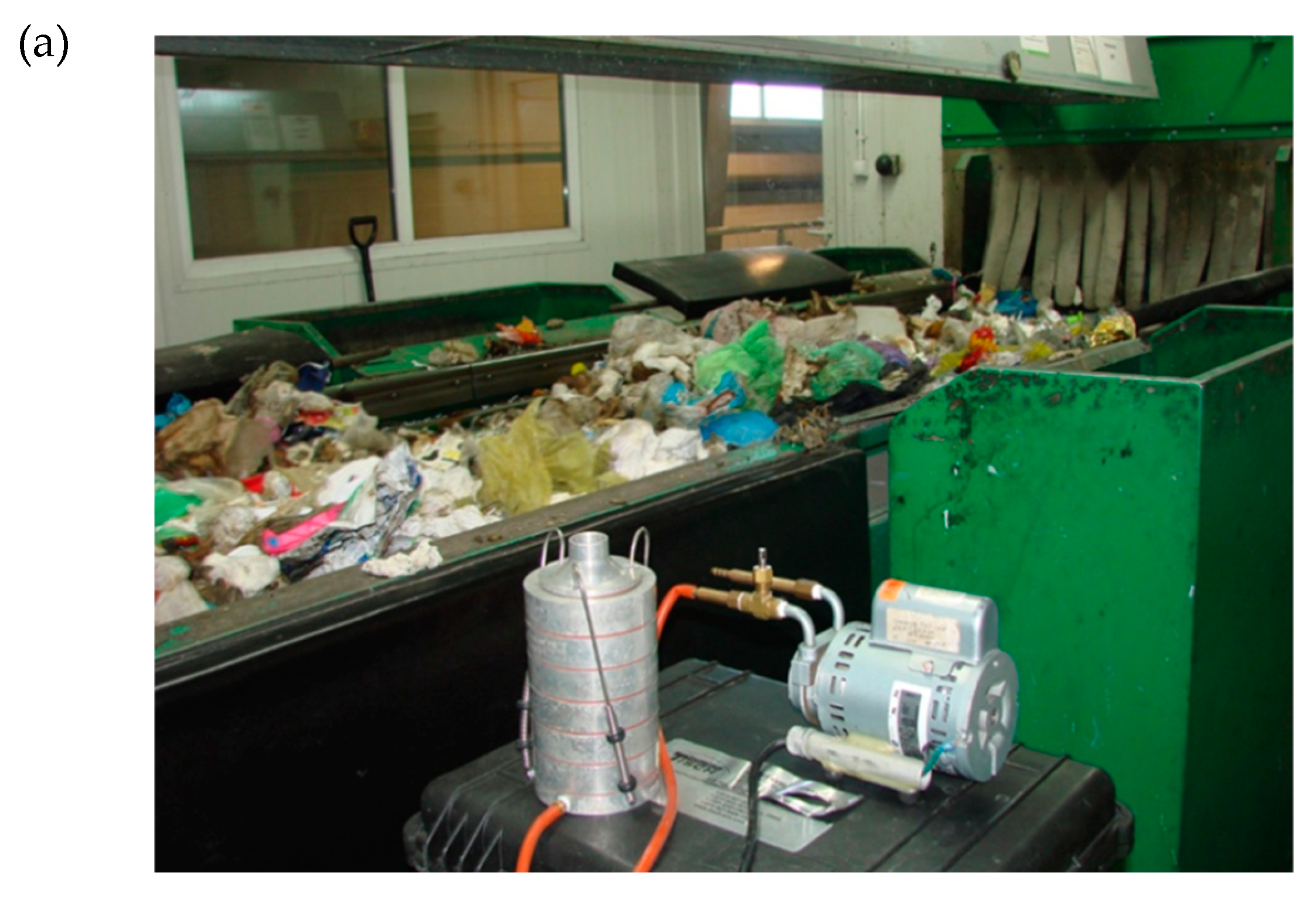
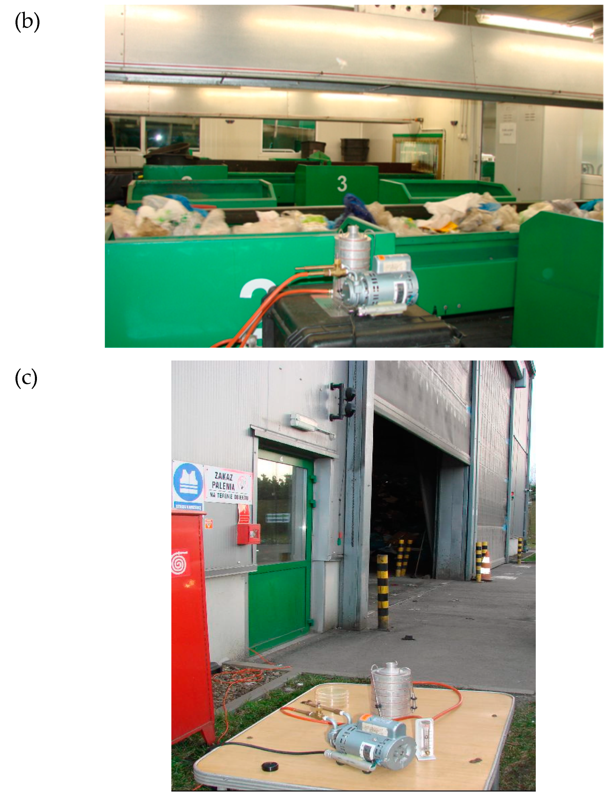
2.1. Sampling Sites
2.2. Sampling and Analytical Methods
2.3. Bacteria Identification and Multi-Antibiotic Resistance (MAR) Test
3. Results and Discussion
3.1. Quantity of Bacterial Aerosol (BA) of Two Cabins of Sorting Plant and Outdoor Air
3.2. Particle Size Distribution (PSD) of Bacterial Aerosol (BA) in Two Cabins of Sorting Plant (PSP; QCSP) and Outdoor Air
3.3. Quality and Antibiotic Resistance of Bacterial Aerosol (BA) in Two Cabins of Sorting Plant (PSP; QCSP)
4. Conclusions
Author Contributions
Funding
Acknowledgments
Conflicts of Interest
References
- Jiayu, C.; Qiaoqiao, R.; Feilong, C.; Chen, L.; Jiguo, W.; Zhendong, W.; Lingyun, C.; Liu, R.; Guoxia, Z. Microbiology Community Structure in Bioaerosols and the Respiratory Diseases. J. Environ. Sci. Public Health 2019, 3, 347–357. [Google Scholar] [CrossRef]
- Pearson, C.; Littlewood, E.; Douglas, P.; Robertson, S.; Gant, T.W.; Hansell, A.L. Exposures and health outcomes in relation to bioaerosol emissions from composting facilities: A systematic review of occupational and community studies. J. Toxicol. Environ. Health. Part. B Crit. Rev. 2015, 18, 43–69. [Google Scholar] [CrossRef] [PubMed]
- Fung, F.; Hughson, W.G. Health effects of indoor fungal bioaerosol exposure. Appl. Occup. Environ. Hyg. 2003, 18, 535–544. [Google Scholar] [CrossRef] [PubMed]
- Douwes, J.; Thorne, P.; Pearce, N.; Heederik, D. Bioaerosol health effects and exposure assessment: Progress and prospects. Ann. Occup. Hyg. 2003, 47, 187–200. [Google Scholar] [PubMed] [Green Version]
- Kim, K.H.; Kabir, E.; Jahan, S.A. Airborne bioaerosols and their impact on human health. J. Environ. Sci. 2018, 67, 23–35. [Google Scholar] [CrossRef]
- Górny, R.L. Microbial Aerosols: Sources, Properties, Health Effects, Exposure Assessment—A Review. KONA Powder Part. J. 2020. [Google Scholar] [CrossRef] [Green Version]
- Wéry, N. Bioaerosols from composting facilities-a review. Front. Cell. Infect. Microbiol. 2014. [Google Scholar] [CrossRef]
- Wood, R.A.; Burchett, M.D.; Orwell, R.A.; Tarran, J.; Torpy, F. Plant/soil capacities to remove harmful substances from polluted indoor air. J. Horticul. Sci. Biotechnol. 2002, 71, 120–129. [Google Scholar] [CrossRef]
- Brągoszewska, E.; Mainka, A.; Pastuszka, J.; Lizończyk, K.; Desta, Y. Assessment of Bacterial Aerosol in a Preschool, Primary School and High School in Poland. Atmosphere 2018, 9, 87. [Google Scholar] [CrossRef] [Green Version]
- Directive 2000/54/EC of the European Parliament and of the Council of 18 September 2000 on the protection of workers from risks related to exposure to biological agents at work. Off. J. Eur. Commun. 2000, 262, 21–45.
- Regulation of the Minister of Health dated April 22, 2005 (Journal of Laws of 2005 No. 81, item 716, as amended and Journal of Laws 2008, No. 48, item 288), in Polish. Available online: http://prawo.sejm.gov.pl/isap.nsf/download.xsp/WDU20050810716/O/D20050716.pdf (accessed on 10 December 2019).
- Karkman, A.; Pärnänen, K.; Larsson, D.G.J. Fecal pollution can explain antibiotic resistance gene abundances in anthropogenically impacted environments. Nat. Commun. 2019. [Google Scholar] [CrossRef] [PubMed]
- Wright, G.D. Antibiotic resistance in the environment: A link to the clinic? Curr. Opin. Microbiol. 2010, 13, 589–594. [Google Scholar] [CrossRef] [PubMed]
- Martinez, J.L. The role of natural environments in the evolution of resistance traits in pathogenic bacteria. Proc. Biol. Sci. 2009, 276, 2521–2530. [Google Scholar] [CrossRef] [PubMed] [Green Version]
- McKinney, C.W.; Pruden, A. Ultraviolet disinfection of antibiotic resistant bacteria and their antibiotic resistance genes in water and wastewater. Environ. Sci. Technol. 2012, 46, 13393–13400. [Google Scholar] [CrossRef]
- Nevalainen, A.; Pastuszka, J.; Liebhaber, F.; Willeke, K. Performance of bioaerosol samplers: Collection characteristics and sampler design considerations. Atmos. Environ. Part A Gen. Top. 1992, 26, 531–540. [Google Scholar] [CrossRef]
- Brągoszewska, E.; Biedroń, I.; Hryb, W. Air Quality and Potential Health Risk Impacts of Exposure to Bacterial Aerosol in a Waste Sorting Plant Located in the Mountain Region of Southern Poland, Around Which There Are Numerous Rural Areas. Atmosphere 2019, 10, 360. [Google Scholar] [CrossRef] [Green Version]
- Brągoszewska, E.; Biedroń, I.; Kozielska, B.; Pastuszka, J.S. Microbiological indoor air quality in an office building in Gliwice, Poland: Analysis of the case study. Air Qual. Atmos. Health 2018. [Google Scholar] [CrossRef] [Green Version]
- Bragoszewska, E.; Biedroń, I. Indoor air quality and potential health risk impacts of exposure to antibiotic resistant bacteria in an office rooms in southern poland. Int. J. Environ. Res. Public Health 2018. [Google Scholar] [CrossRef] [Green Version]
- Skowroń, J.; Górny, R. Harmful biological agents. In The Interdepartmental Commission for Maximum Admissible Concentrations and Intensities for Agents Harmful to Health in the Working Environment: Limit Values; Augustyńska, D., Pośniak, M., Eds.; Central Institute for Labour Protection—National Research Institute: Warszawa, Polish, 2012. (In Polish) [Google Scholar]
- Lehtinen, J.; Tolvanen, O.; Nivukoski, U.; Veijanen, A.; Hänninen, K. Occupational hygiene in terms of volatile organic compounds (VOCs) and bioaerosols at two solid waste management plants in Finland. Waste Manag. 2013. [Google Scholar] [CrossRef]
- Park, D.U.; Ryu, S.H.; Kim, S.B.; Yoon, C.S. An Assessment of Dust, Endotoxin, and Microorganism Exposure during Waste Collection and Sorting. J. Air Waste Manag. Assoc. 2011. [CrossRef] [Green Version]
- Owen, M.K.; Ensor, D.S.; Sparks, L.E. Airborne particle sizes and sources found in indoor air. Atmos. Environ. Part A Gen. Top. 1992. [Google Scholar] [CrossRef]
- Lacey, J.; Dutkiewicz, J. Bioaerosols and occupational lung disease. J. Aerosol Sci. 1994. [Google Scholar] [CrossRef]
- Faridi, S.; Hassanvand, M.S.; Naddafi, K.; Yunesian, M.; Nabizadeh, R.; Sowlat, M.H.; Kashani, H.; Gholampour, A.; Niazi, S.; Zare, A.; et al. Indoor/outdoor relationships of bioaerosol concentrations in a retirement home and a school dormitory. Environ. Sci. Pollut. Res. 2015, 22, 8190–8200. [Google Scholar] [CrossRef]
- Dutkiewicz, J.; Śpiewak, R.; Jabłoński, L.; Szymańska, J. Biological Occupational Risk Factors. Classification, Exposed Occupational Groups, Measurement, Prevention; Ad Punctum: Lublin, Poland, 2007. (In Polish) [Google Scholar]
- Heo, Y.; Park, J.; Lim, S.I.; Hur, H.G.; Kim, D.; Park, K. Size-resolved culturable airborne bacteria sampled in rice field, sanitary landfill, and waste incineration sites. J. Environ. Monit. 2010. [Google Scholar] [CrossRef]
- Ebrahimi, K.; Alipour, M.; Yahyapour, Y. Evaluation of antibiotic resistance pattern in Staphylococcus saprophyticus isolated from patients with urinary tract infection using real-time PCR. Int. J. Mol. Clin. Microbiol. 2018, 8, 975–981. [Google Scholar]
- Raz, R.; Colodner, R.; Kunin, C.M. Who Are You-Staphylococcus saprophyticus? Clin. Infect. Dis. 2005, 40, 896–898. [Google Scholar] [CrossRef]
- Kuroda, M.; Yamashita, A.; Hirakawa, H.; Kumano, M.; Morikawa, K.; Higashide, M.; Maruyama, A.; Inose, Y.; Matoba, K.; Toh, H.; et al. Whole genome sequence of Staphylococcus saprophyticus reveals the pathogenesis of uncomplicated urinary tract infection. Proc. Natl. Acad. Sci. U S A 2005. [Google Scholar] [CrossRef] [Green Version]
- Flores-Mireles, A.L.; Walker, J.N.; Caparon, M.; Hultgren, S.J. Urinary tract infections: Epidemiology, mechanisms of infection and treatment options. Nat. Rev. Microbiol. 2015. [Google Scholar] [CrossRef]
- Martins, K.B.; Ferreira, A.M.; Pereira, V.C.; Pinheiro, L.; de Oliveira, A.; de Lourdes Ribeiro de Souza da Cunha, M. In vitro Effects of Antimicrobial Agents on Planktonic and Biofilm Forms of Staphylococcus saprophyticus Isolated From Patients With Urinary Tract Infections. Front. Microbiol. 2019, 10, 40. [Google Scholar] [CrossRef]
- Oliveira, J.F.P.; Cipullo, J.P.; Burdmann, E.A. Nefrotoxicidade dos aminoglicosídeos. Brazilian Journal of Cardiovascular Surgery 2006, 22, 444–452. [Google Scholar] [CrossRef]
- Ehlers, S.; Merrill, S.A. Staphylococcus saprophyticus; PublPearls Publishing: Treasure Island, FL, USA, 2018. [Google Scholar]
- Flynn, E.H.; Sigal, M.V.; Wiley, P.F.; Gerzon, K. Erythromycin. I. Properties and Degradation Studies. J. Am. Chem. Soc. 1954, 76, 3121–3131. [Google Scholar] [CrossRef]
- Waisbren, B.A.; Crowley, W. Nitrofurantoin: Clinical and laboratory evaluation. A.M.A. Arch. Intern. Med. 1955, 95, 653–661. [Google Scholar] [CrossRef]
- Levine, D.P. Vancomycin: A History. Clin. Infect. Dis. 2006, 42, S5–S12. [Google Scholar] [CrossRef]
- Roe, F.J.C. Metronidazole: View of uses and toxicity. J. Antimicrob. Chemother. 1977, 3, 205–212. [Google Scholar] [CrossRef]
- Fairbrother, R.W.; Williams, B.L. Two new antibiotics. Lancet 1956, 268, 1177–1179. [Google Scholar] [CrossRef]
- Bisacchi, G.S.; Manchester, J.I. A New-Class Antibacterial-Almost. Lessons in Drug Discovery and Development: A Critical Analysis of More than 50 Years of Effort toward ATPase Inhibitors of DNA Gyrase and Topoisomerase IV. ACS Infect. Diseases 2015. [Google Scholar] [CrossRef]
- Eliopoulos, G.M.; Huovinen, P. Resistance to Trimethoprim-Sulfamethoxazole. Clin. Infect. Dis. 2001. [Google Scholar] [CrossRef] [Green Version]
- Mira, P.M.; Crona, K.; Greene, D.; Meza, J.C.; Sturmfels, B.; Barlow, M. Rational design of antibiotic treatment plans: A treatment strategy for managing evolution and reversing resistance. PLoS ONE 2015. [Google Scholar] [CrossRef]
- Li, J.J. History of drug discovery. In Drug Discovery: Practices, Processes, and Perspectives; Li, J.J., Corey, E.J., Eds.; Wiley: New York, NY, USA, 2013. [Google Scholar]
- Emmerson, A.M. The quinolones: Decades of development and use. J. Antimicrob. Chemother. 2003. [Google Scholar] [CrossRef] [Green Version]
- Sensi, P. History of the development of rifampin. Rev. Infect. Dis. 1983. [Google Scholar] [CrossRef]
- Neu, H.C.; Winshell, E.B. Semisynthetic Penicillin 6-[d(—)-α-Carboxy-3-Thienylacetamido] Penicillanic Acid Active Against Pseudomonas In Vitro. Appl. Environ. Microbiol. 1971, 21, 66–70. [Google Scholar] [CrossRef] [Green Version]
- Brogden, R.N.; Avery, G.S. New Antibiotics: Epicillin, Minocycline and Spectinomycin A summary of their antibacterial activity, pharmacokinetic properties and therapeutic efficacy. Drugs 1972. [Google Scholar] [CrossRef]
- Fuller, A.T.; Mellows, G.; Woolford, M.; Banks, G.T.; Barrow, K.D.; Chain, E.B. Pseudomonic acid: An antibiotic produced by Pseudomonas fluorescens. Nature 1971, 234, 416–417. [Google Scholar] [CrossRef]
- Sutherland, R.; Boon, R.J.; Griffin, K.E.; Masters, P.J.; Slocombe, B.; White, A.R. Antibacterial activity of mupirocin (pseudomonic acid), a new antibiotic for topical use. Antimicrob. Agents Chemother. 1985, 27, 495–498. [Google Scholar] [CrossRef] [Green Version]
- Geddes, A.M.; Schnurr, L.P.; Ball, A.P.; Mcghie, D.; Brookes, G.R.; Wise, R. Cefoxitin: A hospital study. Brit. Med. J. 1977. [Google Scholar] [CrossRef] [Green Version]
- Buck, R.E.; Price, K.E. Cefadroxil, a new broad spectrum cephalosporin. Antimicrob. Agents Chemother. 1977, 11, 324–330. [Google Scholar] [CrossRef] [Green Version]
- Winston, D.J.; Murphy, W.; Young, L.S.; Hewitt, W.L. Piperacillin therapy for serious bacterial infections. Am. J. Med. 1980, 69, 255–261. [Google Scholar] [CrossRef]
- Fischer, J.; Robin Ganellin, C. Analogue-based Drug Discovery; WILEY-VCH Verlag GmbH & Co. KGaA: Weinheim, Germany, 2006. [Google Scholar]
- Ito, A.; Hirai, K.; Inque, M. In vitro antibacterial activity of AM-715, a new nalidixic acid analog. Antimicrob. Agents Chemother. 1980, 17, 103–108. [Google Scholar] [CrossRef] [Green Version]
- Leigh, D.A.; Emmanuel, F.X.S. The treatment of Pseudomonas aeruginosa urinary tract infections with norfloxacin. J. Antimicrob. Chemother. 1984, 13, 85–88. [Google Scholar] [CrossRef]
- Childs, S.J. Aztreonam in the treatment of urinary tract infection. Am. J. Med. 1985, 78, 44–46. [Google Scholar] [CrossRef]
- Zhanel, G.G.; Simor, A.E.; Vercaigne, L.; Mandell, L. Imipenem and meropenem: Comparison of in vitro activity, pharmacokinetics, clinical trials and adverse effects. Can. J. Infect. Dis. 1998, 9, 215–228. [Google Scholar] [CrossRef] [PubMed]
- Parenti, F.; Beretta, G.; Berti, M.; Arioli, V. Teichomycins, New Antibiotics from Actinoplanes Teichomyceticus Nov. SP. I. Description of the Producer Strain, Fermentation Studies and Biological Properties. J. Antibiot. 1978. [Google Scholar] [CrossRef] [PubMed] [Green Version]
- Pryka, R.D.; Rodvold, K.A.; Rotschafer, J.C. Teicoplanin: An investigational glycopeptide antibiotic. Clin. Pharm. 1988, 7, 647–658. [Google Scholar]
- Amrol, D. Single-dose azithromycin microsphere formulation: A novel delivery system for antibiotics. Int. J. Nanomed. 2007. [Google Scholar] [CrossRef]
- Whitman, M.S.; Tunkel, A.R. Azithromycin and Clarithromycin: Overview and Comparison with Erythromycin. Infect. Control. Hosp. Epidemiol. 1992. [Google Scholar] [CrossRef]
- Livermore, D.M.; Sefton, A.M.; Scott, G.M. Properties and potential of ertapenem. J. Antimicro. Chem. 2003, 52, 331–334. [Google Scholar] [CrossRef]
- Hilas, O.; Ezzo, D.C.; Jodlowski, T.Z. Doripenem (doribax), a new carbapenem antibacterial agent. Pharm. Ther. 2008, 33, 134–180. [Google Scholar]
- Lounsbury, N.; Reeber, M.G.; Mina, G.; Chbib, C. A mini-review on ceftaroline in bacteremia patients with methicillin-resistant Staphylococcus aureus (MRSA) infections. Antibiotics 2019. [Google Scholar] [CrossRef] [Green Version]
- Momtaz, F.; Ali, M.H.; Hossain, M.N.; Foysal, M.J.; Sumiya, M.K.; Islam, K. Characterisation of multidrug-resistant Alcaligenes faecalis strain AF1 isolated from patient of RUTIs: A study from Bangladesh. J. Clin. Diagn. Res. 2018. [Google Scholar] [CrossRef]
- Filipe, M.; Reimer, Å.; Matuschek, E.; Paul, M.; Pelkonen, T.; Riesbeck, K. Fluoroquinolone-resistant Alcaligenes faecalis related to chronic suppurative otitis media, Angola. Emerg. Infect. Dis. 2017, 23, 1740. [Google Scholar] [CrossRef] [Green Version]
- Bizet, J.; Bizet, C. Strains of Alcaligenes faecalis from clinical material. J. Infect. 1997, 35, 167–169. [Google Scholar] [CrossRef]
- Kau, A.L.; Martin, S.M.; Lyon, W.; Hayes, E.; Caparon, M.G.; Hultgren, S.J. Enterococcus faecalis tropism for the kidneys in the urinary tract of C57BL/6J mice. Infect. Immun. 2005, 73, 2461–2468. [Google Scholar] [CrossRef] [Green Version]
- Rudy, M.; Nowakowska, M.; Wiechuła, B.; Zientara, M.; Radosz-Komoniewska, H. Antibiotic susceptibility analysis of Enterococcus spp. isolated from urine. Przegl Lek 2004, 61, 473–476. [Google Scholar]
- Azadi, D.; Dibaj, R.; Pourchangiz, M.; Daei-Naser, A.; Shojaei, H. First report of isolation of Mycobacterium canariasense from hospital water supplies. Scand. J. Infect. Dis. 2014, 46, 792–796. [Google Scholar] [CrossRef]
- Keikha, M. Case report of isolation of Mycobacterium setense from a hospital water supply. Environ. Dis. 2018, 3, 52. [Google Scholar] [CrossRef]
- Tille, P. Bailey & Scott’s Diagnostic Microbiology, 14th ed.; Elsevier Health Sciences Division: St. Louis, MP, USA, 2014; eBook; ISBN 9780323083287. [Google Scholar]
- Azadi, D.; Shojaei, H.; Pourchangiz, M.; Dibaj, R.; Davarpanah, M.; Naser, A.D. Species diversity and molecular characterization of nontuberculous mycobacteria in hospital water system of a developing country, Iran. Microb. Pathog. 2016, 100, 62–69. [Google Scholar] [CrossRef]
- Bengtsson-Palme, J.; Kristiansson, E.; Larsson, D.G.J. Environmental factors influencing the development and spread of antibiotic resistance. FEMS Microbiol. Rev. 2018. [Google Scholar] [CrossRef]
- Majchrzycka, K.; Okrasa, M.; Jachowicz, A.; Szulc, J.; Gutarowska, B. Microbial growth on dust-loaded filtering materials used for the protection of respiratory tract as a factor affecting filtration efficiency. Int. J. Environ. Res. Public Health 2018, 15, 1902. [Google Scholar] [CrossRef] [Green Version]


| PSP | QCSP | OUT | |
|---|---|---|---|
| Total average concentration | 1.81 × 10³ | 1.25 × 10³ | 1.14 × 10³ |
| Min value | 1.49 × 10³ | 8.6 × 10² | 6.9 × 10² |
| Max value | 2.7 × 10³ | 1.9 × 10³ | 1.7 × 10³ |
| Indoor/outdoor ratio | 1.6 | 1.1 | - |
| SD | 8.0 × 10² | 6.2 × 10² | 5.1 × 10² |
| PSP | QCSP | OUT | I/O for PSP | I/O for QCSP | |
|---|---|---|---|---|---|
| 0.65–1.1 | 6.36 × 10² | 3.26 × 10² | 1.59 × 10² | 4.0 | 2.1 |
| >1.1–2.1 | 6.17 × 10² | 3.76 × 10² | 2.05 × 10² | 3.0 | 1.8 |
| >2.1–3.3 | 3.45 × 10² | 2.14 × 10² | 2.62 × 10² | 1.3 | 0.8 |
| >3.3–4.7 | 0.55 × 10² | 1.63 × 10² | 3.19 × 10² | 0.2 | 0.5 |
| >4.7–7.0 | 1.09 × 10² | 1.0 × 10² | 1.37 × 10² | 0.8 | 0.7 |
| > 7.0 | 0.55 × 10² | 0.75 × 10² | 0.57 × 10² | 1.0 | 1.3 |
| Approved for Medical Use | Antibiotic | Staphylococcus saprophyticus | Enterecoccus faecalis | Alcaligenes faecalis spp. feacalis | Micrococcus luteus E. | Mycobacterium setense |
|---|---|---|---|---|---|---|
| 1944 | Neomycin [33] | |||||
| 1952 | Erythromycin [35] | |||||
| 1952 | Nitrofurantoin [36] | |||||
| 1956 | Vancomycin [37] | |||||
| 1960 | Metronidazole [38] | |||||
| 1961 | Novobiocin [39,40] | |||||
| 1962 | Trimethoprim [41] | |||||
| 1963 | Ampicillin [42] | |||||
| 1963 | Gentamicin [33] | |||||
| 1967 | Doxycycline [43] | |||||
| 1967 | Nalidixic acid [44] | |||||
| 1968 | Rifampicin [45] | |||||
| 1968 | Tobramycin [33] | |||||
| 1968 | Trimethoprim/sulph [41] | |||||
| 1970 | Ticarcillin [46] | |||||
| 1971 | Minocycline [47] | |||||
| 1971 | Mupirocin [48,49] | |||||
| 1972 | Amoxycillin [42] | |||||
| 1975 | Netilmicin [33] | |||||
| 1976 | Amikacinv [33] | |||||
| 1977 | Cefoxitin [50] | |||||
| 1978 | Cefadroxil [51] | |||||
| 1979 | Cefaclor [42] | |||||
| 1981 | Piperacillin [52,53] | |||||
| 1983 | Norfloxacinv [54,55] | |||||
| 1985 | Ceftazidime [42] | |||||
| 1985 | Ofloxacin [53] | |||||
| 1985 | Aztreonam [56] | |||||
| 1986 | Ciprofloxacin [53] | |||||
| 1987 | Imipenem [57] | |||||
| 1988 | Teicolpanin [58,59] | |||||
| 1991 | Azithromycin [60,61] | |||||
| 1996 | Cefepime [42] | |||||
| 2001 | Ertapenem [62] | |||||
| 2005 | Doripenem [63] | |||||
| 2010 | Ceftaroline [64] |
© 2020 by the authors. Licensee MDPI, Basel, Switzerland. This article is an open access article distributed under the terms and conditions of the Creative Commons Attribution (CC BY) license (http://creativecommons.org/licenses/by/4.0/).
Share and Cite
Brągoszewska, E.; Biedroń, I.; Hryb, W. Microbiological Air Quality and Drug Resistance in Airborne Bacteria Isolated from a Waste Sorting Plant Located in Poland―A Case Study. Microorganisms 2020, 8, 202. https://0-doi-org.brum.beds.ac.uk/10.3390/microorganisms8020202
Brągoszewska E, Biedroń I, Hryb W. Microbiological Air Quality and Drug Resistance in Airborne Bacteria Isolated from a Waste Sorting Plant Located in Poland―A Case Study. Microorganisms. 2020; 8(2):202. https://0-doi-org.brum.beds.ac.uk/10.3390/microorganisms8020202
Chicago/Turabian StyleBrągoszewska, Ewa, Izabela Biedroń, and Wojciech Hryb. 2020. "Microbiological Air Quality and Drug Resistance in Airborne Bacteria Isolated from a Waste Sorting Plant Located in Poland―A Case Study" Microorganisms 8, no. 2: 202. https://0-doi-org.brum.beds.ac.uk/10.3390/microorganisms8020202