Article

Diversity and Antimicrobial Resistance in the Streptococcus bovis/Streptococcus equinus Complex (SBSEC) Isolated from Korean Domestic Ruminants

1
Infectious Disease Research Center, Korea Research Institute of Bioscience and Biotechnology, Daejeon 34141, Korea
2
Division of Animal and Dairy Sciences, College of Agriculture and Life Science, Chungnam National University, Daejeon 34134, Korea
*
Authors to whom correspondence should be addressed.
These authors equally contributed to this work.
Submission received: 25 November 2020 / Revised: 24 December 2020 / Accepted: 31 December 2020 / Published: 4 January 2021
(This article belongs to the Special Issue Epidemiology and Pathogenicity of Animal-Adapted Streptococci)

Abstract

:
S. bovis/S. equinus complex (SBSEC) includes lactic acid-producing bacteria considered as the causative agent associated with acute rumen lactic acidosis in intensive ruminants. Considering the limited information on the detailed characteristics and diversity of SBSEC in Korea and the emergence of antimicrobial resistance (AMR), we investigated the diversity of SBSEC from domestic ruminants and verified the presence of antimicrobial resistance genes (ARGs) against several antimicrobials with their phenotypic resistance. Among 51 SBSEC isolates collected, two SBSEC members (S. equinus and S. lutetiensis) were identified; sodA-based phylogenetic analyses and comparisons of overall genome relatedness revealed potential plasticity and diversity. The AMR rates of these SBSEC against erythromycin, clindamycin, and tetracycline were relatively lower than those of other SBSEC isolates of a clinical origin. An investigation of the ARGs against those antimicrobials indicated that tetracycline resistance of SBSECs generally correlated with the presence of tet(M)-possessing Tn916-like transposon. However, no correlation between the presence of ARGs and phenotypic resistance to erythromycin and clindamycin was observed. Although a limited number of animals and their SBSEC isolates were examined, this study provides insights into the potential intraspecies biodiversity of ruminant-origin SBSEC and the current status on antimicrobial resistance of the bacteria in the Korean livestock industry.

1. Introduction

The intensive management systems in the current livestock industry encourage the use of high-concentrate diets rather than high-forage diets in ruminants to enhance productivity and cost efficiency [1,2,3]. The rumen-bacterial community largely depends on the type of feed ingested by the host ruminants and rapid changes in the rumen bacterial community can significantly affect animal health and productivity [4,5]. Feeding ruminants with high-concentrate diets increases the level of non-fibrous carbohydrates, which promotes the proliferation of lactic acid producing amylolytic bacteria (e.g., Streptococcus bovis and Lactobacillus spp.) [6,7], with the accumulation of lactate ultimately leading to a rapid decrease in ruminal pH, causing acute rumen acidosis [8,9]. Rumen acidosis is reported to be associated with several clinical signs including intake depression, reduced fiber digestion, milk fat depression, diarrhea, ruminitis, lameness, liver abscesses, inflammation, pneumonia, and even death and thus, it is considered one of the most important metabolic disorders in intensive ruminants [10].
Streptococcus (S.) bovis, synonymized with S. equinus and currently recognized as S. bovis/S. equinus complex (SBSEC), is commonly found in the digestive tract of humans and ruminants. In addition to ruminants and humans, SBSEC have also been reported in companion animals, livestock (poultry and pigs), and their food products, as well as in wild animals (birds, marsupials, and aquatic mammals) [11,12]. Previously, the SBSEC have been divided into three biotypes designated as biotype I, biotype II/1, and biotype II/2 however, the taxonomy of the SBSEC has undergone several taxonomical changes over the past 20 years in accordance with the description of new species (or subspecies) originally grouped as S. bovis [13,14]. The most recent taxonomical revision based on genetic biomarkers (e.g., 16S rRNA, sodA, and groEL) describes SBSEC as comprising seven species (or subspecies) including S. equinus, S. lutetiensis (previously S. infantarius subsp. coli), S. infantarius subsp. infantarius, S. alactolyticus, S. gallolyticus subsp. pasteurianus, S. gallolyticus subsp. macedonicus, and S. gallolyticus subsp. galloyticus [15]. Although most SBSEC have been described as commensal bacteria, some members are associated with infective endocarditis and colorectal cancer in humans and animals however, the mechanisms by which these members shift from commensal organisms to pathogens remain unclear [16].
In ruminants, SBSECs produce lactic acid when they grow rapidly with sufficient amounts of non-fibrous carbohydrates and are considered one of the important causative agents associated with rumen acidosis [9,11,17]. Recent studies on the rumen microbiome in cattle have provided strong evidence that the SBSEC is an important contributor to ruminal acidosis [18,19] and given the potential causative role of bacteria, numerous strategies to specifically prevent their overgrowth in feedlot cattle have been reported [9]. Interestingly, the physiological and genetic diversities of bovine-origin SBSEC have been addressed [20,21] and genetic heterogeneity in SBSEC, especially in S. equinus isolates, have been described recently [22]. However, the potential plasticity and diversity of SBSEC in domestic ruminants has not been well investigated so far.
Antimicrobials agents (or antibiotics) have been used in the global livestock industry for the prevention and control of diseases, as well as growth promoters however, the emergence of antimicrobial resistance (AMR) and widespread antimicrobial resistance genes (ARGs) have led to serious industrial and public health concerns due to potential health risks to humans and animals [23,24]. Several classes of antimicrobial agents (e.g., tetracyclines, quinolones, aminoglycosides, and macrolides) are widely used in domestic animals and the dissemination of ARGs associated with these antibiotics (e.g., the erm, tetR, and qnr clusters) has been frequently reported from different geographical regions [25,26]. Previous studies have also described the emergence of AMR in some SBSEC strains in livestock products [27,28,29]. However, information regarding the recent prevalence of AMRs in ruminal SBSEC isolates in the global livestock industry is limited compared with that in human medicine [15].
Although the rumen bacterial diversity of Korean domestic ruminants has been investigated [30,31], the diversity and potential AMRs of SBSEC in these animal species have not been elucidated. Therefore, in this study, we investigated the diversity of SBSEC isolates from three Korean domestic ruminant species: Hanwoo steers (Bos taurus coreanae), Holstein dairy cattle (Bos taurus), and Korean native goats (Capra hircus coreanae). We also verified the presence of ARGs associated with several classes of antimicrobials that are widely used in domestic ruminants with their phenotypic resistance. To the best of our knowledge, this is the first report to assess the diversity of SBSEC from Korean domestic ruminants.

2. Materials and Methods

2.1. Animal Care

This study was conducted at the Center for Animal Science Research, Chungnam National University, Korea. The animal use and protocols for this experiment were reviewed and approved by the Chungnam National University Animal Research Ethics Committee (IACUC approval Nos. CNU-00455 (April, 2014) and CNU-01021 (April, 2018)).

2.2. Collection of Rumen Fluids

The rumen fluid samples of bovine (Holstein dairy cattle and Hanwoo) and caprine (Korean native goat) individuals reared in the Center for Animal Science Research, Chungnam National University (Korea), were collected via an oral stomach tube or a rumen cannula. First, the rumen fluids of 4 non-pregnant, non-lactating Hanwoo cows (546 ± 33.6 kg), 4 non-pregnant, non-lactating Holstein dairy cows (516 ± 42.7 kg), and 4 Korean native goats (19 ± 1.4 kg), individually housed, were collected via an oral stomach tube as previously described [31]. In addition, the rumen fluid samples of 2 individually housed non-pregnant and non-lactating Holstein dairy cows fitted with a permanent fistula (698 ± 148.5 kg) were collected. The animals were fed 700 g/kg of corn silage and 300 g/kg of commercial concentrate mix twice a day, and the chemical compositions of the corn silage and concentrate mix are presented in Table S1. Drinking water was provided ad libitum to the animals throughout the experimental period. The collected rumen fluids were immediately transferred to the laboratory for the isolation of SBSEC.

2.3. Bacterial Isolation and Identification

Sterile swabs were used to collect specimens from the rumen fluid samples. Bacteria were isolated using the standard dilution plating technique on the agar media with two different compositions of carbohydrates containing clarified rumen fluids [30,32] and Brain Heart Infusion agar (BHIA; Difco, Detroit, MI), followed by incubation at 37 °C for 24 h. A total of 5 white or orange-colored opaque colonies on each plate were randomly selected and subcultured three times. Then, the isolated bacteria were preferentially identified using the 16S rRNA sequencing analysis. Bacterial genomic DNA was isolated using the DNeasy Blood & Tissue Kit (Qiagen Korea Ltd., Seoul, Korea), according to the manufacturer’s protocol. The 16S rRNA gene was amplified using universal primers 27F and 1492R, and the amplicons were sequenced using universal primers 785F and 907R (Table S2). Overall, the PCR amplifications in this study were performed using the Maxime PCR PreMix kit (Intron Biotechnology, Seongnam, Korea), and all PCR products were purified using the QIAquick Gel Extraction Kit (Qiagen Korea Ltd., Seoul, Korea) before sequencing by Macrogen Inc. (Seoul, Korea). To exclude the repeated isolation of the same bacterial strain, the isolates sharing 100% 16S rRNA identity from the same rumen fluid sample were considered as a single strain in this study. Thereafter, the biochemical characteristics of isolates identified as members of the genus Streptococcus were analyzed using the API 20 Strep system (bioMérieux Inc., Marcy l’ Étoile, France) following the manufacturer’s protocol. All confirmed Streptococcus isolates were stored in Brain Heart Infusion broth (Difco, Detroit, MI) supplemented with 10% glycerol at −80 °C until use.

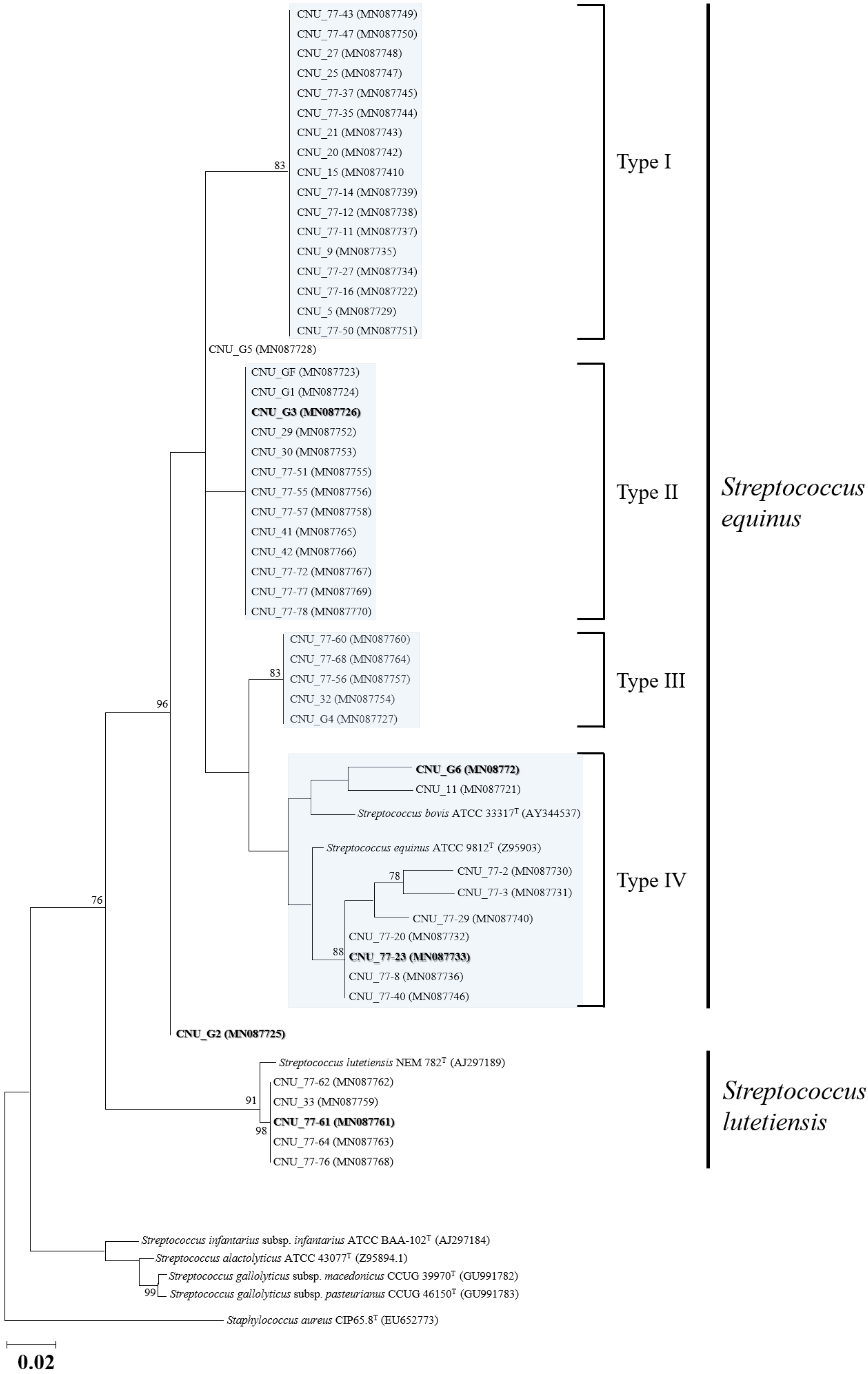
2.4. Species Discrimination and Phylogenetic Analysis

The obtained Streptococcus isolates were cultured overnight on BHIA at 37 °C. For species discrimination, the sodA gene, encoding the manganese-dependent superoxide dismutase, was amplified and sequenced using the primers d1/d2 (Table S2) [33,34]. The amplified and sequenced sodA gene of the isolates were respectively compared with other SBSEC strains including the type strains in the GenBank database by BLAST searches (www.ncbi.nlm.nih.gov/BLAST). Furthermore, the sodA sequences of the isolates were aligned with representative sequences from each SBSEC type strain using ClustalX (version 2.1) [35] and BioEdit Sequence Alignment Editor (version 7.1.0.3) [36]. The datasets were then analyzed phylogenetically using MEGAX (ver. 10.0) [37]. Phylogenic trees were constructed using the Neighbor-Joining (NJ) and Maximum-Likelihood (ML) methods, and the reliability of the trees was assessed using 1000 bootstrap replicates.

2.5. Antimicrobial Susceptibility Test

Antimicrobial susceptibility of the isolated SBSEC bacteria was evaluated using the disk diffusion method according to the guidelines of the Clinical and Laboratory Standards Institute (CLSI) [38,39,40]. In total, 12 antimicrobial agents (Oxoid Ltd., Basingstoke, UK) from 11 classes were used as follows: Aminoglycosides [gentamicin (10 μg)], carbapenems [imipenem (10 μg)], cephalosporins [cephalothin (30 μg)], fluoroquinolones [levofloxacin (5 μg)], glycopeptides [vancomycin (30 μg)], lincosamides [lincomycin (15 μg)], macrolides [erythromycin (15 μg)], oxazolidinones [linezolid (30 μg)], penicillins and β-lactam/β-lactamase inhibitor combinations [oxacillin (1 μg), penicillin (10 μg)], phenicols [chloramphenicol (30 μg)], and tetracyclines [tetracycline (30 μg)]. The minimum inhibitory concentrations (MICs) of four selected antimicrobial agents [gentamicin (256–0.016 μg), erythromycin (256–0.016 μg), clindamycin (256–0.016 μg), and tetracycline (256–0.016 μg)] were determined using MIC Evaluator Strips (Liofilchem®, Teramo, Italy). Standard disk diffusion and MIC tests were conducted on the Muller-Hilton Blood Agar (Synergy Innovation, Seongnam, Korea) at 37 °C for 24 h and the isolates were categorized as susceptible, intermediate, and resistant based on the interpretive criteria of the CLSI guidelines [40,41,42]. For quality control, Streptococcus pneumoniae ATCC 49619 were used.

2.6. Genome Sequencing of the SBSEC Isolates

Based on the phylogenetic analysis, a total of 5 representative SBSEC isolates were selected and submitted for further genomic investigations. The genomes of the selected SBSEC strains (CNU_77-23, CNU_77-61, CNU_G2, CNU_G3, and CNU_G6) were sequenced using a hybrid approach on a PacBio RS II system (Pacific Biosciences) by constructing a 20-kb SMRTbellTM template library and on the HiSeq X-10 platform (Illumina) by preparing a DNA library using the TruSeq nano DNA library prep kit (Illumina). Genome assembly of the filtered PacBio reads was performed using the HGAP (v3.0) pipeline, and the Illumina paired-end 150-bp reads were mapped using BWA-MEM (v0.7.15) and errors were corrected using Pilon (v1.21) with default parameters. Annotation was performed with the NCBI Prokaryotic Genome Annotation Pipeline (http://0-www-ncbi-nlm-nih-gov.brum.beds.ac.uk/books/NBK174280/). The L-lactate dehydrogenase gene, which is responsible for lactic acid production, was manually searched in the sequenced SBSEC genomes by BLAST (www.ncbi.nlm.nih.gov/BLAST). Moreover, the presence of genetic determinants related to antibiotic resistance was screened using the Comprehensive Antibiotic Resistance Database (https://card.mcmaster.ca/) and/or the ARG-ANNOT database (http://en.mediterranee-infection.com/article.php?laref=283&titre=arg-annot). Finally, to assess the genomic relatedness of the five selected SBSEC isolates with other Streptococcus species in the SBSEC, the average nucleotide identity (ANI) was analyzed using OrthoANI (https://www.ezbiocloud.net/tools/othoani) against several other type strains of species (or subspecies) available in the GenBank database.

2.7. Determination of Antimicrobial-resistant Genes and L-lactate Dehydrogenase Genes in the SBSEC Isolates

The presence of genetic determinants conferring resistance to macrolides, lincosamides, and tetracyclines in the SBSEC isolates were investigated by PCR and sequencing analyses. Detections of resistance genes for macrolides [erm(A), erm(B), erm(C), and mef(A)], lincosamides [lnu(C)], and tetracyclines [the putative conjugative Tn916-like transposon and tet(M)] were determined by conventional PCR and those for tet(O), tet(Q), and tet(S) were determined by multiplex PCR using previously described primers and protocols [33,41,42,43,44,45,46]. Moreover, the presence of genetic determinants responsible for lactic acid production (L-lactate dehydrogenase, ldh) was analyzed by PCR and sequencing analyses. The primer pair for the PCR amplification was generated from the annotated ldh genes encoded in the genome of the S. equinus strain CNU_G6 (GenBank accession No. CP046629; ldh region 1571618–1572607) using Primer 3 (http://bioinfo.ut.ee/primer3-0.4.0/primer3/). The primer sequences, amplicon sizes, and annealing temperatures used are summarized in Table S2. Strains yielding amplicons of the expected size were sequenced and the obtained nucleotide and deduced amino acid sequences were compared to those of genes from other Streptococcus spp. including SBSEC, available in the GenBank database.

2.8. Accession Numbers of Nucleotide Sequences and Strain Deposition

All the 16S rRNA, sodA, tet(M), lnu(C), and putative conjugative Tn916-like transposon genes of the SBSEC isolates in this study have been deposited in the GenBank database and the accession numbers are provided in Table 1. The complete genome sequences of the representative SBSEC isolates have been deposited in GenBank database under accession numbers CP046628 (CNU_77-23), CP046875 (CNU_77-61), CP046919 (CNU_G2), CP046624 (CNU_G3), and CP046629 (CNU_G6), respectively. A living axenic culture of the 51 SBSEC isolates has been deposited in the Korean Culture Center of Microorganisms (KCCM) and the deposition numbers are provided in Table 1.

3. Results

3.1. SBSEC Isolation and Identification

Among the bacteria collected during the surveillance studies between 2014 and 2019, a total of 51 isolates from the rumen fluid of Holstein dairy cattle (n = 15), Hanwoo steers (n = 17), and Korean goats (n = 19), were identified as Streptococcus spp. based on 16S rRNA sequencing analysis. The biochemical characteristics of the bacterial isolates were analyzed using the API 20 Strep system (bioMérieux Inc., Marcy l’ Étoile, France) and the results are summarized in Table S3. Briefly, all the isolates showed positive results for leucine aminopeptidase, raffinose, amidon, and glycogen, and most isolates (except strain CNU_77-2) were positive for lactose fermentation, which produces lactate.
During the bacterial identification in this study, the taxonomical positions of the 51 Streptococcus spp. isolates were not clearly discriminated by 16S rRNA sequence analyses therefore, the sodA gene, reported as one of the most reliable biomarkers for identifying SBSEC species [14,47], was used for species classifications in this study. Accordingly, all 51 isolates were confirmed to belong to the SBSEC and a total of two Streptococcus species including S. equinus (n = 46) and S. lutetiensis (n = 5) were identified. From the Hanwoo and Holstein dairy cattle, a total of 32 S. equinus strains (24 and eight each) were identified whereas a total of 14 S. equinus and five S. lutetiensis strains were obtained from Korean native goats (Table 1). The determined 16S rRNA and sodA sequences of the 51 SBSEC strains were deposited in the GenBank database and are listed in Table 2. Phylogenetic analyses based on the obtained sodA genes using the ML method revealed that the 46 S. equinus isolates were divided into four major types (type I, II, III, and IV) and two individual strains (CNU_G2 and CNU_G5), which were not clustered with the other isolates (Figure 1). Among the four major types, the reference strains of S. equinus (ATCC 9812T (Z95903) and ATCC 33317T (AY344537), which were previously reported as S. bovis) were assigned to type IV by clustering only with some other bovine-originated SBSEC isolates. Moreover, five other isolates, which were obtained from caprine animals and classified as S. lutetiensis (CNU_33, CNU_77-61, CNU_77-62, CNU_77-64, and CNU_77-76), were well clustered with the type strain of the species, S. lutetiensis NEM 782T (AJ297189). A similar result was also obtained from the phylogenetic tree generated using the NJ method.

3.2. Antimicrobial Susceptibility of the SBSEC Isolates

The resistance profiles of the 51 SBSEC isolates to several antibiotic classes was evaluated using the disc diffusion and MIC methods based on the CLSI guidelines. In this study, none of the SBSEC isolates were resistant to oxacillin, penicillin, cephalothin, and imipenem furthermore, they were generally susceptible (including intermediate) to aminoglycosides (gentamicin, resistance rate 21.6% (11/51)), fluoroquinolones (levofloxacin, resistance rate 23.5% (12/51)), macrolides (erythromycin, resistance rate 2.0% (1/51)), lincosamides (clindamycin, resistance rate 0% (0/51)), and tetracycline (tetracycline, resistance rate 15.7% (8/51)) (Table 3). However, large proportions of SBSEC isolates were categorized as ‘non-susceptible’ against glycopeptides (vancomycin, non-susceptible rate 84.3% (43/51)) and oxazolidinones (linezolid, non-susceptible rate 94.1% (48/51)) according to the CLSI guidelines [38,39]. Although a total of 12 SBSEC strains (23.5%) were not resistant to any of the antimicrobials used in this study, all isolates were not susceptible to at least one antimicrobial agent, and 25.5% of the isolates (13/51) were resistant to more than two antimicrobial agents tested in this study. Moreover, 5.9% of the isolates (3/51) were resistant to three different classes of antimicrobial agents and two isolates (CNU_77-23 and CNU_77-43) were resistant to four different classes of antimicrobial agents. The overall antimicrobial resistance profiles of all SBSEC isolates tested are summarized in Table 3.

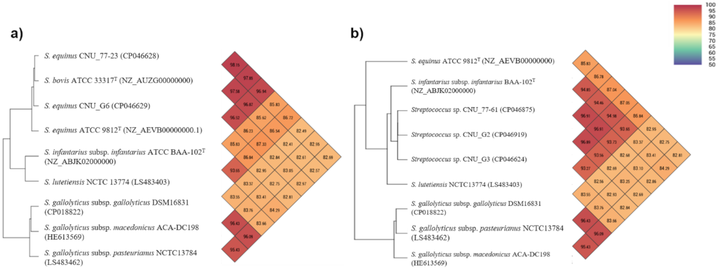
3.3. Genomic Features of the SBSEC Isolates

In this study, we selected and sequenced the whole genome of four representative S. equinus isolates (CNU_G3 which grouped in type II; CNU_G6 which clustered with ATCC 33317T, that was previously reported as S. bovis in type IV; CNU_77-23 which clustered with the type strain of S. equinus (ATCC 9812T) in type IV; and CNU_G2 which was not clustered with any other S. equinus strains used in this study) for the following reasons: First, to elucidate their plasticity and diversity in the S. equinus group, and second, to obtain the reference genomes to determine the genetic determinants related to the production of lactic acid and antimicrobial resistances that could be used for further overall screening in the 51 SBSEC isolates. Additionally, one strain of S. lutetiensis isolated from caprine (CNU_77-61) was also chosen and submitted for further genome sequencing and analysis.
The genomes of the five selected isolates comprised 1.9–2.0 Mbp, consisting of one chromosome each (1.8–1.9 Mbp with 37.4–37.9 G + C content (%)). Although the presence of plasmids in the SBSEC is known to be relatively rare [11], the three strains (CNU_G2, CNU_G3, and CNU_G6) identified as S. equinus possessed a single plasmid, but no antibiotic-resistant or virulence-related genes were detected in it. Detailed features of the sequenced genome and plasmids are indicated in Table 4. Genomic similarities among the five selected isolates and other available type strains of the SBSEC were assessed using the ANI comparisons and the results indicated that the genome of strains CNU_77-23 and CNU_G6 were the most similar to that of S. bovis ATCC 33317T with > 96.0% ANI values (Figure 2a). However, S. equinus strains CNU_G2 and CNU_G3 and the S. lutetiensis strain CNU_77-61 showed <95.0% ANI values against any of the type strains of the SBSEC including S. equinus ATCC 9812T and S. lutetiensis NCTC 13774T. Moreover, the resultant phylogeny based on the OrthoANI values also indicated that these three SBSEC strains were clearly differentiated from the type strains of S. equinus and S. lutetiensis, and were rather closely related to the available genome of S. infantarius subsp. infantarius (GenBank accession No. NZ_ABJK02000000) (Figure 2b). Although the three SBSEC isolates were identified as S. equinus and S. lutetiensis based on the 16S rRNA and sodA gene analyses, these results strongly indicated that the three SBSEC strains (CNU_G2, CNU_G3, and CNU_77-61) could not be classified as S. equinus and S. lutetiensis, and might be categorized as potential new species (or subspecies) in the SBSEC.

3.4. Antimicrobial-Resistant Genes and L-lactate Dehydrogenase Genes in the SBSEC Isolates

The presence of genetic determinants conferring resistance to macrolides, lincosamides, and tetracyclines in the five genome-sequenced SBSEC isolates (CNU_77-23, CNU 77-61, CNU_G2, CNU_G3, and CNU_G6) was preliminarily screened by manual searches using available databases. As a result, the lincosamide-resistance gene, lnu(C), was found to be encoded in the bacterial chromosome of strain CNU_77-23 and CNU_G3 however, none of the genomes showed the known AMR genes for macrolides [erm(A), erm(B), erm(C), and mef(A)]. Interestingly, the putative conjugative Tn916-like transposon that harbored the tetracycline-resistance gene, tet(M), was encoded in the bacterial chromosome of all the sequenced SBSEC isolates except CNU_G3 and the Tn916-like transposons (~18 kb) found in the SBSEC genomes were almost identical (>99%) to those found in the Enterococcus faecalis DS16 transposon Tn916 (U09422.1; 18,032 bp), and its implicated tet(M) genes in the isolates were also very similar (>99%) to those from various G(+) bacterial species available in the GenBank database. Therefore, we screened the presence of lnu(C), erm(A), erm(B), erm(C), mef(A), tet (O), tet (Q), tet (S), and Tn916-like transposon including the tet(M) gene in the remaining 46 isolates and the results indicated that 20.0% (10/51) and 33.3% (17/51) of the SBSEC isolates possessed lnu(C) and the Tn916-like transposon including the tet(M) gene, respectively. Among these, a total of three isolates (CNU_77-23, CNU_GF, and CNU_G1) possessed both the lnu(C) and tet(M) genes, however, erm(A), erm(B), erm(C), mef(A), tet (O), tet (Q), and tet (S) were not detected in any of the SBSEC isolates. Interestingly, all five S. lutetiensis isolates (CNU_33, CNU_77-61, CNU_77-62, CNU_77-64, and CNU_77-76) obtained from caprine possessed the Tn916-like transposon but tet(M) was not found.
We also assessed the presence of the ldh gene related to lactic acid production, in the sequenced SBSEC genomes, and found it to be encoded in the chromosome of all the sequenced SBSEC genomes. The annotated ldh genes (990 bp) in the SBSEC isolates were respectively compared with the fructose-1,6-diphosphate-dependent L-lactate dehydrogenase in S. equinus JB1 (U60997.1) [48], and the nucleotide sequence (99.1–99.8% identities) and deduced amino acid sequence (99.7–100% identities) of these ldh genes were almost homologous to those of strain JB1. Therefore, we screened the presence of the ldh gene in the remaining 46 isolates using ldh-F/R PCR primers designed in this study, and the results indicated that all the 51 SBSEC strains possessed ldh, suggesting that our isolates may have a strong potential to produce lactic acid as was shown in the results of biochemical tests using the API 20 Strep system.

4. Discussion

For several decades, the SBSEC has been recognized as commensal bacteria that inhabit the gastrointestinal tract of ruminants and humans, but numerous studies have provided evidence that the complex is an important contributor to ruminal acidosis in domestic ruminants and are also associated with serious infections and colorectal cancer in humans and animals, thus highlighting the common importance of SBSEC members in the livestock industry and human health [15,16]. Although the exact economic damage caused by SBSEC in domestic animals is not currently clear, it is generally understood that the bacteria can affect animal health, productivity leading to important losses in the livestock industry [9]. Along with ruminants, SBSEC have been reported from various other domestic animals, their food products, and from companion wild animals however, only limited numbers of prevalence studies have been conducted on birds, cattle, and lambs [16].
Although S. bovis was synonymized with S. equinus and is currently recognized as the SBSEC, the name is still commonly used in ruminant livestock research [49,50,51]. However, researchers in the area have already recognized the physiological and genetic diversities of bovine-origin S. bovis isolates [20,21] and genetic heterogeneity in SBSEC has been described recently, especially in S. equinus isolates [22]. Nevertheless, the potential plasticity and diversity of SBSECs in domestic ruminants have not been well investigated so far. In this milieu, we preferentially aimed to investigate the prevalence and diversity of SBSEC inhabiting Korean domestic ruminants.
In this study, a total of 51 SBSEC strains (Holstein dairy cattle (n = 15), Hanwoo (n = 17), and Korean goats (n = 19)) were isolated from collected rumen fluids. As expected, most of the isolates (except strain CNU_77-2) were positive for lactose fermentation which produces lactate, thus suggesting that these isolates have strong potential to cause ruminal acidosis in animals. However, no significant differences in biochemical characteristics between isolates from the three animal species was observed except for esculin, which was positive only in some isolates (n = 5) from Hanwoo steers (Table S3). Esculin hydrolysis has been considered an important diagnostic factor to differentiate enterococci and group D streptococci (mainly SBSEC) from non-group D viridans group streptococci [52] and several SBSEC strains have been identified successfully using esculin hydrolyzing activity [53]. However, based on the results of our study, the SBSEC strains isolated from Korean domestic ruminants seem to have relatively low esculin hydrolyzing activity (9.8%), suggesting that the bile-esculin test might not be suitable for the presumptive identification of SBSEC, at least from Korean domestic ruminants.
For species discrimination of the 51 SBSEC strains collected in this study, both the 16S rRNA and sodA genes were respectively obtained and compared with the seven-type-SBSEC strains. In accordance with previous reports [14,47,54,55,56], partial sodA gene sequencing was more discriminant than 16S rRNA for SBSEC species identification in this study. Therefore, the species of the 51 SBSEC isolates were finally determined by sodA gene-based identification, and as a result, all 51 isolates were confirmed to belong to SBSEC, with a total of two Streptococcus species including S. equinus (n = 46) and S. lutetiensis (n = 5) identified. Interestingly, S. lutetiensis was only isolated from the rumen fluids of goats rather than those from dairy cattle and Hanwoo in this study. Although it is difficult to directly compare the presence of SBSEC members in a single animal species due to the difficulties encountered over the years in the correct identification of SBSEC strains to the species (or subspecies) level by phenotypic and genotypic methods, the potential presence of S. lutetiensis (Previously classified as S. infantarius subsp. coli) in domestic ruminants have been only addressed in caprine and their food products [57,58]. In this study, we also confirmed the presence of S. lutetiensis only in the rumen of Korean goats by direct isolation and sodA gene-based identification rather that from bovine, thus hypothesizing that the species might be one of the distinguished important commensal SBSEC in caprine. However, due to the limited number of animals and SBSEC isolates in this study, additional studies focusing on the prevalence and characteristics of SBSEC in the rumen of goats by direct bacterial isolation will be necessary in the future.
The 46 S. equinus strains were isolated from all three animal species. Although most of the our SBSEC isolates were identified as S. equinus based on a sodA gene comparison, phylogenetic analyses have indicated that these isolates could be divided as several different types, thus revealing their potential plasticity and diversity and the S. equinus isolates were divided as four major types and two individual strains which were not clustered with other isolates, regardless of their source animal species (Figure 1). Among the four major types, S. equinus ATCC 9812T and S. bovis ATCC 33317T (now synonymized to S. equinus) were assigned to type IV and some other bovine-originated SBSEC isolates were clustered together. Therefore, we selected and sequenced the whole genome of the four representative S. equinus isolates (CNU_G2, CNU_G3, CNU_G6, and CNU_77-23) to elucidate their plasticity and diversity in the S. equinus group. As expected, the genome-based ANI comparison revealed that the genome of S. equinus strains CNU_G2 and CNU_G3 showed less than 95.0% ANI values against any of the type strains in the SBSEC including S. equinus ATCC 9812T. To date, it has been generally accepted that ANI values > 95–96% are the threshold for species delineation [59,60,61] and our results of genome-based ANI comparison also support the potential heterogeneity of S. equinus, which has been addressed previously [22]. Similar results were also observed upon the ANI comparison of S. lutetiensis CNU_77-61, which showed < 95.0% ANI values against any of the type strains in the SBSEC including S. lutetiensis NCTC 13774T even though it was classified as S. lutetiensis based on sodA gene-based identification. Although only a limited number of domestic ruminants from a single farm and their SBSEC isolates were examined in this study, these results strongly suggest that SBSECs, at least in S. equinus and S. lutetiensis, are considerably more diverse than previously thought and that the current taxonomic assignments of these two species remains a subject to debate, requiring re-evaluation in the future. Moreover, the three SBSEC strains (CNU_G2, CNU_G3, and CNU_77-61) might be categorized as the potential new species (or subspecies) in the SBSEC and its further studies are currently in progress.
To date, the emergence of AMR bacteria and widespread ARGs has led to serious public health and livestock industry concerns due to potential health risks to humans and animals [23,24]. Furthermore, SBSEC have been reported as one of the most AMR species among streptococci [11]. Although most previous studies evaluating AMR in SBSEC have been focused on isolates of clinical origin, they have clearly indicated high AMR incidence rates against several classes of commercial antibiotics (e.g., clindamycin, erythromycin, and tetracycline) especially in S. gallolyticus subsp. galloyticus, S. gallolyticus subsp. pasteurianus, S. infantarius subsp. infantarius, and S. lutetiensis [22,34,35,62,63,64]. These results suggest that SBSEC could be a potential reservoir of various ARGs that can be transmitted to other opportunistic pathogens and other strains in the complex. However, only a few studies are available on AMR SBSEC isolates from animal origins, focusing on S. gallolyticus, and these present variable resistance rates [28,29]. The incidence of AMR and ARGs in S. equinus and S. lutetiensis of an animal origin has not yet been investigated. In this study, the AMR rates of SBSEC isolated from Korean domestic ruminants were relatively lower than those from other SBSEC isolates of clinical origin and the resistance rates on erythromycin, clindamycin, and tetracycline were estimated to be 2.0% (1/51), 0% (0/51), and 15.7% (8/51), respectively. Moreover, the resistance rates of our SBSEC isolates against the three antibiotics were much lower than those reported previously in various streptococcal species including incompletely classified S. bovis isolated from milk samples in Korea [27,49]. Along with the phenotypic evaluations of AMRs in SBSEC isolates, we also investigated the presence of ARGs associated with resistance to erythromycin, clindamycin, and tetracycline. Several recent reports have indicated that the dominant erythromycin resistance-associated gene in various streptococcal species isolated from domestic animals was erm(B) [65,66] and that resistance in the SBSEC of human clinical origins was mostly associated with erm(B) and mef(A) [28,29,45,62,67]. However, as expected, erm(A), erm(B), erm(C), and mef(A) were not detected in this study, and only the erythromycin-resistant S. equinus strain CNU_77-43 might have another unveiled AMR mechanism against the antibiotic. Although the erm variants are mostly associated with cross-resistance to macrolides-lincosamides-type B streptogramins (MLSB), the bacteria in this genus also contain genes associated with specific resistance to lincosamides, which are commonly used in dairy cattle to treat mastitis [66]. The genes responsible for resistance to lincosamides are lnu class genes, which encode nucleotidyltransferases and cause enzymatic inactivation of the antibiotic [68]. Among the nine lnu class genes reported to date (http://faculty.washington.edu/marilynr/), the lnu(C) is relatively little investigated however, recent reports have demonstrated its presence in the livestock industry [69]. In this study, we confirmed the presence of lnu(C) during genome analysis in some SBSEC isolates (strain CNU_77-23 and CNU_G3) even though they were not phenotypically resistant to lincosamides (clindamycin). Therefore, we have screened the presence of lnu(C) in all isolates, furthermore, the results also indicated the considerable appearance rate (20.0%) of the genetic determinants although the strains did not show phenotypic resistance to the antibiotics. These phenomena showing a lack of correlation between the presence of ARGs and degree of antimicrobial susceptibility in bacteria have been described previously [66,70,71,72] and thus, further studies on the mechanisms of AMR SBSEC presenting in domestic ruminants are urgently needed.
Different from the results on erythromycin and clindamycin, the phenotypical tetracycline resistance of the S. equinus isolates was generally correlated with the presence of tet(M) and Tn916-like transposon genes except for one strain (CNU_77-29), which showed a low MIC value (0.50 μg/mL) (Table 4). In general, the tet(M) gene has been reported to be associated with the conjugative transposons Tn916, Tn1545, and other related transposons [73] and until recently, this gene was mainly considered to have originated from human-originated S. agalactiae [74]. However, several recent studies on the surveillance of tetR genes in various streptococcal species isolated from domestic animals have reported the frequent presence of tet(M) genes in the United States, China, and Europe [66,70,72]. In this study, we only detected the presence of Tn916-like transposon-mediated tet(M) genes among the S. equinus isolates even though the emergence of several other tetR genes (e.g., tet(L) and tet(O)) has been reported from other SBSEC isolates [28,45,62]. Based on these results, although only a limited number of SBSEC isolates was examined in this study, the protection of ribosomal proteins by tet(M) might be one of the main tetracycline-resistance mechanisms in the Korean SBSEC. Additionally, although vancomycin resistance in other SBSEC isolates from animal fecal samples has been rarely reported and its resistance mechanisms were mainly associated with the vanB gene [15], a large proportion of SBSEC isolates was categorized as non-susceptible against the antibiotic however, the two groups of van resistance operons (D-Ala-D-Lac ligase (vanA, vanB, vanD, and vanM) and D-Ala-D-Ser ligase (vanC, vanE, vanG, vanL, and vanN)) were not detected during our manual search in the five sequenced genomes. Therefore, the phenotypical resistance in our SBSEC isolates might be assumed to be associated with other unknown genetic determinants or point mutations, and further research uncovering its resistance mechanisms will be necessary in the future.
Despite raising concerns regarding their potential pathogenicity and carcinogenic properties, some SBSEC members with a dairy origin have been directly supplied as probiotics to young domestic ruminants to support ruminal development by establishing an anaerobic rumen microbiota for efficient feed digestion [75,76,77,78]. However, the fact that ARGs were found in several SBSEC isolates from different geographical origins is worrying as the bacteria in domestic ruminant could be a potential reservoir of these genes and horizontal gene transfer via transposon within the same genus may lead to serious concerns in the global livestock industry, including Korea. Undeniably, SBSEC are one of the most important commensal rumen bacterial species affecting the health and productivity of domestic animals. Although only a limited number of domestic ruminants from a single farm and their SBSEC isolates were examined, the presence of ARGs, which can potentially be transmitted to other opportunistic pathogens in the animals, was verified in this study. Therefore, nation-wide continuous monitoring of AMR strain emergence and ARG acquisition in SBSEC will be needed in Korean domestic ruminants.

Supplementary Materials

The following are available online at https://0-www-mdpi-com.brum.beds.ac.uk/2076-2607/9/1/98/s1, Table S1. Chemical composition (g/kg DM or as stated) of the ruminant diet fed to two fistulated Holstein cow; Table S2. Primers used in this study; Table S3. Biochemical characterization of SBSEC used in this study.

Author Contributions

Conceptualization, S.Y.P., J.H.K. and S.S.; data curation, S.Y.P. and M.L.; formal analysis, S.Y.P. and M.L.; funding acquisition, J.H.K. and S.S.; investigation, S.Y.P., S.R.L., H.K., and Y.S.L.; methodology, S.Y.P., S.R.L., H.K., and Y.S.L.; project administration, J.H.K.; resources, S.Y.P., and M.L.; supervision, J.H.K. and S.S.; writing—original draft, S.Y.P., M.L., and J.H.K.; and writing—review and editing, S.Y.P., J.H.K., and S.S. All authors have read and agreed to the published version of the manuscript.

Funding

This research was supported by the KRIBB Initiative programs, the National Research Foundation (NRF) of Korea (NRF-2020R1I1A2068827), and academic research funds from Chungnam National University.

Institutional Review Board Statement

The animal use and protocols for this experiment were reviewed and approved by the Chungnam National University Animal Research Ethics Committee (IACUC approval Nos. CNU-00455 and CNU-01021).

Conflicts of Interest

The authors declare that they have no conflicts of interest.

References

  1. Jouany, J.; Michalet-Doreau, B.; Doreau, M. Manipulation of the rumen ecosystem to support high-performance beef cattle-Review. Asian-Australas. J. Anim. Sci. 2000, 13, 96–114. [Google Scholar]
  2. Tajima, K.; Arai, S.; Ogata, K.; Nagamine, T.; Matsui, H.; Nakamura, M.; Aminov, R.I.; Benno, Y. Rumen bacterial community transition during adaptation to high-grain diet. Anaerobe 2000, 6, 273–284. [Google Scholar] [CrossRef]
  3. Chen, L.; Shen, Y.; Wang, C.; Ding, L.; Zhao, F.; Wang, M.; Fu, J.; Wang, H. Megasphaera elsdenii lactate degradation pattern shifts in rumen acidosis models. Front. Microbiol. 2019, 10, 162. [Google Scholar] [CrossRef] [PubMed] [Green Version]
  4. Hungate, R.E. The Rumen and Its Microbes; Elsevier: Amsterdam, The Netherlands, 2013. [Google Scholar]
  5. Russell, J.B. Rumen Microbiology and Its Role in Ruminant Nutrition; Department of Microbiology, Cornell University: Ithaca, NY, USA, 2002. [Google Scholar]
  6. Freer, S.N. Purification and characterization of the extracellular alpha-amylase from Streptococcus bovis JB1. Appl. Environ. Microbiol. 1993, 59, 1398–1402. [Google Scholar] [CrossRef] [Green Version]
  7. Asanuma, N.; Hino, T. Regulation of fermentation in a ruminal bacterium, Streptococcus bovis, with special reference to rumen acidosis. Anim. Sci. J. 2002, 73, 313–325. [Google Scholar] [CrossRef]
  8. Enemark, J.M. The monitoring, prevention and treatment of sub-acute ruminal acidosis (SARA): A review. Vet. J. 2008, 176, 32–43. [Google Scholar] [CrossRef]
  9. Herrera, P.; Kwon, Y.M.; Ricke, S.C. Ecology and pathogenicity of gastrointestinal Streptococcus bovis. Anaerobe 2009, 15, 44–54. [Google Scholar] [CrossRef] [PubMed]
  10. Lean, I.; Wade, L.; Curtis, M.; Porter, J. New approaches to control of ruminal acidosis in dairy cattle. Asian Australas. J. Anim. Sci. 2000, 13, 266–269. [Google Scholar]
  11. Jans, C.; Meile, L.; Lacroix, C.; Stevens, M.J. Genomics, evolution, and molecular epidemiology of the Streptococcus bovis/Streptococcus equinus complex (SBSEC). Infect. Genet. Evol. 2015, 33, 419–436. [Google Scholar] [CrossRef]
  12. Jans, C.; Meile, L.; Kaindi, D.W.M.; Kogi-Makau, W.; Lamuka, P.; Renault, P.; Kreikemeyer, B.; Lacroix, C.; Hattendorf, J.; Zinsstag, J. African fermented dairy products—Overview of predominant technologically important microorganisms focusing on African Streptococcus infantarius variants and potential future applications for enhanced food safety and security. Int. J. Food Microbiol. 2017, 250, 27–36. [Google Scholar] [CrossRef]
  13. Parker, M.; Ball, L.C. Streptococci and aerococci associated with systemic infection in man. J. Med. Microbiol. 1976, 9, 275–302. [Google Scholar] [CrossRef] [PubMed]
  14. Dekker, J.P.; Lau, A.F. An update on the Streptococcus bovis group: Classification, identification, and disease associations. J. Clin. Microbiol. 2016, 54, 1694–1699. [Google Scholar] [CrossRef] [PubMed] [Green Version]
  15. Pompilio, A.; Di Bonaventura, G.; Gherardi, G. An overview on Streptococcus bovis/Streptococcus equinus complex isolates: Identification to the species/subspecies level and antibiotic resistance. Int. J. Mol. Sci. 2019, 20, 480. [Google Scholar] [CrossRef] [PubMed] [Green Version]
  16. Jans, C.; Boleij, A. The road to infection: Host-microbe interactions defining the pathogenicity of Streptococcus bovis/Streptococcus equinus complex members. Front. Microbiol. 2018, 9, 603. [Google Scholar] [CrossRef] [Green Version]
  17. Nagaraja, T.; Titgemeyer, E. Ruminal acidosis in beef cattle: The current microbiological and nutritional outlook. J. Dairy Sci. 2007, 90, E17–E38. [Google Scholar] [CrossRef] [Green Version]
  18. Fernando, S.C.; Purvis, H.T.; Najar, F.Z.; Sukharnikov, L.O.; Krehbiel, C.R.; Nagaraja, T.G.; Roe, B.A.; DeSilva, U. Rumen microbial population dynamics during adaptation to a high-grain diet. Appl. Environ. Microbiol. 2010, 76, 7482–7490. [Google Scholar] [CrossRef] [Green Version]
  19. Petri, R.M.; Schwaiger, T.; Penner, G.B.; Beauchemin, K.A.; Forster, R.J.; McKinnon, J.J.; McAllister, T.A. Characterization of the core rumen microbiome in cattle during transition from forage to concentrate as well as during and after an acidotic challenge. PLoS ONE 2013, 8, e83424. [Google Scholar] [CrossRef] [Green Version]
  20. Jarvis, G.N.; Kurtovic, A.; Hay, A.G.; Russell, J.B. The physiological and genetic diversity of bovine Streptococcus bovis strains. FEMS Microbiol. Ecol. 2001, 35, 49–56. [Google Scholar] [CrossRef]
  21. Klieve, A.V.; Heck, G.L.; Prance, M.A.; Shu, Q. Genetic homogeneity and phage susceptibility of ruminal strains of Streptococcus bovis isolated in Australia. Lett. Appl. Microbiol. 1999, 29, 108–112. [Google Scholar] [CrossRef]
  22. Jans, C.; de Wouters, T.; Bonfoh, B.; Lacroix, C.; Kaindi, D.W.M.; Anderegg, J.; Böck, D.; Vitali, S.; Schmid, T.; Isenring, J. Phylogenetic, epidemiological and functional analyses of the Streptococcus bovis/Streptococcus equinus complex through an overarching MLST scheme. BMC Microbiol. 2016, 16, 117. [Google Scholar] [CrossRef] [Green Version]
  23. Marshall, B.M.; Levy, S.B. Food animals and antimicrobials: Impacts on human health. Clin. Microbiol. Rev. 2011, 24, 718–733. [Google Scholar] [CrossRef] [PubMed] [Green Version]
  24. Zhao, Y.; Yang, Q.E.; Zhou, X.; Wang, F.-H.; Muurinen, J.; Virta, M.P.; Brandt, K.K.; Zhu, Y.-G. Antibiotic resistome in the livestock and aquaculture industries: Status and solutions. Crit. Rev. Environ. Sci. Technol. 2020, 1–38. [Google Scholar] [CrossRef]
  25. Economou, V.; Gousia, P. Agriculture and food animals as a source of antimicrobial-resistant bacteria. Infect. Drug Resist. 2015, 8, 49. [Google Scholar] [CrossRef] [PubMed] [Green Version]
  26. Wang, N.; Guo, X.; Yan, Z.; Wang, W.; Chen, B.; Ge, F.; Ye, B. A comprehensive analysis on spread and distribution characteristic of antibiotic resistance genes in livestock farms of Southeastern China. PLoS ONE 2016, 11, e0156889. [Google Scholar] [CrossRef] [PubMed]
  27. Nam, H.-M.; Lim, S.-K.; Kang, H.-M.; Kim, J.-M.; Moon, J.-S.; Jang, K.-C.; Joo, Y.-S.; Kang, M.-I.; Jung, S.-C. Antimicrobial resistance of streptococci isolated from mastitic bovine milk samples in Korea. J. Vet. Diagn. Investig. 2009, 21, 698–701. [Google Scholar] [CrossRef]
  28. Nomoto, R.; Le, H.T.T.; Sekizaki, T.; Osawa, R. Antimicrobial susceptibility of Streptococcus gallolyticus isolated from humans and animals. Jpn. J. Infect Dis. 2013, 66, 334–336. [Google Scholar] [CrossRef] [Green Version]
  29. Romero-Hernández, B.; Tedim, A.P.; Sánchez-Herrero, J.F.; Librado, P.; Rozas, J.; Muñoz, G.; Baquero, F.; Cantón, R.; Del Campo, R. Streptococcus gallolyticus subsp. gallolyticus from human and animal origins: Genetic diversity, antimicrobial susceptibility, and characterization of a vancomycin-resistant calf isolate carrying a vanA-Tn1546-like element. Antimicrob. Agents Chemother. 2015, 59, 2006–2015. [Google Scholar]
  30. Lee, M. Diversity of Rumen Bacterial Community and Streptococcus bovis Strains in Korean Domestic Ruminants Fed a High-Concentrate Diet. Master’s Thesis, Chungnam National University, Daejeon, Korea, 2016. [Google Scholar]
  31. Lee, M.; Jeong, S.; Seo, J.; Seo, S. Changes in the ruminal fermentation and bacterial community structure by a sudden change to a high-concentrate diet in Korean domestic ruminants. Asian-Australas. J. Anim. Sci. 2019, 32, 92. [Google Scholar] [CrossRef] [Green Version]
  32. Makkar, H.P.; McSweeney, C.S. Methods in Gut Microbial Ecology for Ruminants; Springer: Dordrecht, The Netherlands, 2005. [Google Scholar]
  33. Poyart, C.; Quesne, G.; Trieu-Cuot, P. Taxonomic dissection of the Streptococcus bovis group by analysis of manganese-dependent superoxide dismutase gene (sodA) sequences: Reclassification of ‘Streptococcus infantarius subsp. coli’ as Streptococcus lutetiensis sp. nov. and of Streptococcus bovis biotype 11.2 as Streptococcus pasteurianus sp. nov. Int. J. Syst. Evol. Microbiol. 2002, 52, 1247–1255. [Google Scholar]
  34. Romero, B.; Morosini, M.-I.; Loza, E.; Rodríguez-Baños, M.; Navas, E.; Cantón, R.; del Campo, R. Reidentification of Streptococcus bovis isolates causing bacteremia according to the new taxonomy criteria: Still an issue? J. Clin. Microbiol. 2011, 49, 3228–3233. [Google Scholar] [CrossRef] [Green Version]
  35. Larkin, M.A.; Blackshields, G.; Brown, N.P.; Chenna, R.; McGettigan, P.A.; McWilliam, H.; Valentin, F.; Wallace, I.M.; Wilm, A.; Lopez, R. Clustal W and Clustal X version 2.0. Bioinformatics 2007, 23, 2947–2948. [Google Scholar] [CrossRef] [PubMed] [Green Version]
  36. Hall, T.A. BioEdit: A user-friendly biological sequence alignment editor and analysis program for Windows 95/98/NT. Nucleic Acids Symp. Ser. 1999, 41, 95–98. [Google Scholar]
  37. Kumar, S.; Stecher, G.; Li, M.; Knyaz, C.; Tamura, K. MEGA X: Molecular evolutionary genetics analysis across computing platforms. Mol. Biol. Evol. 2018, 35, 1547–1549. [Google Scholar] [CrossRef]
  38. Clinical and Laboratory Standards Institute. Performance Standards for Antimicrobial Disk and Dilution Susceptibility Tests for Bacteria Isolated from Animals M31-A2, 2nd ed.; Clinical and Laboratory Standards Institute: Wayne, PA, USA, 2002. [Google Scholar]
  39. Clinical and Laboratory Standards Institute. Performance Standards for Antimicrobial Disk and Dilution Susceptibility Tests for Bacteria Isolated from Animals VET01S, 3rd ed.; Clinical and Laboratory Standards Institute: Wayne, NJ, USA, 2015. [Google Scholar]
  40. Clinical and Laboratory Standards Institute. Performance Standards for Antimicrobial Susceptibility Testing M100S, 29th ed.; Clinical and Laboratory Standards Institute: Wayne, PA, USA, 2019. [Google Scholar]
  41. Jun, L. Characterization of Antibiotic Resistant Genes Carried by Fish Pathogens in Korea. Ph.D. Thesis, Pukyong National University, Busan, Korea, 2010. [Google Scholar]
  42. Lüthje, P.; Schwarz, S. Molecular basis of resistance to macrolides and lincosamides among staphylococci and streptococci from various animal sources collected in the resistance monitoring program BfT-GermVet. Int. J. Antimicrob. Agents 2007, 29, 528–535. [Google Scholar] [CrossRef] [PubMed]
  43. Li, L.; Olsen, R.H.; Shi, L.; Ye, L.; He, J.; Meng, H. Characterization of a plasmid carrying cat, ermB and tetS genes in a foodborne Listeria monocytogenes strain and uptake of the plasmid by cariogenic Streptococcus mutans. Int. J. Food Microbiol. 2016, 238, 68–71. [Google Scholar] [CrossRef] [PubMed]
  44. Volokhov, D.; Chizhikov, V.; Chumakov, K.; Rasooly, A. Microarray analysis of erythromycin resistance determinants. J. Appl. Microbiol. 2003, 95, 787–798. [Google Scholar] [CrossRef] [PubMed]
  45. Leclercq, R.; Huet, C.; Picherot, M.; Trieu-Cuot, P.; Poyart, C. Genetic basis of antibiotic resistance in clinical isolates of Streptococcus gallolyticus (Streptococcus bovis). Antimicrob. Agents Chemother. 2005, 49, 1646–1648. [Google Scholar] [CrossRef] [Green Version]
  46. De Vries, L.E.; Christensen, H.; Skov, R.L.; Aarestrup, F.M.; Agersø, Y. Diversity of the tetracycline resistance gene tet(M) and identification of Tn916-and Tn5801-like (Tn 6014) transposons in Staphylococcus aureus from humans and animals. J. Antimicrob. Chemother. 2009, 64, 490–500. [Google Scholar] [CrossRef] [Green Version]
  47. Hinse, D.; Vollmer, T.; Erhard, M.; Welker, M.; Moore, E.; Kleesiek, K.; Dreier, J. Differentiation of species of the Streptococcus bovis/equinus-complex by MALDI-TOF mass spectrometry in comparison to sodA sequence analyses. Syst. Appl. Microbiol. 2011, 34, 52–57. [Google Scholar] [CrossRef]
  48. Wyckoff, H.; Chow, J.; Whitehead, T.; Cotta, M. Cloning, sequence, and expression of the L-(+) lactate dehydrogenase of Streptococcus bovis. Curr. Microbiol. 1997, 34, 367–373. [Google Scholar] [CrossRef]
  49. Yun, M.-J.; Yoon, S.; Lee, Y.J. Monitoring and characteristics of major mastitis pathogens from bulk tank milk in Korea. Animals 2020, 10, 1562. [Google Scholar] [CrossRef] [PubMed]
  50. Kim, H.; Lee, H.G.; Kwon, I.; Seo, J. Characterization of endolysin LyJH307 with antimicrobial activity against Streptococcus bovis. Animals 2020, 10, 963. [Google Scholar] [CrossRef] [PubMed]
  51. Peng, Q.; Cheng, L.; Kang, K.; Tian, G.; Al-Mamun, M.; Xue, B.; Wang, L.; Zou, H.; Gicheha, M.G.; Wang, Z. Effects of yeast and yeast cell wall polysaccharides supplementation on beef cattle growth performance, rumen microbial populations and lipopolysaccharides production. J. Integr. Agric. 2020, 19, 810–819. [Google Scholar] [CrossRef]
  52. Chuard, C.; Reller, L. Bile-esculin test for presumptive identification of enterococci and streptococci: Effects of bile concentration, inoculation technique, and incubation time. J. Clin. Microbiol. 1998, 36, 1135–1136. [Google Scholar] [CrossRef] [PubMed] [Green Version]
  53. Devriese, L.; Hommez, J.; Laevens, H.; Pot, B.; Vandamme, P.; Haesebrouck, F. Identification of aesculin-hydrolyzing streptococci, lactococci, aerococci and enterococci from subclinical intramammary infections in dairy cows. Vet. Microbiol. 1999, 70, 87–94. [Google Scholar] [CrossRef]
  54. Sasaki, E.; Osawa, R.; Nishitani, Y.; Whiley, R.A. Development of a diagnostic PCR assay targeting the Mn-dependent superoxide dismutase gene (sodA) for identification of Streptococcus gallolyticus. J. Clin. Microbiol. 2004, 42, 1360–1362. [Google Scholar] [CrossRef] [PubMed] [Green Version]
  55. Hoshino, T.; Fujiwara, T.; Kilian, M. Use of phylogenetic and phenotypic analyses to identify nonhemolytic streptococci isolated from bacteremic patients. J. Clin. Microbiol. 2005, 43, 6073–6085. [Google Scholar] [CrossRef] [PubMed] [Green Version]
  56. Teles, C.; Smith, A.; Ramage, G.; Lang, S. Identification of clinically relevant viridans group streptococci by phenotypic and genotypic analysis. Eur. J. Clin. Microbiol. Infect. Dis. 2011, 30, 243–250. [Google Scholar] [CrossRef] [Green Version]
  57. Aphale, D.; Kulkarni, A. Modifications and optimization of manual methods for polymerase chain reaction and 16S rRNA gene sequencing quality community DNA extraction from goat rumen digesta. Vet. World 2018, 11, 990. [Google Scholar] [CrossRef]
  58. Rosa, N.M.; Agnoletti, F.; Lollai, S.; Tola, S. Comparison of PCR-RFLP, API® 20 Strep and MALDI-TOF MS for identification of Streptococcus spp. collected from sheep and goat milk samples. Small Rumin. Res. 2019, 180, 35–40. [Google Scholar] [CrossRef]
  59. Goris, J.; Konstantinidis, K.T.; Klappenbach, J.A.; Coenye, T.; Vandamme, P.; Tiedje, J.M. DNA–DNA hybridization values and their relationship to whole-genome sequence similarities. Int. J. Sys. Evol. Microbiol. 2007, 57, 81–91. [Google Scholar] [CrossRef] [PubMed] [Green Version]
  60. Richter, M.; Rosselló-Móra, R. Shifting the genomic gold standard for the prokaryotic species definition. Proc. Natl. Acad. Sci. USA 2009, 106, 19126–19131. [Google Scholar] [CrossRef] [PubMed] [Green Version]
  61. Auch, A.F.; von Jan, M.; Klenk, H.-P.; Göker, M. Digital DNA-DNA hybridization for microbial species delineation by means of genome-to-genome sequence comparison. Stand. Genom. Sci. 2010, 2, 117–134. [Google Scholar] [CrossRef] [Green Version]
  62. Gherardi, G.; Palmieri, C.; Marini, E.; Pompilio, A.; Crocetta, V.; Di Bonaventura, G.; Creti, R.; Facinelli, B. Identification, antimicrobial resistance and molecular characterization of the human emerging pathogen Streptococcus gallolyticus subsp. pasteurianus. Diagn. Microbiol. Infec. Dis. 2016, 86, 329–335. [Google Scholar] [CrossRef]
  63. Streit, J.M.; Steenbergen, J.N.; Thorne, G.M.; Alder, J.; Jones, R.N. Daptomycin tested against 915 bloodstream isolates of viridans group streptococci (eight species) and Streptococcus bovis. J. Antimicrob. Chemother. 2005, 55, 574–578. [Google Scholar] [CrossRef] [PubMed]
  64. Teng, L.-J.; Hsueh, P.-R.; Ho, S.-W.; Luh, K.-T. High prevalence of inducible erythromycin resistance among Streptococcus bovis isolates in Taiwan. Antimicrob. Agents Chemother. 2001, 45, 3362–3365. [Google Scholar] [CrossRef] [Green Version]
  65. Rato, M.G.; Bexiga, R.; Florindo, C.; Cavaco, L.M.; Vilela, C.L.; Santos-Sanches, I. Antimicrobial resistance and molecular epidemiology of streptococci from bovine mastitis. Vet. Microbiol. 2013, 161, 286–294. [Google Scholar] [CrossRef]
  66. Kaczorek, E.; Małaczewska, J.; Wójcik, R.; Rękawek, W.; Siwicki, A. Phenotypic and genotypic antimicrobial susceptibility pattern of Streptococcus spp. isolated from cases of clinical mastitis in dairy cattle in Poland. J. Dairy Sci. 2017, 100, 6442–6453. [Google Scholar] [CrossRef]
  67. Rodríguez-Avial, I.; Rodríguez-Avial, C.; Culebras, E.; Picazo, J.J. In vitro activity of telithromycin against viridans group streptococci and Streptococcus bovis isolated from blood: Antimicrobial susceptibility patterns in different groups of species. Antimicrob. Agents Chemother. 2005, 49, 820–823. [Google Scholar] [CrossRef] [Green Version]
  68. Haenni, M.; Lupo, A.; Madec, J.Y. Antimicrobial resistance in Streptococcus spp. Antimicrob. Resist. Bact. Livest. Companion Anim. 2018, 159–184. [Google Scholar] [CrossRef]
  69. Tang, Y.; Jiang, Q.; Tang, H.; Wang, Z.; Yin, Y.; Ren, F.; Kong, L.; Jiao, X.; Huang, J. Characterization and prevalence of Campylobacter spp. from broiler chicken rearing period to the slaughtering process in eastern China. Front. Vet. Sci. 2020, 7, 227. [Google Scholar] [CrossRef] [PubMed]
  70. Gao, J.; Yu, F.-Q.; Luo, L.-P.; He, J.-Z.; Hou, R.-G.; Zhang, H.-Q.; Li, S.-M.; Su, J.-L.; Han, B. Antibiotic resistance of Streptococcus agalactiae from cows with mastitis. Vet. J. 2012, 194, 423–424. [Google Scholar] [CrossRef] [PubMed]
  71. McDougall, S.; Hussein, H.; Petrovski, K. Antimicrobial resistance in Staphylococcus aureus, Streptococcus uberis and Streptococcus dysgalactiae from dairy cows with mastitis. N. Z. Vet. J. 2014, 62, 68–76. [Google Scholar] [CrossRef] [PubMed]
  72. Ruegg, P.; Oliveira, L.; Jin, W.; Okwumabua, O. Phenotypic antimicrobial susceptibility and occurrence of selected resistance genes in gram-positive mastitis pathogens isolated from Wisconsin dairy cows. J. Dairy Sci. 2015, 98, 4521–4534. [Google Scholar] [CrossRef]
  73. Rice, L.B. Tn916 family conjugative transposons and dissemination of antimicrobial resistance determinants. Antimicrob. Agents Chemother. 1998, 42, 1871–1877. [Google Scholar] [CrossRef] [PubMed] [Green Version]
  74. Dogan, B.; Schukken, Y.; Santisteban, C.; Boor, K.J. Distribution of serotypes and antimicrobial resistance genes among Streptococcus agalactiae isolates from bovine and human hosts. J. Clin. Microbiol. 2005, 43, 5899–5906. [Google Scholar] [CrossRef] [Green Version]
  75. Khaldi, T.E.M.; Kebouchi, M.; Soligot, C.; Gomri, M.A.; Kharroub, K.; Le Roux, Y.; Roux, E. Streptococcus macedonicus strains isolated from traditional fermented milks: Resistance to gastrointestinal environment and adhesion ability. Appl. Microbiol. Biotechnol. 2019, 103, 2759–2771. [Google Scholar] [CrossRef]
  76. Kmet, V.; Flint, H.; Wallace, R. Probiotics and manipulation of rumen development and function. Arch. Anim. Nutr. 1993, 44, 1–10. [Google Scholar] [CrossRef]
  77. Kumar, K.; Chaudhary, L.; Agarwal, N.; Kamra, D. Effect of supplementing Streptococcus gallolyticus strain TDGB 406 on rumen fermentation and microbial profile of goats fed on oak leaves. Anim. Nutr. Feed Technol. 2016, 16, 495–503. [Google Scholar] [CrossRef]
  78. Tarrah, A.; da Silva Duarte, V.; Pakroo, S.; Corich, V.; Giacomini, A. Genomic and phenotypic assessments of safety and probiotic properties of Streptococcus macedonicus strains of dairy origin. Food Res. Int. 2020, 130, 108931. [Google Scholar] [CrossRef]
Figure 1. Maximum-likelihood phylogenetic tree based on sodA nucleotide sequences showing the relationships of all Streptococcus isolates reported in this study with seven representative sodA sequences in SBSEC and the outgroup Staphylococcus aureus CIP65.8T. The blue square boxes denote the presence of four major types (type I, II, III, and IV) of S. equinus isolates obtained in this study. The scale bar represents 0.02 nucleotide substitutions per site. The five representative genomes are emphasized in bold.
Microorganisms 09 00098 g001
Figure 2. Overall genome relatedness and heatmaps generated with the OrthoANI values calculated using the complete sequenced genomes of S. equinus strains CNU_77-23 and CNU_G6 (a) and the other three SBSEC strains CNU_77-61, CNU_G2, and CNU_G3 (b) compared with other sequenced genomes of SBSEC strains available in the GenBank database. The results of each two-strain comparison are given where the diagonals departing from each strain meet, e.g., the OrthoANI value between S. equinus CNU_77-23 and S. bovis ATCC 33317T is 97.85%. (2-column fitting image).
Microorganisms 09 00098 g002
Table 1. S. bovis/S. equinus complex (SBSEC) isolates obtained in this study.
No.Bacterial StrainsHostIsolate
Year
16S rRNA Nos. *sodA Gene Nos. *Deposition No. **
1Streptococcus equinus CNU_5Bos taurus2014MN075416MN087729KCCM 90360
2Streptococcus equinus CNU_77-2Bos taurus2014MN075447MN087730KCCM 90361
3Streptococcus equinus CNU_77-3Bos taurus2014MN075448MN087731KCCM 90362
4Streptococcus equinus CNU_11Bos taurus2014MN075419MN087721KCCM 90363
5Streptococcus equinus CNU_77-16Bos taurus2014MN075450MN087722KCCM 90364
6Streptococcus equinus CNU_77-20Bos taurus2014MN075451MN087732KCCM 90365
7Streptococcus equinus CNU_77-23Bos taurus2014MN075454MN087733KCCM 90366
8Streptococcus equinus CNU_77-27Bos taurus2014MN075455MN087734KCCM 90367
9Streptococcus equinus CNU_GFBos taurus2018MN075408MN087723KCCM 90354
10Streptococcus equinus CNU_G1Bos taurus2019MN075409MN087724KCCM 90381
11Streptococcus equinus CNU_G2Bos taurus2019MN075410MN087725KCCM 90355
12Streptococcus equinus CNU_G3Bos taurus2019MN075411MN087726KCCM 90356
13Streptococcus equinus CNU_G4Bos taurus2019MN075412MN087727KCCM 90357
14Streptococcus equinus CNU_G5Bos taurus2019MN075413MN087728KCCM 90358
15Streptococcus equinus CNU_G6Bos taurus2019MN075414MN087720KCCM 90359
16Streptococcus equinus CNU_9Bos taurus coreanae2014MN075423MN087735KCCM 90368
17Streptococcus equinus CNU_77-8Bos taurus coreanae2014MN075456MN087736KCCM 90369
18Streptococcus equinus CNU_77-11Bos taurus coreanae2014MN075459MN087737KCCM 90370
19Streptococcus equinus CNU_77-12Bos taurus coreanae2014MN075460MN087738KCCM 90371
20Streptococcus equinus CNU_77-14Bos taurus coreanae2014MN075461MN087739KCCM 90372
21Streptococcus equinus CNU_77-29Bos taurus coreanae2014MN075462MN087740KCCM 90373
22Streptococcus equinus CNU_15Bos taurus coreanae2014MN075424MN087741KCCM 90374
23Streptococcus equinus CNU_20Bos taurus coreanae2014MN075427MN087742KCCM 90375
24Streptococcus equinus CNU_21Bos taurus coreanae2014MN075428MN087743KCCM 90376
25Streptococcus equinus CNU_77-35Bos taurus coreanae2014MN075465MN087744KCCM 90377
26Streptococcus equinus CNU_77-37Bos taurus coreanae2014MN075467MN087745KCCM 90378
27Streptococcus equinus CNU_77-40Bos taurus coreanae2014MN075468MN087746KCCM 90379
28Streptococcus equinus CNU_25Bos taurus coreanae2014MN075431MN087747KCCM 90380
29Streptococcus equinus CNU_27Bos taurus coreanae2014MN075433MN087748KCCM 90382
30Streptococcus equinus CNU_77-43Bos taurus coreanae2014MN075470MN087749KCCM 90383
31Streptococcus equinus CNU_77-47Bos taurus coreanae2014MN075473MN087750KCCM 90384
32Streptococcus equinus CNU_77-50Bos taurus coreanae2014MN075475MN087751KCCM 90385
33Streptococcus equinus CNU_29Capra aegagrus hircus2014MN075435MN087752KCCM 90386
34Streptococcus equinus CNU_30Capra aegagrus hircus2014MN075436MN087753KCCM 90387
35Streptococcus equinus CNU_32Capra aegagrus hircus2014MN075438MN087754KCCM 90388
36Streptococcus equinus CNU_77-51Capra aegagrus hircus2014MN075476MN087755KCCM 90389
37Streptococcus equinus CNU_77-55Capra aegagrus hircus2014MN075478MN087756KCCM 90390
38Streptococcus equinus CNU_77-56Capra aegagrus hircus2014MN075479MN087757KCCM 90391
39Streptococcus equinus CNU_77-57Capra aegagrus hircus2014MN075480MN087758KCCM 90392
40Streptococcus equinus CNU_77-60Capra aegagrus hircus2014MN075481MN087760KCCM 90394
41Streptococcus equinus CNU_77-68Capra aegagrus hircus2014MN075488MN087764KCCM 90398
42Streptococcus equinus CNU_41Capra aegagrus hircus2014MN075445MN087765KCCM 90399
43Streptococcus equinus CNU_42Capra aegagrus hircus2014MN075446MN087766KCCM 90400
44Streptococcus equinus CNU_77-72Capra aegagrus hircus2014MN075490MN087767KCCM 90401
45Streptococcus equinus CNU_77-77Capra aegagrus hircus2014MN075492MN087769KCCM 90403
46Streptococcus equinus CNU_77-78Capra aegagrus hircus2014MN075493MN087770KCCM 90404
47Streptococcus lutetiensis CNU_33Capra aegagrus hircus2014MN075439MN087759KCCM 90393
48Streptococcus lutetiensis CNU_77-61Capra aegagrus hircus2014MN075482MN087761KCCM 90395
49Streptococcus lutetiensis CNU_77-62Capra aegagrus hircus2014MN075483MN087762KCCM 90396
50Streptococcus lutetiensis CNU_77-64Capra aegagrus hircus2014MN075485MN087763KCCM 90397
51Streptococcus lutetiensis CNU_77-76Capra aegagrus hircus2014MN075491MN087768KCCM 90402
* Descriptions of the species of SBSEC isolates in Table 1 were based on the 16S rRNA and sodA gene sequence comparisons. ** KCCM, Korean Culture Center of Microorganisms.
Table 2. List of antimicrobial resistance (AMR)-related genes and ldh genes detected in the SBSEC isolates in this study *.
Bacterial StrainTetracyclineLincosamidesMLSb **Ldh ***
tet(M)tet(O)tet(Q)tet(S)Tn916-like transposaselnu(C)erm(A)erm(C)erm(B)mef(A)
Streptococcus equinus
CNU_5----------+
CNU_77-2+
(MT949160)
---+
(MT949172)
-----+
CNU_77-3+
(MT949161)
---+
(MT949173)
-----+
CNU_11+
(MT949162)
---+
(MT949174)
-----+
CNU_77-16----------+
CNU_77-20+
(MT949163)
---+
(MT949175)
-----+
CNU_77-23+
(MT949164)
---+
(MT949176)
+
(MT949150)
----+
CNU_77-27----------+
CNU_GF+
(MT949165)
---+
(MT949177)
+
(MT949151)
----+
CNU_G1+
(MT949166)
---+
(MT949178)
+
(MT949152)
----+
CNU_G2+
(MT949167)
---+
(MT949179)
-----+
CNU_G3-----+
(MT949153)
----+
CNU_G4-----+
(MT949154)
----+
CNU_G5+
(MT949168)
---+
(MT949180)
-----+
CNU_G6+
(MT949169)
---+
(MT949181)
-----+
CNU_9----------+
CNU_77-8-----+
(MT949155)
----+
CNU_77-11----------+
CNU_77-12----------+
CNU_77-14----------+
CNU_77-29+
(MT949170)
---+
(MT949182)
-----+
CNU_15----------+
CNU_20----------+
CNU_21----------+
CNU_77-35----------+
CNU_77-37----------+
CNU_77-40+
(MT949171)
---+
(MT949183)
-----+
CNU_25----------+
CNU_27----------+
CNU_77-43----------+
CNU_77-47----------+
CNU_77-50----------+
CNU_29----------+
CNU_30----------+
CNU_32-----+
(MT949156)
----+
CNU_77-51----------+
CNU_77-55----------+
CNU_77-56-----+
(MT949157)
----+
CNU_77-57----------+
CNU_77-60-----+
(MT949158)
----+
CNU_77-68-----+
(MT949159)
----+
CNU_41----------+
CNU_42----------+
CNU_77-72----------+
CNU_77-77----------+
CNU_77-78----------+
Streptococcus lutetiensis
CNU_33----+
(MT949184)
-----+
CNU_77-61----+
(MT949185)
-----+
CNU_77-62----+
(MT949186)
-----+
CNU_77-64----+
(MT949187)
-----+
CNU_77-76----+
(MT949188)
-----+
* All sequenced and confirmed AMR-related genes and ldh genes were deposited in the GenBank database. ** MLSb: macrolide-lincosamide-streptogramin group. *** ldh: L(+)-lactate dehydrogenase gene.
Table 3. Antimicrobial-resistance profile of 51 SBSEC isolates in this study.
Strains Antimicrobial Agents [μg (Disks) or mg/L (MIC)]
Am *CarbCepFqLin *Ma *PenicillinsPheTet *GlyOxa
CN
(0.016–256)
IPM
(10)
KF
(30)
LEV
(5)
CD
(0.016–256)
E
(0.016–256)
OX
(1)
P
(10)
C
(30)
TE
(0.016–256)
VA
(30)
LZD
(30)
Streptococcus equinus
CNU_53 0.0940.19 3
CNU_77-22 0.0640.047 12
CNU_77-31.5 0.0470.50 16
CNU_115 0.0640.19 4
CNU_77-1612 0.0640.19 4
CNU_77-204 0.0640.50 32
CNU_77-2316 10.25 32
CNU_77-2712 0.0940.25 3
CNU_GF12 0.1250.19 6
CNU_G112 0.1250.094 32
CNU_G216 0.1250.19 12
CNU_G36 0.50.19 6
CNU_G416 0.0470.19 0.75
CNU_G58 0.0640.064 3
CNU_G64 0.0940.094 32
CNU_924 2.50.50 3
CNU_77-84 0.250.125 4
CNU_77-1124 0.0940.38 3
CNU_77-128 0.0940.38 4
CNU_77-148 0.190.38 4
CNU_77-293 0.0640.094 0.50
CNU_1512 0.0940.50 3
CNU_208 0.1250.19 2
CNU_218 0.190.50 3
CNU_77-3532 0.190.50 3
CNU_77-3712 0.250.19 4
CNU_77-404 0.0940.19 48
CNU_258 0.1250.75 2
CNU_2712 0.1250.50 3
CNU_77-4324 0.386 6
CNU_77-4724 0.0940.50 3
CNU_77-5012 0.1250.50 3
CNU_296 0.0940.19 2
CNU_306 0.1250.25 0.5
CNU_326 0.1250.19 0.75
CNU_77-516 0.0940.19 0.5
CNU_77-558 0.0940.25 0.5
CNU_77-5612 0.190.19 0.5
CNU_77-5716 0.1250.0125 3
CNU_77-6024 0.1250.25 3
CNU_77-6848 0.1250.19 1
CNU_416 0.0470.25 3
CNU_428 0.1250.125 1
CNU_77-728 0.0940.125 3
CNU_77-776 0.0940.032 0.50
CNU_77-788 0.0640.125 0.38
Streptococcus lutetiensis
CNU_3312 0.1250.032 1
CNU_77-618 0.190.25 3
CNU_77-6212 0.0640.19 3
CNU_77-648 0.0940.25 2
CNU_77-765 0.190.25 3
Susceptible15.7%
(8/51)
100%
(51/51)
90.2%
(46/51)
0%
(0/51)
94.1%
(48/51)
72.5%
(37/51)
98.0%
(50/51)
9.8%
(5/51)
3.9%
(2/51)
29.4%
(15/51)
15.7%
(8/51)
5.9%
(3/51)
Intermediate62.7%
(32/51)
0%
(0/51)
9.8%
(5/51)
76.5%
(39/51)
5.9%
(3/51)
25.5%
(13/51)
2.0%
(1/51)
90.2%
(46/51)
47.1%
(24/51)
54.9%
(28/51)
84.3% **
(43/51)
94.1% **
(48/51)
Resistant21.6%
(11/51)
0%
(0/51)
0%
(0/51)
23.5%
(12/51)
0%
(0/51)
2.0%
(1/51)
0%
(0/51)
0%
(0/51)
49.0%
(25/51)
15.7%
(8/51)
The category of antibiotic susceptibility is indicated as follows: Dark gray, resistant; light gray, intermediate; white, susceptible. Am, Aminoglycosides; CN, Gentamicin; Carb, Carbapenems; IPM, Imipenem; Cep, Cephalosporins; KF, Cephalothin; Fq, Fluoroquinolones; LEV, Levofloxacin; Lin, Lincosamides; CD, Clindamycin; Ma, Macrolides; E, Erythromycin; OX, Oxacillin; P, Penicillin; Phe, Phenicols; C, Chloramphenicol; Tet, Tetracyclines; TE, Tetracycline; Gly, Glycopeptides; VA, Vancomycin; Oxa, Oxazolidinones; LZD, Linezolid. * The results of MIC evaluations are shown in numbers. ** According to the CLSI guidelines, the results of susceptibilities against vancomycin and linezolid in Viridans group Streptococci were referred as susceptible and non-susceptible (replacing intermediate and resistant).
Table 4. The genomic features of five selected SBSEC isolates.
FeatureStrains
CNU_77-23
(CP046628)
CNU_77-61
(CP046875)
CNU_G2
(CP046919)
CNU_G3
(CP046624)
CNU_G6
(CP046629)
Size (bp)1,911,8741,917,8331,960,4911,864,2021,910,720
G + C content (%)37.437.837.937.837.4
Conitgs11222
plasmids--111
Total genes18831910205618851948
tRNAs7070706970
rRNAs2121212121
ncRNAs44444
Protein-coding genes17711778192617591834
Pseudogenes1737353219
Publisher’s Note: MDPI stays neutral with regard to jurisdictional claims in published maps and institutional affiliations.

