
Molecular Characterization of Blastocystis sp. in Camelus bactrianus in Northwestern China

 ,
,
Abstract
:Simple Summary
Abstract
1. Introduction
2. Materials and Methods
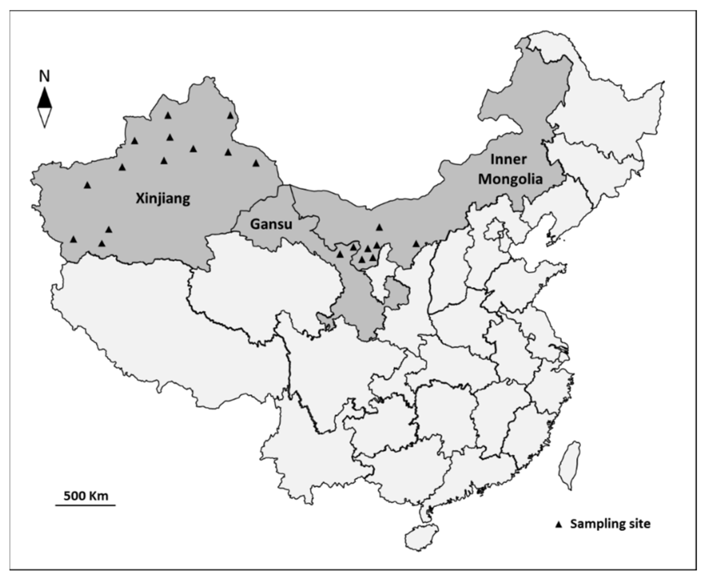
2.1. Sampling
2.2. Genomic DNA Extraction
2.3. PCR Amplification and Cloning
2.4. Sequencing and Sequence Analysis
2.5. Statistical Analysis
2.6. Nucleotide Sequence Accession Numbers
3. Results and Discussion
4. Conclusions
Supplementary Materials
Author Contributions
Funding
Institutional Review Board Statement
Data Availability Statement
Conflicts of Interest
References
- Tan, K.S.W. New Insights on Classification, Identification, and Clinical Relevance of Blastocystis spp. Clin. Microbiol. Rev. 2008, 21, 639–665. [Google Scholar] [CrossRef] [Green Version]
- Tsaousis, A.D.; de Choudens, S.O.; Gentekaki, E.; Long, S.; Gaston, D.; Stechmann, A.; Vinella, D.; Py, B.; Fontecave, M.; Barras, F.; et al. Evolution of Fe/S cluster biogenesis in the anaerobic parasite Blastocystis. Proc. Natl. Acad. Sci. USA 2012, 109, 10426–10431. [Google Scholar] [CrossRef] [Green Version]
- Deng, L.; Wojciech, L.; Gascoigne, N.R.J.; Peng, G.; Tan, K.S.W. New insights into the interactions between Blastocystis, the gut microbiota, and host immunity. PLoS Pathog. 2021, 17, e1009253. [Google Scholar] [CrossRef] [PubMed]
- Jeremiah, S.; Parija, S.C. Blastocystis: Taxonomy, biology and virulence. Trop. Parasitol. 2013, 3, 17–25. [Google Scholar] [CrossRef] [PubMed] [Green Version]
- Reh, L.; Muadica, A.S.; Köster, P.C.; Balasegaram, S.; Verlander, N.Q.; Chércoles, E.R.; Carmena, D. Substantial prevalence of enteroparasites Cryptosporidium spp., Giardia duodenalis and Blastocystis sp. in asymptomatic schoolchildren in Madrid, Spain, November 2017 to June 2018. Eurosurveillance 2019, 24, 1900241. [Google Scholar] [CrossRef] [PubMed]
- Villamizar, X.; Higuera, A.; Herrera, G.; Vasquez-A, L.R.; Buitron, L.; Muñoz, L.M.; Gonzalez-C, F.E.; Lopez, M.C.; Giraldo, J.C.; Ramírez, J.D. Molecular and descriptive epidemiology of intestinal protozoan parasites of children and their pets in Cauca, Colombia: A cross-sectional study. BMC Infect. Dis. 2019, 19, 190. [Google Scholar] [CrossRef] [PubMed] [Green Version]
- Sánchez, A.; Munoz, M.; Gómez, N.; Tabares, J.; Segura, L.; Salazar, Á.; Restrepo, C.; Ruíz, M.; Reyes, P.; Qian, Y.; et al. Molecular Epidemiology of Giardia, Blastocystis and Cryptosporidium among Indigenous Children from the Colombian Amazon Basin. Front. Microbiol. 2017, 8, 248. [Google Scholar] [CrossRef]
- Collins, S.M. A role for the gut microbiota in IBS. Nat. Rev. Gastroenterol. Hepatol. 2014, 11, 497–505. [Google Scholar] [CrossRef]
- Dogruman-Al, F.; Turk, S.; Adiyaman-Korkmaz, G.; Hananel, A.; Levi, L.; Kopelowitz, J.; Babai, O.; Gross, S.; Greenberg, Z.; Herschkovitz, Y.; et al. A novel ELISA test for laboratory diagnosis of Blastocystis spp. in human stool specimens. Parasitol. Res. 2014, 114, 495–500. [Google Scholar] [CrossRef] [PubMed]
- Ajjampur, S.S.R.; Tan, K.S.W. Pathogenic mechanisms in Blastocystis spp.—Interpreting results from in vitro and in vivo studies. Parasitol. Int. 2016, 65, 772–779. [Google Scholar] [CrossRef]
- Kumarasamy, V.; Kuppusamy, U.R.; Jayalakshmi, P.; Samudi, C.; Ragavan, N.D.; Kumar, S. Exacerbation of colon carcinogenesis by Blastocystis sp. PLoS ONE 2017, 12, e0183097. [Google Scholar] [CrossRef] [Green Version]
- Andersen, L.O.B.; Stensvold, C.R. Blastocystis in Health and Disease: Are We Moving from a Clinical to a Public Health Per-spective? J. Clin. Microbiol. 2016, 54, 524–528. [Google Scholar] [CrossRef] [PubMed] [Green Version]
- Krogsgaard, L.R.; Engsbro, A.L.; Stensvold, C.R.; Nielsen, H.V.; Bytzer, P. The Prevalence of Intestinal Parasites Is Not Greater Among Individuals with Irritable Bowel Syndrome: A Population-based Case-control Study. Clin. Gastroenterol. Hepatol. 2015, 13, 507–513.e2. [Google Scholar] [CrossRef] [PubMed]
- Maloney, J.G.; Molokin, A.; da Cunha, M.J.R.; Cury, M.C.; Santin, M. Blastocystis subtype distribution in domestic and cap-tive wild bird species from Brazil using next generation amplicon sequencing. Parasite Epidemiol. Control 2020, 9, e00138. [Google Scholar] [CrossRef] [PubMed]
- Stensvold, C.R.; Clark, C.G. Pre-empting Pandora’s Box: Blastocystis Subtypes Revisited. Trends Parasitol. 2020, 36, 229–232. [Google Scholar] [CrossRef] [PubMed]
- Maloney, J.; Jang, Y.; Molokin, A.; George, N.; Santin, M. Wide Genetic Diversity of Blastocystis in White-Tailed Deer (Odocoileus virginianus) from Maryland, USA. Microorganisms 2021, 9, 1343. [Google Scholar] [CrossRef] [PubMed]
- Higuera, A.; Herrera, G.; Jiménez, P.; Garcia-Corredor, D.; Pulido-Medellin, M.; Bulla, D.; Pinilla, J.; Moreno-Pérez, D.; Maloney, J.; Santín, M.; et al. Identification of multiple Blastocystis subtypes in domestic animals from Colombia using amplicon-based Next generation sequencing. Front. Vet. Sci. 2021, 8, 732129. [Google Scholar] [CrossRef]
- Maloney, J.G.; da Cunha, M.J.R.; Molokin, A.; Cury, M.C.; Santin, M. Next-generation sequencing reveals wide genetic diver-sity of Blastocystis subtypes in chickens including potentially zoonotic subtypes. Parasitol. Res. 2021, 120, 2219–2231. [Google Scholar] [CrossRef]
- Stensvold, C.R.; Clark, C.G. Current status of Blastocystis: A personal view. Parasitol. Int. 2016, 65, 763–771. [Google Scholar] [CrossRef]
- Ramírez, J.D.; Sánchez, A.; Hernandez, D.C.; Flórez, C.; Bernal, M.C.; Giraldo, J.C.; Reyes, P.; López, M.C.; García, L.; Cooper, P.; et al. Geographic distribution of human Blastocystis subtypes in South America. Infect. Genet. Evol. 2016, 41, 32–35. [Google Scholar] [CrossRef]
- Khaled, S.; Gantois, N.; Ly, A.T.; Senghor, S.; Even, G.; Dautel, E.; Dejager, R.; Sawant, M.; Baydoun, M.; Benam-rouz-Vanneste, S.; et al. Prevalence and subtype distribution of Blastocystis sp. in Senegalese school children. Microorganisms 2020, 8, 1408. [Google Scholar] [CrossRef]
- Liu, Z. Studies on the haematology and trace element status of adult Bactrian camels (Camelus bactrianus) in China. Vet. Res. Commun. 2003, 27, 397–405. (In Chinese) [Google Scholar]
- Wu, H.; Guang, X.; Al-Fageeh, M.B.; Cao, J.; Pan, S.; Zhou, H.; Zhang, L.; AbuTarboush, M.H.; Xing, Y.; Xie, Z.; et al. Camelid genomes reveal evolution and adaptation to desert environments. Nat. Commun. 2014, 5, 5188. [Google Scholar] [CrossRef] [Green Version]
- Yang, L.; Fu, Y.H.; Zhang, Z.X.; Feng, X.H.; Yang, J. Nutritional value, edible quality and processing status of camel meat. Meat Res. 2018, 32, 55–60. (In Chinese) [Google Scholar]
- Zhang, L.N.; Wang, N.L.; Zhang, K.; Wang, J.J.; Di, D.L.; Liu, Y.; Liu, Y.W. Analysis and evaluation of nutritional components in camel blood. Modern Food 2018, 19, 249–253. (In Chinese) [Google Scholar]
- China Bureau of Statistics. China Statistical Yearbook; China Statistics Press: Beijing, China, 2019. (In Chinese) [Google Scholar]
- Kong, J.C.; Li, C.L.; Ma, C.; Yang, X.X. Investigation report on camel parasitosis in Northwest China. J. North. Uni. Nat. 1987, 1, 213–226. (In Chinese) [Google Scholar]
- Hao, X.Y.; Sun, J.Q.; Li, H.X.; Huo, Y.F. Investigation on parasite of camel in Bayannaoer League. Chin. J. Vet. Parasitol. 2002, 1, 33–34. (In Chinese) [Google Scholar]
- Zhu, X.J.; Wang, J.Z.; Ren, A.H.; Se, L.; Zhang, X.R. Investigation report on parasite of camel in Alxa Right Banner. J. Gan. Agri. Uni. 1983, 2, 11–12. (In Chinese) [Google Scholar]
- Liu, X.; Zhou, X.; Zhong, Z.; Deng, J.; Chen, W.; Cao, S.; Fu, H.; Zuo, Z.; Hu, Y.; Peng, G. Multilocus genotype and subtype analysis of Cryptosporidium andersoni derived from a Bactrian camel (Camelus bactrianus) in China. Parasitol. Res. 2014, 113, 2129–2136. [Google Scholar] [CrossRef] [PubMed]
- Wang, M.; Wang, Y.; Meng, P.; Ye, Q.; Zhang, D. Toxoplasma gondii infection in Bactrian camel (Camelus bactrianus) in China. Vet. Parasitol. 2012, 192, 288–289. [Google Scholar] [CrossRef]
- Qi, M.; Li, J.; Zhao, A.; Cui, Z.; Wei, Z.; Jing, B.; Zhang, L. Host specificity of Enterocytozoon bieneusi genotypes in Bactrian camels (Camelus bactrianus) in China. Parasites Vectors 2018, 11, 219. [Google Scholar] [CrossRef] [PubMed] [Green Version]
- Alfellani, M.A.; Taner-Mulla, D.; Jacob, A.; Imeede, C.A.; Yoshikawa, H.; Stensvold, C.R.; Clark, C.G. Genetic Diversity of Blastocystis in Livestock and Zoo Animals. Protist 2013, 164, 497–509. [Google Scholar] [CrossRef] [PubMed] [Green Version]
- Mokhtar, A.; Youssef, A. Subtype analysis of Blastocystis spp. isolated from domestic mammals and poultry and its relation to transmission to their in-contact humans in Ismailia governorate, Egypt. Parasitol. United J. 2018, 11, 90–98. [Google Scholar] [CrossRef]
- Stenzel, D.; Cassidy, M.; Boreham, P. Morphology of Blastocystis sp. isolated from circus animals. Int. J. Parasitol. 1993, 23, 685–687. [Google Scholar] [CrossRef]
- Santín, M.; Gomez_Munoz, M.T.; Solano-Aguilar, G.; Fayer, R. Development of a new PCR protocol to detect and subtype Blastocystis spp. from humans and animals. Parasitol. Res. 2011, 109, 205–212. [Google Scholar] [CrossRef] [PubMed]
- Tamura, K.; Stecher, G.; Peterson, D.; Filipski, A.; Kumar, S. MEGA6: Molecular Evolutionary Genetics Analysis version 6.0. Mol. Biol. Evol. 2013, 30, 2725–2729. [Google Scholar] [CrossRef] [Green Version]
- Maloney, J.G.; Lombard, J.E.; Urie, N.J.; Shivley, C.B.; Santin, M. Zoonotic and genetically diverse subtypes of Blastocystis in US pre-weaned dairy heifer calves. Parasitol. Res. 2018, 118, 575–582. [Google Scholar] [CrossRef] [PubMed]
- Zhao, G.H.; Hu, X.F.; Liu, T.L.; Hu, R.S.; Yu, Z.Q.; Yang, W.B.; Wu, Y.L.; Yu, S.K.; Song, J.K. Molecular characterization of Blastocystis sp. in captive wild animals in Qinling Mountains. Parasitol. Res. 2017, 116, 2327–2333. [Google Scholar] [CrossRef]
- Rauff-Adedotun, A.A.; Zain, S.N.M.; Haziqah, M.T.F. Current status of Blastocystis sp. in animals from Southeast Asia: A review. Parasitol. Res. 2020, 119, 3559–3570. [Google Scholar] [CrossRef]
- Moura, R.G.F.; de Oliveira-Silva, M.B.; Pedrosa, A.L.; Nascentes, G.A.N.; Cabrine-Santos, M. Occurrence of Blastocystis spp. in domestic animals in Triangulo Mineiro area of Brazil. Rev. Soc. Bras. Med. Trop. 2018, 51, 240–243. [Google Scholar] [CrossRef] [Green Version]
- Hastutiek, P.; Yuniarti, W.M.; Djaeri, M.; Lastuti, N.D.R.; Suprihati, E.; Suwanti, L.T. Prevalence and diversity of gastrointestinal protozoa in Madura cattle at Bangkalan Regency, East Java, Indonesia. Vet. World 2019, 12, 198–204. [Google Scholar] [CrossRef] [PubMed]
- Wang, R.; Zhang, Y.; Jiang, Y.; Xing, J.; Tao, D.; Qi, M. First report of Blastocystis infection in pigs from large farms in Xinjiang. China. J. Eukaryot. Microbiol. 2020, 67, 642–647. [Google Scholar] [CrossRef] [PubMed]
- Song, J.K.; Yin, Y.L.; Yuan, Y.J.; Tang, H.; Ren, G.J.; Zhang, H.J.; Li, Z.X.; Zhang, Y.M.; Zhao, G.H. First genotyping of Blastocystis sp. in dairy, meat, and cashmere goats in northwestern China. Acta Trop. 2017, 176, 277–282. [Google Scholar] [CrossRef] [PubMed]
- Tan, T.C.; Tan, P.C.; Sharma, R.; Sugnaseelan, S.; Suresh, K.G. Genetic diversity of caprine Blastocystis from Peninsular Ma-laysia. Parasitol. Res. 2013, 112, 85–89. [Google Scholar] [CrossRef] [PubMed]
- Udonsom, R.; Prasertbun, R.; Mahittikorn, A.; Mori, H.; Changbunjong, T.; Komalamisra, C.; Pintong, A.; Sukthana, Y. Blas-tocystis infection and subtype distribution in humans, cattle, goats and pigs in central and western Thailand. Infect. Genet. Evol. 2018, 65, 107–111. [Google Scholar] [CrossRef] [PubMed]
- Navarro, C.; Dominguez-Marquez, M.V.; Garijo-Toledo, M.M.; Vega-Garcia, S.; Fernandez-Barredo, S.; Perez-Gracia, M.T.; Garcia, A.; Borras, R.; Gomez-Munoz, M.T. High prevalence of Blastocystis sp. in pigs reared under intensive growing systems: Frequency of ribotypes and associated risk factors. Vet. Parasitol. 2008, 153, 347–358. [Google Scholar] [CrossRef]
- Han, J.Q.; Li, Z.; Zou, Y.; Pu, L.H.; Zhu, X.Q.; Zou, F.C.; Huang, C.-Q. Prevalence, Molecular Characterization and Risk Factors of Blastocystis sp. from Farmed Pigs in Yunnan Province, Southwestern China. Acta Parasitol. 2020, 65, 1005–1010. [Google Scholar] [CrossRef]
- Wang, W.; Bielefeldt-Ohmann, H.; Traub, R.J.; Cuttell, L.; Owen, H. Location and Pathogenic Potential of Blastocystis in the Porcine Intestine. PLoS ONE 2014, 9, e103962. [Google Scholar] [CrossRef] [Green Version]
- Masuda, A.; Sumiyoshi, T.; Ohtaki, T.; Matsumoto, J. Prevalence and molecular subtyping of Blastocystis from dairy cattle in Kanagawa, Japan. Parasitol. Int. 2018, 67, 702–705. [Google Scholar] [CrossRef]
- Greige, S.; El Safadi, D.; Khaled, S.; Gantois, N.; Baydoun, M.; Chemaly, M.; Benamrouz-Vanneste, S.; Chabé, M.; Osman, M.; Certad, G.; et al. First report on the prevalence and subtype distribution of Blastocystis sp. in dairy cattle in Lebanon and assessment of zoonotic transmission. Acta Trop. 2019, 194, 23–29. [Google Scholar] [CrossRef]
- Suwanti, L.T.; Susana, Y.; Hastutiek, P.; Suprihati, E.; Lastuti, N.D.R. Blastocystis spp. subtype 10 infected beef cattle in Kamal and Socah, Bangkalan, Madura, Indonesia. Vet. World 2020, 13, 231–237. [Google Scholar] [CrossRef] [Green Version]
- Li, W.C.; Wang, K.; Gu, Y. Occurrence of Blastocystis sp. and Pentatrichomonas hominis in sheep and goats in China. Parasites Vectors 2018, 11, 1–7. [Google Scholar] [CrossRef] [PubMed] [Green Version]
- Arpitha, G.; Sreekumar, C.; Latha, B.R.; Bharathi, M.V. Prevalence and staining characteristics of Blastocystis isolates from food animals in Tamil Nadu. Vet. Parasitol. Reg. Stud. Rep. 2018, 11, 61–65. [Google Scholar] [CrossRef] [PubMed]
- Ghimire, T.R.; Bhattarai, N. A survey of gastrointestinal parasites of goats in a goat market in Kathmandu, Nepal. J. Parasit. Dis. 2019, 43, 686–695. [Google Scholar] [CrossRef]
- Roberts, T.; Stark, D.; Harkness, J.; Ellis, J. Subtype distribution of Blastocystis isolates from a variety of animals from New South Wales, Australia. Vet. Parasitol. 2013, 196, 85–89. [Google Scholar] [CrossRef] [PubMed]
- Quilez, J.; Clavel, A.; Sanchez-Acedo, C.; Causape, A. Detection of Blastocystis sp. in pigs in Aragon (Spain). Vet. Parasitol. 1995, 56, 345–348. [Google Scholar] [CrossRef]
- Badparva, E.; Sadraee, J.; Kheirandish, F. Genetic Diversity of Blastocystis Isolated from Cattle in Khorramabad, Iran. Jundishapur J. Microbiol. 2015, 8, e14810. [Google Scholar] [CrossRef] [PubMed] [Green Version]
- Lee, H.; Lee, S.H.; Seo, M.G.; Kim, H.Y.; Kim, J.W.; Lee, Y.R.; Kim, J.H.; Kwon, O.D.; Kwak, D. Occurrence and genetic diversity of Blastocystis in Korean cattle. Vet. Parasitol. 2018, 258, 70–73. [Google Scholar] [CrossRef]
- Aynur, Z.E.; Güçlü, Ö.; Yıldız, I.; Aynur, H.; Ertabaklar, H.; Bozdoğan, B.; Ertuğ, S. Molecular characterization of Blastocystis in cattle in Turkey. Parasitol. Res. 2019, 118, 1055–1059. [Google Scholar] [CrossRef]
- Deng, L.; Chai, Y.; Zhou, Z.; Liu, H.; Zhong, Z.; Hu, Y.; Fu, H.; Yue, C.; Peng, G. Epidemiology of Blastocystis sp. infection in China: A systematic review. Parasite 2019, 26, 41. [Google Scholar] [CrossRef] [Green Version]
- Hublin, J.S.; Maloney, J.G.; Santin, M. Blastocystis in domesticated and wild mammals and birds. Res. Vet. Sci. 2020, 135, 260–282. [Google Scholar] [CrossRef] [PubMed]
- Abarca, N.; Santín, M.; Ortega, S.; Maloney, J.G.; George, N.S.; Molokin, A.; Cardona, G.A.; Dashti, A.; Köster, P.C.; Bailo, B.; et al. Molecular detection and characterization of Blastocystis sp. and Enterocytozoon bieneusi in cattle in Northern Spain. Vet. Sci. 2021, 8, 191. [Google Scholar] [CrossRef] [PubMed]


| Geographical Areas | Sampling Sites | No. Examined | No. Positive (%) | Subtypes (No.) |
|---|---|---|---|---|
| Gansu | Minqin | 63 | 16 (30.2) | ST10 (5); ST14 (3); ST24 (4); ST 25 (2); ST30 (1); PT-ST (1) |
| Yongchang | 39 | 20 (51.3) | ST10 (2); ST14 (2); ST 25 (1); ST30 (6); PT-ST (9) | |
| Subtotal | 102 | 39 (38.2) | ST10 (7); ST14 (5); ST24 (4); ST 25 (3); ST30 (7); PT-ST (10) | |
| Inner Mongolia | Bayanhaote | 82 | 11 (13.4) | ST10 (2); ST14 (2); ST21 (1); ST24 (1); ST26 (1); ST30 (2); PT-ST (2) |
| Suhaitu | 27 | 6 (22.2) | ST24 (1); ST25 (1); ST30 (1); PT-ST (3) | |
| Gilantai | 24 | 2 (8.3) | ST24 (1); ST30 (1) | |
| Shariburidu | 27 | 12 (44.4) | ST10 (1); ST14 (2); ST24 (2); ST25 (2); ST30 (2); PT-ST (3) | |
| Tenggeli | 19 | 0 (0) | 0 | |
| Ordos | 13 | 1 (7.7) | ST25 (1) | |
| Subtotal | 192 | 32 (16.7) | ST10 (3); ST14 (4); ST21 (1); ST24 (5); ST25 (4); ST26 (1); ST30 (6); PT-ST (8) | |
| Xinjiang | Bachu | 16 | 0 (0) | 0 |
| Wensu | 24 | 0 (0) | 0 | |
| Qitai | 19 | 5 (26.3) | ST10 (1); ST14 (3); ST30 (1) | |
| Pishan | 17 | 5 (29.4) | ST10 (3); ST14 (2) | |
| Barkol Kazakh | 58 | 7 (12.1) | ST10 (5); ST14 (1); ST24 (1) | |
| Qinghe | 57 | 9 (15.8) | ST10 (4); ST24 (2); ST30 (1); PT-ST (2) | |
| Shihezi | 60 | 17 (28.3) | ST10 (9); ST14 (5); ST24 (1) | |
| Qapqal Xibe | 12 | 1 (8.3) | ST10 (1) | |
| Hejing | 16 | 0 (0) | 0 | |
| Wushi | 10 | 6 (60.0) | ST10 (5); ST14 (1) | |
| Tarbagatay | 16 | 0 (0) | 0 | |
| Hotan | 17 | 12 (70.6) | ST10 (8); ST30 (4) | |
| Qira | 22 | 6 (27.3) | ST10 (3); ST24 (1); ST25 (1); PT-ST (1) | |
| Subtotal | 344 | 68 (19.8) | ST10 (39); ST14 (12); ST24 (5); ST25 (1); ST30 (6); PT-ST (3) | |
| Total | 638 | 139 (21.8) | ST10 (49); ST14 (21); ST21 (1); ST24 (14); ST25 (8); ST26 (1); ST30 (19); PT-ST (21) |
| Age (Years) | No. Examined | No. Positive (%) | Subtypes (No.) |
|---|---|---|---|
| <2 | 61 | 18 (29.5) | ST10 (5); ST14 (5); ST21 (1); ST26 (1); ST30 (3); PT-ST (1) |
| 2–6 | 28 | 14 (50.0) | ST10 (2); ST14 (1); ST24 (2); ST25 (2); ST30 (1); PT-ST (5) |
| >6 | 549 | 107 (19.5) | ST10 (42); ST14 (15); ST24 (12); ST25 (6); ST30 (15); PT-ST (15) |
| Total | 638 | 139 (21.8) | ST10 (49); ST14 (21); ST21 (1); ST24 (14); ST25 (8); ST26 (1); ST30 (19); PT-ST (21) |
Publisher’s Note: MDPI stays neutral with regard to jurisdictional claims in published maps and institutional affiliations. |
© 2021 by the authors. Licensee MDPI, Basel, Switzerland. This article is an open access article distributed under the terms and conditions of the Creative Commons Attribution (CC BY) license (https://creativecommons.org/licenses/by/4.0/).
Share and Cite
Yang, X.; Li, Y.; Wang, Y.; Wang, J.; Lai, P.; Li, Y.; Song, J.; Qi, M.; Zhao, G. Molecular Characterization of Blastocystis sp. in Camelus bactrianus in Northwestern China. Animals 2021, 11, 3016. https://0-doi-org.brum.beds.ac.uk/10.3390/ani11113016
Yang X, Li Y, Wang Y, Wang J, Lai P, Li Y, Song J, Qi M, Zhao G. Molecular Characterization of Blastocystis sp. in Camelus bactrianus in Northwestern China. Animals. 2021; 11(11):3016. https://0-doi-org.brum.beds.ac.uk/10.3390/ani11113016
Chicago/Turabian StyleYang, Xin, Yunhui Li, Yuxin Wang, Junwei Wang, Peng Lai, Yuan Li, Junke Song, Meng Qi, and Guanghui Zhao. 2021. "Molecular Characterization of Blastocystis sp. in Camelus bactrianus in Northwestern China" Animals 11, no. 11: 3016. https://0-doi-org.brum.beds.ac.uk/10.3390/ani11113016