Article

Molecular Characterization of Anaplasma spp. among Dairy, Cashmere, and Meat Goats in Shaanxi Province, Northwestern China

1
College of Veterinary Medicine, Northwest A&F University, Yangling, Xianyang 712100, China
2
College of Veterinary Medicine, Nanjing Agricultural University, Nanjing 210095, China
*
Authors to whom correspondence should be addressed.
These authors contributed equally to this work.
Submission received: 27 April 2022 / Revised: 15 June 2022 / Accepted: 15 June 2022 / Published: 17 June 2022
(This article belongs to the Collection Advances in Parasite Epidemiology and Population Genetics)

Abstract

:

Simple Summary

Anaplasmosis is an important tick-borne disease caused by Anaplasma spp., significantly threating public health safety and breeding industry. The present study reported the occurrence of Anaplasma infection among dairy, cashmere, and meat goats in Shaanxi province, northwestern China, with the total prevalence of 58.5% (298/509) in goats. Anaplasma phagocytophilum, A. bovis, and A. ovis were the dominant species in meat, dairy, and cashmere goats, respectively, with the absence of A. ovis in meat goats. Furthermore, the different influencing factors (production categories, species, regions, and ages) were analyzed, and statistically significant differences were found. Frequent occurrence of Anaplasma in this study indicated One Health-based intervention approaches were urgently needed to block the transmission between humans and animals.

Abstract

Anaplasma spp. are important tick-borne pathogens endangering the health of humans and various animals. Although several studies have reported Anaplasma infection in livestock in China, little is known about the impact of production categories on the occurrence of Anaplasma species. In the present study, PCR tools targeting the 16S rRNA and msp4 genes were applied to investigate the prevalence of Anaplasma spp. in 509 blood samples of dairy (n = 249), cashmere (n = 139), and meat (n = 121) goats from Shaanxi province. The prevalence of Anaplasma spp. was 58.5% (298/509) in goats, and significant differences (p < 0.001) were identified in the prevalence among production categories, with the highest in meat goats (84.3%, 102/121), followed by cashmere goats (58.3%, 81/139) and dairy goats (46.2%, 115/249). Significant differences (p < 0.001) in prevalence were also found among sampling sites and age groups. Meanwhile, the prevalence was 36.9% (188/509) for A. phagocytophilum, 36.1% (184/509) for A. bovis, and 11.0% (56/509) for A. ovis, and significant differences (p < 0.001) in prevalence of A. phagocytophilum, A. bovis and A. ovis were recognized among production categories and sampling sites. A. phagocytophilum, A. bovis and A. ovis were dominant species in meat, dairy, and cashmere goats, respectively, and A. ovis was absent in meat goats. Co-infections were found in 124 (24.4%) investigated samples. Goats aged < 2, 3–6, and 7–12 months, and goats from Qingjian and Zhenba were risk factors associated with the occurrence of Anaplasma. Phylogenetic analysis indicated separate clades for the distribution of A. phagocytophilum from different ruminant, reflecting potential host adaption within this species. This study reported the colonization occurrence of Anaplasma spp. among production categories in goats in Shaanxi province and enriched our knowledge on the transmission of Anaplasma spp. in goats in China. Considering the existence of zoonotic A. phagocytophilum in goats in this study and previous reports, interventions based on One Health are needed to be developed to control the transmission of Anaplasma spp. between humans and animals.

1. Introduction

Anaplasma spp. are emerging tick-borne pathogens infecting humans and animals which largely distribute in tropical, subtropical, and some temperate regions [1,2,3]. Currently, Anaplasma phagocytophilum, A. bovis, A. ovis, A. marginale, A. centrale, A. platys, and A. capra are recognized species within Anaplasma [1,4]. Of them, A. phagocytophilum, A. bovis, and A. ovis are common pathogenic species causing anaplasmosis. A. phagocytophilum can infect neutrophils of humans and various animals [1,5], causing human granulocytic anaplasmosis (HGA) and tick-borne fever in ruminants [6,7]. A. bovis is mainly found in mononuclear cells of many animals, resulting in anaplasmosis with fever, weight loss, depression, abortion, or even death [8]. A. ovis can infect erythrocytes of small ruminant [2,9], leading to symptoms including hemolytic anemia and fever [10].
As important tick-borne pathogens, Anaplasma spp. significantly affect the health of humans and animals [9]. A. phagocytophilum majorly leads to HGA, a common disease leading to fever in patients. HGA was first reported in an American patient with tick bite history in 1990, and subsequently found in over 14 European countries [6]. According to the data from CDC of USA and Morbidity and Mortality Weekly Reports, over 15,952 HGA cases have been reported in America since 1995. The occurrence of HGA cases increased 12 times from 2001 to 2011, and the disease caused life threatening illness and death in approximately 3% and 1% of infection cases, respectively [11,12,13]. Meanwhile, the World Organization for Animal Health (WOAH) has listed anaplasmosis as a notifiable disease due to its socio-economic impact and international trade restrictions [14].
Knowledge on the distribution and composition of Anaplasma spp. can shed the light for the prevention and control of anaplasmosis. With the application of PCR-based techniques targeting the 16S rRNA, msp4, gltA, and groEL genes, single and/or mixed infections of Anaplasma species, such as A. phagocytophilum, A. bovis, and A. ovis, have been recognized in various animals, ticks, and humans [3,15,16,17].
Recently, several studies have reported Anaplasma infection in livestock in China [18,19,20,21,22,23,24], however, little is known on the occurrence of Anaplasma spp. among production categories. Goats are important economic livestock in China, providing meat, cashmere, and dairy with high quality in daily life. Shaanxi province is one of the main goat breeding areas in China, with a total of 7.22 million goats in 2020 (http://data.stats.gov.cn/, accessed on 20 May 2022). Notably, there exist three main production categories (dairy, cashmere, and meat) for goats in Shaanxi, and the dairy goat stocks and goat milk production of Shaanxi province account for 48.2% and 61.4% of the national total, respectively (http://nynct.shaanxi.gov.cn/, accessed on 20 May 2022). To understand the distribution characteristics of Anaplasma spp. among production categories in goats in Shaanxi province, the present study investigated the prevalence of A. phagocytophilum, A. bovis, and A. ovis in dairy, cashmere, and meat goats of Shaanxi province in northwestern China, and assessed the zoonotic potential of these pathogens in goats.

2. Materials and Methods
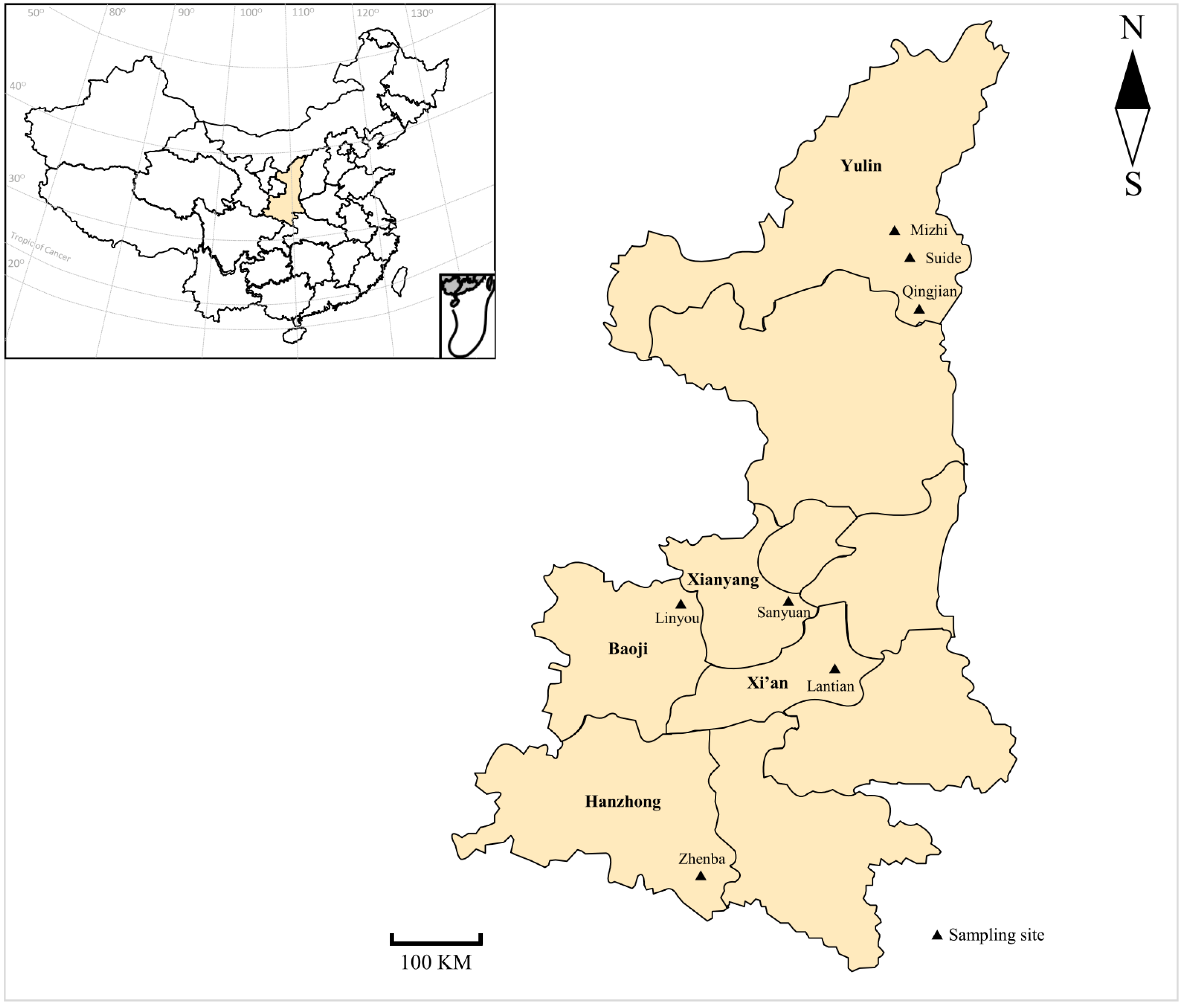
2.1. Sampling

To investigate the prevalence and species composition of Anaplasma in meat, cashmere, and dairy goats in Shaanxi province, we collected samples from representative farms in seven main breeding sampling sites, with 10~20% of animals sampled for each farm (Figure 1). From April to July 2017, a total of 509 blood samples of dairy (n = 249), cashmere (n = 139), and meat (n = 121) goats were collected from 15 representative farms (Table 1). Due to the larger scale of dairy goats compared with other two production categories, we collected more farms and more samples from dairy goats (Table 1). Blood samples were collected from the jugular vein of each animal and placed into separate tubes containing 1.5% Ethylenediaminetetraacetic acid tripotassium (EDTA-3K) with basic information (e.g., sampling sites, breeds, and ages), immediately transported to the department of parasitology of Northwest A&F University under cool condition, and then kept at 4 °C for further analysis.

2.2. Genomic DNA Extraction

The Blood Genomic DNA Isolation Kit (Sangon Biotech, Shanghai, China) was applied to extract genomic DNA (gDNA) samples from approximately 100 μL blood of each sample according to the procedures of the manufacturer, and the gDNA samples were kept at −20 °C until further analysis. Meanwhile, Anaplasma negative blood samples preserved in our lab were also applied for genomic DNA extraction to be used as negative control in PCR amplification.

2.3. PCR Amplification

The occurrence of A. phagocytophilum and A. bovis was identified using nested-PCR targeting a ~551 bp fragment and a ~641 bp fragment of the 16S rRNA gene as reported, respectively [25,26] (Table 2). Nested PCRs were carried out in a 25 μL reaction mixture containing 1 × Ex Taq Buffer (Mg2+ free), 2 mM MgCl2, 0.2 mM dNTP Mixture, 1 U TaKaRa Ex Taq, 0.4 μΜ each primer, 1 μL gDNA for the primary PCR, or 1 μL primary PCR product for the secondary PCR under the following conditions for both primary and secondary PCRs: an initial denaturing at 94 °C for 5 min, followed by 35 cycles of 94 °C for 30 s, 55 °C for 1 min, and 72 °C for 1 min, and a final extension at 72 °C for 10 min. A. ovis DNA was identified by PCR amplification targeting a ~867 bp fragment of the msp4 gene [27] (Table 2). PCRs were conducted in a 25 μL reaction mixture containing 1 × Ex Taq Buffer (Mg2+ free), 2 mM MgCl2, 0.2 mM dNTP Mixture, 1 U TaKaRa Ex Taq, 0.4 μΜ each primer, and 1 μL gDNA under the following conditions: an initial denaturing at 94 °C for 30 s, followed by 40 cycles of 94 °C for 30 s, 60 °C for 30 s, and 68 °C for 1 min, and a final extension at 68 °C for 10 min. A negative control without Anaplasma was used in each PCR amplification. Positive PCR products of the msp4 gene and secondary nested-PCR products of the 16S rRNA gene will appear a band of the expected size under a UV transilluminator after 1% agarose gel electrophoresis. A Good Laboratory Practice was followed to avoid contamination in each step during the whole experiment [28].

2.4. Sequencing and Sequence Analysis

All positive amplicons were sequenced at forward direction by Sangon Biotech (Shanghai, China) using an ABI PRISM 3730XL DNA Analyzer (Applied Biosystems, Bedford, MA, USA). The obtained sequences were identified to be A. phagocytophilum 16S rRNA gene, A. bovis 16S rRNA gene, or A. ovis msp4 gene by BLAST analysis within NCBI (https://blast.ncbi.nlm.nih.gov/Blast.cgi; accessed on 15 April 2021). To assess the phylogenetic placement of Anaplasma species found in this study, phylogenetic trees were constructed by using the neighbor-joining (NJ) method with the Kimura 2-parameter model and the calculation of substitution rates with the bootstrap evaluation of 1000 replicates within software MEGA V6.0 [29].

2.5. Statistical Analysis

Data analysis was conducted using the package R V4.0.5 (https://www.r-project.org/; accessed on 7 July 2021) and RStudio V1.1.463 (http://rstudio.com/; accessed on 7 July 2021). A chi-square test was applied to analyze differences in prevalence of Anaplasma spp. by production categories, age groups, or sampling sites. Logistic regression was used to analyze the association between risk factors and the occurrence of Anaplasma spp. in dairy, cashmere, and meat goats. Initially, univariate analysis was used to assess the strength of association. Then, a multivariate model was built using variables with p ≤ 0.2, with p < 0.05 being recognized as significant in the final model. Odds ratio (OR) with 95% confidence intervals (CI) was analyzed for the identification of risk factors for the occurrence of Anaplasma spp. in goats.

2.6. Nucleotide Sequence Accession Numbers

Representative nucleotide sequences of A. phagocytophilum 16S rRNA gene, A. bovis 16S rRNA gene, and A. ovis msp4 gene in the present study were available in GenBank™ under the accession numbers of MZ489423-MZ489426, MZ489427-MZ489430, and MZ502497-MZ502499, respectively.

3. Results

3.1. Prevalence of Anaplasma in Goats in Shaanxi Province

Of the 509 blood samples, a total of 298 (58.5%) samples were positive for Anaplasma spp. in goats based on the PCR-sequencing tools targeting the 16S rRNA gene and the msp4 gene (Table 3). Among the three production categories in goats, the highest prevalence (84.3%, 102/121) of Anaplasma spp. was found in meat goats, which was significantly higher than that in cashmere goats (58.3%, 81/139) (χ2 = 21.018, df = 1, p < 0.001) and dairy goats (46.2%, 115/249) (χ2 = 48.773, df = 1, p < 0.001), and significant difference was also identified between cashmere goats and dairy goats (χ2 = 5.215, df = 1, p = 0.022) (Table 4 and Table 5). Meanwhile, significant differences in prevalence were also identified among age groups (χ2 = 17.659, df = 3, p = 0.001) (Table 5), with the highest prevalence (80.0%, 12/15) in goats aged <2 months, followed by 7–12 months (75.0%, 45/60), >12 months (58.8%, 197/335), and 3–6 months (44.4%, 44/99) (Table 3).
In the univariate analysis, there existed association between dairy goat (OR = 5.26, p < 0.001) and occurrence of Anaplasma, especially for A. phagocytophilum (OR = 0.52, p = 0.001) and A. ovis (OR = 0.07, p < 0.001), and there also existed association between meat goat (OR = 0.36, p < 0.001) and occurrence of Anaplasma, especially for A. phagocytophilum (OR = 9.10, p < 0.001) and A. bovis (OR = 2.40, p < 0.001). Meanwhile, goats aged 3–6 months (OR = 0.49, p = 0.002) and 7–12 months (OR = 2.32, p = 0.007) were more likely to be infected with Anaplasma, especially for A. phagocytophilum (OR = 0.58, p = 0.028) and A. bovis (OR = 0.53, p = 0.013) for goats from 3–6 months, and A. ovis (OR = 2.95, p = 0.002) for goats from 7–12 months. In addition, goats from Lantian (OR = 0.35, p < 0.001), Qingjian (OR = 2.95, p = 0.008), and Zhenba (OR = 5.26, p < 0.001) were more likely to be infected with Anaplasma, especially for A. phagocytophilum (OR = 0.42, p = 0.031), A. bovis (OR = 2.76, p = 0.003), and A. ovis (OR = 9.74, p < 0.001) for goats from Qingjian, A. phagocytophilum (OR = 9.10, p < 0.001) and A. bovis (OR = 2.40, p < 0.001) for goats from Zhenba, A. phagocytophilum (OR = 0.64, p = 0.045) for goats from Lantian (Table 6). Further analysis applying the multivariate model indicated that there was no association between production categories and the occurrence of Anaplasma. However, goats aged < 2 months (OR = 0.52, p = 0.004), 3–6 months (OR = 0.26, p = 0.017), and 7–12 (OR = 0.27, p < 0.001) months were risk factors associated with the occurrence of Anaplasma, especially for A. bovis (OR = 5.06, p < 0.001) for goats aged < 2 months. In addition, goats from Qingjian (OR = 9.80, p = 0.004) and Zhenba (OR = 8.94, p < 0.001) were risk factors associated with the occurrence of Anaplasma, especially for A. phagocytophilum (OR = 12.84, p < 0.001) for goats from Zhenba (Table 6).

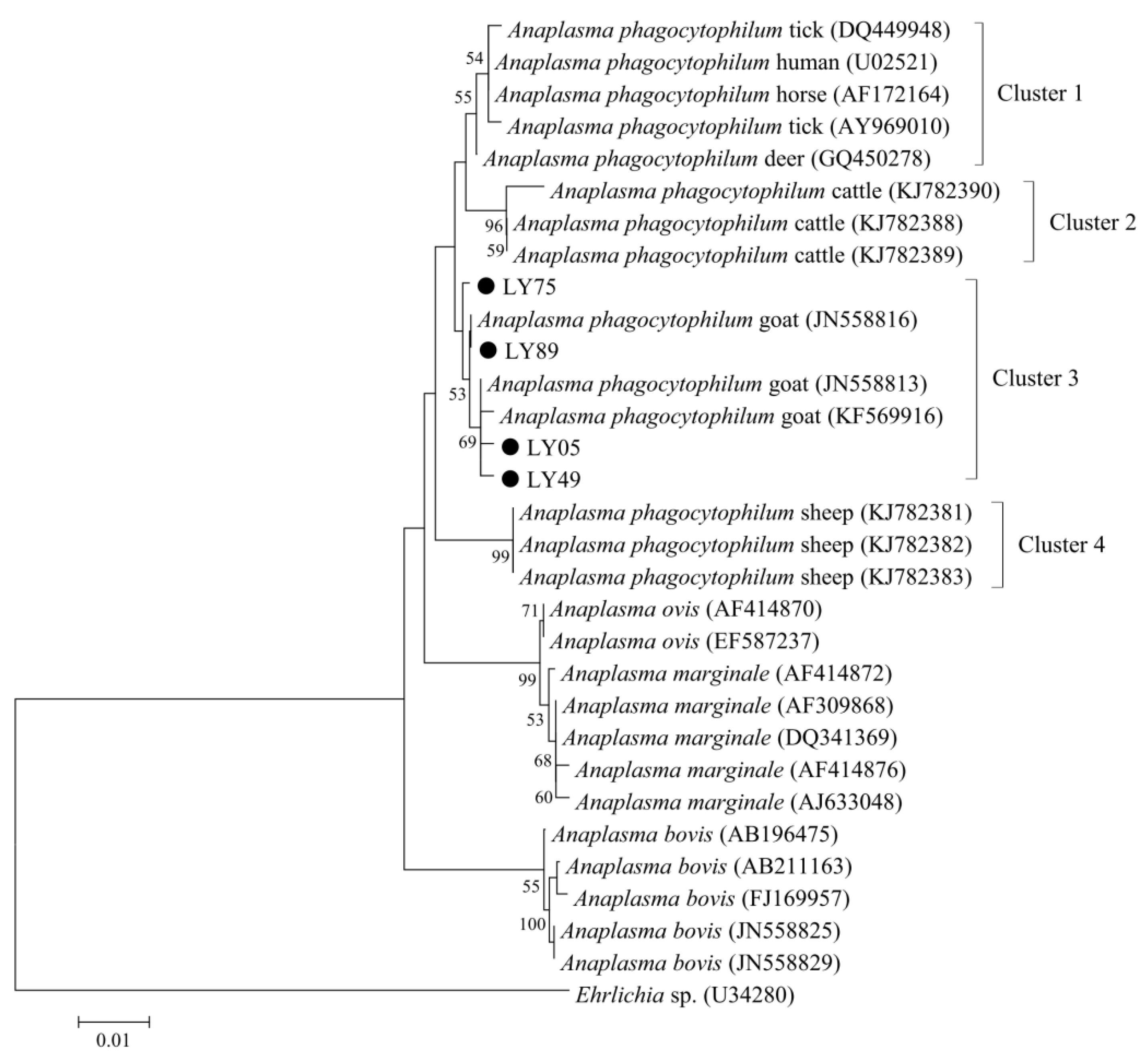
3.2. Anaplasma phagocytophilum in Goats in Shaanxi Province

In the present study, the prevalence of A. phagocytophilum in goats was 36.9% (188/509) (Table 3). All the production categories were positive for A. phagocytophilum with the highest prevalence in meat goats (75.2%, 91/121), followed by dairy goats (29.3%, 73/249) and cashmere goats (17.3%, 24/139), and significant differences were found among production categories (χ2 = 105.376, df = 2, p < 0.001) (Table 3 and Table 5). Significant differences in the prevalence of A. phagocytophilum were also found among sampling sites (χ2 = 187.211, df = 14, p < 0.001). Except for Farm 1, 4, 7, 8, and 9, all the remaining 10 sampling sites were positive for A. phagocytophilum, with the highest prevalence in Farm 14 (84.6%) and lowest in Farm 3 (5.9%), and significant differences in prevalence were identified among these sampling sites (χ2 = 96.340, df = 9, p < 0.001) (Table 3 and Table 5). Besides, A. phagocytophilum infection was frequently identified in all age groups of goats, with the highest prevalence in animals aged 7–12 months (43.3%, 26/60), followed by >12 months (39.1%, 131/335), 3–6 months (27.3%, 27/99), and <2 months (26.7%, 4/15) (Table 3).
To assess the genetic diversity of A. phagocytophilum in goats, all the positive samples for A. phagocytophilum at the 16S rRNA locus were sequenced, and a total of 184 sequences were obtained in the present study. Based on the BLAST analysis at NCBI and sequence alignment in MEGA V6.0, four types of A. phagocytophilum 16S rRNA sequences, namely LY05 (6), LY49 (3), LY75 (170), and LY89 (5), were identified in goats, with 99.2–99.6% sequence identity among these sequence types. Phylogenetic analysis indicated that four A. phagocytophilum 16S rRNA sequences in the present study and other reference sequences from goats were included in the same clade, while sequences from cattle and sheep formed other two separated clades (Figure 2).

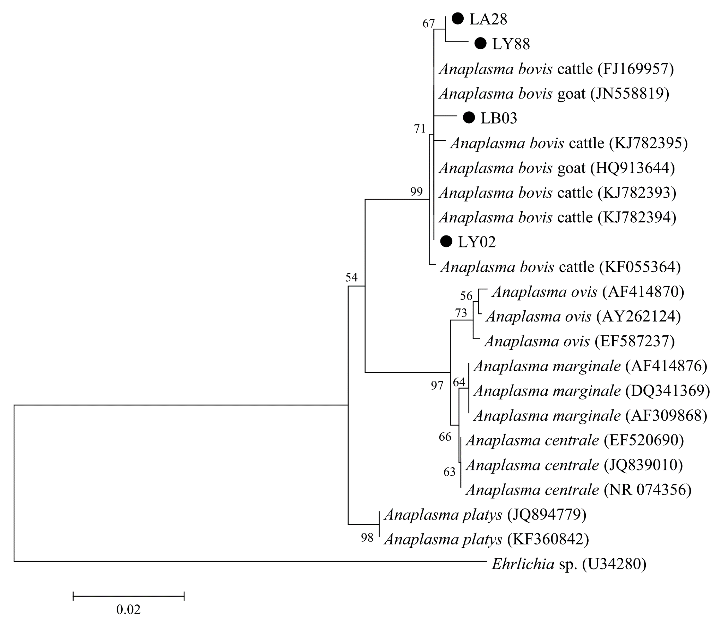
3.3. Anaplasma bovis in Goats in Shaanxi Province

In the present study, the prevalence of A. bovis in goats was 36.1% (184/509) (Table 3). All the production categories were positive for A. bovis with the highest in meat goats (52.1%, 63/121), followed by dairy goats (33.3%, 83/249) and cashmere goats (27.3%, 38/139), and significant differences were found among production categories (χ2 = 18.812, df = 2, p < 0.001) (Table 3 and Table 5). Significant differences in the prevalence of A. bovis were also found among sampling sites (χ2 = 126.079, df = 14, p < 0.001). Except for Farms 1 and 4, all the remaining 13 sampling sites were positive for A. bovis, with the highest prevalence in Farm 13 (82.5%) and lowest in Farm 8 (4.5%), and significant differences were identified among these sampling sites (χ2 = 98.871, df = 12, p < 0.001) (Table 3 and Table 5). Meanwhile, A. bovis infection was commonly found in all age groups of goats, with the highest prevalence in animals aged <2 months (80.0%, 12/15), followed by 7–12 months (45.0%, 27/60), >12 months (35.8%, 120/335), and 3–6 months (25.3%, 25/99) (Table 3).
Further, a total of 184 samples positive for A. bovis were successfully sequenced, and the obtained 184 sequences formed four sequence types, namely LA28 (4), LB03 (2), LY02 (172), and LY88 (6), with 98.7–99.7% sequence identities to each other. Phylogenetic analysis indicated that A. bovis samples from goats and cattle were included in the same clade (Figure 3).

3.4. Anaplasma ovis in Goats in Shaanxi Province

The prevalence of A. ovis in goats was 11% (56/509) in the present study, with the highest prevalence of 37.4% (cashmere) to the lowest of 0% (meat). Significant differences in prevalence were found among three production categories (χ2 = 136.408, df = 2, p < 0.001) (Table 3 and Table 5). Meanwhile, significant differences in the prevalence of A. ovis were also found among sampling sites (χ2 = 167.124, df = 14, p < 0.001). Five farms were positive for A. ovis, with the highest prevalence in Farm 9 (54.1%) and lowest in Farm 6 (4.4%), and significant differences were identified among these sampling sites (χ2 = 48.265, df = 4, p < 0.001) (Table 3 and Table 5). Meanwhile, A. ovis infection was frequently identified in all age groups of goats, with the highest prevalence in animals aged <2 months (26.7%, 4/15), followed by 7–12 months (23.3%, 14/60), 3–6 months (14.1%, 14/99), and >12 months (7.2%, 24/335) (Table 3).
Based on the PCR-sequencing of A. ovis msp4 gene, a total of three sequence types, namely MZ16 (30), MZ25 (18), and SDC23 (8), were recognized in the obtained 56 sequences, with 99.7–99.8% sequence similarity among these sequence types. Further phylogenetic analysis based on the msp4 gene indicated that MZ16, MZ25, SDC23, and reference sequences were included in the same clade, showing high sequence similarity among A. ovis from divergent areas in China (Figure 4).

4. Discussion

Anaplasma spp. are important zoonotic pathogens with worldwide distribution in humans and various animals [4,8,19,25,30,31,32,33]. To understand the occurrence of Anaplasma spp. among production categories, the present study investigated the prevalence of A. phagocytophilum, A. bovis, and A. ovis in dairy, cashmere, and meat goats in seven sampling sites within five main breeding areas in Shaanxi, northwestern China by using PCR-sequencing tools based on the 16S rRNA gene and the msp4 gene. Of the 509 blood samples, a total of 298 (58.5%) samples were positive for Anaplasma spp. in goats in the present study, and statistically significant differences were found in the prevalence of Anaplasma spp. among production categories.
A. phagocytophilum is recognized as one emerging tick-borne pathogen of public health in humans [34]. Besides humans, a variety of animals, e.g., cattle, sheep, goats, deer, horses, cats, dogs, and rats are also susceptible to this pathogen, and infected hosts can show a series of symptoms, including high fever, anorexia, and weight loss [20,24,35,36,37,38]. In the present study, the prevalence of A. phagocytophilum in goats was 36.9% (188/509) (Table 2), which was consistent with that in goats in Gansu (38.5%) [39], but higher than that in other provinces in China (5.7–30.8%) [18,19,20,21,24]. Recently, one study reported a higher average prevalence of 71.5% for A. phagocytophilum in goats in Weinan city, Shaanxi [22]. The differences in the occurrence rates of A. phagocytophilum in goats were likely affected by multiple factors, including different detection methods, geographic regions, sampling sizes, and animal management practice [40,41].
Previously, several reports indicated the existence of differences in host infectivity among divergent A. phagocytophilum strains [42,43,44]. Phylogenetic analysis indicated that four A. phagocytophilum 16S rRNA sequences in the present study and other reference sequences from goats were included in the same clade, while sequences from cattle and sheep formed two separated clades, suggesting the possible host adaptation for the distribution of A. phagocytophilum in these domesticated animals (Figure 2). Similar results have also been identified in A. phagocytophilum-positive samples from sheep and cattle in one previous study, reflected by the formation of separate clades for this pathogen from sheep and cattle by phylogenetic analysis [19].
In the present study, the 16S rRNA gene was applied for the genetic characterization of A. phagocytophilum of goats and found variants among different domesticated animals by phylogenetic analysis. The conserved 16S rRNA gene has been widely applied to detect A. phagocytophilum infection in humans and ruminants, identifying variants within this pathogen. However, the variants of the 16S rRNA are controversial due to the low discriminatory power. Therefore, several molecular markers, such as groESL, ankA, and Msp2, were tested and likely showed better function in distinguishing variant of divergent pathogenicity or origins compared with 16S rRNA [45]. The drhm gene was previously recognized as a maker for the pathogenicity of A. phagocytophilum, but it was not useful for several European A. phagocytophilum strains [46]. To increase the resolution in the characterization of A. phagocytophilum, variable number tandem repeat (VNTR), multi-locus sequence typing (MLST), and other multi-locus sequence analysis were used, which could significantly contribute to the comprehensive understanding of A. phagocytophilum [45,47].
A. bovis can infect a variety of mammals, including cattle, sheep, goats, deer, cats, and dogs [18,25,48,49,50,51]. In the present study, the prevalence of A. bovis in goats was 36.1% (184/509) (Table 2), which was lower than that in Tunisia (72.0%) [8], but higher than that in other regions in China (12.3–20.3%) [18,21]. Previously, some studies reported lower prevalence of 9.5% for A. bovis in goats in Xi’an city, Shaanxi [52], and higher prevalence of 62.1% in Weinan city, Shaanxi [22]. Animal management practices with the good application of drugs and procedures against ectoparasite possibly contribute to the low prevalence of A. bovis in goats since the common occurrence of this pathogen in several ticks, including Rh. microplus, Ha. longicornis, and Ixode crenulatus [41,53]. Contrary to the distribution of A. phagocytophilum, A. bovis samples from goats and cattle were included in the same clade by phylogenetic analysis, suggesting the possible absence of host segregation for the distribution of A. bovis (Figure 3), which was in accordance with the previous report in sheep and cattle [19]. Nevertheless, potential geographic differences for the distribution of A. bovis were identified in goats in China [22].
The average prevalence of A. ovis (11%, 56/509) in goats in the present study was lower than that in France (52%) [54], Mongolia (71.3%) [55], and other regions in China, e.g., Guizhou (17.8%), Xinjiang (40.5%), Zhejiang (26.3%) [18,19], but higher than that in Heilongjiang (2.6%), Henan (8.7%), and Hubei (7.2%) in China [18,21]. One previous study reported lower prevalence of A. ovis (0.9%) in goats in Xi’an city, Shaanxi [56], and the other study found higher prevalence of A. ovis (25.2%) in goats in Weinan city, Shaanxi [22]. Sampling sites, breeds, animal management practices, as well as other multiple factors, may lead to the differences in the prevalence of A. ovis among studies [40,41]. Notably, the occurrence of ticks (e.g., Rhipicephalus bursat) may also influence the prevalence of A. ovis in goats [54]. Among those three production categories in the present study, A. ovis was absent in meat goats, which was likely caused by the specific species composition of ticks in the studied regions due to the possible biased distribution of Anaplasma spp. among divergent ticks [9,54]. As an important major surface protein of Anaplasma spp., the msp4 gene is likely to have higher evolution rate under the selective pressure of host immune system compared with other genes [27]. In the present study, limited variation was observed within the sequences of the msp4 gene among A. ovis isolates, which was in accordance with the previous reports, reflected by low genetic diversity of A. ovis isolates from Xinjiang and Shaanxi [19,22].
Notably, co-infections of two to three Anaplasma species were identified in the present study (24.4%, 124/509), which was also commonly found in previous reports [18,22,49]. Significant differences were found among co-infections of A. phagocytophilum and A. bovis, A. bovis and A. ovis, and A. phagocytophilum and A. ovis2 = 134.594, df = 2, p < 0.001), with the co-infection of A. phagocytophilum and A. bovis being the dominant one (83.1%, 103/124). Interesting, co-infection of A. phagocytophilum, A. bovis, and A. ovis was recognized in 6 samples from dairy and cashmere goats, which has also been reported in 6 samples from goats in a previous report [18].
Previous reports indicated that the age was an influence factor for the infection of Anaplasma [8,23]. In the present study, significant difference was found among ages, with the highest prevalence in young animals under 2 months, which may be caused by low immunity or few samples tested in these animals. However, a higher prevalence of Anaplasma species in old goats (≥2 years) than young goats (<2 years) was found in one study in Anhui, China [23]. The difference in prevalence could be old animals being exposed to tick infection for a longer time. More investigations with a wide age range and geographic region settings are needed to further understand the influence of ages on Anaplasma infection.

5. Conclusions

In the present study, Anaplasma spp. were found in goats, with a prevalence of 58.5%, and significant differences in prevalence were found in animals from different production categories and age groups. A. phagocytophilum, A. bovis, and A. ovis were dominant species in meat, dairy, and cashmere goats, respectively, and A. ovis was absent in meat goats. Meanwhile, co-infections with two or three pathogens were frequently recognized. Frequent occurrence of A. phagocytophilum, A. bovis, and A. ovis in goats in this study indicates that there possibly exist potential risks for the zoonotic transmission between humans and animals due to the appearance of those pathogens in humans in previous reports. These findings can provide baseline information for the understanding of the transmission and zoonotic potential of Anaplasma spp. in goats. Considering the public health risks to humans of zoonotic A. phagocytophilum being found in goats, interventions based on One Health, such as eliminating ticks, maintaining good farm husbandry practices, and keeping personal hygiene, are needed to reduce the transmission of this pathogen from goats to humans and other animals. Certainly, there exist several limitations in the present work, and several questions should be addressed in future studies. For example, did the distribution and species composition of ticks effect the prevalence of Anaplasma spp. in goats? Are tick species and/or burdens related to the distribution of Anaplasma species or not? Why were young children more susceptible to Anaplasma then elder animals? Answers to these questions will assist us to comprehensively illustrate the transmission of Anaplasma between humans and animals.

Author Contributions

Conceptualization, G.Z. and J.S.; methodology, X.Y., M.F. and Z.Y.; software, J.W.; writing—original draft preparation, X.Y.; writing—review and editing, X.Y. and G.Z.; visualization, X.Y. and G.Z.; supervision, G.Z. and J.S.; project administration, G.Z. and J.S. All authors have read and agreed to the published version of the manuscript.

Funding

This work was funded by the grants from the Innovation Support Plan of Shaanxi Province (grant no. 2021TD-31), Key Research and Development Program of Shaanxi Province (grant no. 2022NY-097) and Project of Science and Technology Plan in Yulin City (grant no. CXY-2020-077).

Institutional Review Board Statement

This study was conducted under the approval and instructions of the ethics committee of Northwest A&F University and the animal ethics requirements of the People’s Republic of China.

Informed Consent Statement

Not applicable.

Data Availability Statement

Data is contained within the article.

Acknowledgments

We thank Weijian Wang from College of Veterinary Medicine, South China Agricultural University, for assistance in statistical analysis of this article.

Conflicts of Interest

The authors declare no conflict of interest.

References

  1. Dumler, J.S.; Barbet, A.F.; Bekker, C.P.; Dasch, G.A.; Palmer, G.H.; Ray, S.C.; Rikihisa, Y.; Rurangirwa, F.R. Reorganization of genera in the families Rickettsiaceae and Anaplasmataceae in the order Rickettsiales: Unification of some species of Ehrlichia with Anaplasma, Cowdria with Ehrlichia and Ehrlichia with Neorickettsia, descriptions of six new species combinations and designation of Ehrlichia equi and ‘HGE agent’ as subjective synonyms of Ehrlichia phagocytophila. Int. J. Syst. Evol. Microbiol. 2001, 51, 2145–2165. [Google Scholar] [PubMed] [Green Version]
  2. Kocan, K.M.; de la Fuente, J.; Guglielmone, A.A.; Meléndez, R.D. Antigens and alternatives for control of Anaplasma marginale infection in cattle. Clin. Microbiol. Rev. 2003, 16, 698–712. [Google Scholar] [CrossRef] [PubMed] [Green Version]
  3. Madison-Antenucci, S.; Kramer, L.D.; Gebhardt, L.L.; Kauffman, E. Emerging Tick-Borne Diseases. Clin. Microbiol. Rev. 2020, 33, e00083-18. [Google Scholar] [CrossRef] [PubMed]
  4. Li, H.; Zheng, Y.C.; Ma, L.; Jia, N.; Jiang, B.G.; Jiang, R.R.; Huo, Q.B.; Wang, Y.W.; Liu, H.B.; Chu, Y.L.; et al. Human infection with a novel tick-borne Anaplasma species in China: A surveillance study. Lancet. Infect. Dis. 2015, 15, 663–670. [Google Scholar] [CrossRef]
  5. Kocan, K.M.; de la Fuente, J.; Cabezas-Cruz, A. The genus Anaplasma: New challenges after reclassification. Rev. Sci. Tech. 2015, 34, 577–586. [Google Scholar] [CrossRef] [PubMed]
  6. Matei, I.A.; Estrada-Peña, A.; Cutler, S.J.; Vayssier-Taussat, M.; Varela-Castro, L.; Potkonjak, A.; Zeller, H.; Mihalca, A.D. A review on the eco-epidemiology and clinical management of human granulocytic anaplasmosis and its agent in Europe. Parasit. Vectors 2019, 12, 599. [Google Scholar] [CrossRef]
  7. Woldehiwet, Z. The natural history of Anaplasma phagocytophilum. Vet. Parasitol. 2010, 167, 108–122. [Google Scholar] [CrossRef]
  8. Ben Said, M.; Belkahia, H.; Karaoud, M.; Bousrih, M.; Yahiaoui, M.; Daaloul-Jedidi, M.; Messadi, L. First molecular survey of Anaplasma bovis in small ruminants from Tunisia. Vet. Microbiol. 2015, 179, 322–326. [Google Scholar] [CrossRef]
  9. Battilani, M.; De Arcangeli, S.; Balboni, A.; Dondi, F. Genetic diversity and molecular epidemiology of Anaplasma. Infect. Genet. Evol. 2017, 49, 195–211. [Google Scholar] [CrossRef]
  10. Hornok, S.; Elek, V.; de la Fuente, J.; Naranjo, V.; Farkas, R.; Majoros, G.; Földvári, G. First serological and molecular evidence on the endemicity of Anaplasma ovis and A. marginale in Hungary. Vet. Microbiol. 2007, 122, 316–322. [Google Scholar] [CrossRef]
  11. Bakken, J.S.; Dumler, J.S. Human granulocytic anaplasmosis. Infect. Dis. Clin. 2015, 29, 341–355. [Google Scholar] [CrossRef] [PubMed] [Green Version]
  12. Chapman, A.S.; Bakken, J.S.; Folk, S.M.; Paddock, C.D.; Bloch, K.C.; Krusell, A.; Sexton, D.J.; Buckingham, S.C.; Marshall, G.S.; Storch, A.G.; et al. Diagnosis and management of tickborne rickettsial diseases: Rocky Mountain spotted fever, ehrlichioses, and anaplasmosis—United States: A practical guide for physicians and other health-care and public health professionals. MMWR Recomm. Rep. 2006, 55, 1–27. [Google Scholar] [PubMed]
  13. Wormser, G.P.; Dattwyler, R.J.; Shapiro, E.D.; Halperin, J.J.; Steere, A.C.; Klempner, M.S.; Krause, P.J.; Bakken, J.S.; Strle, F.; Stanek, G.; et al. The clinical assessment, treatment, and prevention of lyme disease, human granulocytic anaplasmosis, and babesiosis: Clinical practice guidelines by the Infectious Diseases Society of America. Clin. Infect. Dis. 2006, 43, 1089–1134. [Google Scholar] [CrossRef] [PubMed]
  14. WOAH. Manual of Standards for Diagnostic Tests and Vaccines for Terrestrial Animals. 2008. Available online: https://www.oie.int/en/what-we-do/standards/codes-and-manuals/ (accessed on 20 July 2021).
  15. Kolo, A.O.; Collins, N.E.; Brayton, K.A.; Chaisi, M.; Blumberg, L.; Frean, J.; Gall, C.A.; Wentzel, M.J.; Wills-Berriman, S.; Boni, L.D.; et al. Anaplasma phagocytophilum and other Anaplasma spp. in various hosts in the Mnisi Community, Mpumalanga Province, South Africa. Microorganisms 2020, 8, 1812. [Google Scholar] [CrossRef]
  16. Hosseini-Chegeni, A.; Tavakoli, M.; Goudarzi, G.; Telmadarraiy, Z.; Sharifdini, M.; Faghihi, F.; Ghanbari, M.K. Molecular detection of Anaplasma marginale and Anaplasma ovis (Rickettsiales: Anaplasmataceae) in Ixodid Tick Species in Iran. Arch. Razi. Inst. 2020, 75, 39–46. [Google Scholar]
  17. Sun, X.F.; Zhao, L.; Wen, H.L.; Luo, L.M.; Yu, X.J. Anaplasma species in China. Lancet Infect. Dis. 2015, 15, 1263–1264. [Google Scholar] [CrossRef] [Green Version]
  18. Liu, Z.; Ma, M.; Wang, Z.; Wang, J.; Peng, Y.; Li, Y.; Guan, G.; Luo, J.; Yin, H. Molecular survey and genetic identification of Anaplasma species in goats from central and southern China. Appl. Environ. Microbiol. 2012, 78, 464–470. [Google Scholar] [CrossRef] [Green Version]
  19. Yang, J.; Li, Y.; Liu, Z.; Liu, J.; Niu, Q.; Ren, Q.; Chen, Z.; Guan, G.; Luo, J.; Yin, H. Molecular detection and characterization of Anaplasma spp. in sheep and cattle from Xinjiang, northwest China. Parasit. Vectors 2015, 8, 108. [Google Scholar] [CrossRef] [Green Version]
  20. Yang, J.; Liu, Z.; Niu, Q.; Liu, J.; Han, R.; Guan, G.; Li, Y.; Liu, G.; Luo, J.; Yin, H. Anaplasma phagocytophilum in sheep and goats in central and southeastern China. Parasit. Vectors 2016, 9, 593. [Google Scholar] [CrossRef] [Green Version]
  21. Shi, Y.; Yang, J.; Guan, G.; Liu, Z.; Luo, J.; Song, M. Molecular investigation of Anaplasma species in sheep from Heilongjiang Province, northeast China identified four Anaplasma species and a novel genotype of Anaplasma capra. Parasitol. Int. 2020, 76, 102072. [Google Scholar] [CrossRef]
  22. Wang, K.; Yan, Y.; Zhou, Y.; Zhao, S.; Jian, F.; Wang, R.; Zhang, L.; Ning, C. Seasonal dynamics of Anaplasma spp. in goats in warm-temperate zone of China. Ticks Tick-Borne Dis. 2021, 12, 101673. [Google Scholar] [CrossRef] [PubMed]
  23. Yang, B.; Sun, E.; Wen, Y.; Ye, C.; Liu, F.; Jiang, P.; Tao, X. Molecular evidence of coinfection of Anaplasma species in small ruminants from Anhui Province, China. Parasitol. Int. 2019, 71, 143–146. [Google Scholar] [CrossRef] [PubMed]
  24. Zhan, L.; Cao, W.C.; Jiang, J.F.; Zhang, X.A.; Wu, X.M.; Zhang, W.Y.; Liu, W.; Zuo, S.Q.; Cao, Z.W.; Yang, H.; et al. Anaplasma phagocytophilum in livestock and small rodents. Vet. Microbiol. 2010, 144, 405–408. [Google Scholar] [CrossRef] [PubMed]
  25. Kawahara, M.; Rikihisa, Y.; Lin, Q.; Isogai, E.; Tahara, K.; Itagaki, A.; Hiramitsu, Y.; Tajima, T. Novel genetic variants of Anaplasma phagocytophilum, Anaplasma bovis, Anaplasma centrale, and a novel Ehrlichia sp. in wild deer and ticks on two major islands in Japan. Appl. Environ. Microbiol. 2006, 72, 1102–1109. [Google Scholar] [CrossRef] [Green Version]
  26. Barlough, J.E.; Madigan, J.E.; DeRock, E.; Bigornia, L. Nested polymerase chain reaction for detection of Ehrlichia equi genomic DNA in horses and ticks (Ixodes pacificus). Vet. Parasitol. 1996, 63, 319–329. [Google Scholar] [CrossRef]
  27. de la Fuente, J.; Atkinson, M.W.; Naranjo, V.; Fernández de Mera, I.G.; Mangold, A.J.; Keating, K.A.; Kocan, K.M. Sequence analysis of the msp4 gene of Anaplasma ovis strains. Vet. Microbiol. 2007, 119, 375–381. [Google Scholar] [CrossRef]
  28. Akyar, I. GLP: Good Laboratory Practice. In Modern Approaches to Quality Control; Eldin, A.B., Ed.; IntechOpen Limited: London, UK, 2011; pp. 35–56. [Google Scholar]
  29. Tamura, K.; Stecher, G.; Peterson, D.; Filipski, A.; Kumar, S. MEGA6: Molecular evolutionary genetics analysis version 6.0. Mol. Biol. Evol. 2013, 30, 2725–2729. [Google Scholar] [CrossRef] [Green Version]
  30. Shi, K.; Li, J.; Yan, Y.; Chen, Q.; Wang, K.; Zhou, Y.; Li, D.; Chen, Y.; Yu, F.; Peng, Y.; et al. Dogs as New Hosts for the emerging zoonotic pathogen Anaplasma capra in China. Front. Cell. Infect. Microbiol. 2019, 9, 394. [Google Scholar] [CrossRef]
  31. Guimarães, A.; Raimundo, J.M.; Peixoto, M.P.; da Silva, C.B.; Pires, M.S.; Santos, H.A.; Baldani, C.D. Molecular detection, characterization of Anaplasma spp. in domestic cats from Rio de Janeiro state. Acta Trop. 2019, 191, 239–242. [Google Scholar] [CrossRef]
  32. Félix, M.L.; Armúa-Fernández, M.T.; Parodi, P.; Bazzano, V.; Mangold, A.J.; Venzal, J.M. Detection of a putative novel genotype of Anaplasma in gray-brocket deer (Mazama gouazoubira) from Uruguay. Exp. Appl. Acarol. 2020, 81, 575–583. [Google Scholar] [CrossRef]
  33. Seo, M.G.; Kwon, O.D.; Kwak, D. Anaplasma bovis infection in a horse: First clinical report and molecular analysis. Vet. Microbiol. 2019, 233, 47–51. [Google Scholar] [CrossRef] [PubMed]
  34. Chen, S.M.; Dumler, J.S.; Bakken, J.S.; Walker, D.H. Identification of a granulocytotropic Ehrlichia species as the etiologic agent of human disease. J. Clin. Microbiol. 1994, 32, 589–595. [Google Scholar] [CrossRef] [PubMed] [Green Version]
  35. Poitout, F.M.; Shinozaki, J.K.; Stockwell, P.J.; Holland, C.J.; Shukla, S.K. Genetic variants of Anaplasma phagocytophilum infecting dogs in Western Washington State. J. Clin. Microbiol. 2005, 43, 796–801. [Google Scholar] [CrossRef] [PubMed] [Green Version]
  36. Zobba, R.; Ben Said, M.; Belkahia, H.; Pittau, M.; Cacciotto, C.; Pinna Parpaglia, M.L.; Messadi, L.; Alberti, A. Molecular epidemiology of Anaplasma spp. related to A. phagocytophilum in Mediterranean small ruminants. Acta Trop. 2020, 202, 105286. [Google Scholar] [CrossRef]
  37. Ebani, V.; Cerri, D.; Fratini, F.; Ampola, M.; Andreani, E. Seroprevalence of Anaplasma phagocytophilum in domestic and wild animals from central Italy. New Microbiol. 2008, 31, 371–375. [Google Scholar]
  38. Cicculli, V.; Capai, L.; Quilichini, Y.; Masse, S.; Fernández-Alvarez, A.; Minodier, L.; Bompard, P.; Charrel, R.; Falchi, A. Molecular investigation of tick-borne pathogens in ixodid ticks infesting domestic animals (cattle and sheep) and small rodents (black rats) of Corsica, France. Ticks Tick-Borne Dis. 2019, 10, 606–613. [Google Scholar] [CrossRef]
  39. Yang, J.; Liu, Z.; Guan, G.; Liu, Q.; Li, Y.; Chen, Z.; Ma, M.; Liu, A.; Ren, Q.; Luo, J.; et al. Prevalence of Anaplasma phagocytophilum in ruminants, rodents and ticks in Gansu, north-western China. J. Med. Microbiol. 2013, 62, 254–258. [Google Scholar] [CrossRef] [Green Version]
  40. Belkahia, H.; Said, M.B.; El Hamdi, S.; Yahiaoui, M.; Gharbi, M.; Daaloul-Jedidi, M.; Mhadhbi, M.; Jedidi, M.; Darghouth, M.A.; Klabi, I.; et al. First molecular identification and genetic characterization of Anaplasma ovis in sheep from Tunisia. Small Rumin. Res. 2014, 121, 404–410. [Google Scholar] [CrossRef]
  41. Torina, A.; Alongi, A.; Naranjo, V.; Estrada-Peña, A.; Vicente, J.; Scimeca, S.; Marino, A.M.; Salina, F.; Caracappa, S.; de la Fuente, J. Prevalence and genotypes of Anaplasma species and habitat suitability for ticks in a Mediterranean ecosystem. Appl. Environ. Microbiol. 2008, 74, 7578–7584. [Google Scholar] [CrossRef] [Green Version]
  42. Jahfari, S.; Coipan, E.C.; Fonville, M.; van Leeuwen, A.D.; Hengeveld, P.; Heylen, D.; Heyman, P.; van Maanen, C.; Butler, C.M.; Földvári, G.; et al. Circulation of four Anaplasma phagocytophilum ecotypes in Europe. Parasit. Vectors 2014, 7, 365. [Google Scholar] [CrossRef] [Green Version]
  43. Foley, J.E.; Nieto, N.C.; Massung, R.; Barbet, A.; Madigan, J.; Brown, R.N. Distinct ecologically relevant strains of Anaplasma phagocytophilum. Emerg. Infect. Dis. 2009, 15, 842–843. [Google Scholar] [CrossRef] [PubMed]
  44. Massung, R.F.; Mather, T.N.; Levin, M.L. Reservoir competency of goats for the Ap-variant 1 strain of Anaplasma phagocytophilum. Infect. Immun. 2006, 74, 1373–1375. [Google Scholar] [CrossRef] [PubMed] [Green Version]
  45. Dugat, T.; Lagrée, A.C.; Maillard, R.; Boulouis, H.J.; Haddad, N. Opening the black box of Anaplasma phagocytophilum diversity: Current situation and future perspectives. Front. Cell. Infect. Microbiol. 2015, 5, 61. [Google Scholar] [CrossRef] [PubMed]
  46. Langenwalder, D.B.; Schmidt, S.; Silaghi, C.; Skuballa, J.; Pantchev, N.; Matei, I.A.; Mihalca, A.D.; Gilli, U.; Zajkowska, J.; Ganter, M.; et al. The absence of the drhm gene is not a marker for human-pathogenicity in European Anaplasma phagocytophilum strains. Parasit. Vectors 2020, 13, 238. [Google Scholar] [CrossRef]
  47. Rar, V.; Tkachev, S.; Tikunova, N. Genetic diversity of Anaplasma bacteria: Twenty years later. Infect. Genet. Evol. 2021, 91, 104833. [Google Scholar] [CrossRef]
  48. Ooshiro, M.; Zakimi, S.; Matsukawa, Y.; Katagiri, Y.; Inokuma, H. Detection of Anaplasma bovis and Anaplasma phagocytophilum from cattle on Yonaguni Island, Okinawa, Japan. Vet. Parasitol. 2008, 154, 360–364. [Google Scholar] [CrossRef]
  49. Wang, J.; Zhang, Y.; Cui, Y.; Yan, Y.; Wang, X.; Wang, R.; Jian, F.; Zhang, L.; Ning, C. A rapid, simple and sensitive loop-mediated isothermal amplification method to detect Anaplasma bovis in sheep and goats samples. Parasitol. Int. 2018, 67, 70–73. [Google Scholar] [CrossRef]
  50. Sakamoto, L.; Ichikawa, Y.; Sakata, Y.; Matsumoto, K.; Inokuma, H. Detection of Anaplasma bovis DNA in the peripheral blood of domestic dogs in Japan. Jpn. J. Infect. Dis. 2010, 63, 349–352. [Google Scholar] [CrossRef]
  51. Tateno, M.; Nishio, T.; Sakuma, M.; Nakanishi, N.; Izawa, M.; Asari, Y.; Okamura, M.; Maruyama, S.; Miyama, T.S.; Setoguchi, A.; et al. Molecular epidemiologic survey of Bartonella, Ehrlichia, and Anaplasma infections in Japanese Iriomote and Tsushima leopard cats. J. Wildl. Dis. 2013, 49, 646–652. [Google Scholar] [CrossRef] [Green Version]
  52. Guo, W.P.; Tie, W.F.; Meng, S.; Li, D.; Wang, J.L.; Du, L.Y.; Xie, G.C. Extensive genetic diversity of Anaplasma bovis in ruminants in Xi’an, China. Ticks Tick-Borne Dis. 2020, 11, 101477. [Google Scholar] [CrossRef]
  53. Guo, W.P.; Wang, X.; Li, Y.N.; Xu, G.; Wang, Y.H.; Zhou, E.M. GroEL gene typing and genetic diversity of Anaplasma bovis in ticks in Shaanxi, China. Infect. Genet. Evol. 2019, 74, 103927. [Google Scholar] [CrossRef] [PubMed]
  54. Cabezas-Cruz, A.; Gallois, M.; Fontugne, M.; Allain, E.; Denoual, M.; Moutailler, S.; Devillers, E.; Zientara, S.; Memmi, M.; Chauvin, A.; et al. Epidemiology and genetic diversity of Anaplasma ovis in goats in Corsica, France. Parasit. Vectors 2019, 12, 3. [Google Scholar] [CrossRef] [PubMed] [Green Version]
  55. Enkhtaivan, B.; Narantsatsral, S.; Davaasuren, B.; Otgonsuren, D.; Amgalanbaatar, T.; Uuganbayar, E.; Zoljargal, M.; Myagmarsuren, P.; Suganuma, K.; Molefe, N.I.; et al. Molecular detection of Anaplasma ovis in small ruminants and ixodid ticks from Mongolia. Parasitol. Int. 2019, 69, 47–53. [Google Scholar] [CrossRef] [PubMed]
  56. Guo, W.P.; Huang, B.; Zhao, Q.; Xu, G.; Liu, B.; Wang, Y.H.; Zhou, E.M. Human-pathogenic Anaplasma spp., and Rickettsia spp. in animals in Xi’an, China. PLoS Negl. Trop. Dis. 2018, 12, e0006916. [Google Scholar] [CrossRef] [Green Version]
Figure 1. Geographical distribution of sampling sites in Shaanxi province in the present study.
Animals 12 01566 g001
Figure 2. Phylogenetic relationships of Anaplasma phagocytophilum in the present study (black circle before the sample name) with reference sequences from GenBankTM based on the sequence analysis of the 16S rRNA gene by neighbor-joining analysis using the Kimura 2-parameter model. Bootstrap values (>50) are indicated at the nodes. Scale bar indicates 0.01 nucleotide substitutions/site. Ehrlichia sp. (U34280) is used as the outgroup.
Animals 12 01566 g002
Figure 3. Phylogenetic relationships of Anaplasma bovis in the present study (black circle before the sample name) with reference sequences from GenBank™ based on the sequence analysis of the 16S rRNA gene by neighbor-joining analysis using the Kimura 2-parameter model. Bootstrap values (>50) are indicated at the nodes. Scale bar indicates 0.02 nucleotide substitutions/site. Ehrlichia sp. (U34280) is used as the outgroup.
Animals 12 01566 g003
Figure 4. Phylogenetic relationships of Anaplasma ovis in the present study (black circle before the sample name) with reference sequences from GenBankTM based on the sequence analysis of the msp4 gene by neighbor-joining analysis using the Kimura 2-parameter model. Bootstrap values (>50) are indicated at the nodes. Scale bar indicates 0.02 nucleotide substitutions/site. Anaplasma centrale (AF428090) is used as the outgroup.
Animals 12 01566 g004
Table 1. Information on the samples collected in the present study.
CategoriesSampling SitesFarmsManagementNo. TotalNo. Tested
Guanzhong dairy goatSanyuanFarm 1Good20026
LantianFarm 2Medium28039
Farm 3Good15017
Farm 4Good14615
Farm 5Poor24027
LinyouFarm 6Poor50091
Farm 7Good10012
Saanen dairy goatSanyuanFarm 8Good12022
Shaanbei white cashmere goatMizhiFarm 9Medium20037
SuideFarm 10Poor22039
Farm 11Good20024
QingjianFarm 12Medium20039
Shaannan white goatZhenbaFarm 13Poor23040
Farm 14Poor21039
Farm 15Poor21042
Good: good animal husbandry practice with regular usage of drugs against ectoparasite; Medium: medium animal husbandry practice with random usage of drugs against ectoparasite; Poor: poor animal husbandry practice with little usage of drugs against ectoparasite.
Table 2. Primers for PCR analysis in the present study.
PathogenTarget GenePrimerSequence (5′–3′)Amplicon Size (bp)Reference
A. phagocytophilum16S rRNAEE1TCCTGGCTCAGAACGAACGCTGGCGGC1430[25,26]
EE2AGTCACTGACCCAACCTTAAATGGCTG
SSAP2fGCTGAATGTGGGGATAATTTAT641
SSAP2rATGGCTGCTTCCTTTCGGTTA
A. bovis16S rRNAEE1TCCTGGCTCAGAACGAACGCTGGCGGC1430[25,26]
EE2AGTCACTGACCCAACCTTAAATGGCTG
AB1fCTCGTAGCTTGCTATGAGAAC551
AB1rTCTCCCGGACTCCAGTCTG
A. ovismsp4MSP4fCCGGATCCTTAGCTGAACAGGAATCTTGC867[27]
MSP4rGGGAGCTCCTATGAATTACAGAGAATTGTTTAC
Table 3. Occurrence of Anaplasma spp. in dairy, cashmere, and meat goats in Shaanxi province.
FactorProduction Categories No. TestedNo. Positive (%)
TotalAP aAB bAO cAP + AB dAB + AO eAP + AO fAP + AB + AO g
Production
Guanzhong dairy goatFarm 1260 (0)0 (0)0 (0)0 (0)0 (0)0 (0)0 (0)0 (0)
Farm 23923 (59.0)13 (33.3)18 (46.2)0 (0)8 (20.5)0 (0)0 (0)0 (0)
Farm 3171 (5.9)1 (5.9)1 (5.9)0 (0)1 (5.9)0 (0)0 (0)0 (0)
Farm 4150 (0)0 (0)0 (0)0 (0)0 (0)0 (0)0 (0)0 (0)
Farm 52722 (81.5)21 (77.8)15 (55.6)0 (0)14 (51.9)0 (0)0 (0)0 (0)
Farm 69165 (71.4)38 (41.8)45 (49.5)4 (4.4)19 (20.9)0 (0)1 (1.1)1 (1.1)
Farm 7123 (25.0)0 (0)3 (25.0)0 (0)0 (0)0 (0)0 (0)0 (0)
Saanen dairy goatFarm 8221 (4.5)0 (0)1 (4.5)0 (0)0 (0)0 (0)0 (0)0 (0)
Subtotal 249115 (46.2)73 (29.3)83 (33.3)4 (1.6)42 (16.9)0 (0)1 (0.4)1 (0.4)
Shaanbei white cashmere goatFarm 93720 (54.1)0 (0)2 (5.4)20 (54.1)0 (0)2 (5.4)0 (0)0 (0)
Farm 103931 (79.5)8 (20.5)23 (59.0)18 (46.2)3 (7.7)6 (15.4)1 (2.6)4 (10.3)
Farm 11246 (25.0)2 (8.3)3 (12.5)4 (16.7)1 (4.2)0 (0)0 (0)1 (4.2)
Farm 123924 (61.5)14 (35.9)10 (25.6)10 (25.6)5 (12.8)2 (5.1)3 (7.7)0 (0)
Subtotal 13981 (58.3)24 (17.3)38 (27.3)52 (37.4)9 (6.5)10 (7.2)4 (2.9)5 (3.6)
Shaannan white goatFarm 134036 (90.0)32 (80.0)33 (82.5)0 (0)29 (72.5)0 (0)0 (0)0 (0)
Farm 143934 (87.2)33 (84.6)10 (25.6)0 (0)9 (23.1)0 (0)0 (0)0 (0)
Farm 154232 (76.2)26 (61.9)20 (47.6)0 (0)14 (33.3)0 (0)0 (0)0 (0)
Subtotal 121102 (84.3)91 (75.2)63 (52.1)0 (0)52 (43.0)0 (0)0 (0)0 (0)
Ages
<2 months 1512 (80.0)4 (26.7)12 (80.0)4 (26.7)2 (13.3)2 (13.3)0 (0)2 (13.3)
3–6 months 9944 (44.4)27 (27.3)25 (25.3)14 (14.1)15 (15.2)2 (2.0)3 (3.0)1 (1.0)
7–12 months 6045 (75.0)26 (43.3)27 (45.0)14 (23.3)13 (21.7)4 (6.7)1 (1.7)2 (3.3)
>12 months 335197 (58.8)131 (39.1)120 (35.8)24 (7.2)73 (21.8)2 (0.6)1 (0.3)1 (0.3)
Total 509298 (58.5)188 (36.9)184 (36.1)56 (11.0)103 (20.2)10 (2.0)5 (1.0)6 (1.2)
a/b/cA. phagocytophilum/A. bovis/A. ovis. d/e/f/g Co-infection of A. phagocytophilum and A. bovis/A. bovis and A. ovis/A. phagocytophilum and A. ovis/A. phagocytophilum, A. bovis, and A. ovis.
Table 4. Differences in the occurrence of Anaplasma in each production categories of goats in the present study, as indicated by results of chi-square analysis.
PathogenMeat GoatCashmere GoatDairy Goat
nχ2nχ2OR (95% CI)pnχ2OR (95% CI)p
Any Anaplasma102REF8121.0183.84 (2.12–6.97)<0.00111548.7736.26 (3.61–10.84)<0.001
A. phagocytophilum91REF2488.03814.54 (7.95–26.57)<0.0017369.4867.31 (4.46–11.99)<0.001
A. bovis63REF3816.6512.89 (1.72–4.84)<0.0018311.9622.17 (1.39–3.39)0.001
A. ovis0REF5256.583NA<0.00141.965NA0.161
OR, odds ratio; CI, confidence interval; NA, not available.
Table 5. Differences in the occurrence of Anaplasma in goats among production categories, ages, and sampling sites, as indicated by results of chi-square analysis.
PathogenProduction CategoriesAgesSampling Sites (All)Sampling Sites (Positive)
χ2dfpχ2dfpχ2dfpχ2dfp
Any Anaplasma48.7432<0.00117.65930.001174.90314<0.001117.4412<0.001
A. phagocytophilum105.3762<0.0016.37830.095187.21114<0.00196.349<0.001
A. bovis18.8122<0.00119.6413<0.001126.07914<0.00198.87112<0.001
A. ovis136.4082<0.00119.1133<0.001167.12414<0.00148.2654<0.001
OR, odds ratio; CI, confidence interval; NA, not available.
Table 6. Association between production categories and the occurrence of Anaplasma in four age groups from seven sampling sites, as indicated by univariate and multivariate analyses.
PathgenFactorSpecimens Size of Factor (n)Univariate ModelMultivariate Model
OR95% CIpOR95% CIp
Any Anaplasma (n = 298)Production categories
meat goat1020.360.25–0.52<0.0010.970.35–4.790.982
cashmere goat810.980.66–1.470.939NANANA
dairy goat1155.263.17–9.16<0.0010.790.48–1.270.532
Ages
<2 months122.910.91–12.890.1010.520.07–4.950.004
3–6 months440.490.31–0.760.0020.260.10–0.630.017
7–12 months452.321.29–4.420.0070.270.09–0.79<0.001
>12 months1971.030.71–1.490.869NANANA
Sampling sites
Lantian460.350.23–0.54<0.0010.980.36–4.820.982
Linyou681.490.95–2.360.0860.970.32–4.730.981
Mizhi200.820.42–1.620.565NANANA
Qingjian242.951.39–7.010.0089.82.14–47.590.004
Suide370.60.35–1.030.0622.970.89–10.330.081
Sanyuan10.980.82–1.600.971NANANA
Zhenba1025.263.17–9.16<0.0018.943.45–24.83<0.001
A. phagocytophilum (n = 188)Production categories
meat goat919.15.74–14.79<0.0010.980.86–1.720.98
cashmere goat240.260.16–0.42<0.0010.890.37–2.310.837
dairy goat730.520.36–0.750.0010.410.25–0.690.01
Ages
<2 months40.610.17–1.820.407NANANA
3–6 months270.580.35–0.930.0280.890.29–2.680.836
7–12 months261.350.78–2.330.276NANANA
>12 months1311.320.9–1.940.163.231.32–8.04<0.001
Sampling sites
Lantian350.640.4–0.980.0451.170.67–2.050.575
Linyou380.990.92–1.850.992NANANA
Mizhi00.970.85–1.720.979NANANA
Qingjian140.420.17–0.880.0310.960.67–1.820.979
Suide100.540.29–0.970.0450.960.57–1.790.978
Sanyuan00.970.77–1.590.973NANANA
Zhenba919.15.74–14.79<0.00112.846.53–26.8<0.001
A. bovis (n = 184)Production categories
meat goat632.41.58–3.64<0.0011.970.96–4.820.089
cashmere goat380.580.37–0.880.0121.280.47–2.770.423
dairy goat830.790.55–1.130.1960.450.21–0.860.314
Ages
<2 months127.492.34–33.190.0025.060.88–42.36<0.001
3–6 months250.530.32–0.860.0130.730.32–1.560.645
7–12 months271.520.88–2.620.1310.660.3–1.470.578
>12 months1200.960.66–1.410.831NANANA
Sampling sites
Lantian340.660.42–1.030.071.030.51–2.410.982
Linyou481.731.12–2.690.0141.050.63–2.670.982
Mizhi20.090.01–0.30.0011.050.66–2.830.985
Qingjian102.761.43–5.460.0030.970.65–1.430.981
Suide260.420.21–0.770.0080.980.77–1.690.983
Sanyuan10.970.83–1.690.973NANANA
Zhenba632.41.58–3.64<0.0010.970.89–1.870.981
A. ovis (n = 56)Production categories
meat goat00.980.86–1.650.985NANANA
cashmere goat529.863.69–24.18<0.0010.980.37–4.010.937
dairy goat40.070.02–0.16<0.0011.020.57–2.360.981
Ages
<2 months43.090.83–9.410.0611.030.49–2.450.986
3–6 months141.440.73–2.70.268NANANA
7–12 months142.951.46–5.70.0021.040.63–2.710.986
>12 months240.340.19–0.6<0.0010.960.32–3.970.932
Sampling sites
Lantian00.980.89–1.770.986NANANA
Linyou40.280.08–0.690.0151.030.41–2.090.991
Mizhi2014.256.89–29.94<0.0010.970.41–4.070.992
Qingjian109.744.76–19.91<0.0010.980.37–3.890.994
Suide222.751.36–5.290.0030.980.42–3.880.995
Sanyuan00.960.66–1.710.984NANANA
Zhenba00.960.71–1.690.985NANANA
OR, odds ratio; CI, confidence interval; NA, not available.
Publisher’s Note: MDPI stays neutral with regard to jurisdictional claims in published maps and institutional affiliations.

