
Study of Organic Matter of Unconventional Reservoirs by IR Spectroscopy and IR Microscopy


 , , and
Abstract
:1. Introduction
2. Materials and Methods
2.1. Source Rock and Kerogen
2.2. Kerogen Preparation
2.3. Fourier Transform Infrared Spectroscopy
2.4. Fourier Transform Infrared Microscopy
2.5. Methods for Geochemical Characterization of Samples
3. Results and Discussion
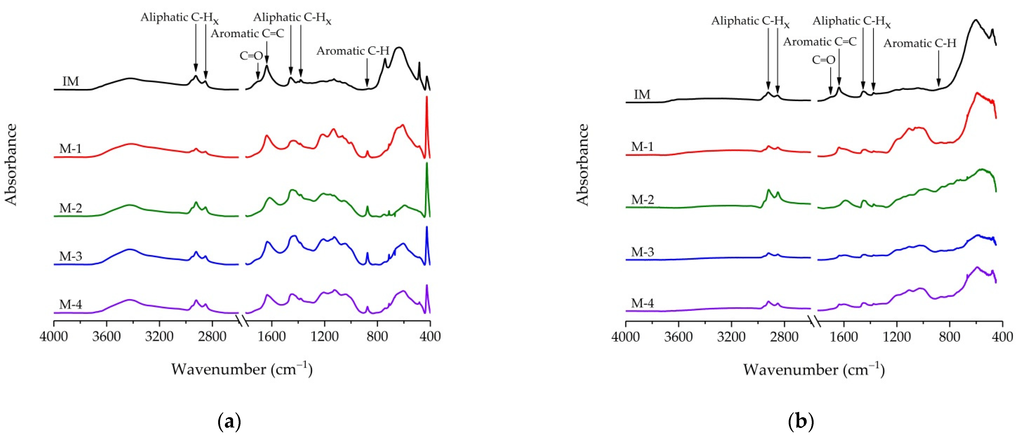
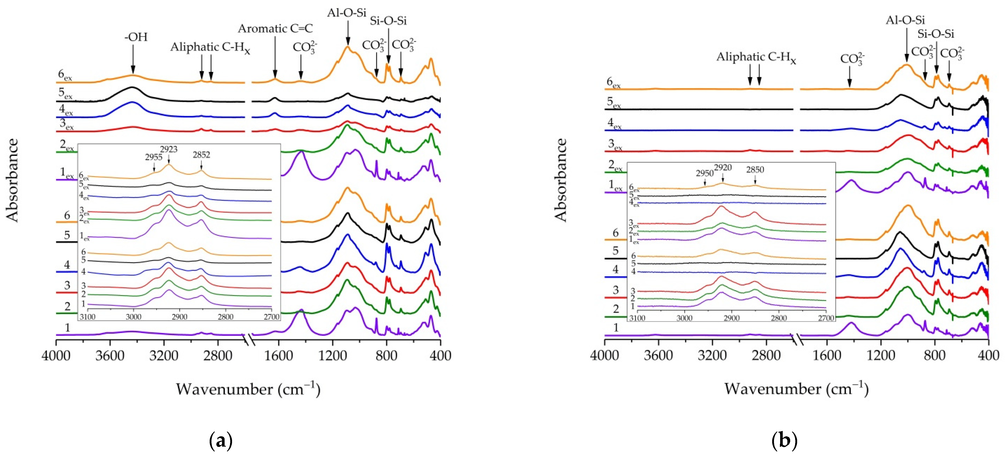
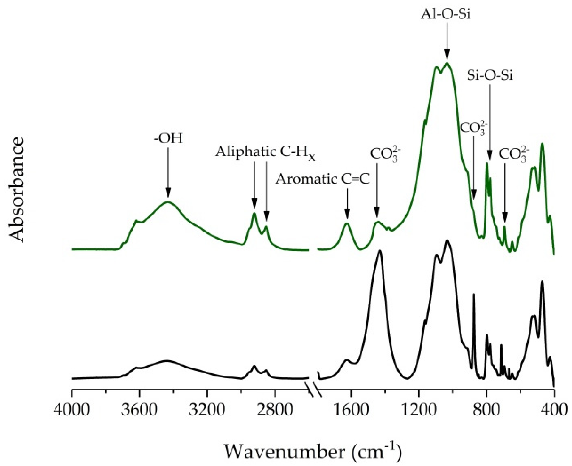
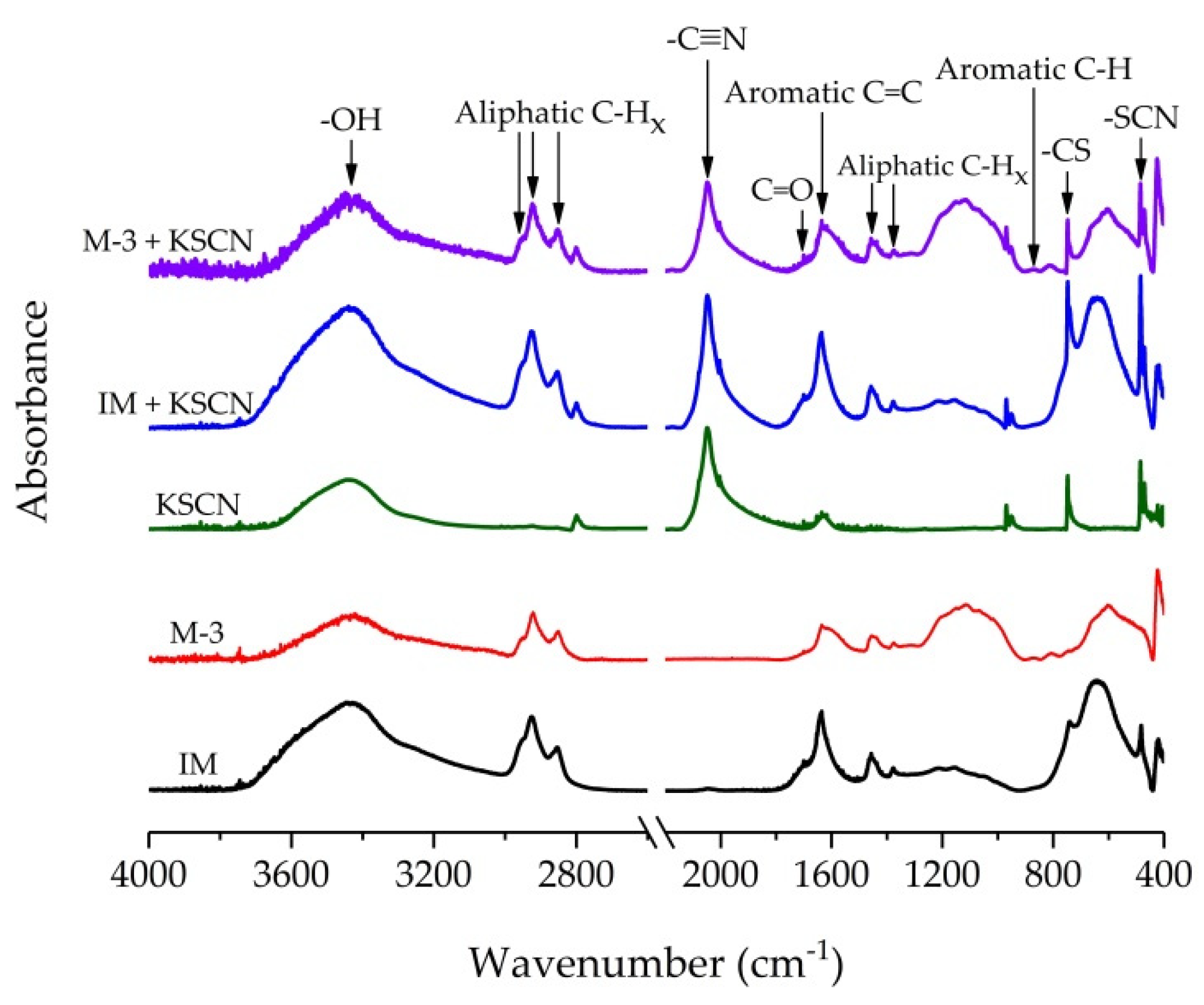
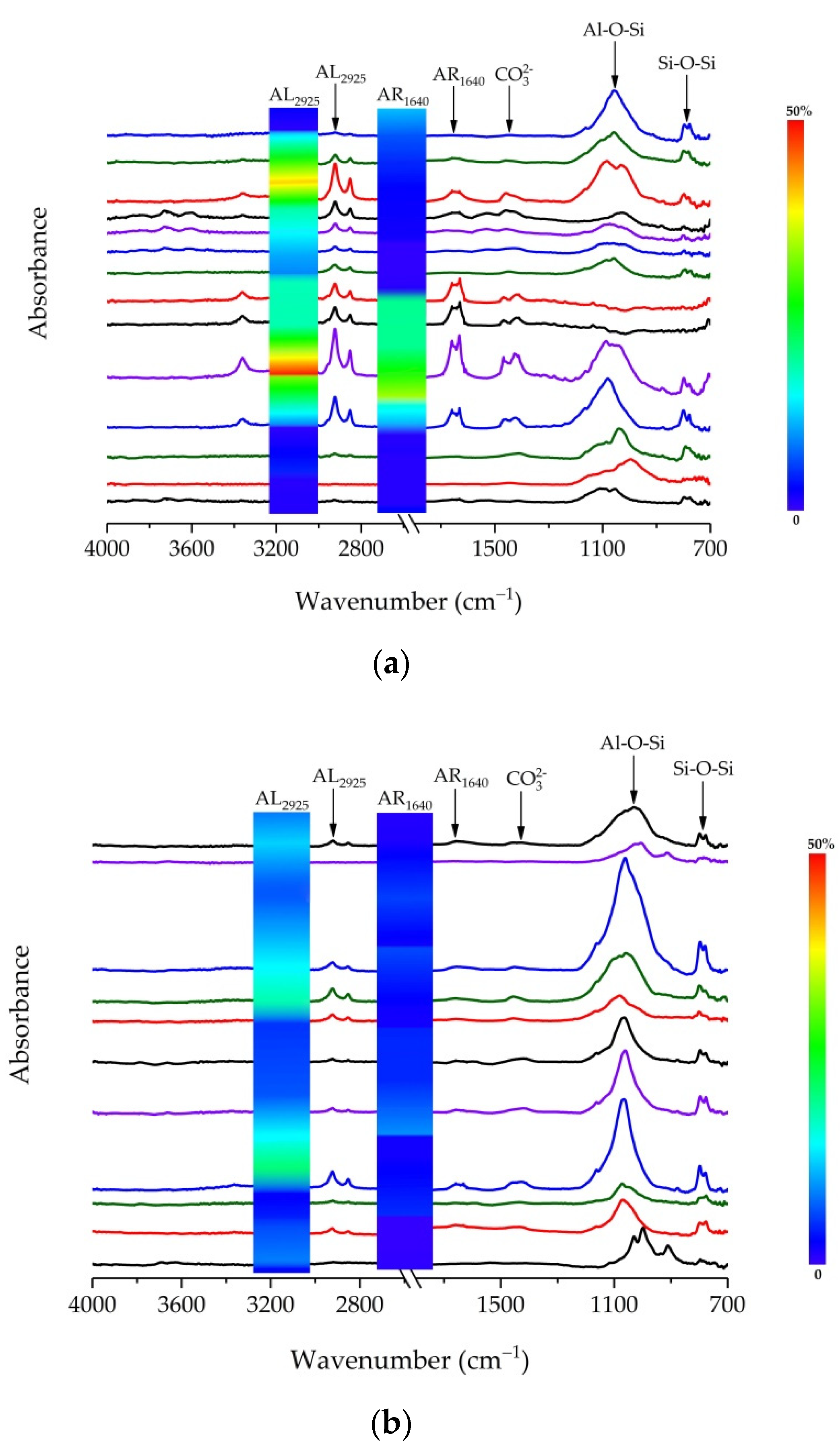
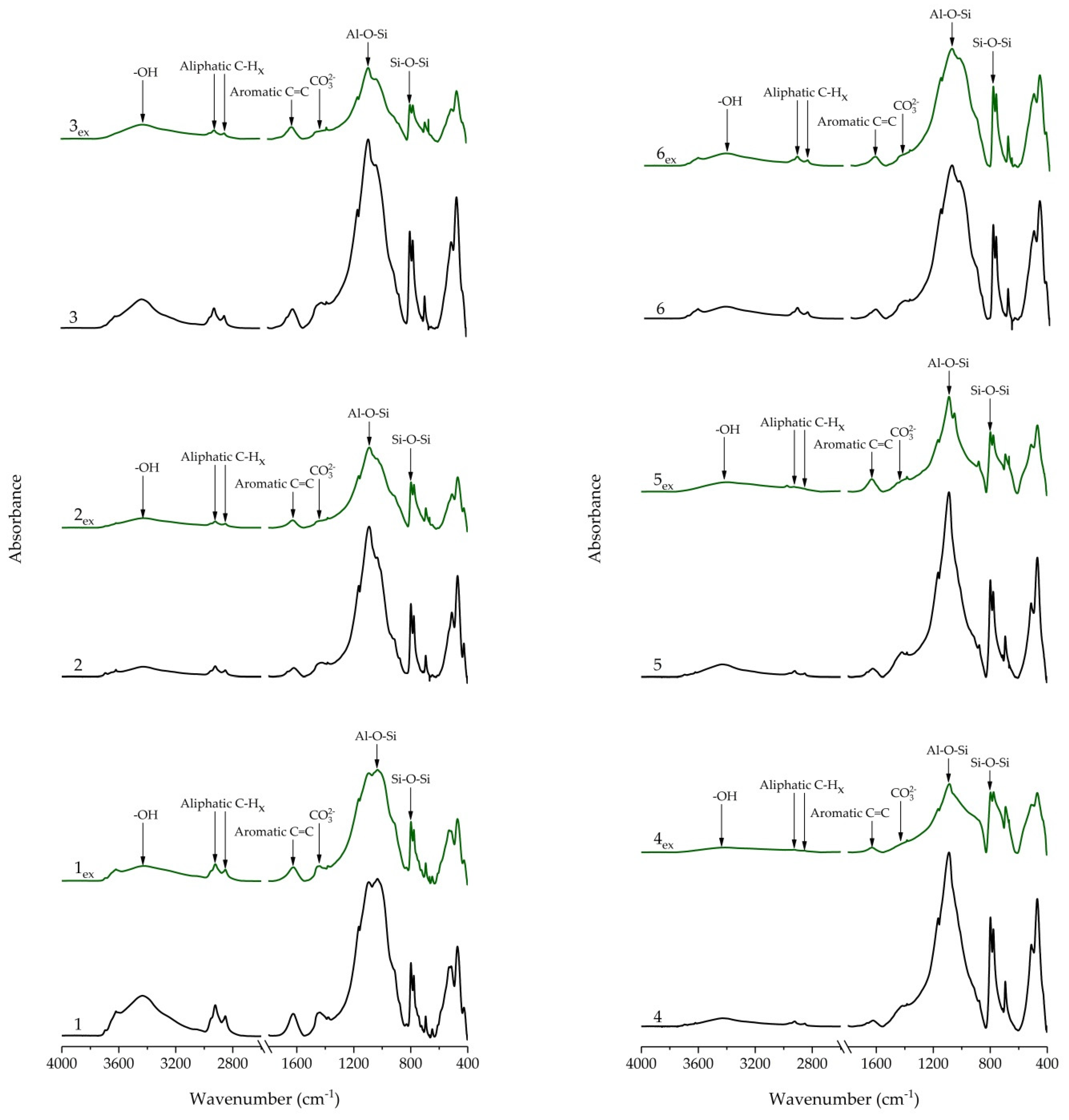
3.1. IR Spectra of Kerogen and Shale Rock Samples
3.2. Evaluation of Organic Matter Content by IR Spectroscopy
3.3. Surface Organic Matter Analysis of Oil Shale Samples by FTIR Microscopy
4. Conclusions
Author Contributions
Funding
Data Availability Statement
Acknowledgments
Conflicts of Interest
Appendix A


References
- Chen, Y.; Mastalerz, M.; Schimmelmann, A. Heterogeneity of shale documented by micro-FTIR and image analysis. J. Microsc. 2014, 256, 177–189. [Google Scholar] [CrossRef] [PubMed]
- Bordenave, M.L. Applied Petroleum Geochemistry; Edition Technips: Paris, France, 1993; p. 524. [Google Scholar]
- Stankiewicz, B.A.; Kruge, M.A.; Mastalerz, M. A geochemical study of macerals from a Miocene lignite and an Eocene bitu-minous coal, Indonesia. Org. Geochem. 1996, 24, 531–545. [Google Scholar] [CrossRef]
- Stankiewicz, B.; Kruge, M.; Mastalerz, M.; Salmon, G.L. Geochemistry of the alginite and amorphous organic matter from Type II-S kerogens. Org. Geochem. 1996, 24, 495–509. [Google Scholar] [CrossRef]
- Maciel, G.; Dennis, L. Comparison of oil shales and kerogen concentrates by 13C nuclear magnetic resonance. Org. Geochem. 1981, 3, 105–109. [Google Scholar] [CrossRef]
- Suggate, R.; Dickinson, W. Carbon NMR of coals: The effects of coal type and rank. Int. J. Coal Geol. 2004, 57, 1–22. [Google Scholar] [CrossRef]
- Dang, S.T.; Sondergeld, C.H.; Rai, C.S. Study of Kerogen Maturity using Fourier Transform Infrared Spectroscopy (FTIR) and Thermogravimetric Analysis (TGA). In Proceedings of the Society of Petroleum Engineers (SPE), Houston, TX, USA, 28–30 September 2015; p. 175149. [Google Scholar]
- Ballard, B.D. Quantitative mineralogy of reservoir rocks using Fourier transform infrared spectroscopy. In Proceedings of the Society of Petroleum Engineers Annual Technical Conference and Exhibition, Anaheim, CA, USA, 11–14 November 2007. [Google Scholar]
- Tarakanova, A.V.; Kardasheva, Y.S.; Isichenko, I.V.; Anisimov, A.V.; Maksimov, A.L.; Karakhanov, E.A. Physicochemical analysis of a kerogen rock (oil shale). Mosc. Univ. Chem. Bull. 2016, 71, 329–335. [Google Scholar] [CrossRef]
- Ganz, H.; Kalkreuth, W. Application of infrared spectroscopy to the classification of kerogen-types and the evolution of source rock and oil shale potentials. Fuel 1987, 66, 708–711. [Google Scholar] [CrossRef]
- Kister, J.; Guiliano, M.; Largeau, C.; Derenne, S.; Casadevall, E. Characterization of chemical structure, degree of maturation and oil potential of Torbanites (type I kerogens) by quantitative FT-i.r. spectroscopy. Fuel 1990, 69, 1356–1361. [Google Scholar] [CrossRef]
- Lin, R.; Ritz, G.P. Reflectance FT-IR Microspectroscopy of Fossil Algae Contained in Organic-Rich Shales. Appl. Spectrosc. 1993, 47, 265–271. [Google Scholar] [CrossRef]
- Landais, P. Statistical determination of geochemical parameters of coal and kerogen macerals from transmission micro-infrared spectroscopy data. Org. Geochem. 1995, 23, 711–720. [Google Scholar] [CrossRef]
- Chen, J.; Ping, L.; Jinchao, L. Using kerogen FTIR parameters for determination of organic facies. Chin. Sci. Bull. 1998, 43, 681–684. [Google Scholar] [CrossRef]
- Petsch, S.; Berner, R.; Eglinton, T. A field study of the chemical weathering of ancient sedimentary organic matter. Org. Geochem. 2000, 31, 475–487. [Google Scholar] [CrossRef]
- Painter, P.C.; Snyder, R.W.; Starsinic, M.; Coleman, M.M.; Kuehn, D.W.; Davis, A. Concerning the Application of FT-IR to the Study of Coal: A Critical Assessment of Band Assignments and the Application of Spectral Analysis Programs. Appl. Spectrosc. 1981, 35, 475–485. [Google Scholar] [CrossRef]
- Painter, P.C.; Starsinic, M.; Squires, E.; Davis, A.A. Concerning the 1600 cm−1 region in the i.r. spectrum of coal. Fuel 1983, 62, 742–744. [Google Scholar] [CrossRef]
- Christy, A.A.; Hopland, A.L.; Barth, T.; Kvalheim, O.M. Quantitative determination of thermal maturity in sedimentary or-ganic matter by diffuse reflectance infrared spectroscopy of asphaltenes. Org. Geochem. 1989, 14, 77–81. [Google Scholar] [CrossRef]
- Lis, G.P.; Mastalerz, M.; Schimmelmann, A.; Lewan, M.D.; Stankiewicz, B.A. FTIR absorption indices for thermal maturity in comparison with vitrinite reflectance R0 in type-II kerogens from Devonian black shales. Org. Geochem. 2005, 36, 1533–1552. [Google Scholar] [CrossRef]
- Lee, H.; Oncel, N.; Liu, B.; Kukay, A.; Altincicek, F.; Varma, R.S.; Shokouhimehr, M.; Ostadhassan, M. Structural Evolution of Organic Matter in Deep Shales by Spectroscopy (1H and 13C Nuclear Magnetic Resonance, X-ray Photoelectron Spectroscopy, and Fourier Transform Infrared) Analysis. Energy Fuels 2020, 34, 2807–2815. [Google Scholar] [CrossRef]
- Labus, M.; Lempart, M. Studies of Polish Paleozoic shale rocks using FTIR and TG/DSC methods. J. Pet. Sci. Eng. 2018, 161, 311–318. [Google Scholar] [CrossRef]
- Cesar, J.; Quintero, K. Organic geochemistry of kerogen from La Luna Formation, Western Venezuelan Basin, using diffuse reflectance–Fourier transform infrared spectroscopy (DRFTIR). Fuel 2020, 282, 118805. [Google Scholar] [CrossRef]
- Mastalerz, M.; Drobniak, A.; Stankiewicz, A.B. Origin, properties, and implications of solid bitumen in source-rock reservoirs: A review. Int. J. Coal Geol. 2018, 195, 14–36. [Google Scholar] [CrossRef]
- Vandenbroucke, M.; Largeau, C. Kerogen origin, evolution and structure. Org. Geochem. 2007, 38, 719–833. [Google Scholar] [CrossRef]
- Guido, A.; Mastandrea, A.; Demasi, F.; Tosti, F.; Russo, F. Organic matter remains in the laminated microfabrics of the Kess-Kess mounds (Hamar Laghdad, Lower Devonian, Morocco). Sediment. Geol. 2012, 263–264, 194–201. [Google Scholar] [CrossRef]
- Mastandrea, A.; Guido, A.; Demasi, F.; Ruffolo, S.A.; Russo, F. The Characterisation of Sedimentary Organic Matter in Carbonates with Fourier-Transform Infrared (FTIR) Spectroscopy. Eng. Geol. Infrastruct. Plan. Eur. 2011, 131, 331–342. [Google Scholar] [CrossRef]
- Bonoldi, L.; Di Paolo, L.; Flego, C. Vibrational spectroscopy assessment of kerogen maturity in organic-rich source rocks. Vib. Spectrosc. 2016, 87, 14–19. [Google Scholar] [CrossRef]
- Washburn, K.E.; Birdwell, J. Multivariate analysis of ATR-FTIR spectra for assessment of oil shale organic geochemical properties. Org. Geochem. 2013, 63, 1–7. [Google Scholar] [CrossRef]
- Pejcic, B.; Bourdet, J.; Piane, C.D.; Li, Z.; Heath, C.; Clennell, B. Investigating the Organic Matter in Shales From the Canning and Perth Basins via Infrared and Raman Spectroscopy. In Proceedings of the 4th Unconventional Resources Technology Conference, Austin, TX, USA, 24–26 July 2017; pp. 24–26. [Google Scholar]
- Pejcic, B.; Heath, C.; Pagès, A.; Normore, L. Analysis of carbonaceous materials in shales using mid-infrared spectroscopy. Vib. Spectrosc. 2021, 112, 103186. [Google Scholar] [CrossRef]
- Washburn, K.E.; Birdwell, J.E.; Foster, M.; Gutierrez, F. Detailed Description of Oil Shale Organic and Mineralogical Hetero-geneity via Fourier Transform Infrared Microscopy. Energy Fuels 2015, 29, 4264–4271. [Google Scholar] [CrossRef]
- Shurygin, B.N.; Nikitenko, B.L.; Devyatov, V.P.; Il’ina, V.I.; Meledina, S.V.; Gaideburova, E.A.; Dzuba, O.S.; Kazakov, A.M.; Mogucheva, N.K. Stratigraphy of Oil and Gas Basins of Siberia. Jurassic System; Publishing House of SB RAS, Department “GEO”: Novosibirsk, Russia, 2000; p. 480. [Google Scholar]
- Baturin, Y.E.; Bochkarev, V.S.; Braduchan, Y.V.; Gurari, F.G.; Dzyuba, O.S.; Iliyna, V.I.; Karagodin, Y.N.; Krasnov, V.I.; Kulakhmetov, N.K.; Meledina, S.V. Conclusions of the Sixth Interdepartmental Stratigraphic Symposium on the Examination and the Acceptance of the Adjusted Stratigraphic Schemes on Mesozoic Deposits of the West Siberia; SNIIGGiMS: Novosibirsk, Russia, 2004; p. 113. [Google Scholar]
- Yuri, Z.N.; Eder, V.; Zamirailova, A. Composition and formation environments of the Upper Jurassic–Lower Cretaceous black shale Bazhenov Formation (the central part of the West Siberian Basin). Mar. Pet. Geol. 2008, 25, 289–306. [Google Scholar] [CrossRef]
- Acholla, F.V.; Orr, W.E. Pyrite removal from kerogen without altering organic matter: The chromous chloride method. Energy Fuels 1993, 7, 406–410. [Google Scholar] [CrossRef]
- Craddock, P.R.; Van Le Doan, T.; Bake, K.; Polyakov, M.; Charsky, A.M.; Pomerantz, A.E. Evolution of Kerogen and Bitumen during Thermal Maturation via Semi-Open Pyrolysis Investigated by Infrared Spectroscopy. Energy Fuels 2015, 29, 2197–2210. [Google Scholar] [CrossRef]
- Wang, S.-H.; Griffiths, P.R. Resolution enhancement of diffuse reflectance i.r. spectra of coals by Fourier self-deconvolution: 1. C-H stretching and bending modes. Fuel 1985, 64, 229–236. [Google Scholar] [CrossRef]
- Sobkowiak, M.; Painter, P. Determination of the aliphatic and aromatic CH contents of coals by FT-i.r.: Studies of coal extracts. Fuel 1992, 71, 1105–1125. [Google Scholar] [CrossRef]
- Espitalie, J.; Deroo, G.; Marquis, F. La pyrolyse rock-eval et ses applications. Revue d’IFP 1985, 40, 563–579. [Google Scholar] [CrossRef]
- Singh, A.K.; Hakimi, M.H.; Kumar, A.; Ahmed, A.; Abidin, N.S.Z.; Kinawy, M.; El Mahdy, O.; Lashin, A. Geochemical and organic petrographic characteristics of high bituminous shales from Gurha mine in Rajasthan, NW India. Sci. Rep. 2020, 10, 1–19. [Google Scholar] [CrossRef] [PubMed]
- ASTM D5373. Standard Test Methods for Determination of Carbon, Hydrogen, and Nitrogen in Analysis Samples of Coal; ASTM International: West Conshohocken, PA, USA, 1993; pp. 1–11. [Google Scholar] [CrossRef]
- Müller, C.M.; Pejcic, B.; Esteban, L.; Piane, C.D.; Raven, M.; Mizaikoff, B. Infrared Attenuated Total Reflectance Spectroscopy: An Innovative Strategy for Analyzing Mineral Components in Energy Relevant Systems. Sci. Rep. 2015, 4, 6764. [Google Scholar] [CrossRef] [PubMed] [Green Version]
- Heath, C.; Pejcic, B.; Piane, C.D.; Esteban, L. Development of far-infrared attenuated total reflectance spectroscopy for the mineralogical analysis of shales. Fuel 2016, 182, 771–779. [Google Scholar] [CrossRef]
- Chen, Y.; Mastalerz, M.; Schimmelmann, A. Characterization of chemical functional groups in macerals across different coal ranks via micro-FTIR spectroscopy. Int. J. Coal Geol. 2012, 104, 22–33. [Google Scholar] [CrossRef]
- He, X.; Liu, X.; Nie, B.; Song, D. FTIR and Raman spectroscopy characterization of functional groups in various rank coals. Fuel 2017, 206, 555–563. [Google Scholar] [CrossRef]
- Skala, D.; Korica, S.; Vitorović, D.; Neumann, H.-J. Determination of kerogen type by using DSC and TG analysis. J. Therm. Anal. Calorim. 1997, 49, 745–753. [Google Scholar] [CrossRef]
- Bulatov, T.; Kozlova, E.; Leushina, E.; Pronina, N.; Panchenko, I.; Kostina, Y.; Spasennykh, M. Specific Layers Containing Highly Oil-Prone Organic Matter in the Bazhenov Formation. In Proceedings of the 29th International Meeting on Organic Geochemistry, European Association of Geoscientists & Engineers. Gothenburg, Sweden, 1–6 September 2019; pp. 1–6. [Google Scholar]
- Dong, Z.; Holditch, S.A.; McVay, D.A.; Ayers, W.B.; Lee, W.J.; Morales, E. Probabilistic Assessment of World Recoverable Shale-Gas Resources. SPE Econ. Manag. 2015, 7, 72–82. [Google Scholar] [CrossRef]


| Sample | Depth, m | TOC, wt.% | Tmax, °C | S1, mgHC/g Rock | S2, mgHC/g Rock | H/C |
|---|---|---|---|---|---|---|
| IM | 2600 | 35.20 | 421 | 2.89 | 189.11 | 1.29 |
| M-1 | 2721 | 34.51 | 440 | 1.34 | 59.02 | 1.19 |
| M-2 | 2725 | 38.46 | 444 | 1.03 | 94.06 | 0.92 |
| M-3 | 2730 | 35.22 | 442 | 2.38 | 55.86 | 0.93 |
| M-4 | 2732 | 27.60 | 439 | 0.71 | 51.92 | 1.02 |
| Sample | Depth, m | TOC, wt.% | Tmax, °C | S1, mgHC/g Rock | S2, mgHC/g Rock | PI | H/C |
|---|---|---|---|---|---|---|---|
| 1 | 2717 | 13.43 | 447 | 4.27 | 52.04 | 0.08 | 0.92 |
| 1ex | 12.43 | 446 | 0.14 | 40.48 | - | 0.91 | |
| 2 | 2719 | 11.62 | 443 | 4. 41 | 37.95 | 0.10 | 1.18 |
| 2ex | 9.58 | 445 | 0.26 | 27.22 | 1.17 | ||
| 3 | 2723 | 12.25 | 446 | 2.48 | 44.67 | 0.05 | 1.18 |
| 3ex | 8.20 | 446 | 0.12 | 43.61 | - | 1.20 | |
| 4 | 2725 | 2.87 | 441 | 3.11 | 4.18 | 0.43 | 0.85 |
| 4ex | 1.97 | 446 | 0.06 | 2.08 | 0.71 | ||
| 5 | 2726 | 2.44 | 446 | 2.20 | 3.47 | 0.39 | 1.58 |
| 5ex | 1.91 | 452 | 0.11 | 2.24 | - | 1.51 | |
| 6 | 2727 | 6.58 | 445 | 1. 49 | 15.38 | 0.09 | 1.32 |
| 6ex | 6.60 | 442 | 0.11 | 17.21 | - | 1.30 |
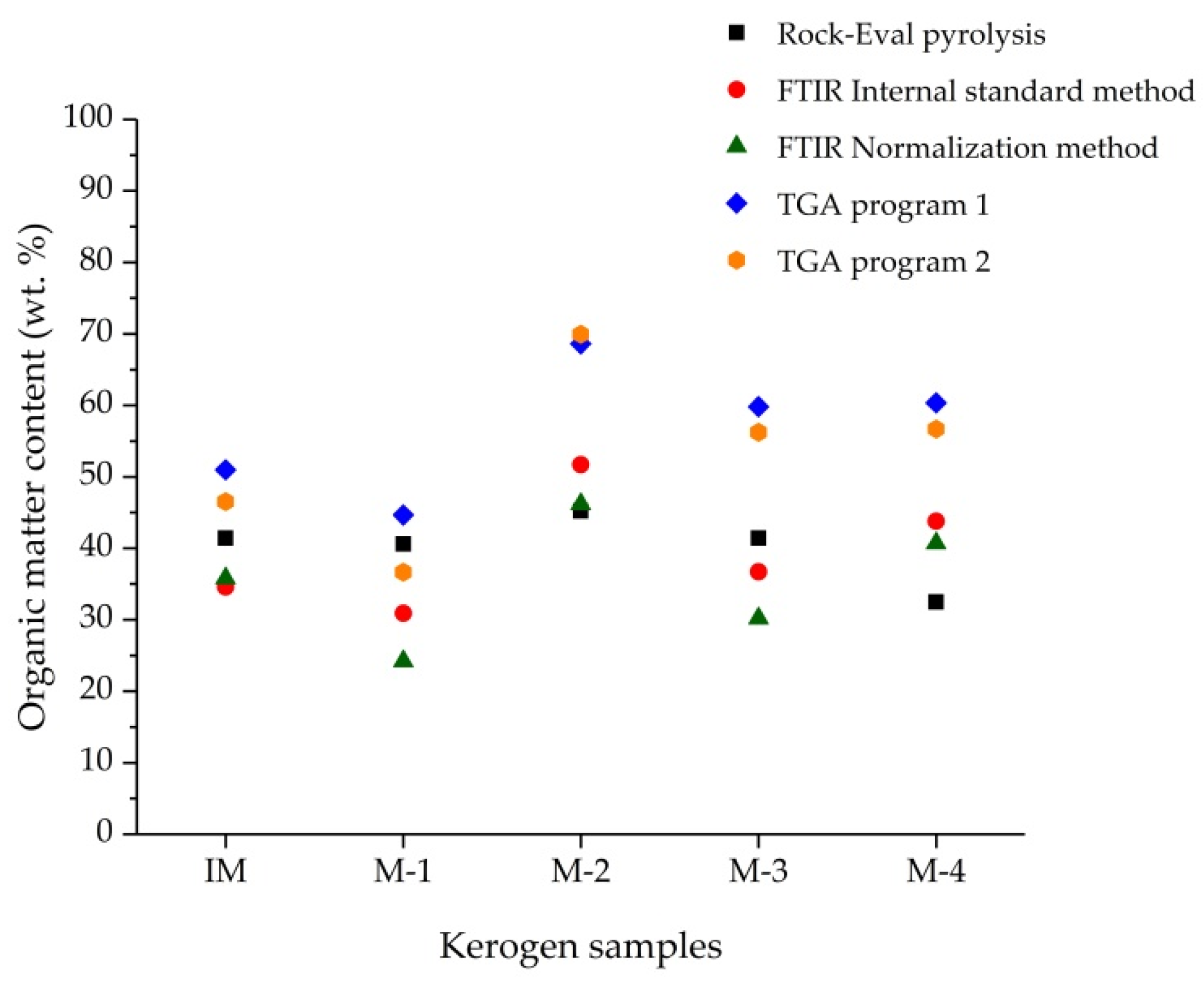
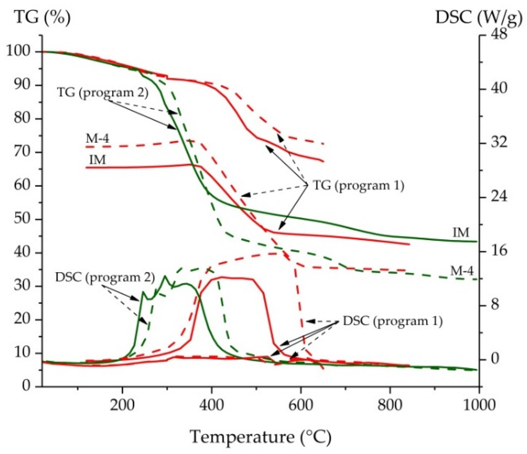
| Sample | TOC by Rock-Eval, wt.% | OM Content Evaluated by FTIR, wt.% | Mass Loss by TGA, wt.% (up to 650 °C) | ||
|---|---|---|---|---|---|
| Internal Standard Method (KSCN) | Normalization Method | Program 1 (N2, Air) | Program 2 (Air) | ||
| IM | 35.20 | 34.6 | 35.8 | 50.97 | 46.54 |
| M-1 | 34.51 | 30.9 * | 24.2 * | 44.66 | 36.66 |
| M-2 | 38.46 | 51.7 * | 46.2 * | 68.62 | 69.93 |
| M-3 | 35.22 | 36.7 | 30.2 | 59.82 | 56.25 |
| M-4 | 27.60 | 43.8 | 40.7 | 60.34 | 56.69 |
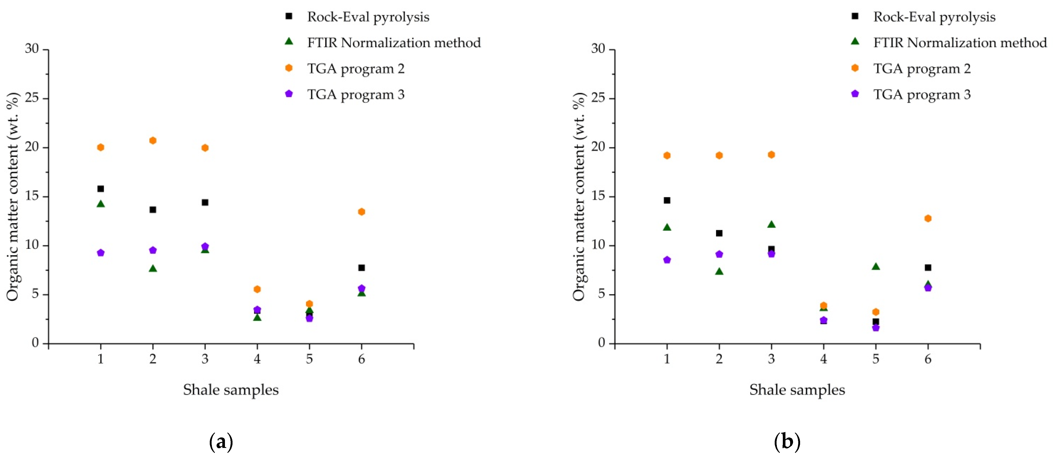
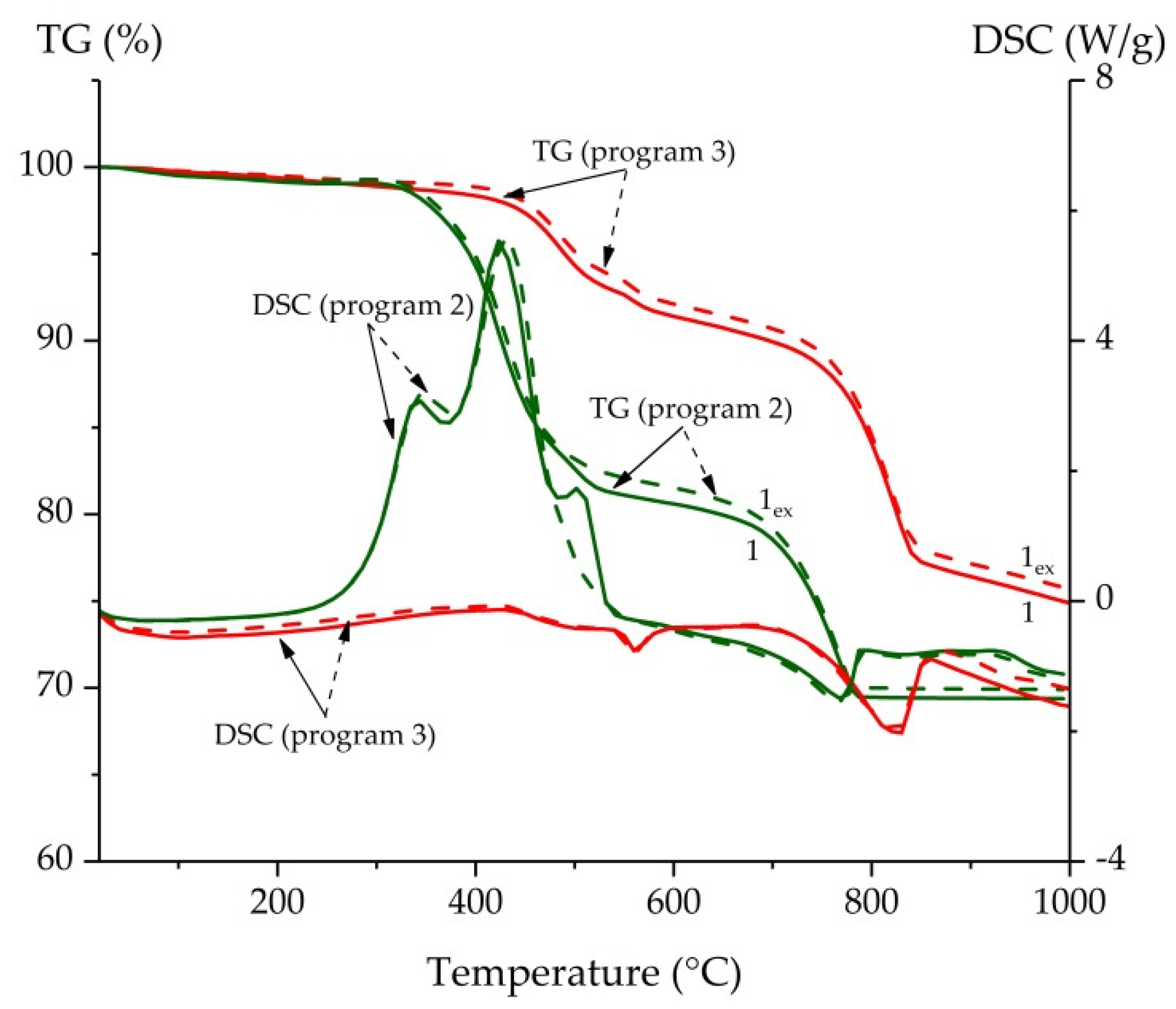
| Sample | AL, AR and Minerals Content Evaluated by FTIR, wt.% | TOC, by Rock-Eval, wt.% | Mass Loss by TGA, wt.% (up to 650 °C) | |||||
|---|---|---|---|---|---|---|---|---|
| Normalization Method | Program 2 (air) | Program 3 (N2) | ||||||
| AL2925 | AR1620 | Clay | Quartz | Other Silicates | ||||
| 1 | 8.3 | 5.9 | 42.4 | 19.5 | 23.9 | 13.43 | 20.04 | 9.27 |
| 1ex | 6.5 | 5.3 | 42.3 | 22.5 | 23.5 | 12.43 | 19.21 | 8.55 |
| 2 | 3.3 | 4.3 | 43.6 | 20.4 | 28.4 | 11.62 | 20.74 | 9.54 |
| 2ex | 3.5 | 3.8 | 42.6 | 23.9 | 26.3 | 9.58 | 19.22 | 9.13 |
| 3 | 4.6 | 4.9 | 41.5 | 20.7 | 28.3 | 12.25 | 19.99 | 9.93 |
| 3ex | 5.1 | 7.0 | 41.1 | 19.6 | 27.2 | 8.20 | 19.29 | 9.15 |
| 4 | 1.2 | 1.4 | 41.4 | 25.7 | 30.2 | 2.87 | 5.56 | 3.47 |
| 4ex | 1.3 | 2.3 | 34.9 | 30.9 | 30.5 | 1.97 | 3.90 | 2.40 |
| 5 | 1.4 | 2.0 | 44.5 | 23.3 | 28.9 | 2.44 | 4.07 | 2.57 |
| 5ex | 2.7 | 5.1 | 39.7 | 24.9 | 27.6 | 1.91 | 3.24 | 1.62 |
| 6 | 2.8 | 2.3 | 39.9 | 24.3 | 30.7 | 6.58 | 13.47 | 5.65 |
| 6ex | 3.0 | 3.0 | 38.9 | 25.9 | 29.6 | 6.60 | 12.79 | 5.69 |
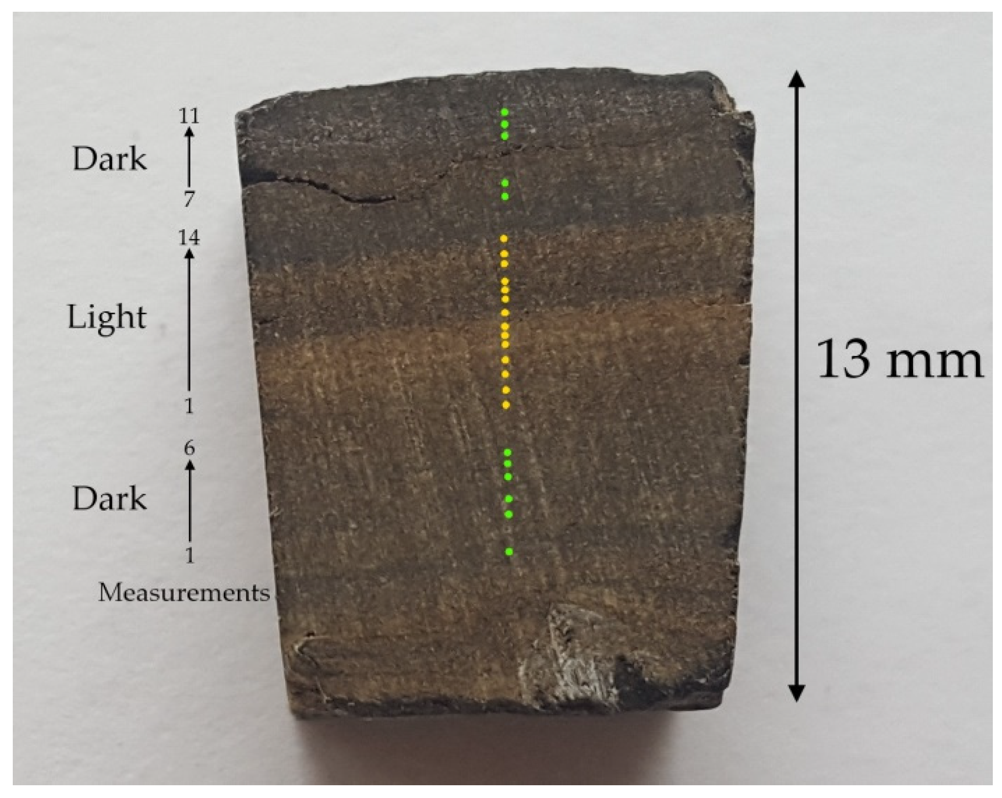
| Lamina | Measurement | OM Content by Micro-FTIR, wt.% | Ratio | Mineral Composition by Micro-FTIR, wt.% | OM Content by TGA, wt.% (Mass Loss up to 650 °C) | |||
|---|---|---|---|---|---|---|---|---|
| AL2920–2928 | AR1629–1640 | AL:AR | Clay | Carbonates | Quartz | Program 3 (Inert) | ||
| Light | 1 | 5.3 | 10.5 | 0.5 | 52.6 | 5.3 | 26.3 | - |
| 2 | 8.0 | 2.0 | 4.0 | 66.0 | 2.0 | 22.0 | - | |
| 3 | 5.6 | 2.8 | 2.0 | 58.3 | 8.3 | 25.0 | - | |
| 4 | 22.8 | 15.2 | 1.5 | 39.1 | 7.6 | 15.2 | - | |
| 5 | 25.6 | 25.6 | 1.0 | 25.6 | 13.2 | 9.9 | - | |
| 6 | 36.4 | 48.5 | 0.8 | 0.0 | 15.2 | 0.0 | - | |
| 7 | 35.3 | 50.0 | 0.7 | 0.0 | 14.7 | 0.0 | - | |
| 8 | 20.0 | 4.0 | 5.0 | 52.0 | 0.0 | 24.0 | - | |
| 9 | 21.1 | 5.3 | 4.0 | 36.8 | 15.8 | 21.1 | - | |
| 10 | 33.3 | 4.8 | 6.9 | 42.9 | 0.0 | 19.0 | - | |
| 11 | 44.4 | 14.8 | 3.0 | 29.6 | 0.0 | 11.1 | - | |
| 12 | 35.2 | 9.9 | 3.6 | 42.3 | 0.0 | 12.7 | - | |
| 13 | 16.2 | 5.4 | 3.0 | 56.8 | 0.0 | 21.6 | - | |
| 14 | 4.3 | 0.0 | - | 71.7 | 0.0 | 23.9 | - | |
| Average ±δ (p = 0.95) | 22.4 ± 7.7 | 14.2 ± 9.4 | 1.5 ± 1.2 | 41.0 ± 12.5 | 5.9 ± 3.7 | 16.6 ± 5.1 | 17.53 | |
| SD | 13.3 | 16.3 | 1.9 | 21.7 | 6.5 | 8.8 | - | |
| Dark | 1 | 4.7 | 0.0 | - | 86.0 | 0.0 | 9.3 | - |
| 2 | 10.7 | 5.4 | 2.0 | 58.9 | 5.4 | 19.6 | - | |
| 3 | 6.7 | 3.3 | 2.0 | 63.3 | 6.7 | 20.0 | - | |
| 4 | 11.2 | 4.2 | 2.7 | 61.5 | 5.6 | 17.5 | - | |
| 5 | 4.5 | 3.4 | 1.3 | 68.5 | 4.5 | 19.1 | - | |
| 6 | 4.7 | 3.1 | 1.5 | 67.2 | 6.3 | 18.8 | - | |
| 7 | 15.8 | 5.3 | 3.0 | 60.5 | 0.0 | 18.4 | - | |
| 8 | 17.8 | 4.1 | 4.3 | 61.6 | 0.0 | 16.4 | - | |
| 9 | 5.3 | 2.0 | 2.6 | 72.7 | 0.0 | 20.0 | - | |
| 10 | 0.0 | 0.0 | - | 81.8 | 0.0 | 18.2 | - | |
| 11 | 8.3 | 5.0 | 1.7 | 61.7 | 3.3 | 21.7 | - | |
| Average ±δ (p = 0.95) | 8.2 ± 3.6 | 3.3 ± 1.3 | 2.1 ± 0.7 | 67.6 ± 6.1 | 2.9 ± 1.9 | 18.1 ± 2.2 | 12.69 | |
| SD | 5.3 | 1.9 | 1.0 | 9.1 | 2.9 | 3.2 | - | |
Publisher’s Note: MDPI stays neutral with regard to jurisdictional claims in published maps and institutional affiliations. |
© 2021 by the authors. Licensee MDPI, Basel, Switzerland. This article is an open access article distributed under the terms and conditions of the Creative Commons Attribution (CC BY) license (https://creativecommons.org/licenses/by/4.0/).
Share and Cite
Tanykova, N.; Petrova, Y.; Kostina, J.; Kozlova, E.; Leushina, E.; Spasennykh, M. Study of Organic Matter of Unconventional Reservoirs by IR Spectroscopy and IR Microscopy. Geosciences 2021, 11, 277. https://0-doi-org.brum.beds.ac.uk/10.3390/geosciences11070277
Tanykova N, Petrova Y, Kostina J, Kozlova E, Leushina E, Spasennykh M. Study of Organic Matter of Unconventional Reservoirs by IR Spectroscopy and IR Microscopy. Geosciences. 2021; 11(7):277. https://0-doi-org.brum.beds.ac.uk/10.3390/geosciences11070277
Chicago/Turabian StyleTanykova, Natalya, Yuliya Petrova, Julia Kostina, Elena Kozlova, Evgenia Leushina, and Mikhail Spasennykh. 2021. "Study of Organic Matter of Unconventional Reservoirs by IR Spectroscopy and IR Microscopy" Geosciences 11, no. 7: 277. https://0-doi-org.brum.beds.ac.uk/10.3390/geosciences11070277