Review

SARS-CoV-2 (COVID-19): New Discoveries and Current Challenges

by
Ghazaleh Jamalipour Soufi
1,
Ali Hekmatnia
1,
Mahmoud Nasrollahzadeh
2,
Nasrin Shafiei
2,
Mohaddeseh Sajjadi
2,
Parisa Iravani
3,
Salman Fallah
4,
Siavash Iravani
5,* and
Rajender S. Varma
6,*
1
Radiology Department, School of Medicine, Isfahan University of Medical Sciences, Isfahan 81746-73461, Iran
2
Department of Chemistry, Faculty of Science, University of Qom, Qom 37185-359, Iran
3
Pediatrics Department, School of Medicine, Isfahan University of Medical Sciences, Isfahan 81746-73461, Iran
4
Physical Medicine and Rehabilitation Department, School of Medicine, Isfahan University of Medical Sciences, Isfahan 81746-73461, Iran
5
Faculty of Pharmacy and Pharmaceutical Sciences, Isfahan University of Medical Sciences, Isfahan 81746-73461, Iran
6
Regional Centre of Advanced Technologies and Materials, Palacký University in Olomouc, Šlechtitelů 27, 783 71 Olomouc, Czech Republic
*
Authors to whom correspondence should be addressed.
Submission received: 21 April 2020 / Revised: 19 May 2020 / Accepted: 20 May 2020 / Published: 26 May 2020
(This article belongs to the Special Issue COVID-19: Impact on Human Health and Behavior)

Abstract

:
SARS-CoV-2 (COVID-19) has today multiplied globally and various governments are attempting to stop the outbreak of the disease escalation into a worldwide health crisis. At this juncture, readiness, candor, clarity, and partaking of data are of paramount importance to speed up factual evaluation and starting pattern control activities, including serendipitous findings. Owing to the involvement of COVID-19, many facts regarding virulence, pathogenesis, and the real viral infection source and/or transmission mode still need to be addressed. The infected patients often present clinical symptoms with fever, dyspnea, fatigue, diarrhea, vomiting, and dry cough, as well as pulmonary, infiltrates on imaging. Extensive measures to decrease person-to-person transmission of COVID-19 are being implemented to prevent, recognize, and control the current outbreak as it is very similar to SARS-CoV in its clinical spectrum, epidemiology, and pathogenicity. In response to this fatal disease and disruptive outbreak, it is extremely vital to expedite the drug development process to treat the disease and vaccines for the prevention of COVID-19 that would help us defeat this pandemic expeditiously. This paper sums up and unifies the study of virological aspects, disease transmission, clinically administered techniques, therapeutics options, managements, future directions, designing of vaccines, and news dissemination pertaining to COVID-19.

Graphical Abstract

1. Introduction

Coronaviruses (CoVs) are one of the main pathogens that largely target the human respiratory system. The world has recently witnessed an outbreak of unidentified pneumonia via a novel coronavirus (2019-nCoV) initially distributed rapidly throughout China and subsequently affecting many other countries, with the elevated hazards of a pandemic. Ever since the appearance of 2019-nCoV disease in Wuhan City (Hubei Province, China) in the last part of 2019 [1,2], it was acknowledged as the main causative agent of the potentially fatal illness. The current outbreak of 2019-nCoV was subsequently termed coronavirus disease 2019 (COVID-19) by World Health Organization (WHO). Based on a great number of infected people that were exposed to the fish, livestock or wild animal markets in Wuhan City [3], it has been suggested that COVID-19 (earlier known as 2019-nCoV) is likely a zoonotic pathogen, which is present in people and diverse animals with a diversity of clinical features and course ranging from asymptomatic infection to minor ailment or brutal/deadly disease necessitating hospitalization. With its high mutation rates, COVID-19 can quickly spread by animal-to-human and/or human-to-human transmission through direct contact or droplets leading to infections in respiratory, cardiovascular, hepatic, gastrointestinal and neurologic system [4,5,6]; the pandemic has spread and escalated quickly. Importantly, on March 19, 2020, investigations showed the pangolin-CoV genome had 91% and 90.6% nucleotide identity with SARS-CoV-2 and Bat-CoV RaTG13, respectively; pangolin has been identified as a possible intermediate host of the 2019-nCoV [7]. Though these findings showed pangolin as the most likely intermediate host for SARS-CoV-2, further studies should be performed for other possible animals as intermediate hosts, and scientific investigations should be continued on this front (Figure 1) [8]. Interestingly, the extent of molecular divergence between SARS-CoV-2 and other related CoVs has been investigated wherein just 4% variability in genomic nucleotides was discerned between SARS-CoV-2 and a bat SARS-related CoV (SARSr-CoV; RaTG13), the difference at neutral sites being 17%. Additionally, it was noted that new variations in functional sites, comprising the receptor-binding domain of the spike detected in SARS-CoV-2 and viruses from pangolin SARSr-CoVs, are because of the natural selection besides recombination [9]. Population genetic evaluations of 103 SARS-CoV-2 genomes demonstrated that these viruses had two major lineages (designated S and L), identified by two various single nucleotide polymorphisms showing nearly complete linkage across the viral strains sequenced until now; L lineage was more prevalent than the S lineage. Undeniably, more comprehensive evaluations are needed about the viral genomic data as well as epidemiological analyses of COVID-19 [9].
As of 31 December 2019, numerous cases of pneumonia patients (at least 27 cases) were admitted into hospitals in Wuhan, where the etiological agent was identified as COVID-19 [2,10]. Subsequently, this first detected batch of cases revealed no exposure and/or even no relationship to Huanan seafood/wet animal wholesale marketplace, and the person-to-person transmission was affirmed; besides that, a variety of nosocomial infections were described in some healthcare professionals. From the published reports, the most clinical symptoms in patients with COVID-19 are shortness of breath (dyspnea), cough, fever, fatigue, diarrhea, vomiting, chest pain/tightness, and in some cases, the loss of smell/taste sensation [8,11,12]. Additionally, severe complications, such as acute heart injury, acute respiratory distress syndrome (ARDS), and secondary infections have been noticed. Besides that, most infected patients with COVID-19 have a history of hypertension, coronary heart illness, chronic bronchitis, diabetes, and cerebral infarction [13]. Some indications have emerged that this viral disease can be the reason for different tissue and organ damages and complications other than the lungs (Figure 2) [8,11]. The evaluations of case reports revealed that the loss of taste and smell sensation can be considered as a predictor for patients infected by SARS-CoV-2, but further evaluations are still required [12]. Interestingly, SARS-CoV-2 potential tropism was investigated by surveying expression of viral entry-associated genes in single-cell RNA-sequencing information from multiple tissues of healthy individuals [14]. These genes are co-expressed in nasal epithelial cells with genes involved in innate immunity; thus pointing out that nasal epithelial cells can be the reservoir of virus in the early viral infection, spread, and clearance [14]. Some important diagnostic techniques and imaging modalities, including quantitative reverse transcription polymerase chain reaction (RT-PCR), high-throughput sequencing technique, enzyme-linked immunosorbent assay (ELISA), Point-of-Care Testing (POCT) IgM/IgG, and computed tomography (CT) imaging are employed for the detection of COVID-19 [15].
Previous disease outbreaks of CoVs comprising SARS-CoV in 2002–2003 (first time; 8-month outbreak) and in 2012, the Middle East respiratory syndrome (MERS)-CoV (after ten years) that originated from China and Middle East countries, respectively, and then were proliferated to diverse parts of the world; they have been considered as agents with major public health threats. SARS-CoV with hospital-obtained infectious or nosocomial cases had a fatality rate of 10% (~774 virus-related deaths) and was transmitted to over 8000 people worldwide (based on information in http://www.who.int/csr/don/12-january-2020-novel-coronavirus-china/en, https://www.who.int/csr/sars/country/table2004_04_21/en/). Since September 2012, MERS-CoV, which emerged in Arabian Peninsula, was transmitted to almost 27 countries with a fatality rate of 35.6% in 2220 laboratory-confirmed infection cases (https://www.who.int/emergencies/mers-cov/en/; 2013). It is believed that all of these emerging infectious illnesses are zoonotic viruses illustrating hospital-acquired and person-to-person or animal-to-person transmission, and all of them lead to a universal transmission caused by using β-CoVs.
The mortality rate of COVID-19 has been evaluated until March 1, 2020. Patients from China (79,968 people) and 7169 beyond China have been recognized positive for COVID-19 [16]; 2873 deaths occurred in Chinese patients, attaining a death rate of 3.6% (95% CI 3.5–3.7), while 104 deaths from COVID-19 have been accounted for outside China (1.5% [1.2–1.7]). Though the estimation of mortality rate is dependent on the number of fatalities comparative with the number of affirmed infection cases, which is not illustrative of the real mortality rate; much earlier infected patients die on some random day, and hence the denominator of the death rate ought to be the overall figure of simultaneously infected patients as the individuals who died. Strikingly, the full denominator stays obscure in light of the fact that asymptomatic cases or patients with extremely mellow indications probably will not be tested and distinguished. Such cases along these lines cannot be incorporated for the calculation of real death rates, as real estimates relate to clinically evident COVID-19 patients. Importantly, in one study, Baud et al. re-evaluated death rates by isolating the death numbers on a certain day by the number of sick people with affirmed COVID-19 disease 14 days prior. On this premise, utilizing WHO information on the combined number of deaths to March 1, 2020, the rates would be 5.6% (95% CI 5.4–5.8) for China and 15.2% (12.5–17.9) outside of China [17]. COVID-19 is proliferating and surging amongst the public and has caused extensive afflictions because of its person-to-person spread. Indeed, the quick increase in confirmed or even suspected cases makes the prevention, detection, and control of COVID-19 very difficult, and thus the infectivity and dispersal speed of COVID-19 has dramatically exceeded compared to SARS- or MERS-CoVs.
It was revealed that viral 3-chymotrypsin-like cysteine protease (3CLpro) enzyme can control CoV replication and is vital for viral life cycle; 3CLpro is an established target for drug discovery regarding SARS- and MERS-CoVs [18]. Recent discoveries have determined that the genome sequence of SARS-CoV-2 is extremely similar to that of SARS-CoV, thus, the 3CLpro sequence is evaluated by researchers for potential antiviral compounds. The studies have revealed that SARS-CoV-2 3CLpro is conserved and share 99.02% sequence identity with SARS-CoV 3CLpro and together with 12 point-mutations. These mutations disrupt important hydrogen bonds and change the receptor binding site of SARS-CoV-2 3CLpro [18].
The COVID-19 eruption also presents critical challenges to the public health research and medical research communities as well as the global economies on par with international financial crisis, which transmitted moderately more than a previous decade. Due to the COVID-19 involvement, several facts regarding virulence, the real source of viral infection, or means of spread still await investigation. With currently accessible data, it is extremely difficult to measure and/or explain the level of human/animal-to-human spread. Herein, we highlight the disease transmission, epidemiology, virology, signs and symptoms, future directions, vaccine design approaches, and therapeutics options to limit/prevent the proliferation of this deadly virus. Further, we allude to some important case reports regarding radiologic imaging assessments of COVID-19 infected patients.

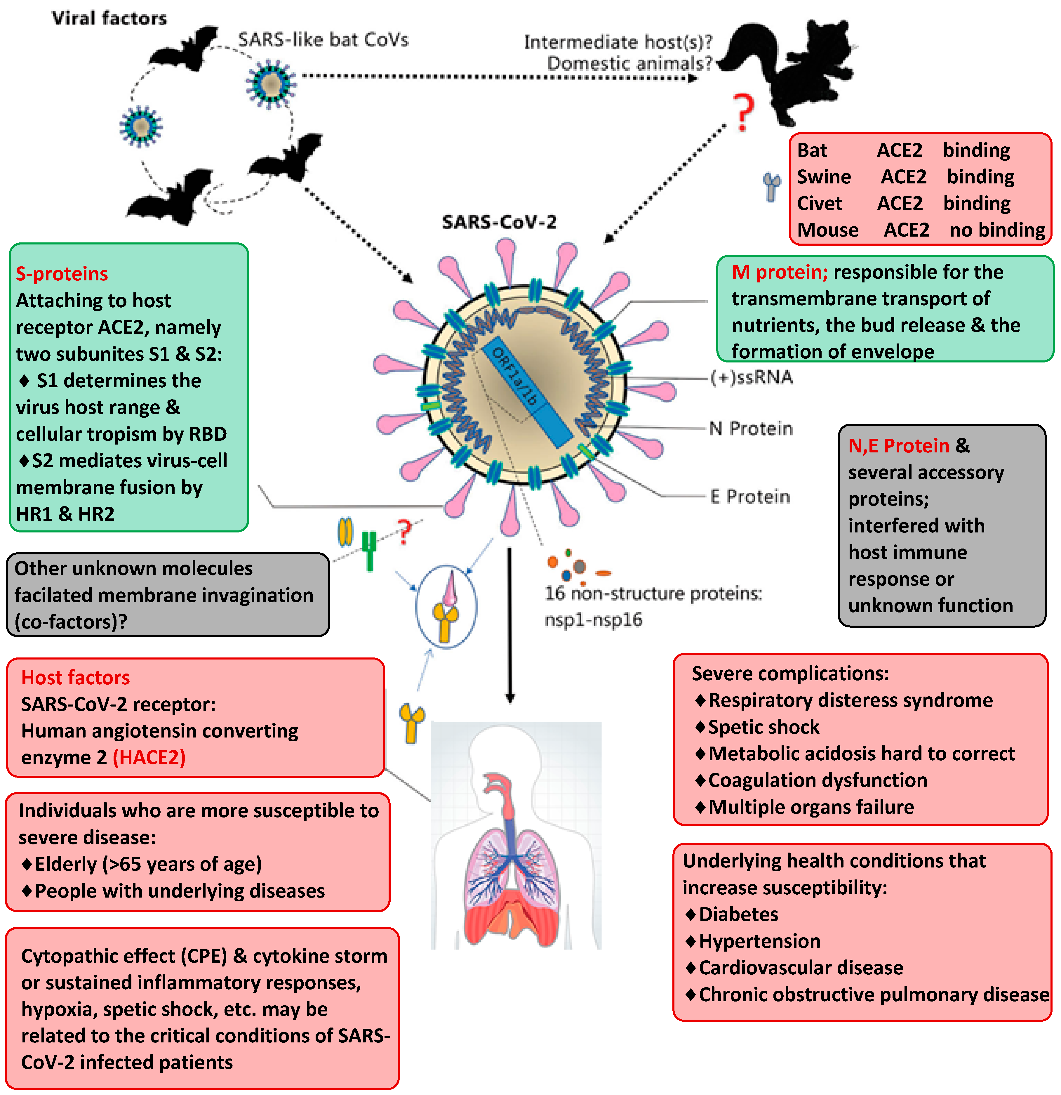
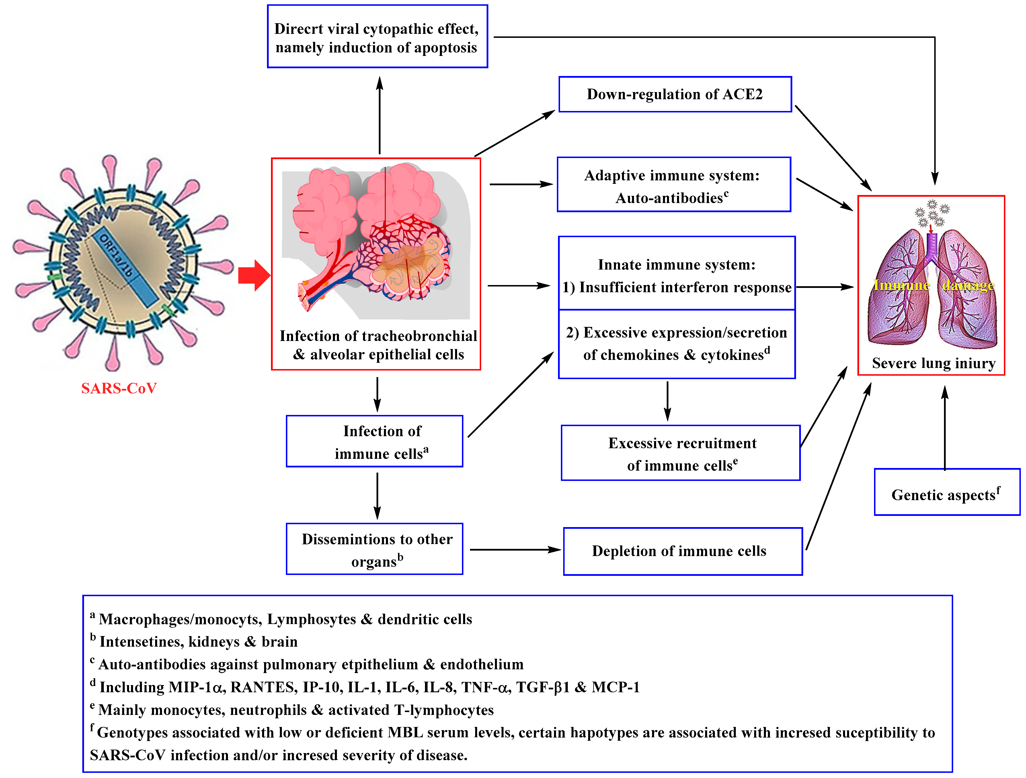
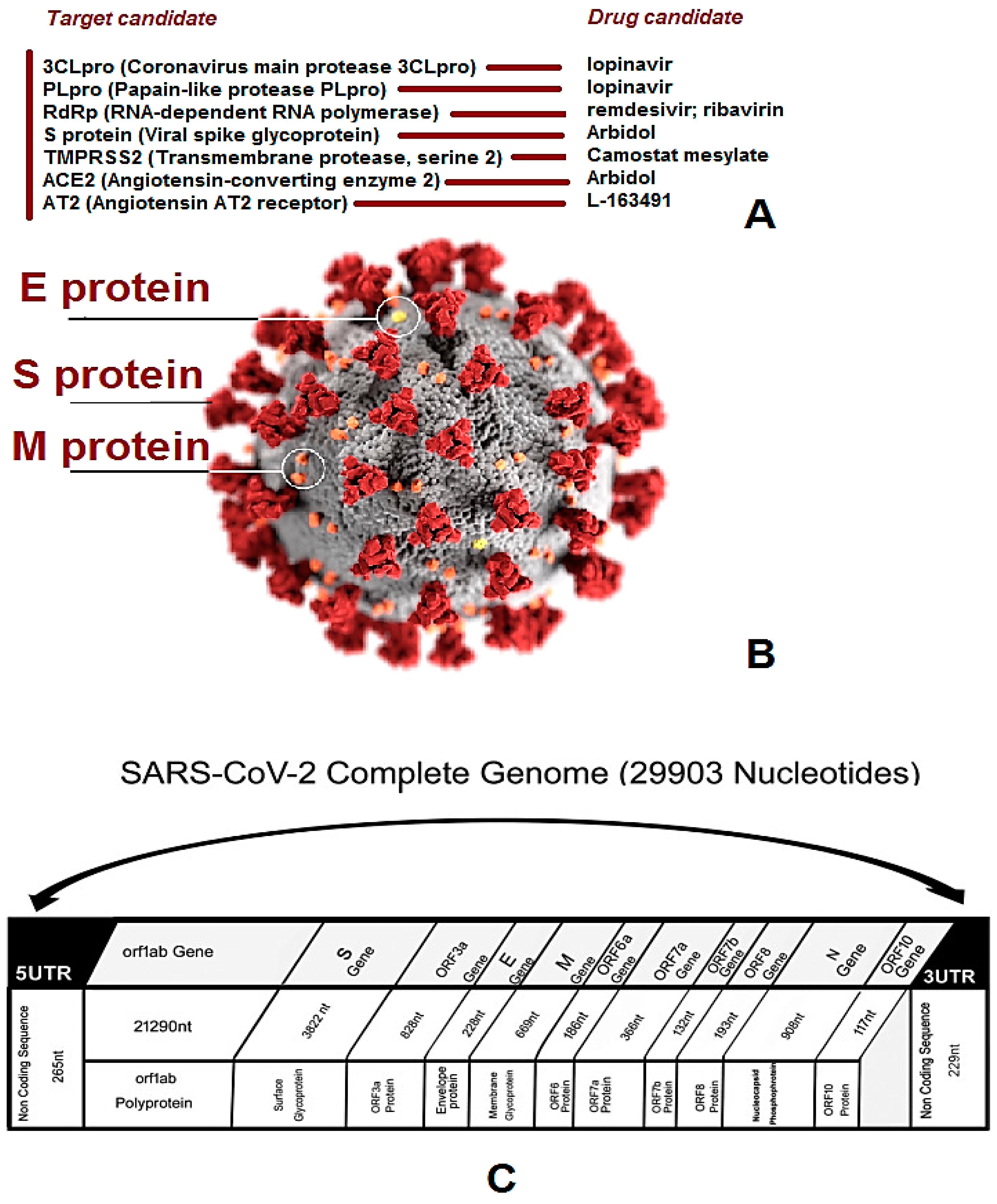
2. Genomics and Virology

CoVs (especially SARS-CoV-2) are a large enveloped non-segmented positive-sense, single-stranded RNA virus, that has caused enteric and respiratory illnesses in humans and/or animals [19]. SARS-CoV-2 is recognized to infect humans/animals, and the additional six CoVs comprise HCoV-229E, HCoV-OC43, HCoV-NL63, SARS-CoV, MERS-CoV, and HCoV-HKU [20]. Generally, HCoV-229E and -NL63 belong to α-CoVs, and the others including SARS-CoV-2 have its place in the genus β-CoVs. Among these outbreaks, the two most known kinds of CoV, HCoV-229E, and HCoV-OC43, have generally caused a mild infection in people with adequate immune systems [21,22]. All CoVs belong to a genus CoV with its excellent mutation rate in the Coronaviridae, and they all are large pleomorphic RNA viruses characteristically encompassing crown-shape peplomers or spikes with 80~160 nm in size and 27~32 kb positive polarity [5]. COVID-19 contains an RNA structure that belongs to the Coronaviridae family (Figure 3) [23].
Among the comparative genome sequences of SARS- and MERS-CoVs, COVID-19 has closer sequence identities with SARS-CoV. Genomic examination illustrates that novel SARS-CoV-2 is in the identical β-CoVs clades as SARS- and MERS-CoVs and can share a highly homological sequence with SARS-CoV, which also leads to acute, rapidly lethal pneumonia. Although the amino acid sequence of SARS-CoV-2 differs from other CoVs, particularly in the 1ab polyprotein regions and surface glycoprotein or spike protein, all the seven CoVs encode a spike protein and surface glycoprotein that can easily bind to the host/cell receptor and thus can mediate viral entrance [26].
Well-known host receptors for β-CoVs contain ACE2 (angiotensin-converting enzyme-2) for SARS-CoV-2 as well as DPP4 (dipeptidyl peptidase-4) for MERS-CoV [27]. According to the SARS-CoV-2 intermediate host is still unclear and finding its probable intermediate host is essential to prevent further transmission of the epidemic. Cell entry is generally an imperative component of cross-species spread, particularly for the β-CoVs. A single region of the spike protein named the receptor-binding domain (RBD), also termed as C-terminal domain (CTD) can be mediated by the interaction with the host/cell receptor. SARS-CoV-2 is supposed to infect the host cells via the interaction between the angiotensin-converting enzyme 2 (ACE2) and the main amino acids of spike protein RBD which can act as the potential intermediate hosts spreading SARS-CoV-2 to animal/human, however, the specific mechanism is uncertain. In this respect, a study developed new approach to functionally test for the RBDs from lineage B β-CoVs in replacing the SARS-CoV spike-RBDs (Figure 4) [28]. Herein, biosynthesizing the RBD of spike sequences is much faster and cheaper than traditional pseudo typing approaches, which rely on the preparation of the full spike sequence (~4 kb) towards CoVs and is very cost prohibitive for the large panels of spikes. Besides that, lineage B RBDs can be divided into functionally separate clades and thus many formerly unappreciated viruses show compatibility with unrecognized receptors on human cells.
In one study, samples obtained from seven patients with serious pneumonia were evaluated for determination of the causative pathogen; five examples were PCR-positive for CoVs. By focusing on PCR, a 29,891-base-pair coronavirus genome was obtained which shared 79.6% sequence identity to SARS-CoV BJ01 (GenBank accession number AY278488.2). High genome inclusion was acquired by remapping the entire reads of this genome and this sequence was analyzed; it was named novel coronavirus (2019-nCoV). Four additional full-length genome sequences of COVID-19 (WIV02, WIV05, WIV06, and WIV07) (GISAID accession numbers EPI_ISL_402127–402130) were found to be over 99.9% indistinguishable from each other and were acquired from four extra cases of patients applying next-generation sequencing and PCR [29].
Wu and co-workers made phylogenetic investigation on the total viral genome and reasoned that the infection was very intensely identified with a gathering of SARS-like coronavirus recently examined from bats in China [30]. In another study, extensive sequence evaluation was performed, and correlation connected to relative synonymous codon usage (RSCU) inclination among various creature species dependent on the COVID-19 RNA genome sequence [4]. The outcomes revealed the infection to be a recombinant infection among the bat coronavirus and an additional coronavirus of obscure inception. Additionally, they proposed that snake is the most plausible creature repository because of the infection’s RSCU predisposition being nearest to snake. Furthermore, Zhu and associates utilized profound learning calculations to investigate the gene sequences of new CoVs and the other different ones to foresee prospective viral hosts [31]. The conclusions showed that minks and bats might be the two likely hosts of the new coronavirus, while minks might be the transitional/intermediate hosts of the infection. The new coronavirus indicated a comparative pattern of disease to different CoVs in humans [31], especially Bat SARS-like CoV, SARS-nCoV, and MERS-nCoV [32]. Subsequent to scrutinizing of CoVs in different vertebrates, it was found that the bat CoVs had the most similar/comparative infection patterns to the new CoVs. In the wake of studying the infection patterns of other vertebrate coronavirus hosts, the prototypes from minks were observed as most similar to those of the new coronavirus [31]. By displaying the spike protein of the receptor for COVID-19, Xu et al. [33] detailed that ACE2 could be the receptor for this virus. Notably, ACE2 has also been the receptor for SARS-nCoV and NL63 [34]. As per their model, the binding strength between COVID-19 and ACE2 is higher than the limit required for viral infection disease but being more vulnerable than that between SARS-nCoV and ACE2. Lu et al. [2] directed the virus infectivity considerations and indicated that ACE2 is basic for COVID-19 to go in the HeLa cells. This information showed that ACE2 is probably going to be the receptor for COVID-19. Furthermore, Zhao et al. [35] investigated normal lung tissue cells from eight people, and found that the main Asian donor has more than fivefold the amount of ACE2 expressing cell ratio than African American and White cases. These outcomes point to a potential increased susceptibility of Asian populace, albeit more proof is expected to reach such a determination.

3. COVID-19—A Serious Threat for Global Health

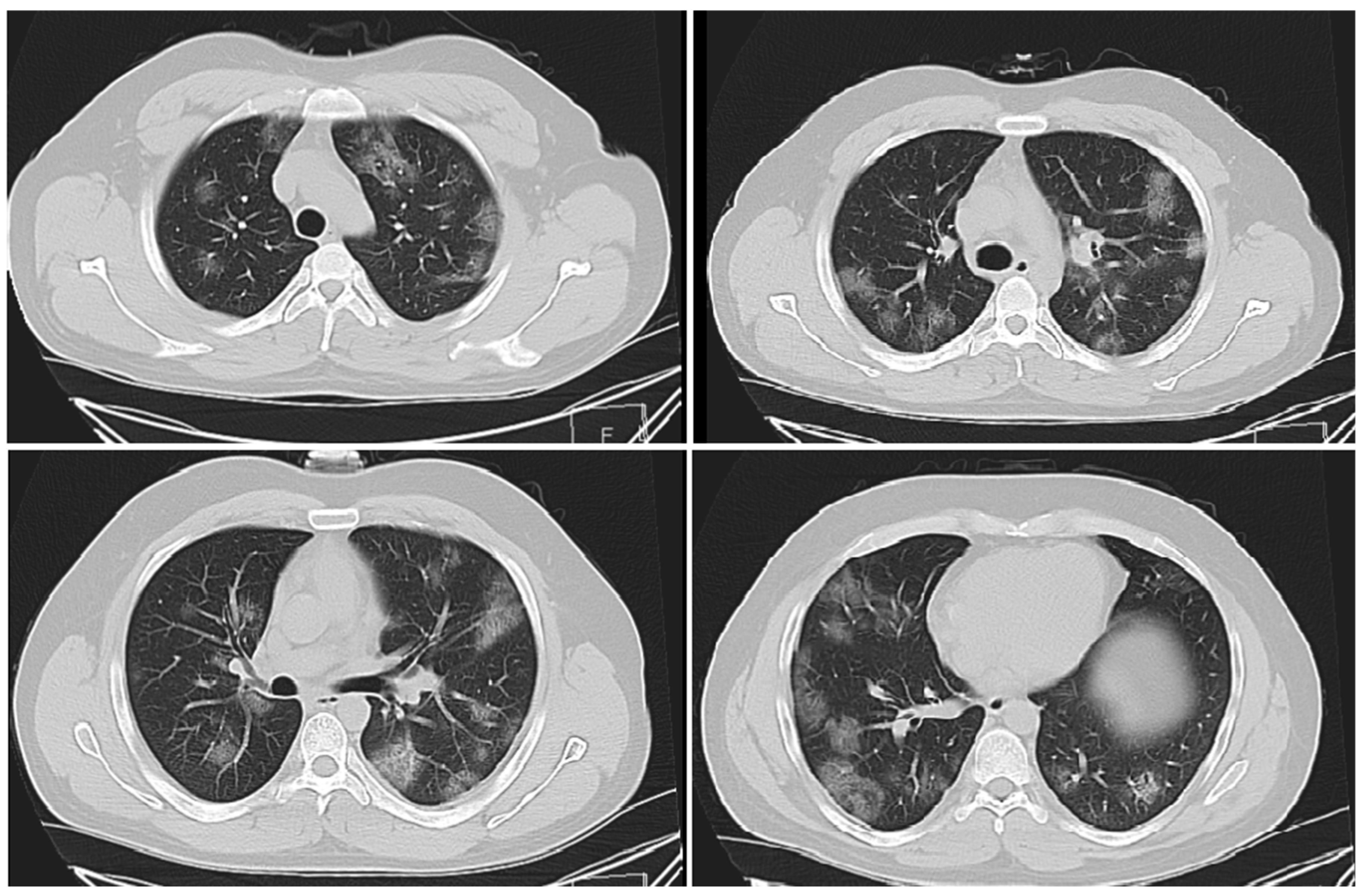
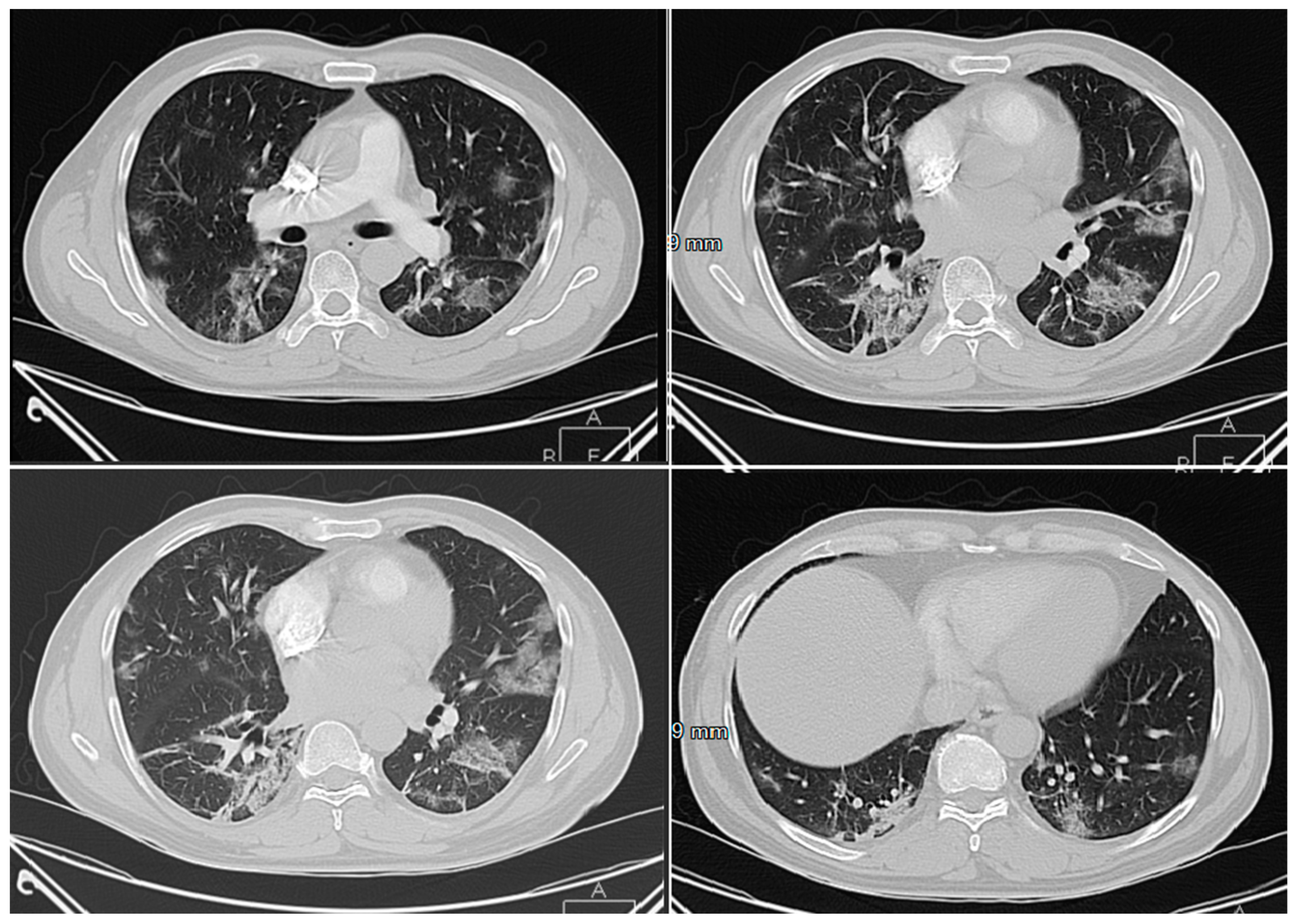
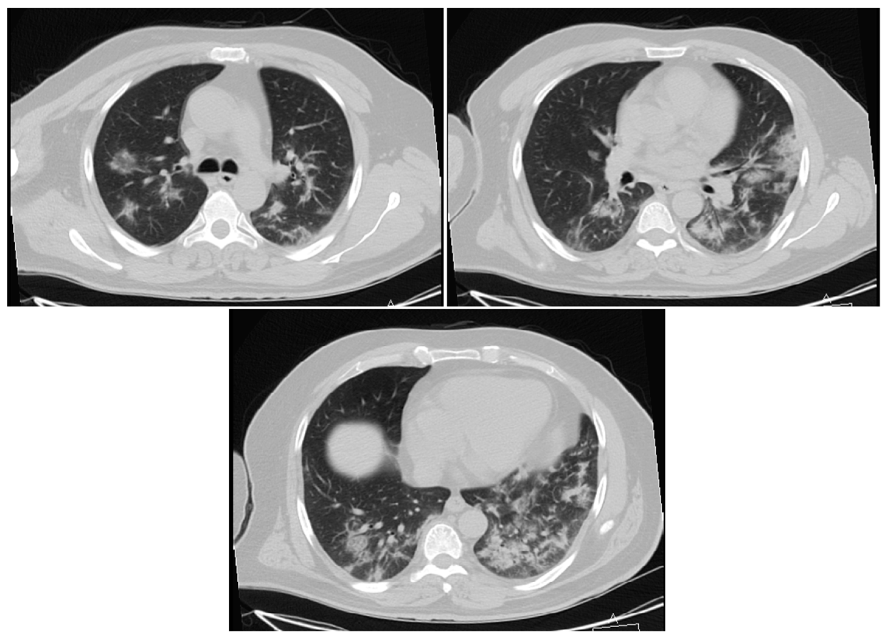
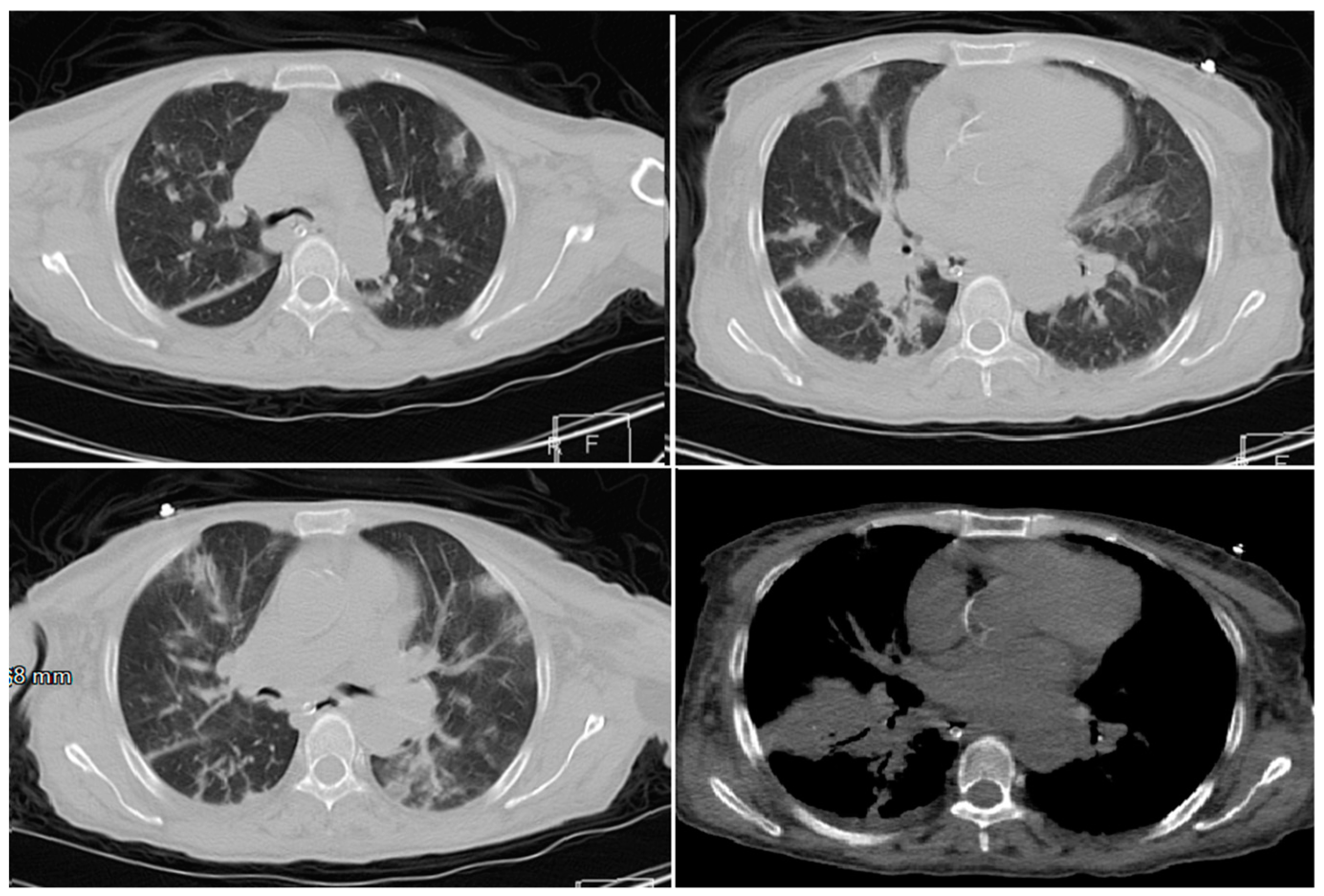
3.1. Respiratory Systems

Severe/acute respiratory syndrome (SARS), recent past communicable illness, mostly is an acute form of bronchopneumonia, which is instigated by new coronavirus—“SARS-CoV”. Researchers studying SARS-CoV, a group of enveloped non-segmented positive, single-strand RNA viruses (or (+)ssRNA) infecting vertebrates, have found that an acute and even life-threatening illness in humans belongs to a distinctive clades of the Sarbecovirus subgenus inside the Orthocoronavirinae subfamily [5]. Among the human-susceptible viruses, HCoV-NL63 or HCoV-229E (among α-CoVs), and HCoV-OC43 or HCoV-HKU1 (among β-CoVs) with low pathogenicity, can lead to gentle respiratory signs like the common cold. SARS/MERS-CoVs (two known β-CoVs) cause potentially fatal and/or severe breathing area infections; it was realized that the SARS-CoV-2 genome sequence was about a 96% match to a bat CoV RaTG13, while it shared about 79% sequence identity to SARS-CoV. It is obvious now that the recent SARS-CoV-2 can utilize ACE2, the same protein receptor as SARS-CoV, to infect people (Figure 5) [3,4,31]. SARS-CoV-2 is a (+)ssRNA CoVs, having around two-thirds of viral RNAs mostly placed in the first open reading frames 1a/b (ORF 1a/b), encoding 16 nonstructural protein (nsps). Host genetic factors can also be influencing the susceptibility to the illness or infection progression (Figure 5). COVID-19 is a serious threat/risk to the elderly people, and those with causal diseases (i.e., diabetes, hypertension, cardiovascular, or chronic obstructive pulmonary illness), and these groups are enormously prone to infection which may quickly turn into ARDS, cytokine storm, septic shock, coagulation dysfunction, metabolic acidosis, or even heading to demise (at least half of these patients) [4].
The lung is the first marked organ of SARS-CoV-2, which can cause potentially fatal respiratory tract infections. In general, deep sequencing techniques and diverse analysis of lower respiratory tract samples designated this as novel COVID-19 infection, which affect the higher respiratory tract (nose, throat, or sinuses) and/or lower respirational tract (windpipe or lungs). Human CoVs can generally cause other illnesses, namely bronchitis or pneumonia, and they may affect the gut. Besides, some patients revealed gastrointestinal symptoms, with vomiting (5.0%) and diarrhea (3.8%). Few patients with COVID-19 infection generally had eminent upper respiratory tract symptoms and/or signs (e.g., sneezing, sore throat, or rhinorrhea), representing that the marked cells of SARS-CoV-2 might locate in the lower airway tissues. Generally, people with COVID-19 infection have a fever and lower respiratory tract manifestations, with the estimated incubation time of 1–14 days (often 3–7 days) [31].
In this respect, a cohort study led by Prof. Huang et al. [31] appraised 41 laboratory- established cases, which revealed that the usual clinical indications comprised fever (98%), dry cough (76%), and myalgia/fatigue (44%), whereas a lesser amount of symptoms were sputum fabrication (28%), headache (8%), haemoptysis (5%), and diarrhea (3%). Altogether, 41 hospitalized patients had pneumonia with irregular respiratory outcomes on chest CT which is important in the diagnosis and/or treatment of lung illnesses. Radiologists generally have distinguished a COVID-19 infection from other viral pneumonia from CT scans of chest with high specificities but moderate sensitivities. Researchers reported 1099 laboratory-affirmed cases from 552 hospitals in mainland China by 29 January 2020 [36]. They found that the common clinical symptoms encompassed cough (67.8%), fever (88.7%), sputum production (33.4%), fatigue (38.1%), sore throat (13.9%), headache (13.6%), and shortness of breath (18.6%). Besides, some patients revealed gastrointestinal symptoms, with vomiting (5.0%) and diarrhea (3.8%). Importantly, cough or fever was the prevailing signs while gastrointestinal indications or upper respiratory symptoms were mostly rare, indicating the specific genetic variances in viral tropism/trend, when contrasted with influenza, SARS-, or MERS-CoVs [37,38,39].
ACE2, located in the person’s lower respiratory region, is well-known as an effective cell receptor for SARS-CoV and SARS-CoV-2, and can regulate both the cross-species and person/animal-to-person transmission. Viral structure and/or genome of SARS-CoV-2 must be considered to address its pathogenetic mechanisms. Commencing from the viral genome RNA, the production of polyproteins 1ab and/or 1a (pp1a, pp1ab) in the host is actualized, which formed a RTC (replication/transcription complex) in a double-membranous vesicle [40]. In this context, the significant genetic discoveries and the main etiological mechanisms of SARS-CoV can be described [41], and hence SARS-CoV-2 can be linked to the role of these nsps. However, many of the features have not still been defined. A series of the changes, cascade, and pathological events form the basis for pathological findings and clinical manifestations at diverse stages of SARS-CoV (Figure 6) [41].
Numerous SARS-related CoVs (SARSr-CoVs) are found in bats (their native pool hosts) [29]. Past investigations have demonstrated that some bat SARSr-CoVs can possibly infect humans. COVID-19 related to a pestilence of severe respirational syndrome in people from Wuhan, China, has been characterized and evaluated. Complete genome sequences were acquired from five patients at a beginning period of the outbreak which were almost alike and revealed 79.6% sequence uniqueness to SARS-CoV [29]. Besides, COVID-19 was found to be 96% indistinguishable to a bat coronavirus on the entire genome basis. Protein sequence evaluations of 7 preserved non-structural proteins areas showed that this viral infection has a place with the types of SARSr-CoV. Moreover, COVID-19 infection segregated from the bronchoalveolar lavage liquid of a fundamentally sick person could be neutralized by sera from a few patients. Outstandingly, it was affirmed that COVID-19 utilizes a similar receptor, angiotensin converting over protein II (ACE2), for cell entry as SARS-CoV [29].
The significant contrast among the Chinese 4th edition guidelines and WHO is the criterion in healing children with COVID-19 disease [42,43]. ARDS is a typical indication in the COVID-19 disease cases; SpO2 and respiration rate check turned out to be significant in the clinical exercise. Additionally, mechanical ventilation is a significant remedy for it, and is suggested in guidelines by both. The plateau pressure limitation, prone position, low tidal volume, and no oscillatory aeriation have been demonstrated as great degree of confirmation. The antiviral infection medications have been suggested in the Chinese fourth edition guideline; an ongoing distribution announced 99 cases in Jinyin-Tan emergency clinic were provided antiviral infection medications, for example, ganciclovir (intravenous, 0.25 g every 12 h,), oseltamivir (Orally, 75 mg every 12 h), and lopinavir and ritonavir tablets (Orally, 500 mg twice day by day), and the impact of the results has been referenced in detail [44]. The utilization of short-term corticosteroids has been suggested in the Chinese fourth version, with the aim to lessen the self-injury by over-responsive immune reaction; additionally, the antibiotic utilization is suggested when the individual has proof of bacterial infections. A few individuals among the 99 events introduced a quick advancement to ARDS, and the advantages of short-term utilization of methylprednisolone 1–2 mg/kg every day and intravenous immunoglobulin were noticed in these individuals [44]. Generally, these two guiding principles can be applied, but in any case, a portion of the substance are as yet questionable, particularly the advantages of steroids and antibiotics. Thus, further examinations ought to be ensued in the clinical practice. In one study, an open-labelled, randomized controlled trial evaluation has been conducted on 48 subjects from Chongqing Public Health Medical Center. Every qualified patient was allotted to an intervention group (methylprednisolone by means of intravenous (IV) injection at a portion of 1–2 mg/kg/day for three days) or a control group with no glucocorticoid intervention arbitrarily, in 1:1 proportion [45]. Individuals in the two gatherings were welcomed after 28 days follow-up that were scheduled at four sequential stay focuses; the clinical improvement rate has been utilized as the essential endpoint. Previously, parenteral and glucocorticoids have been administered to coronavirus-contaminated patients with severe respiratory symptoms; no conclusive indications in the works possibly promote the use of systemic glucocorticoids in truly sick patients with coronavirus-associated serious respirational sickness, or variant forms of extreme respiratory illnesses [45]. Overall, the suitability of adjunctive glucocorticoid treatment utilized in the COVID-19 tainted patient’s managements with severe lower respirational area infections is not well-defined and needs additional investigations.
According the WHO guidelines, the intravenous fluid treatments and oxygen are prescribed for both the mild and extreme instances with COVID-19 [46]; the objectives being SpO2  ≥  90% in non-pregnant adults or children, and SpO2  ≥  92–95% in patients with pregnancy. Powered aeriation has been suggested for the two groups (adults and children) with ARDS with the tide volume recommendation of 6 mL/kg. Adequate hypercapnia and ventilation (longer than 12 h) for every day is proposed for severe ARDS cases. The medications for septic shock and sepsis follow the 2016 universal rule of septic shock and sepsis. In the Chinese fourth release, antiviral medications are prescribed for use in the patients with COVID-19 infections; powered aeriation is prescribed for use in severe and mortal instances. Extracorporeal membrane oxygenation (ECMO) possibly may be utilized to diminish pneumonic disability through the cure time frame [46].

3.1.1. Cases Reports from Iran

The radiologic assessments of SARS-CoV-2 patients revealed extremely suggestive findings in lung high resolution computed tomography (HRCT) scan are bilateral multifocal or unilateral unifocal ground glass opacities, with peripheral or less likely peribronchovascular distribution are found. Additionally, findings which are inconsistent with COVID-19 pneumonia in lung HRCT are, tree-in-bud opacities, centrilobular nodules, predominantly peribronchovascular distribution, nodular opacities, reticular opacities, cavity, lymphadenopathy, and pleural effusion [47].

Case 1

A 38 years old man who worked at a radiology department was referred to the emergency room (ER) of the hospital with a few days of low-grade fever and dry coughs and no past history of underlying disease. The physical examination showed tachypnea, tachycardia, and fever, and the laboratory data revealed positive CRP (C reactive protein) and lymphopenia. Chests MDCT (multislice detector computed tomography) scan without contrast was performed (Figure 7) and PCR test for COVID-19 was positive.

Case 2

A 68 years old man was referred to ER with a history of 3 days high grade fever and dry cough with complaints of difficulty in breathing. In a clinical examination he had tachypnea and fever with pulse oximetry revealing 90% O2 saturation. Laboratory data showed positive CRP, lymphopenia, and increased blood glucose levels, and a chest MDCT scan was performed (Figure 8). PCR test for COVID-19 was found positive.

Case 3

A 53 years old man with no underlying disease was admitted to ER. He complained of dry coughs, myalgia, and fever, and was found to have tachypnea and fever on physical examination. CRP was positive in laboratory data with lymphopenia. Thorax MDCT scan was performed (Figure 9). PCR test for COVID-19 was positive.

Case 4

A 52 years old man was admitted at hospital with a history of fever, dyspnea, and coughs. He had tachypnea and fever. In lab data, positive CRP and lymphopenia were detected (Figure 10). PCR test for COVID-19 was positive.

Case 5

A 59 years old female was referred to ER with loss of consciousness, dyspnea, and fever. She had fever and increased respiratory rate on physical examination. Laboratory data showed positive CRP and lymphopenia (Figure 11). PCR test for COVID-19 was positive.
It is obvious from these cases that radiological evaluations and assessments are crucial for timely management and detection of SARS-CoV-2; the chest radiography is not sensitive for detecting ground-glass opacity, and probably shows normal results in the initial stage of viral infection [48,49], thus not optional as the first-line imaging modality for SARS-CoV-2. Additionally, thin slice chest CT can be applied successfully and efficiently for early detection of SARS-CoV-2 pneumonia [50]. Further, repeating the chest CT is very crucial for detection of lung abnormalities. Though, chest CT results are nonspecific for SARS-CoV-2 detection, CT evaluations have been suggested as an important indication of clinical diagnosis in Iran. Moreover, CT can be employed for detection of the severity of SARS-CoV-2 disease for clinical control and management. Clinical findings revealed that intensive care unit (ICU) patients on admittance frequently had bilateral multiple lobular and sub-segmental consolidation, while non-ICU patients had bilateral ground-glass opacities and sub-segmental consolidation [50]. In patients with severe symptoms, CT revealed diffuse heterogeneous consolidation with ground-glass opacities in bilateral lungs with air bronchial sign and bronchiectasis, showing as “white lung” while most lung lobes are distressed [48,49,50]. Thickening of interlobular septa and bilateral pleura with a small pleural effusion can be detected in these patients [48,49,50]. As a final point, characteristic chest CT imaging is extremely useful for SARS-CoV-2 pneumonia, while reverse-transcription polymerase chain reaction (RT-PCR) is believed as the reference benchmark.

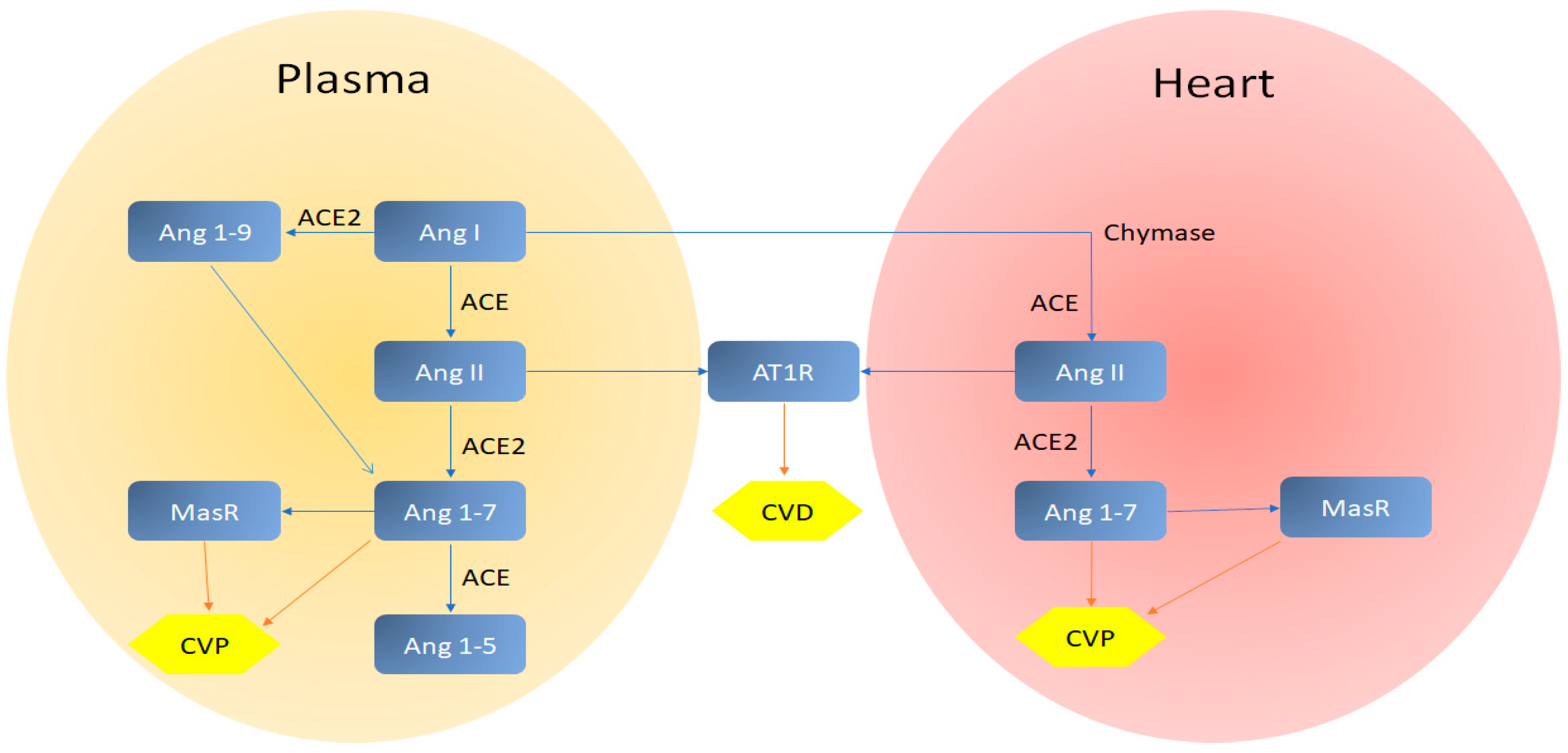
3.2. Cardiovascular System

ACE2 gene, which encodes the angiotensin-converting enzyme-2, has a major function in the immune and cardiovascular systems. It performs a task in the functioning of heart and the occurrence of diabetes mellitus and hypertension. ACE2 has the potential to be a host for CoVs such as SARS-CoV-2 and SARS-CoV wherein infection occurs by fastening of the spike protein (S-protein) from the virus to ACE2, abundantly expressed in the lungs and heart. Therefore, the heart is the second target organ of SARS-CoV-2 after the lungs [16,42,43,51,52,53].
According to a report by the National Health Commission of China (NHC), some patients diagnosed with COVID-19 first felt the cardiovascular symptoms like chest tightness and heart palpitations before respiratory symptoms, like fever and cough; 11.8% of people without underlying cardiovascular ailments (CVD) who died because of COVID-19 had considerable heart damage, with high levels of cardiac troponin I (cTnI) or cardiac arrest over hospitalization [53].
Although the most important appearance of COVID-19 is the respiratory indications, some patients have serious cardiovascular injury. Besides, certain patients might have a higher risk of death due to underlying CVDs; patients with SARS-CoV-2 infection and underlying CVD have an unfavorable prognosis [31,53]. The occurrence of the acute myocardial damage triggered by SARS-CoV-2 infection could be correlated to ACE2 which is broadly expressed in the lungs and cardiovascular system, thus implicating ACE2-related signaling routes being responsible for heart injury. Other anticipated mechanisms of myocardial injury may involve a cytokine storm caused by an imbalanced reaction by type 1 and 2 T helper cells and respiratory dysfunction and hypoxemia instigated by COVID-19 [53]. A study states that several ACE2 variants can reduce the association between S-protein and ACE2 in SARS-CoV or other types of CoVs. Thus, the expression level and pattern of ACE2 in various tissues can be critical for the symptoms, susceptibility, and outcome of the 2019-nCoV/SARS-CoV-2 infection [51]. Another study by Cao et al. [51] ascertains that there is no direct evidence to genetically prove the presence of coronavirus S-protein binding-resistant ACE2 mutants in various populations. However, the East Asian inhabitants have much higher allele frequency (AFs) in the eQTL variants associated with higher ACE2 expression in tissues that may afford diverse susceptibility or response to 2019-nCoV/SARS-CoV-2 among various inhabitants under the comparable circumstances. Nevertheless, the genetic information of different populations about the expression and function of ACE2 is still largely unknown [51].
Different types of CoVs have highly similar structures. Therefore, given the limited information about SARS-CoV-2 and even with the occurrence of amino acid mutations in the 2019-nCoV, this virus and its influences on the cardiovascular system can be highly predicted concerning previous variants of the virus family; MERS-CoV and SARS-CoV. A 12-year follow-up study of 25 patients with SARS-CoV infection revealed that the recovered patients were found to have hyperlipidemia (68%), cardiovascular system abnormalities (44%), and glucose metabolism disorders (60%). Because of the structural similarity of SARS-CoV and SARS-CoV-2, this novel virus may too come with chronic damage to the cardiovascular system [53,54]; the patients with underlying CVD are more likely to be infected by MERS-CoV. Amongst the individuals with acute symptoms and MERS-CoV infection, 50% had diabetes and hypertension, and up to 30% had heart disease. Pneumonitis Diagnosis and Treatment Program for New Coronavirus Infection (Trial Version 4) states that the SARS-CoV-2 infection is more probable to ensue in elderly people, especially those who suffer from diabetes, hypertension, or coronary heart disease. It is also proven that patients with underlying CVD present severe symptoms if infected by SARS-CoV-2 and also account for a considerable percentage of deaths. Another study represents that in COVID-19 patients with severe symptoms, 58% had hypertension, 44% had arrhythmia, and 25% had heart disease [13,31,53,55]. Although the data from previous types of CoVs may help, this does not seem to be enough and the studies about the direct and indirect influences of COVID-19 on the cardiovascular system need to be continued The comprehension of the injury inflicted by SARS-CoV-2 to the cardiovascular system and its mechanisms are of great significance to heal the patient and reduce the mortality.
COVID-19 can cause severe myocardial damage and long-lasting injury to the cardiovascular system, in addition to pneumonia. Although the specific mechanisms of myocardial injury caused by COVID-19 are uncertain, some thought should be given to cardiovascular protection in the cure of COVID-19. The increased secretion of ACE2 in patients with CVD may cause more severe respiratory symptoms compared to other patients [53]. While the counter-regulatory ACE2/Ang-(1–7)/MasR axis mediates cardiovascular protection (CVP), the ACE/Ang II/AT1R axis intercedes CVD (Figure 12) [56].
The utilization of inhibitors of renin-angiotensin-aldosterone system is capable of increasing the amount of ACE2. Since the ACE2 is an operational receptor for SARS-CoV-2, the influences of the utilization of ACE inhibitors or the blockers of angiotensin-receptor should be carefully considered for antihypertension therapy [53]. Comparing non-respiratory complications, MERS-CoV implicates the cardiovascular system more frequently than SARS-CoV. It has been reported that MERS-CoV is capable of instigating heart failure and acute myocarditis. Thus, it can be a concern for COVID-19 patients to develop serious cardiac difficulties such as myocarditis and heart failure. The heart not only show abnormal myocardial enzymes but also indicates structural and functional damage. Further, the immune injury may be the key cause of myocardial damage triggered by the virus infection. The outbreak of myocarditis may occur either early or never [5,16,57]. Myocarditis may represent many symptoms such as chest pain or mild dyspnea and cause heart failure with dilated cardiomyopathy as the main lasting sequela; often, myocarditis ensues from viral infections depicted by the myocarditis pathogenesis [58]. The common knowledge about the cellular and molecular pathogenesis of post-viral and autoimmune myocarditis is founded on the animal models, in which the advancement from severe damage to chronic dilated cardiomyopathy can be explained in 3-stagecourse. Severe injury leads to cardiac damage, revelation of intracellular antigens, namely cardiac myosin, followed by the activation of immune system. Robust inflammation ensues over week-long periods [58]. In predominant cases, the pathogen is removed, and the reaction is down-regulated with little sequelae. However, in some individuals when the virus is not removed it triggers insistent myocyte injury, and heart-exclusive inflammation may endure due to mistaken identification of pathogenic bodies representing endogenous heart antigens [58].
The incidence of cardiovascular symptoms during the progression of COVID-19 may be high, due to the immune system disorders and systemic inflammatory response [53]. Inflammatory levels indicators include interleukin-6 (IL-6), white blood cells (WBC), procalcitonin (PCT) C-reaction protein (CRP), neutrophils, and erythrocyte sedimentation rate (ESR). Increase in numbers of inflammatory cells (WBC and neutrophils) and inflammatory cytokines (IL-6 and CRP) triggered by the progression of COVID-19 can instigate high levels of heart injury. For example, the exceptionally elevated IL-6 level was strongly correlated to the vital signs of COVID-19 and the occurrence of RNAaemia. Consequently, it is important to offer a combination of antiviral and anti-inflammatory treatment, especially for patients with heart injury; baricitinib may play an adequate role. COVID-19 patients with signs of heart injury on reception must be characterized early and cautiously supported by cardiologists to maximally prevent heart injury-related death rate in COVID-19 [59,60,61]. The underlying acute coronary syndrome (ACS) can also worsen the symptoms for SARS-CoV-2 infected people and increase the mortality rate. ACS refers to a range of disorders congruent with acute myocardial ischemia or infarction which are typically because of a sudden decrease in coronary blood flow; it reduces the cardiac functional reserve owing to myocardial ischemia or necrosis. The infection of patients with pre-existing ACS increases the risk of cardiac insufficiency, leading to a sudden deterioration in their condition [53,62].
An indirect harm to the heart may also occur by hypoxemia caused by COVID-19 infection. There are pieces of evidence to prove that hypoxia plays a significant role in the cardiovascular risk development [63,64]. Moreover, COVID-19 patients represent low levels of lymphocyte, albumin, potassium, and eosinophil [65]. It is apparent that optimum levels of potassium ions are important for the functioning and structure of the myocardium [66]. Thus, the lack of some components including potassium in infected people may cause cardiovascular damages as an indirect effect of COVID-19 on the cardiovascular system. For patients with cardiac insufficiency, SARS-CoV-2 infection may serve as a hastening issue to deteriorate the state leading to demise. In addition, the risk of cardiac toxicity during the utilization of antiviral drugs is a concern as several antiviral drugs can begin cardiovascular disorders, arrhythmia, or other cardiac insufficiencies [67].

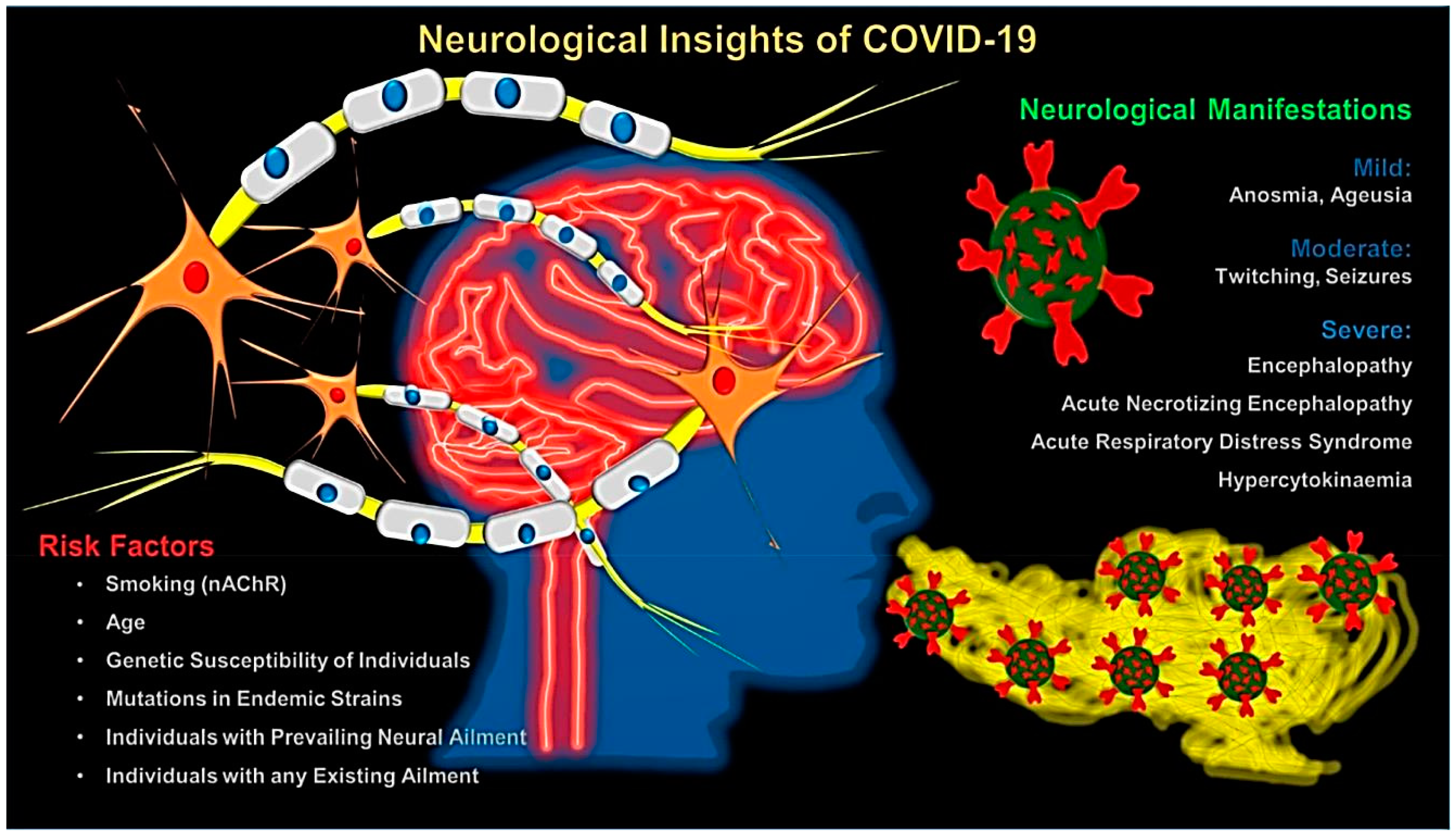
3.3. Central Nervous System (CNS)

Hyposmia and other neurological symptoms have been detected in patients with COVID-19 (Figure 13); patients with COVID-19 infection displayed mild (anosmia and ageusia) to severe (encephalopathy) neurological manifestations [68]. COVID-19 infection (like SARS-CoV) exploits ACE2 receptor to make entry into the cells; this discovery incites the interest of exploring the pronouncement of ACE2 in neurological tissue and deciding the conceivable neurological tissue damages to death inflicted by COIVD-19. The expression of ACE2 receptors by brains which have been recognized over glial cells and neurons makes them potential targets of SARS-CoV-2 [69]. Furthermore, the severe characteristic symptom of patients with this pathogenic virus is respiratory distress; a larger portion of the individuals admitted to the hospital could not breathe impulsively. Furthermore, neurologic signs (e.g., vomiting, headache, and nausea) have been observed in some infected patients; this evidence demonstrates that CoVs are not constantly restricted to the respiratory area and that they might similarly attack the central nervous system (CNS), prompting neurological ailments. The disease from SARS-CoV has been accounted in the brains (evaluated in animal models), where the brainstem was heartily contaminated. Besides, some CoVs have been shown to readily spread by means of a synapse-connected pathway to the medullary cardiorespiratory center from the chemoreceptors and mechanoreceptors in the lung and lower respirational aviation airways [70].

3.4. Gastrointestinal Tract (GI Tract)

Bowel abnormalities and cholestasis have been observed on abdominal imaging of inpatients with COVID-19; patients who went to laparotomy often had ischemia, probably because of small vessel thrombosis [71]. The study reported that 33% of inpatients with COVID-19 had abdominal imaging and 17% had cross-sectional imaging. Additionally, 54% of right upper quadrant ultrasounds showed findings of cholestasis with 31% of CTs exhibiting bowel wall abnormalities. Signs of late ischemia have been detected on 20% of CTs in ICU patients (2.7% of ICU patients), with pathologic correlation suggesting small vessel thrombosis [71].

3.5. COVID-19 and Pregnancy

Previous findings have revealed that SARS during pregnancy is linked to a significant occurrence of adverse maternal and neonatal problems, namely unprompted miscarriage, preterm delivery, application of endotracheal intubation, intrauterine growth restriction, admission to the intensive care unit, renal failure, and disseminated intravascular coagulopathy [72]. Pregnant women with COVID 19 infection have had fewer adverse maternal and neonatal problems so far, but consequences can be expected for those with SARS CoV 1 infection. The clinical features observed in pregnant women with established COVID 19 infection are comparable to those stated for non-pregnant adults with detected COVID 19 infection in the overall population, thus revealing a somewhat hopeful clinical course and consequences for COVID-19 when contrasted to SARS CoV 1 infection [72]. Based on the available data, it appears that pregnant people have the same risk as non-pregnant adults, but they have a higher risk of severe illness when infected with viruses. Thus, important cautions and guidelines should be considered, as still more studies are needed in this regard.

3.6. COVID-19 and Patients with Cancer

Some important challenges for patients with cancers in COVID-19 outbreak are their inability to receive essential medical services (hospitalization and typical medical cares); patients are also advised not to visit hospitals due to the serious infection risk, and this can cause treatment restrictions and consequent problems for patients with advanced cancers (such as tumor progression) [73]. Additionally, adverse effects among patients who receive immune checkpoint inhibitors (for example in severe myocarditis and pneumonitis) are very important and challenging to identify, and might not be cured rapidly, affecting their survival. Under these epidemic conditions, severe protection, online medical counselling, and suitable recognition and therapy of perilous cases are very critical for patients with cancers [73].

4. Therapeutic Options

The outbreak of COVID-19 infection is presently, as of March 2020, influencing the whole world, and many researchers and scientists are desperately aiming to find the suitable vaccine and therapeutic options. Active hexose correlated compound (AHCC) is α-glucan-based standardized mushroom extract which has been broadly explored as an immuno-stimulant for West Nile infection, herpes infection, hepatitis C infection, flu infection, avian flu infection, papillomavirus, hepatitis B infection, and HIV by advancing a controlled and defensive immune response [74]. Despite the fact that the adequacy of AHCC has not yet been explicitly assessed for SARS-CoV-2 sickness, its activity in elevating a defensive reaction to a wide scope of viral infections, and the absence of suitable vaccines, could bolster its utilization in the anticipation of maladies incited by human pathogenic coronavirus, including COVID-19 [74]. Additionally, the most encouraging compound is remdesivir (GS-5734), a prodrug nucleotide analog as of now in clinical trials for Ebola infection disease treatments; it repressed the replication of SARS-CoV and MERS-CoV in tissue cultures, and it showed adequacy in non-human animal models [75]. Additionally, a cocktail mix of the human immunodeficiency infection type 1 (HIV-1) protease inhibitors, lopinavir/ritonavir, and interferon beta (LPV/RTV-INFb) have shown some success in individuals contaminated with SARS-CoV; LPV/RTV-INFb likewise mended clinical parameters in mice and marmosets tainted with MERS-CoV. Astoundingly, the beneficial viability of remdesivir gave off an impression of being better than that of LPV/RTV-INFb against MERS-CoV in a transgenic adapted mice model [75]. The generally high death rates related with SARS- and MERS-CoVs, and SARS-CoV-2, recommend that pro-inflammatory responses may assume a significant role in the disease advancement. It is ambiguous whether the produced inflammatory state ought to be focused on; therapeutics that focus on the coronavirus alone probably will not have the option to turn around exceptionally pathogenic infections [75]. Martinez has reviewed the documented literature to provide a synopsis of therapeutic alternatives that have shown potential in battling SARS-CoV-2 diseases [75].
Recognizable proof of targets is significant for discriminating medications with elevated target explicitness or potentially revealing prevailing medications that could be repurposed to treat SARS-CoV-2 disease [65]. Two viral proteases, 3CLpro and PLpro, are responsible for cleaving the viral peptides into functional units for virus replication and packaging inside the host cells. In this way, drugs focus on these proteases in different viruses, namely HIV drugs (ritonavir and lopinavir), have been explored [76]. Additionally, RdRp is the RNA polymerase accountable for viral RNA synthesis that might be obstructed by existing antiviral medications or medication competitors (e.g., remdesivir) [76]. Possibly, the collaboration of viral S protein with its receptor ACE2 on host cells, and ensuing viral endocytosis into the cells, may likewise be a reasonable drug objective. As an example, the broad-range antiviral medication Arbidol, which works as a virus–host cell fusion inhibitor to prevent viral entry into host cells against flu virus, has gone into a clinical trial for SARS-CoV-2 treatment [65,77]. The protease TMPRSS2 delivered by the host cells assumes a significant function in proteolytic dispensation of S protein grooming to the receptor ACE2 attachment in human cells [78]. It was demonstrated that camostat mesylate, a clinically affirmed TMPRSS2 inhibitor, had the option to block SARS-CoV-2 passage to human cells, showing its prospective as a medication for COVID-19 [65,78]. Significant proteins and their capabilities during the viral infections are summarized in Figure 14 [65,67,77,79,80]. Furthermore, an appealing medication focus among CoVs is the main protease (Mpro, 3CLpro), because of its basic role in preparing the polyproteins translated from the viral RNA. Importantly, the lead compound was developed into an effective inhibitor of the SARS-CoV-2 Mpro, and pharmacokinetic evaluations of the optimized inhibitor showed a pronounced lung tropism and aptness for inhalation method of dispensation [81].
Ongoing investigations have pointed out the conceivable advantage of chloroquine, a widely used antimalarial drug, for treating infected patients by the new emerging coronavirus. Established researchers ought to think about this available data in view of the past investigations with chloroquine in the field of antiviral research [84]. The choice of utilizing chloroquine in the treatment of SARS-CoV-2 should be inspected with guarded considerations, and further evaluations are needed to affirm the adequacy of the proposed treatment and to control acute viral diseases. In one study, after evaluations of hydroxychloroquine and azithromycin as a treatment for COVID-19 in an open-label non-randomized clinical trial, it was revealed that hydroxychloroquine treatment was remarkably related to viral load reduction in SARS-CoV-2 patients and its influence was strengthened by azithromycin [85]. In terms of underlying mechanistic aspects for chloroquine, Devaux et al. [86] suggested that this drug can interfere with ACE2 receptor glycosylation, and therefore prevent SARS-CoV-2 binding to target cells. In another investigation, Wang and Cheng revealed that MERS- and SARS-CoVs upregulated the ACE2 expression in lung tissue, which can increase their replications and spread [87]. Additionally, the possibility of targeting sialic acids on specific cell subtypes by SARS-CoV-2 (like other CoVs), the interaction can be influenced by chloroquine treatment [88]. Further, SARS-CoV-2 molecular crosstalk with its target cell might be changed by chloroquine via the inhibition of kinases (e.g., mitogen-activated protein kinase). On the other hand, chloroquine interfered with proteolytic processing of the M protein and thus altered virion assembly and budding. Lastly, for SARS-CoV-2, chloroquine indirectly acted by decreasing the formation of pro-inflammatory cytokines and/or by triggering anti-SARS-CoV-2 CD8+ T-cells [86]. Although chloroquine can shorten the time of hospital stay and help to reduce the evolution of COVID-19 pneumonia, the side effects of this drug, especially on the cardiovascular system, should be carefully considered, as it can cause cardiac complications, such as conduction disorders, myocardial hypertrophy, and restrictive cardiomyopathy as a rare but severe adverse event leading to demise. Some studies declare that hydroxychloroquine plays a vascular protective function in the context of inflammatory ailments as a safe and well-tolerated drug, but a long-term hydroxychloroquine administration may cause serious cardiac disorders. Therefore, the monitoring of patients who use chloroquine/hydroxychloroquine drugs appears to be vital. A study by Keating et al. [89] proved a cardiotoxicity induced by hydroxychloroquine in a woman (39 years old) with systolic dysfunction and systemic lupus erythematosus (SLE). Diastolic apical 4-chamber sight of patient’s transthoracic echocardiogram proves diffuse thickening of ventricular septum; contrarily, an echocardiogram executed 1 year before depicted normal function of cardiac valvular, a left ventricular ejection fraction of 55%, and modest wall thickening (16-mm septum) [89,90,91,92]. Additionally, the biopsy of tissue by light microscopic examination disclosed diffusely inflated vacuolated myocytes [89]. Echocardiographic results of diffusely stiffened ventricular walls and globally reduced systolic function, caused by chloroquine or hydroxychloroquine, should be a concern as it shows the cardiotoxicity. An endo-myocardial biopsy is necessary to establish the diagnosis. If cardiotoxicity is established, hydroxychloroquine (or related substances) should be stopped [89].
It was shown that chloroquine is capable of inhibiting broadly the endocytosis of nanoparticles through resident macrophages. Thus, this drug can reduce the synthetic nanoparticles accumulation (from various sizes and morphologies) in cell lines and the mononuclear phagocyte system of mice. Additionally, it was disclosed that chloroquine decreased the expression of phosphatidylinositol binding clathrin assembly protein, one of the three most plentiful proteins in clathrin-coated pits. Recently, Hu et al. [93] suggested the potential influences of chloroquine against SARS-CoV-2 falls (with a size of 60–140 nm, and spherical in shape). They mentioned that this drug can generally reduce the capability of cells for clathrin-mediated endocytosis of nanostructures, because of phosphatidylinositol binding clathrin assembly protein suppression [93]. It should be noted that additional evaluations are needed to verify the early discoveries of chloroquine activities in COVID-19 patients, and its prophylactic and/or therapeutic clinical applications with comprehensive analyses regarding viral disease stage and needed doses. Further, comparison investigations are needed regarding the effects of hydroxychloroquine and chloroquine and their efficacy against SARS-CoV-2. Additionally, preclinical evaluations should be accomplished for better determination of their anti-SARS-CoV-2 mechanistic aspects, including endocytosis suppression in the host cells. For instance, in one study, it was revealed that hydroxychloroquine (EC50 = 0.72 μM) had additional potency over chloroquine (EC50 = 5.47 μM) in vitro. Based on physiologically based pharmacokinetic (PBPK) modeling findings, a loading dose of 400 mg twice daily of hydroxychloroquine sulfate was given orally, followed by a maintenance dose of 200 mg given twice daily for four days can be suggested for SARS-CoV-2 infection, as it reached three times the potency of chloroquine phosphate when given 500 mg twice daily five days prior [94]. In another study, Liu et al. [95] showed that hydroxychloroquine can have inhibitory results on SARS-CoV-2 infection in vitro, and predicted that in combination with its anti-inflammatory function, this drug can have suitable antiviral potentials; it still needs additional clinical trials. It should be noticed that hydroxychloroquine is less toxic than chloroquine, but overdose and continued utilization can reason to possible side effects [95].
Small-molecule entities accepted for other human ailments may control the virus-host interaction of new coronavirus. Some important antiviral compounds that may possibly be suggested for COVID-19 inhibition are summarized in Table 1 [65,77]. Additionally, Harrison has summarized some selected repurposed drugs in clinical development for COVID-19 treatment [96]; NIH, U.S National Library of Medicine, https://clinicaltrials.gov is available for additional search and updates in this context.

New Insights from Herbal Medicines

Newer findings have shown that the garlic essential oil may be an invaluable natural antivirus option, which provides preventive effects for the attack of CoVs on the human body, although more investigations are required [101]. A molecular docking method has affirmed the inhibitory influence of the organosulfur compounds present in the garlic essential oil on the host receptor ACE2 protein in the human body. This is an important discovery about coronavirus resistance of individual garlic compounds on the main protease (PDB6LU7) protein of SARS-CoV-2; seventeen organosulfur compounds, comprising 99.4% constituents of the garlic essential oil, had remarkable interactions with the amino acids of the ACE2 protein and the main protease PDB6LU7 of SARS-CoV-2. Interestingly, the significant anti-coronavirus effect was displayed by allyl disulfide and allyl trisulfide, the prominent components in the garlic essential oil (51.3%) Importantly, the docking evaluations have pointed out that there are synergistic interactions of these seventeen constituents with favorable inhibitory effects on the PDB6LU7 and ACE2 proteins [101].
Recent investigation has revealed that glycyrrhizin (a triterpene saponin) may be a potential therapeutic option for COVID-19, due to its valuable pharmacological effects, such as downregulating pro-inflammatory cytokines, binding ACE2, obstructing the intracellular reactive oxygen species (ROS) accumulation, thrombin inhibition, provoking endogenous interferon, and hindering the extra formation of airway exudates [102]. However, clinical evaluations and comprehensive assessments should be carefully designed.

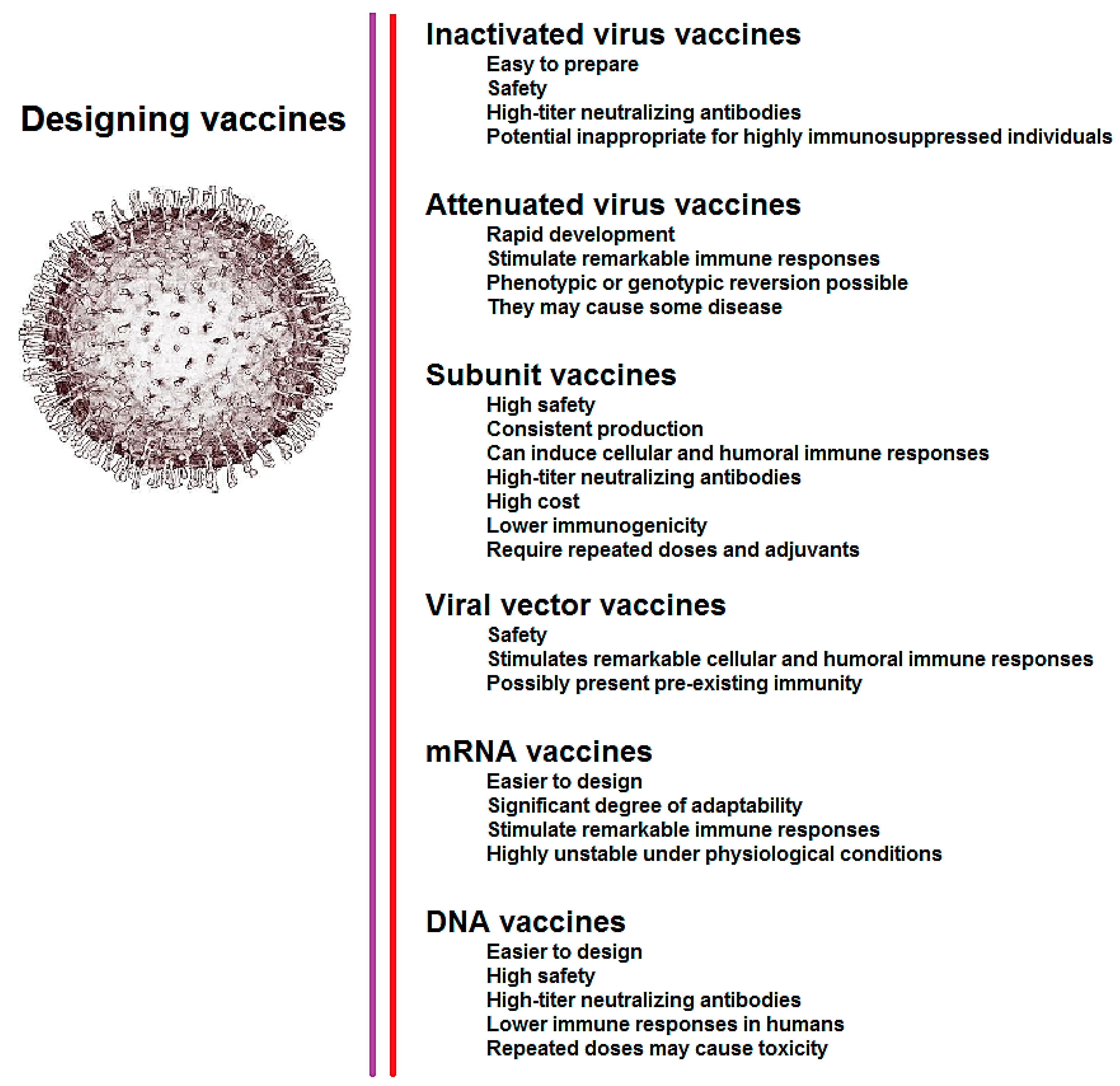
5. Design of Vaccine for COVID-19

Vaccines are imperative for controlling the COVID-19 pandemic, wiping out its proliferation, and at last forestalling its future reappearance. On this front, researchers have evaluated the vaccine patents related to MERS- and SARS-CoVs and to apply recently advancements that may help to design vaccines for SARS-CoV-2; altogether, around 190 patents have been identified regarding SARS and MERS vaccines. The advancements in designing attenuated-virus vaccines should be explored by screening the serially propagated SARS-CoV-2 with reduced pathogenesis, including reduced inadequate neutrophil influx, stimulated minimal lung injury, and accelerated anti-inflammatory cytokine expressions compared with the wild-type virus. The techniques shrouded in patents, which are likewise being utilized for designing COVID-19 vaccines, are mentioned in Figure 15, with their advantages and disadvantages. Importantly, innovative strategies for vaccines, based on the putative protective antigen/peptides originated from SARS-CoV-2, ought to be addressed [103,104]. Indeed, progress is being made at breakneck speed in planning new vaccines, immunizations, and therapeutics to counter COVID-19, as made earlier for MERS- and SARS-CoVs, which could empower endeavors to end this developing viral infection disease. Different systems are contributing to the improvement of CoV vaccines; the greater part of this target being the surface-uncovered spike (S) glycoprotein or S protein as the significant inducer of neutralizing antibodies. A few S-protein-based methodologies have been endeavored for creating CoV immunizations, e.g., the utilization of full-length S protein or expression in virus-like particles (VLP), S1-receptor-binding domain (RBD), and viral vectors or DNA [70,105,106].
Analysts are intently scrutinizing the compelling and reasonable vaccine candidates for the control and prevention of COVID-19 (Table 2). The draft landscape of COVID-19 candidate vaccines (until 15 May 2020) can be found in the following site from WHO documents detail: https://www.who.int/who-documents-detail/draft-landscape-of-covid-19-candidate-vaccines. Outcomes attained from the in vitro examination against COVID-19 are assuring as the medications, remdesivir, and chloroquine were seen as exceptionally powerful in limiting the disease. Clinical trials can be organized among the patients tainted with COVID-19 as these medications are already being used for combating different sicknesses and have entrenched good profiles, thus rendering further assessment of such medications much simpler. S-protein is viewed as a key viral antigen for creating CoV antibodies, as appeared in a few preclinical investigations. Despite hurdles, the concerted efforts are progressing well to improve COVID-19 control or prevention, and treatment. The archived clinical information on various remedial methodologies for CoVs is not much; further research ought to be coordinated toward the investigation of SARS-CoV-2 in reasonable animal models for examining transmission, pathogenesis, and replication [70].

6. Current Challenges and Future Perspectives

The big question is how effectually the 2019-nCoV will spread and how lethal will be the infection? Right now, we just have educated theories, which are probably going to cement in the coming months. Be that as it may, what we distinguish up until this point can reveal some insight into the attributes of the infection and provide a few guidelines on managing this rapidly developing pandemic. Numerous groups considering the viral infection around the globe are examining the situation and are engaged in applying findings acquired from their individual models. Indeed, morbidity shifts in various situations/environments are contingent upon factors like population density, temperature, and infection susceptibility in a populace; thus, increasingly dependable and precise R0 worth ought to show up as more data becomes available in public domain. The expansion in accessibility of PCR evaluation kits and additional medical assets would bring about higher affirmation rate than the factual worth [46]. Importantly, recovered cases with COVID-19 should be further evaluated (especially with RT-PCR test), as “turn positive” of nucleic acid detection by RT-PCR evaluation for COVID-19 was detected after two repeated negative outcomes from these patients, probably because of the false negative of RT-PCR evaluation and protracted nucleic acid change [109].
CT characteristic of SARS-CoV-2 pneumonia contain multifocal bilateral ground-glass opacities with patchy consolidations, prominent peripherally sub-pleural distribution, and favored posterior parts or lower lobe predilection. In this regard, thin slice chest CT can assist early diagnosis, pointer for clinical conclusion, and evaluate/diagnosis disease progression, thus playing a crucial function in initial management, prevention, and control of this pathogenic viral infection [48,49,50]. Further, as indicated by accessible guidelines, the analysis of SARS-CoV-2 ought to be affirmed by RT-PCR or gene sequencing of respiratory or blood examples. In any case, the RT-PCR has an identification rate as low as 30–60%, at preliminary presentation with various constraints and restrictions [10]. Consequently, this deferral in diagnosis and treatment creates a higher risk of infections in a bigger populace. Accordingly, finding a methodology for the early COVID-19 diagnosis is of utmost significance.
Indeed, the antiviral efficacy of therapeutic compounds should be evaluated in comprehensively clinical trials. It is worth mentioning that numerous endeavors have focused on finding host-targeted small molecules against viral infections, but only a handful have attained successful approval by the FDA, as exemplified, for HIV treatment and prevention [110]. Additionally, this pathogenic viral infection challenges the industrial companies and businesses around the world, and the most pressing question is whether clinics and medications in countries will be adequately prepared to handle these deadly viruses. Indeed, many COVID-19 related mitigation and prevention efforts are being undertaken by humans and businesses; these are necessary and very critical but challenging efforts.

7. Conclusions

Unknown infectious illnesses, such as RNA viruses subject to genetic recombination and mutations, and cross-species transmission, will keep on presenting a genuine worldwide health risk, as exemplified by SARS-CoV-2. In spite of two previous significant coronavirus diseases outbreaks caused by the SARS and MERS respiratory sicknesses, the world remains unprepared to adequately deal with the existing SARS-CoV-2 outbreak, as attested by the fact that SARS-CoV-2 has brought about a huge number of deaths around the globe. In this regard, we should be avoiding any superstitions, unscientific beliefs, and folkloric suggestions that may cause more disasters and further spread of the virus. Currently, investigations pertaining to the production of vaccines ought to be centered on anticipation/prevention to reduce the spread of the virus; clinical trials evaluations are still in the early stages. The big question is that how to stimulate the immune response with enough efficiency and safety; it needs to trigger the formation of antibodies blocking viral proteins or prompt body to make T cells recognizing and killing the infected cells. Therapeutic options and approaches for SARS-CoV-2 and their related efficiency should be further evaluated from their mechanistic aspects, metabolic stability, possible serious adverse effects, and important pharmacokinetics and pharmacodynamics issues.

Author Contributions

All authors contributed to writing of original draft. G.J.S., M.N., S.I., and R.S.V. conceptualization, writing—review and editing. All authors have read and agreed to the published version of the manuscript.

Funding

The study was self-funded.

Conflicts of Interest

There is no conflict of interest.

Financial Disclosure

There is no financial disclosure.

Informed Consent

The authors certify that they have obtained all appropriate patient consent forms. The patients understand that their names and initials will not be published.

References

  1. Munster, V.J.; Koopmans, M.; van Doremalen, N.; van Riel, D.; de Wit, E. A novel coronavirus emerging in China—Key questions for impact assessment. N. Engl. J. Med. 2020, 382, 692–694. [Google Scholar] [CrossRef] [PubMed]
  2. Lu, H.; Stratton, C.W.; Tang, Y.W. Outbreak of Pneumonia of Unknown Etiology in Wuhan China: The Mystery and the Miracle. J. Med. Virol. 2020, 92, 401–402. [Google Scholar] [CrossRef] [PubMed] [Green Version]
  3. Zhang, Y.-Z.; Holmes, E.C. A Genomic Perspective on the Origin and Emergence of SARS-CoV-2. Cell 2020, 181, 223–227. [Google Scholar] [CrossRef] [PubMed]
  4. Guo, Y.-R.; Cao, Q.-D.; Hong, Z.-S.; Tan, Y.-Y.; Chen, S.-D.; Jin, H.-J.; Tan, K.-S.; Wang, D.-Y.; Yan, Y. The origin, transmission and clinical therapies on coronavirus disease 2019 (COVID-19) outbreak–An update on the status. Mil. Med. Res. 2020, 7, 1–10. [Google Scholar] [CrossRef] [PubMed] [Green Version]
  5. Yin, Y.; Wunderink, R.G. MERS, SARS and other coronaviruses as causes of pneumonia. Respirology 2018, 23, 130–137. [Google Scholar] [CrossRef] [Green Version]
  6. Carlos, W.G.; Dela Cruz, C.S.; Cao, B.; Pasnick, S.; Jamil, S. Novel Wuhan (2019-nCoV) Coronavirus. Am. J. Respir. Crit. Care Med. 2020, 201, 7–8. [Google Scholar] [CrossRef]
  7. Zhang, T.; Wu, Q.; Zhang, Z. Probable pangolin origin of SARS-CoV-2 associated with the COVID-19 outbreak. Curr. Biol. 2020, 30, 1346–1351. [Google Scholar] [CrossRef] [PubMed]
  8. Abd El-Aziz, T.M.; Stockand, J.D. Recent progress and challenges in drug development against COVID-19 coronavirus (SARS-CoV-2) - an update on the status. Infect. Genet. Evol. 2020, 83, 104327. [Google Scholar] [CrossRef]
  9. Tang, X.; Wu, C.; Li, X.; Song, Y.; Yao, X.; Wu, X.; Duan, Y.; Zhang, H.; Wang, Y.; Qian, Z.; et al. On the origin and continuing evolution of SARS-CoV-2. Natl. Sci. Rev. 2020, nwaa036. [Google Scholar] [CrossRef] [Green Version]
  10. Zhu, N.; Zhang, D.; Wang, W.; Li, X.; Yang, B.; Song, J.; Zhao, X.; Huang, B.; Shi, W.; Lu, R. A novel coronavirus from patients with pneumonia in China, 2019. N. Engl. J. Med. 2020, 382, 727–733. [Google Scholar] [CrossRef]
  11. Wang, L.; Wang, Y.; Ye, D.; Liu, Q. Review of the 2019 novel coronavirus (SARS-CoV-2) based on current evidence. N. Engl. J. Med. 2020. [Google Scholar] [CrossRef]
  12. Menni, C.; Valdes, A.M.; Freidin, M.B.; Ganesh, S.; Moustafa, J.S.E.-S.; Visconti, A.; Hysi, P.; Bowyer, R.C.E.; Mangino, M.; Falchi, M.; et al. Loss of smell and taste in combination with other symptoms is a strong predictor of COVID-19 infection. medRxiv 2020. [Google Scholar] [CrossRef]
  13. Chan, J.F.-W.; Yuan, S.; Kok, K.-H.; To, K.K.-W.; Chu, H.; Yang, J.; Xing, F.; Liu, J.; Yip, C.C.-Y.; Poon, R.W.-S. A familial cluster of pneumonia associated with the 2019 novel coronavirus indicating person-to-person transmission: A study of a family cluster. Lancet 2020, 395, 514–523. [Google Scholar] [CrossRef] [Green Version]
  14. Sungnak, W.; Huang, N.; Bécavin, C.; Berg, M.; Queen, R.; Litvinukova, M.; Talavera-López, C.; Maatz, H.; Reichart, D.; Sampaziotis, F.; et al. SARS-CoV-2 entry factors are highly expressed in nasal epithelial cells together with innate immune genes. Nat. Med. 2020. [Google Scholar] [CrossRef] [PubMed] [Green Version]
  15. Li, X.; Geng, M.; Peng, Y.; Meng, L.; Lu, S. Molecular immune pathogenesis and diagnosis of COVID-19. J. Pharm. Anal. 2020, 10, 102–108. [Google Scholar] [CrossRef] [PubMed]
  16. Zeng, J.H.; Liu, Y.-X.; Yuan, J.; Wang, F.-X.; Wu, W.-B.; Li, J.-X.; Wang, L.-F.; Gao, H.; Wang, Y.; Dong, C.-F. First Case of COVID-19 Infection with Fulminant Myocarditis Complication: Case Report and Insights. Int. J. Antimicrob. Agents 2020. [Google Scholar] [CrossRef]
  17. Baud, D.; Qi, X.; Nielsen-Saines, K.; Musso, D.; Pomar, L.; Favre, G. Real estimates of mortality following COVID-19 infection. Lancet Infect. Dis. 2020. [Google Scholar] [CrossRef] [Green Version]
  18. Tahir ul Qamar, M.; Alqahtani, S.M.; Alamri, M.A.; Chen, L.-L. Structural basis of SARS-CoV-2 3CLpro and anti-COVID-19 drug discovery from medicinal plants. J. Pharm. Anal. 2020, 55, 105924. [Google Scholar] [CrossRef] [PubMed]
  19. Glass, W.G.; Subbarao, K.; Murphy, B.; Murphy, P.M. Mechanisms of host defense following severe acute respiratory syndrome-coronavirus (SARS-CoV) pulmonary infection of mice. J. Immunol. 2004, 173, 4030–4039. [Google Scholar] [CrossRef] [PubMed] [Green Version]
  20. Tang, Q.; Song, Y.; Shi, M.; Cheng, Y.; Zhang, W.; Xia, X.-Q. Inferring the hosts of coronavirus using dual statistical models based on nucleotide composition. Sci. Rep. 2015, 5, 17155. [Google Scholar] [CrossRef]
  21. Woo, P.C.Y.; Huang, Y.; Lau, S.K.P.; Yuen, K.-Y. Coronavirus genomics and bioinformatics analysis. Viruses 2010, 2, 1804–1820. [Google Scholar] [CrossRef] [PubMed] [Green Version]
  22. Peiris, J.S.M.; Lai, S.T.; Poon, L.L.M.; Guan, Y.; Yam, L.Y.C.; Lim, W.; Nicholls, J.; Yee, W.K.S.; Yan, W.W.; Cheung, M.T. Coronavirus as a possible cause of severe acute respiratory syndrome. Lancet 2003, 361, 1319–1325. [Google Scholar] [CrossRef] [Green Version]
  23. Ahmed, T.; Noman, M.; Almatroudi, A.; Shahid, M.; Khurshid, M.; Tariq, F.; ul Qamar, M.T.; Yu, R.; Li, B. Coronavirus Disease 2019 Assosiated Pneumonia in China: Current 23 Status and Future Prospects. 2020; Preprints. [Google Scholar]
  24. ICD-11 (World Health Organization, 2018). Available online: https://www.who.int/classifications/icd/en/ (accessed on 3 February 2020).
  25. Gorbalenya, A.E.; Baker, S.C.; baric, R.S.; de Groot, R.J.; Drosten, C.; Gulyaeva, A.A.; Haagmans, B.I.; Iauber, C.; Ieontovich, A.M.; Neuman, B.W.; et al. The species Severe acute respiratory syndrome-related coronavirus: Classifying 2019-nCoV and naming it SARS-CoV-2. Nat. Microbiol. 2020, 5. [Google Scholar] [CrossRef]
  26. Li, F. Structure, function, and evolution of coronavirus spike proteins. Annu. Rev. Virol. 2016, 3, 237–261. [Google Scholar] [CrossRef] [PubMed] [Green Version]
  27. Raj, V.S.; Mou, H.; Smits, S.L.; Dekkers, D.H.W.; Müller, M.A.; Dijkman, R.; Muth, D.; Demmers, J.A.A.; Zaki, A.; Fouchier, R.A.M. Dipeptidyl peptidase 4 is a functional receptor for the emerging human coronavirus-EMC. Nature 2013, 495, 251–254. [Google Scholar] [CrossRef] [PubMed] [Green Version]
  28. Letko, M.; Marzi, A.; Munster, V. Functional assessment of cell entry and receptor usage for SARS-CoV-2 and other lineage B betacoronaviruses. Nat. Microbiol. 2020, 5, 1–8. [Google Scholar] [CrossRef] [PubMed] [Green Version]
  29. Zhou, P.; Yang, X.-L.; Wang, X.-G.; Hu, B.; Zhang, L.; Zhang, W. A pneumonia outbreak associated with a new coronavirus of probable bat origin. Nature 2020, 579, 270–273. [Google Scholar] [CrossRef] [Green Version]
  30. Li, Q.; Guan, X.; Wu, P.; Wang, X.; Zhou, L.; Tong, Y.; Ren, R.; Leung, K.S.M.; Lau, E.H.Y.; Wong, J.Y. Early transmission dynamics in Wuhan, China, of novel coronavirus–infected pneumonia. N. Engl. J. Med. 2020, 382, 1199–1207. [Google Scholar] [CrossRef]
  31. Huang, C.; Wang, Y.; Li, X.; Ren, L.; Zhao, J.; Hu, Y.; Zhang, L.; Fan, G.; Xu, J.; Gu, X. Clinical features of patients infected with 2019 novel coronavirus in Wuhan, China. Lancet 2020, 395, 497–506. [Google Scholar] [CrossRef] [Green Version]
  32. Cui, J.; Li, F.; Shi, Z.L. Origin and evolution of pathogenic coronaviruses. Nat. Rev. Microbiol. 2019, 17, 181–192. [Google Scholar] [CrossRef] [Green Version]
  33. Xu, X.; Chen, P.; Wang, J.; Feng, J.; Zhou, H.; Li, X.; Zhong, W.; Hao, P. Evolution of the novel coronavirus from the ongoing Wuhan outbreak and modeling of its spike protein for risk of human transmission. Sci. China Life Sci. 2020, 63, 457–460. [Google Scholar] [CrossRef] [PubMed] [Green Version]
  34. Wu, K.; Li, W.; Peng, G.; Li, F. Crystal structure of NL63 respiratory coronavirus receptor-binding domain complexed with its human receptor. Proc. Natl. Acad. Sci. USA 2009, 106, 19970–19974. [Google Scholar] [CrossRef] [PubMed] [Green Version]
  35. Zhao, Y.; Zhao, Z.; Wang, Y.; Zhou, Y.; Ma, Y.; Zuo, W. Single-cell RNA expression profiling of ACE2, the putative receptor of Wuhan 2019-nCov. bioRxiv Prepr. 2020. [Google Scholar] [CrossRef]
  36. Guan, W.-J.; Ni, Z.-Y.; Hu, Y.; Liang, W.-H.; Ou, C.-Q.; He, J.-X.; Liu, L.; Shan, H.; Lei, C.-L.; Hui, D.S.C. Clinical characteristics of coronavirus disease 2019 in China. N. Engl. J. Med. 2020, 382, 1708–1720. [Google Scholar] [CrossRef] [PubMed]
  37. Wang, H.; Xiao, X.; Lu, J.; Chen, Z.; Li, K.; Liu, H.; Luo, L.; Wang, M.; Yang, Z. Factors associated with clinical outcome in 25 patients with avian influenza A (H7N9) infection in Guangzhou, China. BMC Infect. Dis. 2016, 16, 534. [Google Scholar] [CrossRef] [Green Version]
  38. Lee, N.; Hui, D.; Wu, A.; Chan, P.; Cameron, P.; Joynt, G.M.; Ahuja, A.; Yung, M.Y.; Leung, C.B.; To, K.F. A major outbreak of severe acute respiratory syndrome in Hong Kong. N. Engl. J. Med. 2003, 348, 1986–1994. [Google Scholar] [CrossRef]
  39. Assiri, A.; Al-Tawfiq, J.A.; Al-Rabeeah, A.A.; Al-Rabiah, F.A.; Al-Hajjar, S.; Al-Barrak, A.; Flemban, H.; Al-Nassir, W.N.; Balkhy, H.H.; Al-Hakeem, R.F. Epidemiological, demographic, and clinical characteristics of 47 cases of Middle East respiratory syndrome coronavirus disease from Saudi Arabia: A descriptive study. Lancet Infect. Dis. 2013, 13, 752–761. [Google Scholar] [CrossRef] [Green Version]
  40. Sawicki, S.G.; Sawicki, D.L. Coronavirus transcription: A perspective. In Coronavirus Replication and Reverse Genetics; Springer: Berlin/Heidelberg, Germany, 2005; pp. 31–55. [Google Scholar]
  41. Gu, J.; Korteweg, C. Pathology and pathogenesis of severe acute respiratory syndrome. Am. J. Pathol. 2007, 170, 1136–1147. [Google Scholar] [CrossRef] [Green Version]
  42. Li, W.; Moore, M.J.; Vasilieva, N.; Sui, J.; Wong, S.K.; Berne, M.A.; Somasundaran, M.; Sullivan, J.L.; Luzuriaga, K.; Greenough, T.C. Angiotensin-converting enzyme 2 is a functional receptor for the SARS coronavirus. Nature 2003, 426, 450–454. [Google Scholar] [CrossRef] [Green Version]
  43. Lu, R.; Zhao, X.; Li, J.; Niu, P.; Yang, B.; Wu, H.; Wang, W.; Song, H.; Huang, B.; Zhu, N. Genomic characterisation and epidemiology of 2019 novel coronavirus: Implications for virus origins and receptor binding. Lancet 2020, 395, 565–574. [Google Scholar] [CrossRef] [Green Version]
  44. Chen, N.; Zhou, M.; Dong, X.; Qu, J.; Gong, F. Epidemiological and clinical characteristics of 99 cases of 2019 novel coronavirus pneumonia in Wuhan, China: A descriptive study. Lancet 2020, 395, 507–513. [Google Scholar] [PubMed] [Green Version]
  45. Zhou, Y.H.; Qin, Y.Y.; Lu, Y.Q.; Sun, F.; Yang, S.; Harypursat, V.; Tang, S.Q.; Huang, Y.Q.; He, X.Q.; Zeng, Y.M.; et al. Effectiveness of glucocorticoid therapy in patients with severe novel coronavirus pneumonia: Protocol of a randomized controlled trial. Chin. Med. J. (Engl.) 2020. [Google Scholar] [CrossRef] [PubMed]
  46. Cheng, Z.J.; Shan, J. 2019 Novel coronavirus: Where we are and what we know. Infection 2020. [Google Scholar] [CrossRef] [PubMed] [Green Version]
  47. Mahdavi, A.; Khalili, N.; Davarpanah, A.H.; Faghihi, T.; Mahdavi, A.; Haseli, S.; Sabri, A.; Kahkouee, S.; Kazemi, M.A.; Mehrian, P.; et al. Radiologic Management of COVID-19: Preliminary Experience of the Iranian Society of Radiology COVID-19 Consultant Group (ISRCC). Iran. J. Radiol. 2020. [Google Scholar] [CrossRef] [Green Version]
  48. Pan, Y.; Guan, H. Imaging changes in patients with 2019-nCov. Eur. Radiol. 2020. [Google Scholar] [CrossRef] [Green Version]
  49. Ng, M.-Y.; Lee, E.Y.P.; Yang, J.; Yang, F.; Li, X.; Wang, H.; Lui, M.M.-S.; Lo, C.S.-Y.; Leung, B.; Khong, P.-L.; et al. Imaging Profile of the COVID-19 Infection: Radiologic Findings and Literature Review. Radiol. Cardiothorac. Imaging 2020. [Google Scholar] [CrossRef] [Green Version]
  50. Zu, Z.Y.; Jiang, M.D.; Xu, P.P.; Chen, W.; Ni, Q.Q.; Lu, G.M.; Zhang, L.J. Coronavirus Disease 2019 (COVID-19): A Perspective from China. Radiology 2020. [Google Scholar] [CrossRef] [Green Version]
  51. Cao, Y.; Li, L.; Feng, Z.; Wan, S.; Huang, P.; Sun, X.; Wen, F.; Huang, X.; Ning, G.; Wang, W. Comparative genetic analysis of the novel coronavirus (2019-nCoV/SARS-CoV-2) receptor ACE2 in different populations. Cell Discov. 2020, 6, 1–4. [Google Scholar]
  52. Turner, A.J.; Hiscox, J.A.; Hooper, N.M. ACE2: From vasopeptidase to SARS virus receptor. Trends Pharmacol. Sci. 2004, 25, 291–294. [Google Scholar]
  53. Zheng, Y.-Y.; Ma, Y.-T.; Zhang, J.-Y.; Xie, X. COVID-19 and the cardiovascular system. Nat. Rev. Cardiol. 2020, 17, 1–2. [Google Scholar] [CrossRef] [Green Version]
  54. Wu, Q.; Zhou, L.; Sun, X.; Yan, Z.; Hu, C.; Wu, J.; Xu, L.; Li, X.; Liu, H.; Yin, P. Altered lipid metabolism in recovered sars patients twelve years after infection. Sci. Rep. 2017, 7, 1–12. [Google Scholar] [CrossRef]
  55. Badawi, A.; Ryoo, S.G. Prevalence of comorbidities in the Middle East respiratory syndrome coronavirus (MERS-CoV): A systematic review and meta-analysis. Int. J. Infect. Dis. 2016, 49, 129–133. [Google Scholar] [CrossRef] [PubMed] [Green Version]
  56. Basu, R.; Poglitsch, M.; Yogasundaram, H.; Thomas, J.; Rowe, B.H.; Oudit, G.Y. Roles of angiotensin peptides and recombinant human ACE2 in heart failure. J. Am. Coll. Cardiol. 2017, 69, 805–819. [Google Scholar] [CrossRef] [PubMed]
  57. Alhogbani, T. Acute myocarditis associated with novel Middle East respiratory syndrome coronavirus. Ann. Saudi Med. 2016, 36, 78–80. [Google Scholar] [CrossRef] [Green Version]
  58. Cooper Jr, L.T. Myocarditis. N. Engl. J. Med. 2009, 360, 1526–1538. [Google Scholar] [CrossRef] [PubMed] [Green Version]
  59. Chen, X.; Zhao, B.; Qu, Y.; Chen, Y.; Xiong, J.; Feng, Y.; Men, D.; Huang, Q.; Liu, Y.; Yang, B. Detectable serum SARS-CoV-2 viral load (RNAaemia) is closely associated with drastically elevated interleukin 6 (IL-6) level in critically ill COVID-19 patients. medRxiv 2020. [Google Scholar] [CrossRef]
  60. Stebbing, J.; Phelan, A.; Griffin, I.; Tucker, C.; Oechsle, O.; Smith, D.; Richardson, P. COVID-19: Combining antiviral and anti-inflammatory treatments. Lancet Infect. Dis. 2020, 20, 400–402. [Google Scholar] [CrossRef]
  61. Wu, C.; Hu, X.; Song, J.; Du, C.; Xu, J.; Yang, D.; Chen, D.; Zhong, M.; Jiang, J.; Xiong, W. Heart injury signs are associated with higher and earlier mortality in coronavirus disease 2019 (COVID-19). medRxiv 2020. [Google Scholar] [CrossRef] [Green Version]
  62. Amsterdam, E.A.; Wenger, N.K.; Brindis, R.G.; Casey, D.E.; Ganiats, T.G.; Holmes, D.R.; Jaffe, A.S.; Jneid, H.; Kelly, R.F.; Kontos, M.C. 2014 AHA/ACC guideline for the management of patients with non–ST-elevation acute coronary syndromes: Executive summary: A report of the American College of Cardiology/American Heart Association Task Force on Practice Guidelines. J. Am. Coll. Cardiol. 2014, 64, 2645–2687. [Google Scholar] [CrossRef] [Green Version]
  63. Garvey, J.; Taylor, C.; McNicholas, W. Cardiovascular disease in obstructive sleep apnoea syndrome: The role of intermittent hypoxia and inflammation. Eur. Respir. J. 2009, 33, 1195–1205. [Google Scholar] [CrossRef] [Green Version]
  64. Wu, Y.; Wang, T.; Guo, C.; Zhang, D.; Ge, X.; Huang, Z.; Zhou, X.; Li, Y.; Peng, Q.; Li, J. Plasminogen Improves Lung Lesions and Hypoxemia in Patients with COVID-19. Int. J. Med. 2020. [Google Scholar] [CrossRef]
  65. Liu, F.; Xu, A.; Zhang, Y.; Xuan, W.; Yan, T.; Pan, K.; Yu, W.; Zhang, J. Patients of COVID-19 may benefit from sustained lopinavir-combined regimen and the increase of eosinophil may predict the outcome of COVID-19 progression. Int. J. Infect. Dis. 2020, 95, 183–191. [Google Scholar] [CrossRef] [PubMed]
  66. Johnson, C.J.; Peterson, D.R.; Smith, E.K. Myocardial tissue concentrations of magnesium and potassium in men dying suddenly from ischemic heart disease. Am. J. Clin. Nutr. 1979, 32, 967–970. [Google Scholar] [CrossRef] [PubMed] [Green Version]
  67. Sakabe, M.; Yoshioka, R.; Fujiki, A. Sick sinus syndrome induced by interferon and ribavirin therapy in a patient with chronic hepatitis C. J. Cardiol. Cases 2013, 8, 173–175. [Google Scholar] [CrossRef] [PubMed] [Green Version]
  68. Das, G.; Mukherjee, N.; Ghosh, S. Neurological Insights of COVID-19 Pandemic. ACS Chem. Neurosci. 2020. [Google Scholar] [CrossRef]
  69. Netland, J.; Meyerholz, D.K.; Moore, S.; Cassell, M.; Perlman, S. Severe acute respiratory syndrome coronavirus infection causes neuronal death in the absence of encephalitis in mice transgenic for human ACE2. J. Virol. 2008, 82, 7264–7275. [Google Scholar] [CrossRef] [Green Version]
  70. Dhama, K.; Sharun, K.; Tiwari, R.; Dadar, M.; Malik, Y.S.; Singh, K.P.; Chaicumpa, W. COVID-19, an emerging coronavirus infection: Advances and prospects in designing and developing vaccines, immunotherapeutics, and therapeutics. Hum. Vaccines Immunother. 2020, 18, 1–7. [Google Scholar] [CrossRef] [Green Version]
  71. Bhayana, R.; Som, A.; Li, M.D.; Carey, D.E.; Anderson, M.A.; Blake, M.A.; Catalano, O.; Gee, M.S.; Hahn, P.F.; Harisinghani, M.; et al. Abdominal Imaging Findings in COVID-19: Preliminary Observations. Radiology 2020. [Google Scholar] [CrossRef]
  72. Qiao, J. What are the risks of COVID-19 infection in pregnant women? Lancet 2020, 395, 760–762. [Google Scholar] [CrossRef] [Green Version]
  73. Wang, H.; Zhang, L. Risk of COVID-19 for patients with cancer. Lancet Oncol. 2020, 21. [Google Scholar] [CrossRef]
  74. Di Pierro, F.; Bertuccioli, A.; Cavecchia, I. Possible therapeutic role of a highly standardized mixture of active compounds derived from cultured Lentinula edodes mycelia (AHCC) in patients infected with 2019 novel coronavirus. Minerva Gastroenterol. Dietol. 2020. [Google Scholar] [CrossRef]
  75. Martinez, M.A. Compounds with therapeutic potential against novel respiratory 2019 coronavirus. Antimicrob. Agents Chemother. 2020. [Google Scholar] [CrossRef] [PubMed] [Green Version]
  76. Sheahan, T.P.; Sims, A.C.; Leist, S.R.; Schafer, A.; Won, J.; Brown, A.J.; Montgomery, S.A.; Hogg, A.; Babusis, D.; Clarke, M.O.; et al. Comparative therapeutic efficacy of remdesivir and combination lopinavir, ritonavir, and interferon beta against MERS-CoV. Nat. Commun. 2020. [Google Scholar] [CrossRef] [PubMed] [Green Version]
  77. Li, G.; Clercq, E.D. Therapeutic options for the 2019 novel coronavirus (2019-nCoV). Nat. Rev. Drug Discov. 2020, 19, 149–150. [Google Scholar] [CrossRef] [PubMed] [Green Version]
  78. Hoffmann, M.; Kleine-Weber, H.; Schroeder, S.; Kruger, N.; Herrler, T.; Erichsen, S.; Schiergens, T.S.; Herrler, G.; Wu, N.-H.; Nitsche, A.; et al. SARS-CoV-2 Cell Entry Depends on ACE2 and TMPRSS2 and Is Blocked by a Clinically Proven Protease Inhibitor. Cell 2020. [Google Scholar] [CrossRef] [PubMed]
  79. Richardson, P.; Griffin, I.; Tucker, C.; Smith, D.; Oechsle, O.; Phelan, A.; Stebbing, J. Baricitinib as potential treatment for 2019-nCoV acute respiratory disease. Lancet 2020, 395, e30–e31. [Google Scholar] [CrossRef] [Green Version]
  80. De Witt, B.J.; Garrison, E.A.; Champion, H.C.; Kadowitz, P.J. L-163,491 is a partial angiotensin AT(1) receptor agonist in the hindquarters vascular bed of the cat. Eur. J. Pharmacol. 2000, 404, 213–219. [Google Scholar] [CrossRef]
  81. Zhang, L.; Lin, D.; Sun, X.; Curth, U.; Drosten, C.; Sauerhering, L.; Becker, S.; Rox, K.; Hilgenfeld, R. Crystal structure of SARS-CoV-2 main protease provides a basis for design of improved α-ketoamide inhibitors. Science 2020, eabb3405. [Google Scholar] [CrossRef] [Green Version]
  82. The Harvard Gazette, Coronavirus Cases Hit 17,400 and Are Likely to Surge. 2020. Available online: https://news.harvard.edu/gazette/story/2020/02/as-confrmed-cases-ofcoronavirus-surge-path-grows-uncertain/ (accessed on 3 February 2020).
  83. Khailany, R.A.; Safdar, M.; Ozaslan, M. Genomic characterization of a novel SARS-CoV-2. Gene Rep. 2020, 19, 100682. [Google Scholar] [CrossRef]
  84. Touret, F.; de Lamballerie, X. Of chloroquine and COVID-19. Antivir. Res. 2020, 177, 104762. [Google Scholar] [CrossRef]
  85. Gautret, P.; Lagier, J.-C.; Parola, P.; Hoang, V.T.; Meddeb, L.; Mailhe, M.; Doudier, B.; Courjon, J.; Giordanengo, V.; Vieira, V.E.; et al. Hydroxychloroquine and azithromycin as a treatment of COVID-19: Results of an open-label non-randomized clinical trial. Int. J. Antimicrob. Agents 2020, 105949. [Google Scholar] [CrossRef]
  86. Devaux, C.A.; Rolain, J.M.; Colson, P.; Raoult, D. New insights on the antiviral effects of chloroquine against coronavirus: What to expect for COVID-19? Int. J. Antimicrob. Agents 2020, 105938. [Google Scholar] [CrossRef] [PubMed]
  87. Wang, P.H.; Cheng, Y. Increasing host cellular receptor—Angiotensin-converting enzyme 2 (ACE2) expression by coronavirus may facilitate 2019-nCoV infection. bioRxiv 2020. [Google Scholar] [CrossRef] [Green Version]
  88. Bakkers, M.J.G.; Lang, Y.; Feitsma, L.J.; Hulswit, R.J.G.; de Poot, S.A.H.; van Vliet, A.L.W.; Margine, I.; de Groot-Mijnes, J.D.F.; van Kuppeveld, F.J.M.; Langereis, M.A.; et al. Betacoronavirus Adaptation to Humans Involved Progressive Loss of Hemagglutinin-Esterase Lectin Activity. Cell Host Microbe 2017, 21, 356–366. [Google Scholar] [CrossRef] [PubMed] [Green Version]
  89. Keating, R.J.; Bhatia, S.; Amin, S.; Williams, A.; Sinak, L.J.; Edwards, W.D. Hydroxychloroquine-induced cardiotoxicity in a 39-year-old woman with systemic lupus erythematosus and systolic dysfunction. J. Am. Soc. Echocardiogr. 2005, 18, 981.e1–981.e5. [Google Scholar] [CrossRef]
  90. Cervera, A.; Espinosa, G.; Cervera, R.; Font, J.; Ingelmo, M. Cardiac toxicity secondary to long term treatment with chloroquine. Ann. Rheum. Dis. 2001, 60, 301–302. [Google Scholar] [CrossRef] [Green Version]
  91. Chatre, C.; Roubille, F.; Vernhet, H.; Jorgensen, C.; Pers, Y.-M. Cardiac complications attributed to chloroquine and hydroxychloroquine: A systematic review of the literature. Drug Saf. 2018, 41, 919–931. [Google Scholar] [CrossRef]
  92. Colson, P.; Rolain, J.-M.; Lagier, J.-C.; Brouqui, P.; Raoult, D. Chloroquine and Hydroxychloroquine as Available Weapons to Fight COVID-19; Elsevier: Amsterdam, The Netherlands, 2020. [Google Scholar]
  93. Hu, T.Y.; Frieman, M.; Wolfram, J. Insights from nanomedicine into chloroquine efficacy against COVID-19. Nat. Nanotechnol. 2020. [Google Scholar] [CrossRef] [Green Version]
  94. Yao, X.; Ye, F.; Zhang, M.; Cui, C.; Huang, B.; Niu, P.; Liu, X.; Zhao, L.; Dong, E.; Song, C.; et al. In Vitro Antiviral Activity and Projection of Optimized Dosing Design of Hydroxychloroquine for the Treatment of Severe Acute Respiratory Syndrome Coronavirus 2 (SARS-CoV-2). Clin. Infect. Dis. 2020. [Google Scholar] [CrossRef] [Green Version]
  95. Liu, J.; Cao, R.; Xu, M.; Wang, X.; Zhang, H.; Hu, H.; Li, Y.; Hu, Z.; Zhong, W.; Wang, M. Hydroxychloroquine, a less toxic derivative of chloroquine, is effective in inhibiting SARS-CoV-2 infection in vitro. Cell Discov. 2020. [Google Scholar] [CrossRef] [Green Version]
  96. Harrison, C. Coronavirus puts drug repurposing on the fast track. Nat. Biotechnol. 2020. [Google Scholar] [CrossRef] [PubMed] [Green Version]
  97. De Clercq, E. New nucleoside analogues for the treatment of hemorrhagic fever virus infections. Chem. Asian J. 2019, 14, 3962–3968. [Google Scholar] [CrossRef] [PubMed]
  98. Wang, M.; Cao, R.; Zhang, L.; Yang, X.; Liu, J.; Xu, M.; Shi, Z.; Hu, Z.; Zhong, W.; Xiao, G. Remdesivir and chloroquine effectively inhibit the recently emerged novel coronavirus (2019-nCoV) in vitro. Cell Res. 2020, 30, 269–271. [Google Scholar] [CrossRef] [PubMed]
  99. Zumla, A.; Chan, J.F.; Azhar, E.I.; Hui, D.S.; Yuen, K.Y. Coronaviruses—Drug discovery and therapeutic options. Nat. Rev. Drug Discov. 2016, 15, 327–347. [Google Scholar] [CrossRef] [PubMed] [Green Version]
  100. Holshue, M.L.; DeBolt, C.; Lindquist, S.; Lofy, K.H.; Wiesman, J.; Bruce, H.; Spitters, C.; Ericson, K.; Wilkerson, S.; Tural, A.; et al. First case of 2019 novel coronavirus in the United States. N. Engl. J. Med. 2020, 382, 929–936. [Google Scholar] [CrossRef] [PubMed]
  101. Thuy, B.T.P.; My, T.T.A.; Hai, N.T.T.; Hieu, L.T.; Hoa, T.T.; Loan, H.T.P.; Triet, N.T.; Anh, T.T.V.; Quy, P.T.; Tat, P.V.; et al. Investigation into SARS-CoV-2 Resistance of Compounds in Garlic Essential Oil. ACS Omega 2020. [Google Scholar] [CrossRef] [PubMed]
  102. Luo, L.P.D.; Li, J. Pharmacologic perspective: Glycyrrhizin may be an efficacious therapeutic agent for COVID-19. Int. J. Antimicrob. Agents 2020. [Google Scholar] [CrossRef]
  103. Callaway, E. The race for coronavirus vaccines: A graphical guide. Nature 2020, 580, 576–577. [Google Scholar] [CrossRef]
  104. Shang, W.; Yang, Y.; Rao, Y.; Rao, X. The outbreak of SARS-CoV-2 pneumonia calls for viral vaccines. npj Vaccines 2020, 5. [Google Scholar] [CrossRef] [Green Version]
  105. Widjaja, I.; Wang, C.; van Haperen, R.; Gutiérrez-Álvarez, J.; van Dieren, B.; Okba, N.M.A.; Raj, V.S.; Li, W.; Fernandez-Delgado, R.; Grosveld, F. Towards a solution to MERS: Protective human monoclonal antibodies targeting different domains and functions of the MERS-coronavirus spike glycoprotein. Emerg. Microbes Infect. 2019, 8, 516–530. [Google Scholar] [CrossRef]
  106. Li, E.; Yan, F.; Huang, P.; Chi, H.; Xu, S.; Li, G.; Liu, C.; Feng, N.; Wang, H.; Zhao, Y.Y. Characterization of the immune response of MERS-CoV vaccine candidates derived from two different vectors in mice. Viruses 2020, 12, 125. [Google Scholar] [CrossRef] [PubMed] [Green Version]
  107. Roper, R.L.; Rehm, K.E. SARS vaccines: Where are we? Expert Rev. Vaccines 2009, 8, 887–898. [Google Scholar] [CrossRef] [PubMed]
  108. Du, L.; He, Y.; Zhou, Y.; Liu, S.; Zheng, B.J.; Jiang, S. The spike protein of SARS-CoV—A target for vaccine and therapeutic development. Nat. Rev. Microbiol. 2009, 7, 226–236. [Google Scholar] [CrossRef] [PubMed]
  109. Xiao, A.T.; Tong, Y.X.; Zhang, S. False-negative of RT-PCR and prolonged nucleic acid conversion in COVID-19: Rather than recurrence. J. Med. Virol. 2020. [Google Scholar] [CrossRef] [Green Version]
  110. De Clercq, E.; Li, G. Approved antiviral drugs over the past 50 years. Clin. Microbiol. Rev. 2016, 29, 695–747. [Google Scholar] [CrossRef] [Green Version]
Figure 1. Important reservoir and possible interspecies transmission methods of SARS- and MERS-CoVs and SARS-CoV-2. Reproduced with permission from Ref. [8].
Applsci 10 03641 g001
Figure 2. Some common symptoms observed in patients with COVID-19. Reproduced with permission from Ref. [8].
Applsci 10 03641 g002
Figure 3. Naming of virus and its discovery; based on the illnesses current international classification [24]. Redrawn from Ref. [25], An Open Access article (CC BY 4.0).
Applsci 10 03641 g003
Figure 4. β-CoVs lineage B entry with angiotensin-converting enzyme 2 (ACE2) is clade specific: (a) β-CoVs, comprising SARS-CoV, interact with the host–cell receptor through the receptor-binding domain (RBD) in spike (Protein Data Bank ID: 5X5B; 2AJF), (b) Engineered silent mutations in SARS-spike eased replacement of the RBD sequence. SARS-spike amino acid numbers are designated in black towards the silent cloning sites or orange for the RBD, (c) The experimental workflow outline, (d) Western blot of creator cell lysates and concentrated reporter particles. The labels along the top illustrate the origin of the RBD in the SARS-CoV spike protein, (e) Cladogram of the 29 spikes tested. The data are representative of 3 technical replicates. Vertical bars indicate mean values of all three replicates and horizontal bars indicate s.d. Reproduced with permission from Ref. [28].
Applsci 10 03641 g004
Figure 5. Viral, genetic or host factors that influence the SARS-CoV-2 pathogenesis and/or epidemiology, and a summary of the sway of susceptible host factors to infection and disease progression. HR1 and HR2: heptad repeats 1 and 2. Reproduced with permission from Ref. [4]. An Open Access article (CC BY 4.0).
Applsci 10 03641 g005
Figure 6. Important mechanistic aspects contributing to the severe/acute respiratory syndrome (SARS) pathogenesis. TGF-β1: transforming growth factor-β1; MIP-1α: macrophage-inflammatory protein-1α; RANTES: regulated on activation/normal T cell expressed and secreted; TNF-α: tumor necrosis factor-α; MCP-1: monocyte-chemoattractant protein-1; Reproduced with permission from Ref. [41].
Applsci 10 03641 g006
Figure 7. Chest MDCT, lung window, axial plane: Bilateral multi-lobar multifocal ground glass opacities with random peripheral distribution are seen. Some areas of peribronchovascular opacities are also seen with no lymphadenopathy and no pleural effusion.
Applsci 10 03641 g007
Figure 8. Chest MDCT scan, lung window, axial plane: Bilateral multilobar multifocal ground glass opacities with random peripheral and peribronchovascular distribution are seen, more prominent in right lung. Bilateral mild pleural effusion is seen.
Applsci 10 03641 g008
Figure 9. Chest MDCT, lung window, axial plane: Bilateral multilobar multifocal ground glass opacities with random peripheral and peribronchovascular distribution are seen.
Applsci 10 03641 g009
Figure 10. Chest MDCT scan, lung window, axial plane: Bilateral multilobar asymmetrical patchy ground glass opacities with random peribronchovascular and peripheral distribution more severe in left lung are seen.
Applsci 10 03641 g010
Figure 11. Chest MDCT scan, lung window, axial plane: Bilateral multilobar multifocal ground glass and alveolar opacities with random peribronchovascular and peripheral distribution are seen. Right side mild pleural effusion is seen.
Applsci 10 03641 g011
Figure 12. The metabolomic pathways of angiotensin peptide in the heart and plasma. Redrawn from Ref. [56].
Applsci 10 03641 g012
Figure 13. Neurological insights of COVID-19. Reproduced with permission from Ref. [68]. Copyright© 2020 American Chemical Society.
Applsci 10 03641 g013
Figure 14. (A) Target candidates and their related drug candidates. (B) SARS-CoV-2 illustration [82]. (C) Genomic characterization of SARS-CoV-2, reproduced with permission from Ref. [83].
Applsci 10 03641 g014
Figure 15. Some important vaccine strategies and their attributes based on the data from Refs. [103,104,107,108].
Applsci 10 03641 g015
Table 1. Some prominent drugs in clinical evaluations/development stages for COVID-19.
DrugsPropertiesFindingsRefs.
Arbidol (Umifenovir)An antiviral treatment for influenza infection applied in Russia and ChinaS protein/ACE2[65,67]
Favipiravir Antiviral drug against influenza It can successfully restrain the RNA-dependent RNA polymerase of RNA infections (e.g., flu, Ebola, yellow fever, chikungunya, norovirus and enterovirus) and against COVID-19.[97,98]
BaricitinibApproved for the treatment of rheumatoid arthritisInhibitor of janus kinase (JAK) [79]
DarunavirAn antiretroviral medication used to treat and prevent HIV/AIDSNon-peptidic inhibitor of protease[65]
RibavirinHCV and RSV treatmentPotentials for COVID-19 treatment, but should be additionally evaluated [99]
Remdesivir An approved HIV reverse transcriptase inhibitor; for Ebola virus infection Potentials against COVID-19; two phase III trials for COVID-19 (NCT04252664 and NCT04257656)[98,100]
Galidesivir Potentials against HCV; against yellow feverIn preclinical evaluations against SARS and MERS2. Clinical trials should be evaluated its efficacy against COVID-19[99]
Lopinavir and ritonavirProtease inhibitors Active against SARS and MERS. Clinical trials for patients infected with COVID-19; inhibition of the 3-chymotrypsin-like protease (for MERS and SARS)[99]
DisulfiramFor treatment of chronic alcoholismIt inhibits the papain-like protease (for SARS and MERS) in cell cultures, but clinical proof is not sufficient.[99]
GriffithsinA glycoprotein isolated from red alga; it can bind to oligosaccharides on the surface of viral glycoproteins (e.g., SARS-CoV spike and HIV glycoproteins)In phase I evaluation for HIV prevention; further evaluations should be accomplished regarding potentials for COVID-19 treatment/prevention.[99]
Pegylated interferon alfa 2aFor HBV and HCV treatmentsFor stimulation of innate antiviral replies in patients infected with COVID-19; trial evaluations for approving anti-HCV combination of a pegylated interferon plus ribavirin (ChiCTR2000029387); further analyses should be accomplished to clear that pegylated interferon and a nucleoside compound could perform synergistically against COVID-19. [77]
Chloroquine/hydroxychloroquineImmune modulator; antimalarial drugPotentials for inhibitory influences against COVID-19; should be evaluated (in open-label trial evaluation stage). [98]
NitazoxanideDiarrhea treatmentPotentials for COVID-19 inhibition; should be evaluated[98]
Table 2. Some important vaccine candidates for COVID-19.
CandidatesProperties
Moderna; mRNA-1273Lipid nanoparticle-encapsulated mRNA vaccine encoding S protein; Phase I (NCT04283461)
CanSino Biologicals; Ad5-nCoVAdenovirus type 5 vector that expresses S protein; Phase I (NCT04313127)
Inovio Pharmaceuticals; INO-4800DNA plasmid encoding S protein delivered by electroporation; Phase I (NCT04336410)
Shenzhen Geno-Immune Medical Institute; Pathogen-specific artificial antigen-presenting cellArtificial antigen presenting cells altered with lentiviral vector expressing synthetic minigene based on domains of selected viral proteins; Phase I (NCT04299724)
Shenzhen Geno-Immune Medical Institute; LV-SMENP-DCDendritic cells altered with lentiviral vector expressing synthetic minigene based on domains of selected viral proteins; administered with antigen-specific cytotoxic T lymphocytes; Phase I (NCT04276896)
Janssen (Johnson and Johnson)Adenovirus-vectored vaccine using AdVac® and PER.C6® technology
Codagenix/Serum Institute of IndiaLive-attenuated vaccine
NovavaxRecombinant nanoparticle technology
Inovio/Beijing Advaccine Biotechnology Co./CEPIDNA vaccine (INO-4800, based on INO-4700 MERS vaccine)
CureVac/CEPImRNA vaccine

