Article

Real Time In Vivo Confocal Microscopic Analysis of the Enamel Remineralization by Casein Phosphopeptide-Amorphous Calcium Phosphate (CPP-ACP): A Clinical Proof-of-Concept Study

1
Multidisciplinary Department of Medical-Surgical and Dental Specialties, University of Campania Luigi Vanvitelli, Via Luigi de Crecchio, 6. 80138 Naples, Italy
2
Interdisciplinary Department of Medicine, University of Bari “Aldo Moro”, 70121 Bari, Italy
3
Department of Medicine and Surgery, Centre of Neuroscience of Milan, University of Milano-Bicocca, 20126 Milan, Italy
*
Author to whom correspondence should be addressed.
These authors should be considered joint last author.
Submission received: 12 May 2020 / Revised: 3 June 2020 / Accepted: 14 June 2020 / Published: 17 June 2020
(This article belongs to the Special Issue New Trends in Biosciences)

Abstract

:

Featured Application

To date, the remineralizing processes on dental enamel have been studied ex vivo and/or in vitro, but the present paper has proven the feasibility of in vivo reflectance confocal microscopy (RCM) to visualize, at microscopic resolution, these processes in vivo, thus encouraging its clinical application in monitoring responsiveness to enamel therapies.

Abstract

Enamel defects (EDs) are qualitative and/or quantitative disturbances of the dental surface. To date, the responsiveness to remineralizing treatments has been studied ex vivo, on dental sections from extracted teeth. The present research aims to establish if in vivo reflectance confocal laser scanning microscopy is able to visualize the changes in the enamel architecture on living teeth, before, during and after remineralizing treatments by casein phosphopeptide-amorphous calcium phosphate (CPP-ACP). As proof-of-concept study, 17 consecutive children affected by EDs were enrolled and 38 EDs were considered. A CPP-ACP mousse was applied twice a week for 6 weeks and clinical and microscopic images were collected before, during and after the treatment for evaluating the changes occurred. For in vivo microscopic imaging, a reflectance confocal laser scanning microscope (RCM) for in vivo use was adopted. In this study RCM was proven to be able to visualize in vivo and at microscopic resolution the changes occurred during the remineralizing processes without needing for dental extractions and histopathological procedures. This in vivo RCM capability could encourage its clinical application in monitoring responsiveness to enamel therapies.

1. Introduction

“Enamel defects” (EDs) are a series of developmental disturbances, frequently encountered in clinical practice [1] and involving dental enamel during its formation processes and whose clinical aspect may vary in intensity according to the duration and the timing in which the disturbance had occurred [2]. The Commission on Oral Health Research & Epidemiology classification considers two main types of EDs: hypoplasia and opacities. Hypoplasia is a quantitative defect, characterized by a reduction in thickness of the enamel, while opacity is a qualitative defect due to enamel hypomineralization and characterized by demarcated or diffuse discoloured areas [3], clinically appearing whitish, yellow or brown [4].
Teeth affected by EDs are associated with a significantly higher risk of developing caries and/or dental sensitivity proportional to the loss of enamel substance and caused by the exposure of the dentine [5], and can be also considered an aesthetic problem that can compromise the patient’s social life [6]. The conventional treatment of EDs consists in the microabrasion of the enamel with pumice associated with bleaching and topical fluoride application [7], but, during last years, further treatments have been proposed and applied to clinically reduce EDs, such as the combined tin-containing fluoride solution and CO2 laser treatment [8], the use of calcium sodium phosphosilicate agents [9], amelogenin-based peptides [10] and/or mouth rinses containing tricalcium phosphate [11]. Recent literature reports the promising use of “infiltrating agents” with biomimetic and bioactive properties, able not only to act on the surface of the enamel, but also to penetrate through the enamel layers, thus promoting a deeper remineralization. Among these agents, the so-called amorphous calcium phosphate complexes (ACP), added with fluoride (ACFP) [11] and/or with milk-derived proteins (casein phosphopeptide, CPP) [12], are able to inhibit enamel demineralization and promote its remineralization by binding to the calcium and phosphate ions and stabilizing them as ACP [13,14,15].
To date, all these processes and the dental microscopic architecture before, during and after the use of these biomimetic compounds, have been studied in vitro, mainly by transverse microradiography, scanning electron microscopy and confocal microscopy on dental sections of specimens from extracted teeth [16,17,18,19,20,21].
By this way, however, the approach to intraoral clinical conditions in vivo is difficult to completely simulate, given the complex interactions among products, dental surface, oral ecosystem, biofilm, saliva and salivary proteins that characterize the real scenario.
To overcome these limitations, the in vivo reflectance confocal microscopy (RCM) should be used. RCM is an in vivo microscopic imaging technique widely applied in dermatology for over 20 years [22], to non-invasively image the vital tissues at microscopic resolution, in the living context in which they are, not requiring for ex vivo sampling, or staining, and which is free of adverse reactions and not dangerous for both the patient and the operator [23]. RCM is a laser scanning confocal microscopy based on the mechanism of backscattering of light through tissues, which are constitutively composed by substances of different refractive indexes. These differences allow the detector probe and the computer integrated software to image on a monitor horizontal optical slices of the tissue images in a greyscale, at microscopic resolution in real time and without pain or adverse reactions [24,25]. Recently, RCM has been already applied in oral pathology [26], where, after pilot studies to validate the method [27,28], further researches have focused on highlighting the peculiarities of various pathologies affecting the oral mucosa [29], such as oral lichen planus [30,31], pigmented lesions of the lips [32], erosive-ulcerative oral lesions [33] and the main preneoplastic alterations and tumors [34]. Recently, this method has pioneered the in vivo study of dental surfaces, giving encouraging results both in the definition of the characteristics of healthy enamel of deciduous and permanent teeth [35,36], and in the visualization of EDs of various nature and entity [37] up to a depth of approximately 300 µm, which is the limit of penetration of the laser beam of this device.
Therefore, the present study aims to establish if in vivo RCM is also able to detect any changes occurred after remineralizing processes, performed in vivo on a series of permanent teeth in children affected by EDs of various degree.

2. Patients, Materials and Methods

2.1. Patients

A series of consecutive children, with one or more ED, referring at the Dental Clinic of the University of Campania “Luigi Vanvitelli”, Naples, Italy were considered. The exclusion criteria were allergies to milk proteins and/or lactose intolerance. The Internal Ethics Committee has approved this study (#541/2019). All parents legally responsible for each child gave the written consent, after they were informed, to the non-invasive imaging analysis and the CPP-ACP topical applications. Anamnestic data collection, clinical evaluation and EDs classification were performed by two well-trained clinicians with a match of diagnosis over 98%.
EDs were clinically classified according to Ghanim et al. [38], as follows:
  • Opacity, defined as a variation of the normal enamel translucence, clinically expressed by a white/creamy or yellow/brown discoloration, whose distribution may be linear, patchy or diffuse and with indistinct boundaries with the adjacent normal enamel.
  • Hypoplasia, defined as a quantitative defect of the enamel, clinically expressed by areas of partial/total lack of enamel, rounded by smooth boundaries; the degree of enamel missing may correlate with the several clinical appearances: pits (tiny areas, single or multiple, shallow or deep, scattered or in rows); grooves/lines (single or multiple, narrow or wide until 2 mm in diameter/size); diffuse patch (wide areas, more than 2 mm in diameter/size, of partial/complete lack of enamel).

2.2. CPP-ACP Topical Application: Protocol

All children were treated with a CPP-ACP mineralizing agent (Recaldent® Tooth Mousse, GC Europe). A customized oral tray was rinsed thoroughly under running water and then a generous layer of the product was applied, partly in the tray and partly on the affected teeth. After three minutes of contact, the tray was removed and the patient was instructed to distribute the remaining mousse with the tongue for another two minutes, avoiding spitting and swallowing it. Therefore, each individual treatment lasted five minutes. Thereafter, patients had the only recommendation to avoid drinking and eating for at least 30 min.
The same treatment was repeated 2 times per week for a period of 6 weeks. To standardize the procedure, the application of the CPP-ACP mousse has always been performed at the Dental Clinic, under our supervision; while, in order to avoid failures in the remineralizing process, children and their parents were asked to avoid carbonated and/or acid drinks. Their compliance was verified by a daily written report of the beverage intake.
At the beginning (T0) and at the end (T2) of the treatment, clinical pictures were taken by a Canon PowerShot a2000 (Canon Italia) and in vivo imaging was performed by RCM, in order to detect and compare any clinical changes with the respective microscopic features. In detail, the final clinical outcomes (T2) were classified by two independent observers as ED “disappearance”, “reduction”, “invariability” or “worsening”, according to their comparison with their respective (T0) starting point. All the microscopic features have been noted and associated with the respective clinical outcomes. In addition, a further RCM in vivo imaging was performed immediately after the first CPP-ACP application (T1) to define the refractivity of the mousse and its appearance at RCM.

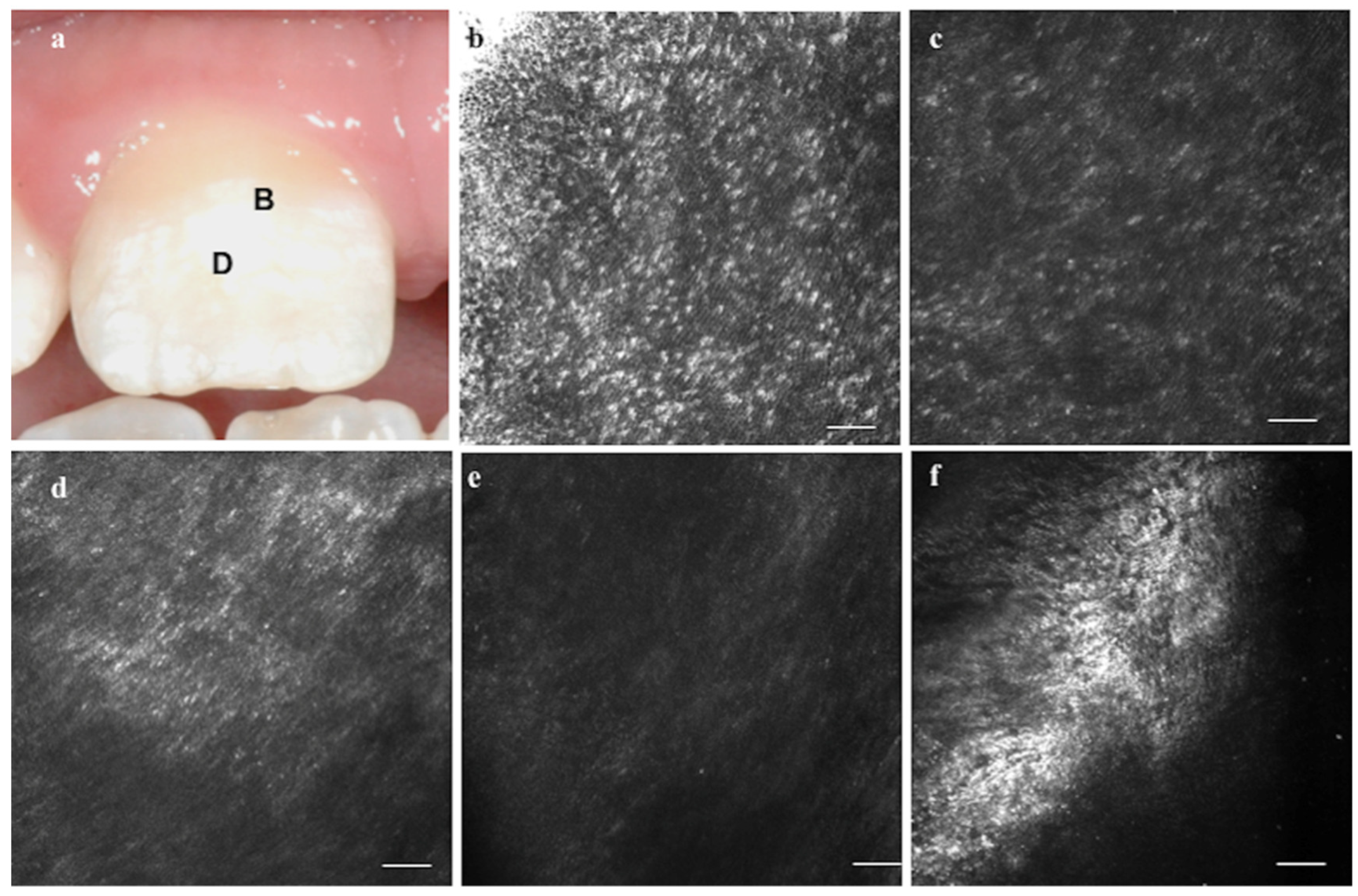
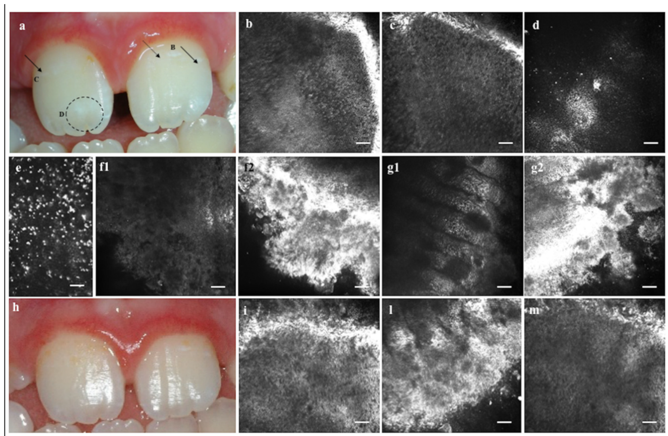
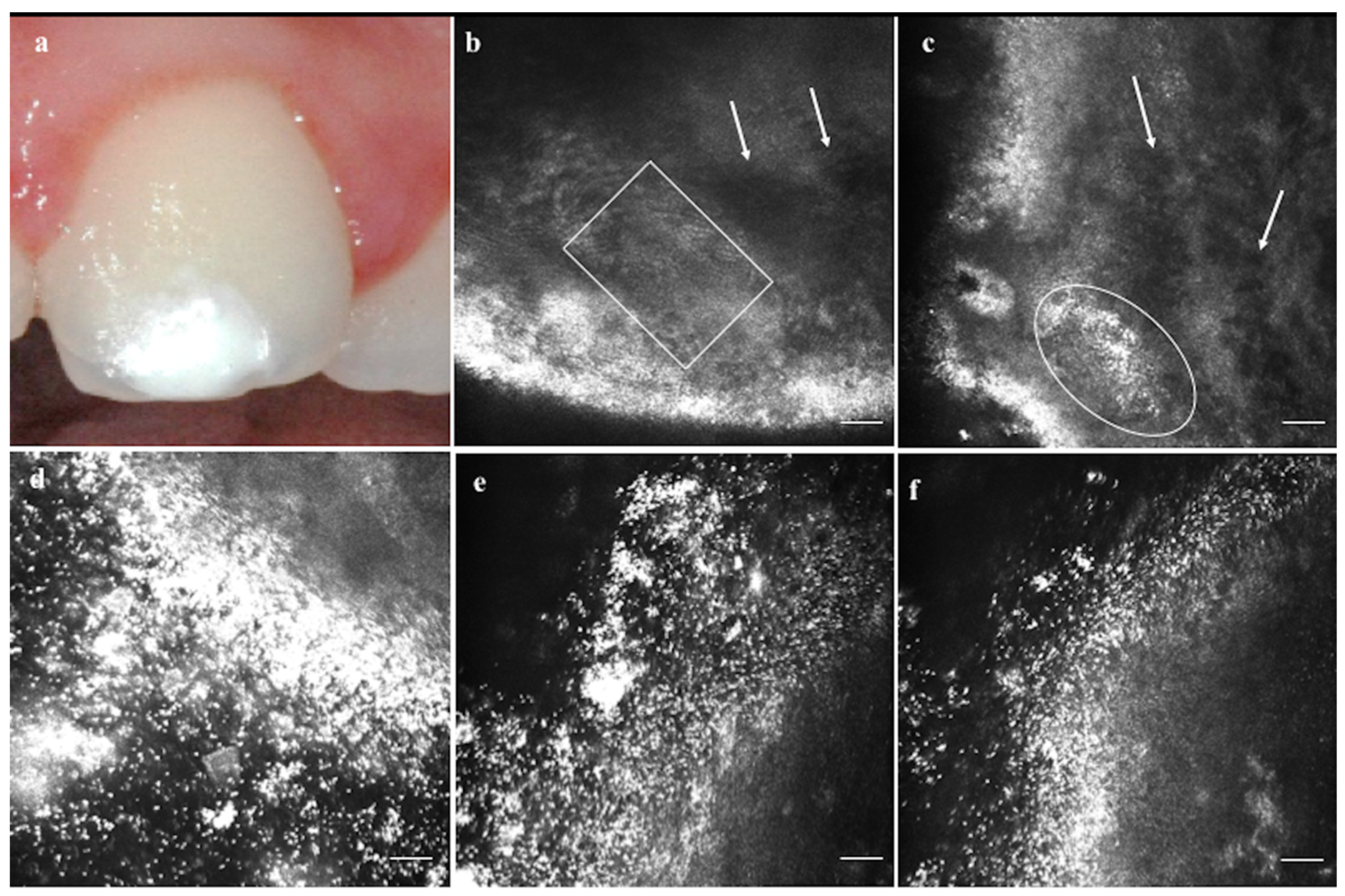
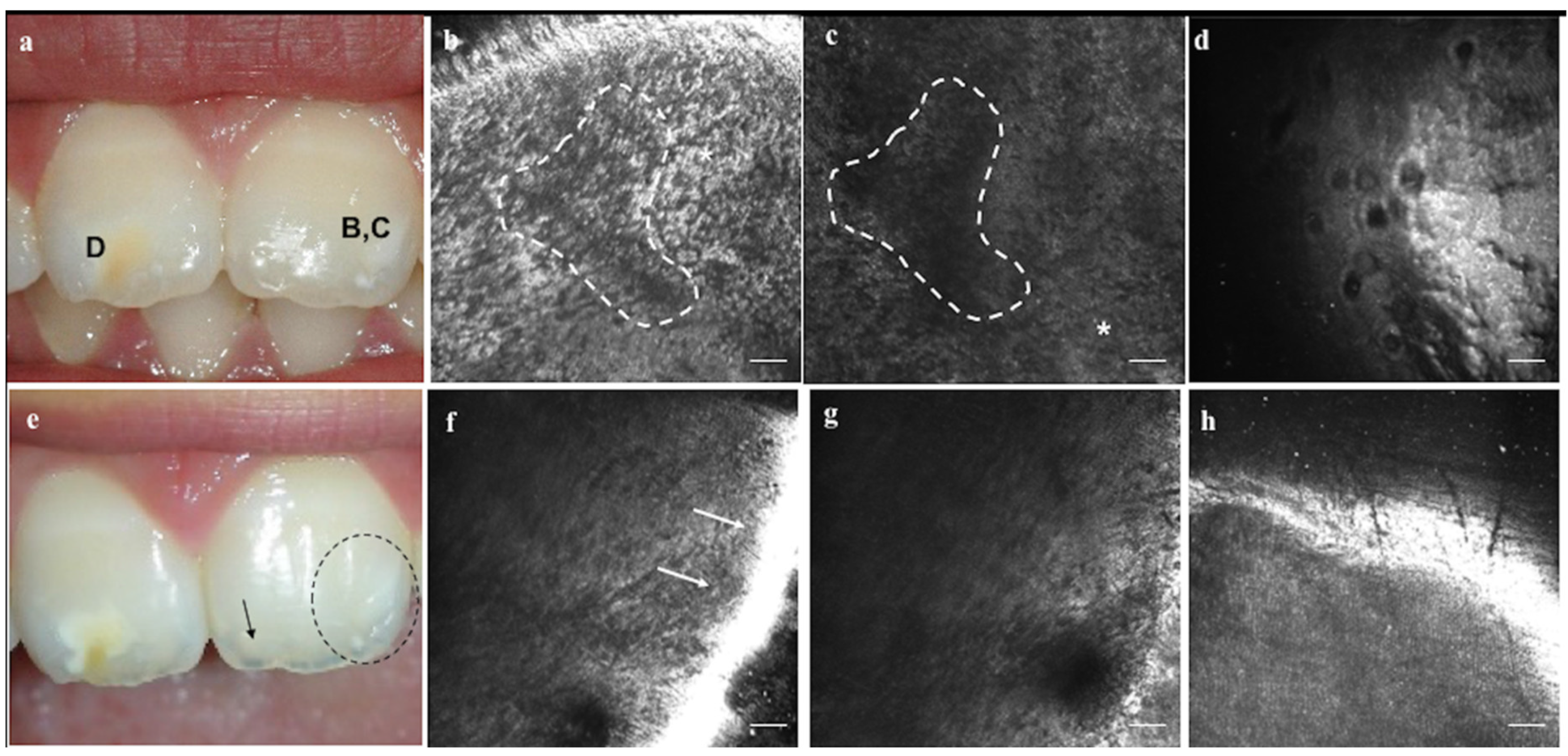
2.3. In Vivo Reflectance Confocal Laser Scanning Microscopy

In order to microscopically evaluate the initial EDs and their changes (disappearance, reduction, invariability or worsening) at the end of the CPP-ACP treatment, a series of in vivo confocal images were collected. For the purpose, a commercially available reflectance confocal microscope (RCM) was used (Vivascope3000®, first version, Lucid, Rochester, NY, USA) to image in vivo the enamel surface, at microscopic resolution, with no pain or discomfort for the patients. Each picture corresponds to a horizontal 500 × 500 µm optical section of the dental enamel, 5 µm in thickness and with a 0.5–1 µm lateral resolution with correspondence to standard histology [26,28]. RCM allows to vertically scan the enamel layers until a maximum depth of 300 µm from the surface. The system operates with a diode laser class 3A (European version) at 830 nm wavelength and with a 309 water immersion objective lens and a 0.9 numerical aperture. The laser power varies from 5 to 10 mW according to the layer to evaluate, and it causes neither tissue damage nor dental heating [28,35].
In order to obtain confocal images of the tissue from the plans parallel to the RCM lens (confocality), the device needs to work in close contact to the dental surface. Ultrasound gel is interposed between the lens and the window, and the window and the tooth, to optimize the incident and the refracted lights transmission. Hence, only vestibular surfaces of front teeth (incisors and canines) could be imaged, because both the size of the RCM sensor and the encumbrance of the handpiece impede to reach the back teeth and lingual dental surfaces.

3. Results

Seventeen consecutive children—7 males and 10 females—aged between 6 and 12 years (8.52 ± 2.00 years) showing one or more EDs were enrolled. Totally, 38 permanent teeth affected by EDs were analyzed: 23 white/creamy opacities and 11 yellow/brown ones; among them, 8 were linear, 16 appeared as small patches and 10 as diffuse areas (Figure 1 and Figure 2). Hypoplasia was found in 4 teeth, with a degree of severity from low-medium (pit, groove and/or lines) in 2 teeth, to severe, in the remaining 2 teeth showing diffuse hypoplastic areas with serious reduction or lack of enamel thickness (Figure 3 and Figure 4).
With regard to the clinical responsiveness to treatment, ED “disappearance” was found in 7 teeth (in 6 linear and 1 patchy opacities); ED “reduction” was reported in 18 teeth (in 2 linear, 10 patchy and 5 diffuse opacities and in 1 low-medium grade hypoplasia); ED “invariability” was found in 13 teeth (in 5 patchy and 5 diffuse opacities, in 1 low-medium grade hypoplasia and in 2 severe hypoplasia); no ED “worsening” was reported.
Before the CPP-ACP treatment (T0), RCM highlighted microscopic differences among the various degrees of ED severity, similarly to what found in our previous studies on in vivo RCM features of healthy enamel [35], the in vivo RCM differences between deciduous and permanent enamel [36] and the main in vivo RCM findings in EDs, both in permanent and deciduous teeth [37]. On RCM imaging, the 34 opacities without severe structural macroscopic defects (Figure 1 and Figure 2), showed a fairly preserved enamel architecture, identifiable at the boundaries of the lesion and along the whole visible layers, 300 µm beneath the surface (Figure 1b,c and Figure 2b,c), while, at the center of the defect, the enamel pattern was replaced by scattered deposits of bright organic substance (Figure 1d,e; Figure 2d).
The T0 RCM pictures of the 2 teeth with low-medium degree of hypoplasia revealed disturbances in enamel arrangement (Figure 3), only occasionally resembling the classical enamel architecture and mainly shown as hyporefractive dark areas with lack of the normal crystalline enamel architecture (Figure 3b) or as hyperrefractive organic matrix accumulation within the whole imaged layers (Figure 3c).
In the remaining 2 severe hypoplastic EDs, T0 RCM imaged wide irregular areas where the prismatic/interprismatic enamel architecture totally lacks along the several depths of view, thus allowing to highlight the presence of some roundish holes, corresponding to dentinal tubules (Figure 4d).
When RCM was performed immediately after CPP-ACP application (T1), the molecular complex could be seen and identified for its high brightness and refractivity as a dotted granular compound (Figure 2e), covering the holes and the lack of tissue both in hypoplasia and in opacities (Figure 2f2,g2).
At the end of the treatment (T2), qualitative defects such as opacities and demineralization of various degrees showed a variable responsiveness to treatment both at clinical examination (Figure 2h and Figure 4e) and on RCM imaging. While they appeared clinically reduced in size and discoloration, the confocal reports showed a higher content of organic substances, expressed by a higher refractivity respect to the ones observed at the starting point (Figure 1f, Figure 2i–m, and Figure 3f).
Hypoplasia showed less responsiveness to treatment: in those two cases with severe lack of enamel, CPP-ACP appeared not to be able to replace the absent enamel, but a partial improvement and re-coloring was possible at the boundaries of the defect in one case of low-medium grade severity (Figure 4f,h).

4. Discussion

The present study aimed to establish the feasibility of in vivo RCM to image the changes in enamel microscopic appearance after CPP-ACP treatments in vivo, on permanent teeth of children affected by EDs of various degree.
As expected, CPP-ACP worked better on demineralized teeth than on hypoplastic ones because of the impossibility, to date, to re-create the enamel prisms where they lack. Indeed, the clinical responsiveness to treatment was higher in opacities than in hypoplastic EDs. The clinical results correlated with RCM imaging confirmed that CPP-ACP mineralization does not re-establish ex novo the normal prismatic enamel architecture where it was initially absent, because of the lack of prismatic formation during the normal amelogenesis. Therefore, according to the RCM in vivo images, the reported clinical improvement (and in some cases the disappearance) of the EDs after CPP-ACP treatment could be justified by the CPP-ACP compounds that stably fulfilled and deposited within the organic compounds of the demineralized enamel. These evidences are in line with what Ferrazzano et al. reported in 2011 on the remineralizing effects of CPP-ACP mousse, revealing that the surfaces were covered with an amorphous deposit, completely obscuring the underlying prismatic structure [21]. Nevertheless, unlike Ferrazzano et al., where the mineralizing CPP-ACP effects were evaluated ex vivo with Scanning Electron Microscopy (SEM) [21], in the present work the entire procedure was performed in vivo, on living teeth. In detail, in vivo RCM demonstrated that CPP-ACP complex mainly allows the organic proteins to adhere to the enamel layers, and to reduce the areas of lack of minerals, by recruiting minerals where initially absent, as previously proven and with similar microscopic pictures reported by further ex vivo studies with confocal laser scanning microscopy (CLSM) and SEM from the works of Llena et al. [18] and Moura-Netto et al. [39].
Moreover, in a case of severe hypoplasia, thanks to the total absence of enamel, dentinal tubules have been imaged in vivo for the first time, with their characteristic shape, similarly to what was depicted ex vivo by Lucchese et al. [16], Moura-Netto et al. [39] and Marzuki et al. [40], thus overcoming the limited depth of penetration on healthy enamel, which impeded, in previous living studies [35,36,37], the imaging of the dentinal tissue.
Although these results are merely qualitative and very preliminary, and a quantitative analysis of the improvements has been excluded due to the low number of subjects enrolled, they confirmed the RCM capability to highlight in vivo microscopic differences of EDs before, during and after remineralizing therapy.
Furthermore, the imaging procedures revealed to be well tolerated by the children and the possibility to watch on the monitor the state of their “teeth” at microscopic resolution has intrigued and motivated children to be more compliant.
In addition, the bad compliance of the patients as bias has been excluded by the feedback from their daily report of drink intake, which strongly motivated the patients to be collaborative.
In conclusion, this novel approach to the study of enamel in vivo, could be applied to evaluate the responsiveness to remineralizing infiltrating agents, as well as to image in vivo the dental restoration interface, the margins of infiltrations, the duration and the deterioration of the compounds, and/or for longitudinal evaluation of non-invasive methods for white spot lesions arrestment, without the need for working on extracted and processed teeth, supporting and supported by the previous evidence of in vitro studies [18,19,20,21].

Author Contributions

Conceptualization, M.C., R.S. (Rosario Serpico) and A.L.; data curation, M.C., F.d.V., D.L. and R.S. (Rossella Santoro); investigation, M.C. and R.S. (Rossella Santoro); methodology, M.C.; project administration, R.S. (Rosario Serpico), R.S. (Rossella Santoro) and A.L.; resources, R.S. (Rosario Serpico); supervision, R.S. (Rosario Serpico), R.S. (Rossella Santoro) and A.L.; validation, F.d.V. and D.L.; writing—original draft, M.C., D.D.S. and F.d.V.; writing—review and editing, M.C., D.L. and A.L. All authors have read and agreed to the published version of the manuscript.

Funding

This research received no external funding

Conflicts of Interest

The authors declare no conflict of interest.

Ethics Approval

All procedures performed in the present study and involving human participants were in accordance with the ethical standards of the institutional and/or national research committee (protocol number #541/2019, Comitato Etico Università della Campania “Luigi Vanvitelli”- Azienda Ospedaliera Universitaria “Luigi Vanvitelli” -AORN “Ospedale dei Colli”) and with the 1964 Helsinki declaration and its later amendments.

Consent to Participate

Informed consent was obtained from all individual participants and their parents/caregivers included in the study.

Consent for Publication

All individual participants and their parents/caregivers included in the study have given their consent to the publication of data and images in accordance with the privacy of the subjects involved, without reporting identifying details.

References

  1. Robles, M.J.; Ruiz, M.; Bravo-Perez, M.; González, E.; Peñalver, M.A. Prevalence of enamel defects in primary and permanent teeth in a group of schoolchildren from Granada (Spain). Med. Oral Patol. Oral Cir. Bucal. 2013, 18, e187–e193. [Google Scholar] [CrossRef]
  2. Contaldo, M.; Luzzi, V.; Ierardo, G.; Raimondo, E.; Boccellino, M.; Ferati, K.; Bexheti-Ferati, A.; Inchingolo, F.; Di Domenico, M.; Serpico, R.; et al. Bisphosphonate-related osteonecrosis of the jaws and dental surgery procedures in children and young people with osteogenesis imperfecta: A systematic review. J. Stomatol. Oral Maxillofac. Surg. 2020. [Google Scholar] [CrossRef]
  3. Commission on Oral Health Research & Epidemiology. A review of the developmental defects of enamel index (DDE Index). Commission on Oral Health, Research & Epidemiology. Report of an FDI Working Group. Int. Dent. J. 1992, 42, 411–426. [Google Scholar]
  4. Farah, R.A.; Monk, B.C.; Swain, M.V.; Drummond, B.K. Protein content of molar-incisor hypomineralisation enamel. J. Dent. 2010, 38, 591–596. [Google Scholar] [CrossRef]
  5. Vargas-Ferreira, F.; Salas, M.M.; Nascimento, G.G.; Tarquinio, S.B.; Faggion, C.M., Jr.; Peres, M.A.; Thomson, W.M.; Demarco, F.F. Association between developmental defects of enamel and dental caries: A systematic review and meta-analysis. J. Dent. 2015, 43, 619–628. [Google Scholar] [CrossRef]
  6. Gupta, T.; Sadana, G.; Rai, H.K. Effect of Esthetic Defects in Anterior Teeth on the Emotional and Social Well-being of Children: A Survey. Int. J. Clin. Pediatr. Dent. 2019, 12, 229–232. [Google Scholar]
  7. Mony, S.; Rao, A.; Shenoy, R.; Suprabha, B.S. Comparative evaluation of the remineralizing efficacy of calcium sodium phosphosilicate agent and fluoride based on quantitative and qualitative analysis. J. Indian Soc. Pedod. Prev. Dent. 2015, 33, 291–295. [Google Scholar]
  8. Esteves-Oliveira, M.; Witulski, N.; Hilgers, R.D.; Apel, C.; Meyer-Lueckel, H.; Eduardo Cde, P. Combined Tin-Containing Fluoride Solution and CO2 Laser Treatment Reduces Enamel Erosion in vitro. Caries Res. 2015, 49, 565–574. [Google Scholar] [CrossRef]
  9. Rirattanapong, P.; Vongsavan, K.; Saengsirinavin, C.; Phuekcharoen, P. Efficacy of fluoride mouthrinse containing tricalcium phosphate on primary enamel lesions: A polarized light microscopic study. Southeast Asian J. Trop. Med. Public Health 2015, 46, 168–174. [Google Scholar]
  10. Lv, X.; Yang, Y.; Han, S.; Li, D.; Tu, H.; Li, W.; Zhou, X.; Zhang, L. Potential of an amelogenin based peptide in promoting reminerlization of initial enamel caries. Arch. Oral Biol. 2015, 60, 1482–1487. [Google Scholar] [CrossRef]
  11. Iafisco, M.; Degli Esposti, L.; Ramírez-Rodríguez, G.B.; Carella, F.; Gómez-Morales, J.; Ionescu, A.C.; Brambilla, E.; Tampieri, A.; Delgado-López, J.M. Fluoride-doped amorphous calcium phosphate nanoparticles as a promising biomimetic material for dental remineralization. Sci. Rep. 2018, 8, 17016. [Google Scholar] [CrossRef] [PubMed]
  12. Kumar, K.; Sreedharan, S. Comparative Evaluation of the Remineralization Potential of Monofluorophosphate, Casein Phosphopeptide-Amorphous Calcium Phosphate and Calcium Sodium Phosphosilicate on Demineralized Enamel Lesions: An In Vitro Study. Cureus 2018, 10, e3059. [Google Scholar] [CrossRef] [PubMed] [Green Version]
  13. Wu, L.; Geng, K.; Gao, Q. Early Caries Preventive Effects of Casein Phosphopeptide-Amorphous Calcium Phosphate (CPP-ACP) Compared with Conventional Fluorides: A Meta-analysis. Oral Health Prev. Dent. 2019, 17, 495–503. [Google Scholar] [PubMed]
  14. Bakry, A.S.; Abbassy, M.A. Increasing the efficiency of CPP-ACP to remineralize enamel white spot lesions. J. Dent. 2018, 76, 52–57. [Google Scholar] [CrossRef] [PubMed]
  15. Huq, N.L.; Myroforidis, H.; Cross, K.J.; Stanton, D.P.; Veith, P.D.; Ward, B.R.; Reynolds, E.C. The Interactions of CPP-ACP with Saliva. Int. J. Mol. Sci. 2016, 17, E915. [Google Scholar] [CrossRef] [PubMed] [Green Version]
  16. Lucchese, A.; Pilolli, G.P.; Petruzzi, M.; Crincoli, V.; Scivetti, M.; Favia, G. Analysis of collagen distribution in human crown dentin by confocal laser scanning microscopy. Ultrastruct. Pathol. 2008, 32, 107–111. [Google Scholar] [CrossRef]
  17. Scivetti, M.; Pilolli, G.P.; Corsalini, M.; Lucchese, A.; Favia, G. Confocal laser scanning microscopy of human cementocytes: Analysis of three-dimensional image reconstruction. Ann. Anat. 2007, 189, 169–174. [Google Scholar] [CrossRef]
  18. Llena, C.; Esteve, I.; Rodríguez-Lozano, F.J.; Forner, L. The application of casein phosphopeptide and amorphous calcium phosphate with fluoride (CPP-ACPF) for restoring mineral loss after dental bleaching with hydrogen or carbamide peroxide: An in vitro study. Ann. Anat. 2019, 225, 48–53. [Google Scholar] [CrossRef]
  19. Angelini Sfalcin, R.; da Silva, J.V.P.; Oliva Pessoa, V.; Santos, J.; Garcia Olivan, S.R.; Porta Santos Fernandes, K.; Melo Deana, A.; Makeeva, I.; Sauro, S.; Kalil Bussadori, S. Remineralization of early enamel caries lesions induced by bioactive particles: An in vitro speckle analysis. Photodiagnosis Photodyn. Ther. 2019, 28, 201–209. [Google Scholar] [CrossRef]
  20. Schwendicke, F.; Al-Abdi, A.; Pascual Moscardó, A.; Ferrando Cascales, A.; Sauro, S. Remineralization effects of conventional and experimental ion-releasing materials in chemically or bacterially-induced dentin caries lesions. Dent. Mater. 2019, 35, 772–779. [Google Scholar] [CrossRef]
  21. Ferrazzano, G.F.; Amato, I.; Cantile, T.; Sangianantoni, G.; Ingenito, A. In vivo remineralising effect of GC tooth mousse on early dental enamel lesions: SEM analysis. Int. Dent. J. 2011, 61, 210–216. [Google Scholar] [CrossRef] [PubMed]
  22. Moscarella, E.; González, S.; Agozzino, M.; Sánchez-Mateos, J.L.; Panetta, C.; Contaldo, M.; Ardigò, M. Pilot study on reflectance confocal microscopy imaging of lichen planus: A real-time, non-invasive aid for clinical diagnosis. J. Eur. Acad. Dermatol. Venereol. 2012, 26, 1258–1265. [Google Scholar] [CrossRef] [PubMed]
  23. Gareau, D.S.; Patel, Y.G.; Rajadhyaksha, M. Introduction to Reflectance Confocal Microscopy. In Reflectance Confocal Microscopy of Cutaneous Tumors: An Atlas with Clinical, Dermoscopic and Histological Correlations; Gonzales, S., Gill, M., Halpern, A.C., Eds.; Informa Healthcare: London, UK, 2008; pp. 1–3. [Google Scholar]
  24. Rajadhyaksha, M.; Grossman, M.; Esterowitz, D.; Webb, R.H.; Anderson, R.R. In vivo confocal scanning laser microscopy of human skin: Melanin provides strong contrast. J. Investig. Dermatol. 1995, 104, 946–952. [Google Scholar] [CrossRef] [Green Version]
  25. Ulrich, M.; Roewert-Huber, J.; González, S.; Rius-Diaz, F.; Stockfleth, E.; Kanitakis, J. Peritumoral clefting in basal cell carcinoma: Correlation of in vivo reflectance confocal microscopy and routine histology. J. Cutan. Pathol. 2011, 38, 190–195. [Google Scholar] [CrossRef]
  26. Lucchese, A.; Gentile, E.; Romano, A.; Maio, C.; Laino, L.; Serpico, R. The potential role of in vivo reflectance confocal microscopy for evaluating oral cavity lesions: A systematic review. J. Oral Pathol. Med. 2016, 45, 723–729. [Google Scholar] [CrossRef]
  27. Contaldo, M.; Poh, C.F.; Guillaud, M.; Lucchese, A.; Rullo, R.; Lam, S.; Serpico, R.; MacAulay, C.E.; Lane, P.M. Oral mucosa optical biopsy by a novel handheld fluorescent confocal microscope specifically developed: Technologic improvements and future prospects. Oral Surg. Oral Med. Oral Pathol. Oral Radiol. 2013, 116, 752–758. [Google Scholar] [CrossRef]
  28. Contaldo, M.; Agozzino, M.; Moscarella, E.; Esposito, S.; Serpico, R.; Ardigò, M. In vivo characterization of healthy oral mucosa by reflectance confocal microscopy: A translational research for optical biopsy. Ultrastruct. Pathol. 2013, 37, 151–158. [Google Scholar] [CrossRef]
  29. Contaldo, M.; Agozzino, M.; Ardigò, M. In vivo reflectance confocal microscopy for oral mucosa assessment. In Non Invasive Diagnostic Techniques in Clinical Dermatology; Berardesca, E., Maibach, H., Wilhelm, K., Eds.; Springer: Berlin/Heidelberg, Germany, 2014; pp. 81–87. [Google Scholar]
  30. Alessi, S.S.; Nico, M.M.; Fernandes, J.D.; Lourenço, S.V. Reflectance confocal microscopy as a new tool in the in vivo evaluation of desquamative gingivitis: Patterns in mucous membrane pemphigoid, pemphigus vulgaris and oral lichen planus. Br. J. Dermatol. 2013, 168, 257–264. [Google Scholar] [CrossRef]
  31. Contaldo, M.; Di Stasio, D.; Petruzzi, M.; Serpico, R.; Lucchese, A. In vivo reflectance confocal microscopy of oral lichen planus. Int. J. Dermatol. 2019, 58, 940–945. [Google Scholar] [CrossRef]
  32. Contaldo, M.; Lajolo, C.; Di Petrillo, M.; Ballini, A.; Inchingolo, F.; Serpico, R.; Romano, A. Analysis of lip pigmentations by reflectance confocal microscopy: Report of two cases. J. Biol. Regul. Homeost. Agents 2019, 33, 19–25. [Google Scholar]
  33. Romano, A.; Santarelli, A.; Lajolo, C.; Della Vella, F.; Mascitti, M.; Serpico, R.; Contaldo, M. Analysis of oral mucosa erosive-ulcerative lesions by reflectance confocal microscopy. J. Biol. Regul. Homeost. Agents 2019, 33, 11–17. [Google Scholar] [PubMed]
  34. Contaldo, M.; Lauritano, D.; Carinci, F.; Romano, A.; Di Stasio, D.; Lajolo, C.; Della Vella, F.; Serpico, R.; Lucchese, A. Intraoral confocal microscopy of suspicious oral lesions: A prospective case series. Int. J. Dermatol. 2020, 59, 82–90. [Google Scholar] [CrossRef] [PubMed]
  35. Contaldo, M.; Serpico, R.; Lucchese, A. In vivo imaging of enamel by reflectance confocal microscopy (RCM): Non-invasive analysis of dental surface. Odontology 2014, 102, 325–329. [Google Scholar] [CrossRef] [PubMed]
  36. Gentile, E.; Di Stasio, D.; Santoro, R.; Contaldo, M.; Salerno, C.; Serpico, R.; Lucchese, A. In vivo microstructural analysis of enamel in permanent and deciduous teeth. Ultrastruct. Pathol. 2015, 39, 131–134. [Google Scholar] [CrossRef] [Green Version]
  37. Contaldo, M.; Di Stasio, D.; Santoro, R.; Laino, L.; Perillo, L.; Petruzzi, M.; Lauritano, D.; Serpico, R.; Lucchese, A. Non-invasive in vivo visualization of enamel defects by reflectance confocal microscopy (RCM). Odontology 2015, 103, 177–184. [Google Scholar] [CrossRef]
  38. Ghanim, A.; Elfrink, M.; Weerheijm, K.; Mariño, R.; Manton, D. A practical method for use in epidemiological studies on enamel hypomineralisation. Eur. Arch. Paediatr. Dent. 2015, 16, 235–246. [Google Scholar] [CrossRef] [Green Version]
  39. Moura-Netto, C.; Mello-Moura, A.C.; Palo, R.M.; Prokopowitsch, I.; Pameijer, C.H.; Marques, M.M. Adaptation and penetration of resin-based root canal sealers in root canals irradiated with high-intensity lasers. J. Biomed. Opt. 2015, 20, 038002. [Google Scholar] [CrossRef] [Green Version]
  40. Marzuki, A.F.; Masudi, S.M. Confocal laser scanning microscopy study of dentinal tubules in dental caries stained with alizarin red. Arch. Orofac. Sci. 2008, 3, 2–6. [Google Scholar]
Figure 1. (a) Initial clinical presentation and T0 (be) and T2 (f) confocal images of a white/cream opacity without structural defects of the two-third of the vestibular surface of the upper right central incisor in an 8-year-old female. (b,c) At T0, at the boundaries of the defect, marked «B» in (a), enamel architecture was preserved and identifiable both at the superficial layers, 10 µm beneath the surface (b), and at the deepest visible layers, 300 µm beneath the surface (c). The diffuse bright points corresponded to the lack of mineral compounds, replaced by scattered deposits of bright organic substance. (d,e) At T0, at the center of the defect, marked as «D» in (a), bright organic matrix was not arranged in the classical interprismatic regular net, neither at the superficial layers, 10 µm beneath the surface (d), nor at the deepest visible layers, 300 µm beneath the surface (e). (f) At T2, enamel prisms still lack at the center of the defect, but the site appeared fulfilled by inhomogeneous and irregular accumulation of bright material, presumably organic casein phosphopeptide (CPP). Scale bars: 100 µm.
Applsci 10 04155 g001
Figure 2. Diffuse white opacities without structural defects of both upper central incisors in an 8-year-old male before and after the casein phosphopeptide-amorphous calcium phosphate (CPP-ACP) treatment. (a) At T0, both teeth showed linear (arrows) and patchy (dotted circle) white opacities. (b,c) T0 reflectance confocal microscopy (RCM) of the linear opacities marked, respectively, as «B» and «C» in (a): the enamel architecture was well preserved few layers below the surface (10 µm below) while sparse dark areas (hyporefractive) expressed the slight disarrangement of the organic interprismatic substance, usually highly refractive. (d) T0 RCM of the small patched opacity marked as «D» in (a): evident deposits of organic substance (hyper-refractive) and inhomogeneous mineralization. (e) T1 RCM of a dental surface, immediately after the CPP-ACP application: the mousse appeared dispersed in the ultrasound gel used to perform RCM imaging, revealing to be a highly refractive granular compound, reflecting the CPP composition. (f,g) RCM before (T0) (f1,g1) and immediately after (T1) (f2,g2) CPP-ACP application at the patchy area of the upper right central incisor, showing the evident differences in organic accumulation within the dark areas. (h) At T2, the patchy opacity of the upper right central incisor seems to have clinically totally disappeared. (i–m) The related T2 RCM images showed a more refractive enamel arrangement through the layers: 10 µm (i), 150 µm (l) and 300 µm (m). Scale bars: 100 µm.
Applsci 10 04155 g002
Figure 3. (a) Clinical presentation of a patchy white opacity with low severe hypoplasia of the vestibular one/half of the upper left central incisor in an 11-year-old male. (b,c) At T0, RCM revealed disturbances in enamel arrangement shown as hyporefractive dark areas with lack of the normal crystalline enamel architecture (white arrows) alternated with partially preserved prismatic/interprismatic architecture (white square) or hyperrefractive organic matrix accumulation (dotted circle) within the whole imaged layers. (df) At T2, the areas previously dark and low refractive, appeared fulfilled by bright refractive material scattered along the entire field of view and within the whole visible layers: 10 µm (d), 150 µm (e) and 300 µm (f). Scale bars: 100 µm.
Applsci 10 04155 g003
Figure 4. (a) T0 clinical picture of a yellow/brown patchy opacity with severe hypoplasia affecting the vestibular one/half of upper right central incisor and multiple, linear and patchy white/creamy opacities of the vestibular one/half of the upper left central incisor in a 9-year-old female. (b,c) T0 RCM of the opacity marked as “B,C” in (a), revealed a partially preserved prismatic/interprismatic architecture, brighter than usual due to the higher organic/inorganic compounds ratio (asterisks) alternated with irregular areas in which the prismatic/interprismatic architecture lacks (dotted areas) at 10 µm (b) and 300 µm (c), respectively, from the surface, corresponding to severe lack of enamel. (d) When the enamel is constitutionally absent, as in “D” marked in (a), the T0 RCM revealed some roundish holes, corresponding to dentinal tubules. (e) At T2, the boundaries of the yellow/brown opacity of the upper right central incisor turned into a white/cream less severe defect, and the opacity of the upper left central incisor was strongly reduced (dotted black circle), and disappeared in some linear areas (black arrow). (f,g) T2 RCM of the “D” area showed a persistent lack of prismatic/interprismatic architecture, but also the improvement in bright/dark contrast, due to the organic compounds fulfilling the interprismatic areas instead of the mineral inorganic part, most strongly visible at the periphery (white arrows) (f) and still with deficiencies in some areas of the center (g). (h) T2 RCM of “B,C” revealed a more homogeneous enamel prisms arrangement. Scale bars: 100 µm.
Applsci 10 04155 g004

