Article

Inhibitory Effects of 4-Hexylresorcinol on Root Resorption Induced by Orthodontic Tooth Movement

1
Department of Orthodontics, School of Dentistry, Seoul National University, Seoul 3080, Korea
2
Department of Oral Biochemistry, College of Dentistry, Gangneung-Wonju National University, Gangneung 28644, Korea
3
Department of Oral and Maxillofacial Surgery, College of Dentistry, Gangneung-Wonju National University, Gangneung 28644, Korea
*
Authors to whom correspondence should be addressed.
Submission received: 22 August 2020 / Revised: 4 September 2020 / Accepted: 8 September 2020 / Published: 10 September 2020

Abstract

:

Featured Application

The administration of 4-hexylresorcinol reduces root resorption during orthodontic tooth movement.

Abstract

Root resorption during orthodontic tooth movement (OTM) is caused by an imbalance between the bone turnover rate and applied mechanical stress. The administration of 4-hexylresorcinol (4HR) increases the bone turnover rate and factors associated with bone formation. Thus, 4HR may show protective activity against root resorption during orthodontic tooth movement (OTM). A total of 40 rats (male: 20; female: 20) were included in this study, and the mandibular first molar was subjected to excessive orthodontic force. The experimental group (n = 20) received 12.8 mg/kg of 4HR every 2 weeks. The controls (n = 20) received a solvent without 4HR. Both groups had the same sex distribution. On Day 28 after the initiation of OTM, all the animals were sacrificed for micro-computed tomography analysis, Western blot analysis, and immunohistochemistry. The ratios of the root length and root volume to the total volume were significantly higher in the experimental group compared to those in the control group (p < 0.05). The expression levels of OPG, RANKL, alkaline phosphatase, and Runx2 in the experimental group according to Western blotting were significantly higher in the experimental group compared to those in the control group (p < 0.05). Their expression was mainly found in the periodontal ligament area. In conclusion, the administration of 4HR decreased the root resorption caused by OTM and increased the expression levels of OPG, RANKL, alkaline phosphatase, and Runx2.

Graphical Abstract

1. Introduction

Orthodontic tooth movement (OTM) can be achieved by the remodeling process of periodontal tissue, including cementum and alveolar bone. When orthodontic force is applied, the pressure induces osteoclastic mineralized tissue degradation on the compression side and mineralization by osteoblasts on the tension side [1]. This mineralized tissue turnover is the coupling mechanism of bone resorption followed by bone formation and is known as a marker that influences OTM [2].
Alveolar bone remodeling is influenced by localized tooth-related factors [3]. Systemic factors such as menopause [4], aging [5], osteoporosis [6], changes in parathyroid hormone levels [7], and levels of steroid [8] or bisphosphonate intake can also influence the overall alveolar bone turnover rate [9]. It is well known that reduced alveolar bone turnover rates can impede the overall velocity of tooth movement during orthodontic treatment [10].
Certain levels of tooth root resorption are an unavoidable consequence caused by orthodontic forces, and there have been many efforts to minimize it [11,12,13,14]. Unfortunately, severe resorption, defined as a reduction that is more than 4 mm or a third of the original length of the root, occurs in 1% to 5% of treated teeth [11]. The etiology of root resorption is multifactorial; there are patient-related factors such as genetic predisposition [12], treatment-related factors including extensive treatment duration or tooth movement [13], and local factors of the tooth itself [14]. However, none of these factors have been clearly proven as the principal cause of root resorption [11]. Bone metabolism not only affects the acceleration of tooth movement but also modifies the periodontal tissue and therefore the extent of root resorption [15]. A low bone turnover rate is considered to be a predisposing factor for root resorption, and high bone turnover is associated with less root resorption [16]. Moreover, studies have shown that periodic vibration [17] and low-level laser therapy (LLLT) stimulation during orthodontic treatment can increase the rate of bone turnover and thus reduce the extent of root resorption [18]. In the case of an ovariectomized mouse model, a lack of estrogen induces the imbalance of the receptor activator of the NF-κB ligand (RANKL)/osteoprotegerin (OPG) system and increases root resorption during OTM [19]. The application of resveratrol in the rat model decreases root resorption during OTM but reduces the amount of OTM [20]. The agent for reducing root resorption during OTM should not inhibit OTM. However, this type of agent is rare.
4-hexylresorcinol (4HR), a phenol derivative with anti-septic and anti-parasitic properties, has been used as a food preservative and as a means to prevent melanosis [21]. It is known to be safe and effective in topical applications for infected skin or mucosa [21]. 4HR suppresses tumor necrosis factor-α [22] and the nuclear factor-κB (NF-κB) signaling pathway [23] related to osteoclast differentiation, which increases bone formation by suppressing osteoclast activity and promoting angiogenesis [24]. 4HR has been incorporated into bone-graft material and has been proven to prevent the formation of foreign-body giant cells [25] and suppress the NF-kB signaling pathway in osteoblasts [26]. Furthermore, it is known to activate transforming growth factor-β1 (TGF-β1) signaling, which induces bone regeneration and remodeling [27]. TGF-β1 activated by 4HR induces angiogenesis independently of hypoxia inducible factor (HIF) [24,28].
In tooth-related research, 4HR increases bone turnover markers in blood samples and accelerates OTM [29]. The administration of 4HR increases mineral deposition during tooth formation and mandibular incisor eruption in rat models [30]. Both bone turnover and mineral deposition during tooth formation are important for preventing root resorption during OTM. However, not much is known about its effects on root resorption during OTM. This study aims to provide insight into the effects of 4-HR application on root resorption during OTM through imaging and histological examination.

2. Materials and Methods

2.1. Animals and Experimental Design

A total of 40 rats (Sprague Dawley) were provided by an animal supplier (Orientbio Inc., Sungnam, Korea). Half were male, and the others were female. All the rats were specifically infection free. The subsequent experimental procedure was approved by the Institutional Animal Care and Use Committee of Gangneung-Wonju National University (GWNU-2020-16 approved at 23 April 2020). The housing and feeding were in accordance with previous publications [29].
The application of the orthodontic appliance was in accordance with previous publications [29]. In brief, indentation was prepared with a sharp diamond bur on the distal surface of the right mandibular first molar for preventing wire loss. A ligature wire was passed through this indentation. The distal end of a coiled spring was ligated with wire. The proximal end of the coil spring was put around the right mandibular incisor. The force initially generated by the coiled spring was 120 g. There were two groups, and each group had 20 rats (10 males and 10 females); as a control, solvent without 4HR was used. The rats in the experimental group received 12.8 mg/kg of 4HR. The injection of 4HR was performed every 2 weeks. An injection agent was prepared just before injection, and the agent was injected into the back skin, subcutaneously. The deactivation of the coiled spring was checked with regard to a predetermined schedule. At 4 weeks after appliance application, blood was sampled from the heart under general anesthesia. Then, all the animals were sacrificed humanely. The following animals were excluded for further analysis: (1) animals showing a loss of the orthodontic appliance during follow-up, and (2) those in which the gap formed by OTM was less than 0.5 mm. A summary of the exclusion process is shown in the flowchart (Figure 1). A biopsy of the mandible was also performed. Histological and molecular biology analyses were conducted.

2.2. Plain X-ray and Micro-Computerized Tomography (mCT)

Both mandibles were collected, and plain X-rays were taken using an intra-oral radiogram sensor. The lengths of four roots in the first mandibular molar were measured using SigmaScan Pro (SPSS Inc., Chicago, IL, USA). The central two roots were interposed in a plain X-ray, and the outermost border was used for comparison. The ratio for each root length between the affected side and the non-affected side was calculated.
The hemi-mandibles were sent to Genoss (Suwon, Korea) and the Korea National University of Transportation (Chungju, Korea) for analysis with mCT. The subsequent procedures were conducted in accordance with our previous publication [29]. In brief, hemi-mandibles were loaded on the mCT scanner (SkyScan1173, Bruker, Kontich, Belgium). The source voltage was 130 kV, and the image pixel size was 13.85 μm. Three-dimensional reconstruction was performed with the subtraction of the surrounding bony structure. The total volume (TV) and root volume (RV) of the mandibular first molar were measured according to previous publications [19]. The ratio of RV to TV was calculated and compared between groups.

2.3. Immunohistochemical Determination and Western Blot Analysis in Mandible Samples

To assess the expression of OPG, RANKL, alkaline phosphatase (AP), and runt-related transcription factor 2 (Runx2) in the mandible samples, we performed immunohistochemical staining using anti-OPG, anti-RANKL, anti-alkaline phosphatase, and anti-Runx2 antibodies (Santa Cruz Biotech, Santa Cruz, CA, USA). The detailed procedure was in accordance with our previous publications [24,25]. A silane-coated glass slide was used for preventing tissue detachment during the staining procedure. After hydration, sections were treated with trypsin for 5 min. Then, 30% H2O2 was applied on the sections for 7 min. After washing with phosphate buffered saline (PBS) twice, protein blocking was performed for 1 h with a ready-to-use solution (Serum-Free Protein Block, Dako North America Inc., Carpinteria, CA, USA). Then, primary antibodies (dilution ratio 1:100) were applied on the tissue section. Incubation with a primary antibody was performed at 4 °C overnight. Three washes were performed with PBS. A universal secondary antibody (Dako REAL™ EnVision™/HRP, Rabbit/Mouse; Dako North America Inc.) was conjugated under a humid chamber. Three washes were performed with PBS again. After removing the unreacted secondary antibody, the slides were stained with a chromogen (Dako REAL™ DAB+ Chromogen and Dako REAL™ Substrate Buffer; Dako North America Inc.).
The other mandible samples (n = 5 for each group) were collected in a cryovial and stored in a deep freezer (−70 °C). For the extraction of tissue protein, frozen tissue was crushed and mixed with a tissue-protein-extraction reagent buffer with a protease inhibitor cocktail. Subsequently, Western blot analysis was performed as described previously [29].

2.4. Statistical Analysis

The comparison between groups was performed with the independent-samples t-test. The level of significance was set at <0.05.

3. Results

3.1. The Application of 4HR Inhibited Root Resorption during OTM

The ratios of the root length at 4 weeks after OTM in the male control group were 0.94 ± 0.08, 0.81 ± 0.10, and 0.81 ± 0.10 in the distal, central, and mesial roots, respectively (Figure 2). Those in the male experimental group were 1.00 ± 0.09, 0.95 ± 0.11, and 0.99 ± 0.06 in the distal, central, and mesial roots, respectively. The difference between groups was statistically significant in the central and mesial roots (p = 0.017 and 0.001, respectively).
The ratios of the root length at 4 weeks after OTM in the female control group were 0.94 ± 0.06, 0.82 ± 0.03, and 0.76 ± 0.06 in the distal, central, and mesial roots, respectively. Those in the female experimental group were 1.00 ± 0.07, 0.95 ± 0.08, and 0.98 ± 0.09 in the distal, central, and mesial roots, respectively. The difference between the groups was statistically significant in the central and mesial roots (p = 0.005 and 0.001, respectively).
The results of the mCT analysis were in accordance with the results for plain film (Figure 3). Three-dimensionally reconstructed images demonstrated that root resorption was severe in the central roots. Multiple lacunas were observed on the surface of the mesial root in the high-magnification view. These lacunas were not confined to the surface of the compression side.
The RV and TV were calculated from the mCT, and their ratio was compared between groups (Table 1). The comparison of the percentages of RV to TV showed a statistically significant difference between groups (p = 0.002 and 0.006 for male and female, respectively).

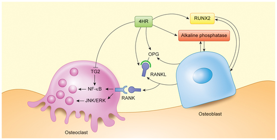
3.2. 4HR Increased OPG, RANKL, AP, and Runx2 Expression in Tissue

The administration of 4HR increased the expression of OPG, RANKL, Runx2, and AP in both sexes of the rats (Figure 4). Full length blots were shown in Figure S1. The relative expression of OPG, RANKL, Runx2, and AP in the male control group was 1.00 ± 0.15, 1.11 ± 0.10, 1.12 ± 0.20, and 1.32 ± 0.42, respectively. That in the male experimental group was 1.90 ± 0.13, 1.78 ± 0.06, 1.83 ± 0.23, and 2.34 ± 0.13, respectively. The difference between the groups was significantly different for OPG, RANKL, Runx2, and AP (p = 0.014, 0.009, 0.015, and 0.016, respectively). The OPG-to-RANKL ratios for males were 0.91 ± 0.17 and 1.07 ± 0.05 for the control and experimental groups, respectively (p > 0.05).
The relative expression of OPG, RANKL, Runx2, and AP in the female control group was 0.81 ± 0.04, 0.94 ± 0.13, 1.16 ± 0.75, and 1.52 ± 0.16, respectively. That in the male experimental group was 2.29 ± 0.38, 1.56 ± 0.13, 2.32 ± 0.60, and 2.01 ± 0.84, respectively. The difference between the groups was significantly different for OPG and RANKL (p = 0.040 and 0.007, respectively). The OPG-to-RANKL ratios for females were 0.87 ± 0.12 and 1.48 ± 0.34 for the control and experimental groups, respectively (p = 0.041).
Upon histological analysis, the findings for active root resorption were similar in both groups. However, the area of active root resorption was more frequently observed in the control group (Figure 5a). The thickness of the cementum was thinner in the middle area of the root surface. Discontinuous cementum was frequently found in the control group. The results of the immunohistochemical staining were in accordance with the results of the Western blotting (Figure 5b). Positive reactions to AP, OPG, RANKL, and Runx2 were mainly found in the periodontal ligament area.

4. Discussion

Root resorption during OTM has been considered an unavoidable phenomenon [11]. However, severe resorption is considered to be a complication and has been reported in 1% to 5% of OTM procedures [11]. Accordingly, reducing root resorption during OTM is an important issue in orthodontics. In this study, the administration of 4HR to rat models resulted in reduced root resorption induced by OTM (Figure 2 and Figure 3). The bone turnover markers AP, OPG, RANKL, and Runx2 showed elevated expression levels in the 4HR-administered group (Figure 4 and Figure 5). To the best of our knowledge, this is the first report about reducing root resorption during OTM by 4HR administration.
Several studies have been conducted on the effects of various drugs or chemicals on the rate of tooth movement and root resorption [31]. Bisphosphonate binds to hydroxyapatite on the absorbing surface of bone and inhibits bone resorption through a mechanism that inhibits osteoclast migration and action and induces osteoclast apoptosis [32]. It has been reported that the administration of bisphosphonate reduces root resorption and tooth movement [33] due to its anti-inflammatory action and inhibition of osteoclastic activity [34]. Bisphosphonate administration has the advantage of reducing root resorption during OTM, but it significantly interferes with OTM [35]. Prednisolone administration also decreases root resorption and the rate of tooth movement [36]. Controversial results have been reported for the effect of non-steroidal anti-inflammatory drug (NSAID) administration on root resorption [36]. It is believed that some types of NSAIDs inhibit root resorption [31] and tooth movement [37]. Lithium promotes reduced osteoclast formation by promoting the Wnt/β-catenin signaling system [37]. As the concentration of lithium increases, root resorption and tooth movement decrease in animal experiments [38]. As described above, most of the substances administered to reduce root resorption through a mechanism that reduces osteoclast activity have tended to decrease tooth movement speed as well as root resorption [38]. The 4HR used in this study resulted in root resorption being reduced compared to that in the control group (Figure 2 and Figure 3). When looking at the numbers of exclusions because of a low rate of tooth movement, they were four for the experimental group and five for the control group (Figure 1). The administration of 4HR accelerates OTM in ovariectomized rat models [29].
The bone remodeling process is balanced through the coupling of bone resorption by osteoclasts and bone formation by osteoblasts. Medications or dietary supplements that decrease osteoclastic activity suppress root resorption but also hinder OTM [31]. The substances that increase bone turnover rate and increase both osteoblastic and osteoclastic activity may diminish root resorption and promote tooth movement. The effect of parathyroid hormone (PTH) is different according to the treatment method that is either catabolic or anabolic [39]. The long-term intermittent injection of PTH facilitates the repair of root resorption [40]. Intermittent PTH administration increases the expression levels of both RANKL and insulin-like growth factor-1, indicating that intermittent PTH stimulates both osteoclasts and osteoblasts [7]. LLLT may inhibit orthodontically induced root resorption [18] but accelerates OTM [41]. LLLT accelerates the bone remodeling process by stimulating both osteoblastic and osteoclastic activity during OTM [42]. 4HR facilitates OTM in ovariectomized rat models [29]. In our study, 4HR increased the bone turnover markers, which were RANKL, OPG, Runx2, and AP, in the experimental group more than in the control group (Figure 4). An increased bone turnover rate might inhibit root resorption (Figure 2).
The main physiological role of the RANKL/RANK/OPG system is regulating bone remodeling. Functional osteoclast formation and bone resorption are dependent on the RANKL/OPG ratio expressed by osteoblastic cells and RANK expression by osteoclast precursor cells [43]. The OPG/RANKL/RANK system may influence root resorption during OTM, as well [15]. Odontoclasts are multinucleated cells located on root dentin being resorbed. RANKL expression was detected not only in osteoclasts but also in odontoclasts, so there seems to be a common regulatory mechanism between osteoclasts and odontoclasts [44]. If root resorption is more severe, the expression of RANKL increases [45]. The amount of RANKL is an important factor for root resorption, but the OPG/RANKL ratio and the concentration of OPG are also important for hard-tissue repair [40]. In this study, the expression of both RANKL and OPG may have contributed in inhibiting root resorption in the experimental group (Figure 2 and Figure 3).
Rats have been used frequently as a model to study OTM, despite the morphological and physiological differences in the periodontal ligament and alveolar bone compared to humans [46]. In this study, the experimental teeth included the lower first molar. Although anterior teeth are known to be the most vulnerable teeth for root resorption [47], since the anterior teeth of the rats continue to grow, they are not suitable as an experimental model for root resorption. As heavier forces were applied to teeth, greater root resorption occurred [48]. A large force of 120 g was used to examine the effect on root resorption in this study. Because a human molar is approximately 20 times larger than a rat molar [49], the effect of a 120 g force on a rat molar is comparable with that of a very heavy force of 2400 g on a human molar. This is a force that is large enough to cause root resorption. In this study, it was found that the root resorption induced by a very heavy force was alleviated by 4HR (Figure 2 and Figure 3). Further research is needed to investigate the effect of 4HR on root resorption under normal orthodontic force.
Root resorption during OTM is closely associated with the OPG-to-RANKL ratio [50]. In this study, both OPG and RANKL were shown to be elevated in their expression by 4HR administration (Figure 4). Considering that 4HR inhibits the NF-κB pathway [22,26], the effects on osteoclasts and cementoclasts by the elevated expression of RANKL are restricted by hampering its downstream signaling and by the blocking of 4HR. TGF-β1 inhibits mineralized tissue resorption by elevating the OPG level [51]. Interestingly, 4HR is a strong inducer of TGF-β1 [21,51]. Cementum is a barrier to root resorption, and its resorption is dependent on the OPG-to-RANKL ratio [50]. In this study, the experimental group showed a higher OPG-to-RANKL ratio compared to the control group (p < 0.05 for the female group, Figure 4b). Despite the elevation of both bone-formation and bone-resorption markers in the alveolus supports in a previous study [29], the systemic effects of 4HR injection were not evaluated, and the mechanisms of how 4HR affects the signaling pathway of bone metabolism is still unclear. Further study is necessary to elucidate the in vivo mechanism of 4HR’s activity. 4HR has been accorded a GRAS (generally recognized as safe) status as an oral health care agent [52]. There are some issues for the estrogenic activity of 4HR [53], but a recent study reported that 4HR behaves differently from other xeno-estrogens [54].
The limitations of this study were as follows: First, rats have a continuous erupting incisor and no premolar. In addition, the root morphology of the mandibular first molar is very different from that in humans. Accordingly, the direct translation of this result to clinical application should be considered as preliminary. Second, this study focused on bone turnover markers to interpret the mechanism of 4HR’s effects. However, these markers might influence the turnover of other calcified tissue such as cementum. Actually, all the surfaces that showed active root resorption had a broken cemental line (Figure 5a). Secondary cementum has bone-like structures with cementoblasts. Without breaking the cemental line, dentin resorption would be impossible [55]. Interestingly, mineralized tissue formation during tooth eruption is enhanced by 4HR administration according to our recent research [30]. RANKL/OPG is also involved in the turnover of cementum [50]. In this study, the expression levels of RANKL and OPG were increased by 4HR administration (Figure 4). Therefore, 4HR might increase cementum formation during OTM, and this might be one mechanism of reducing root resorption. In addition, increasing M2-type macrophages is associated with reduced root resorption [55]. 4HR is a potent M2-polarizing agent [24,56]. Third, heavy force applied on the root surface might induce an inflammatory reaction and subsequent pH drop. An acidic environment can decalcify mineralized tissue without the help of cells. Direct resorption without cells was not examined in this study. Further investigation for the clinical application of 4HR in orthodontic treatment is necessary.

5. Conclusions

In this study, the administration of 4HR decreased the root resorption of the mandibular first molar caused by OTM and increased the expression levels of OPG, RANKL, alkaline phosphatase, and Runx2 in the mandible.

Supplementary Materials

The following are available online at https://0-www-mdpi-com.brum.beds.ac.uk/2076-3417/10/18/6313/s1, Figure S1: Full length blot of Figure 4. Application of 4HR increased the expression level of OPG, RANKL, alkaline phosphatase, and Runx2.

Author Contributions

J.-K.J. contributed to the conceptualization, data curation, formal analysis, and writing of the original draft. D.-W.K. contributed to the data curation, formal analysis, and investigation. S.-G.K. contributed to the formal analysis, funding acquisition, methodology, writing of the original draft, and review and editing. T.-W.K. contributed to the formal analysis and review and editing. All authors approved the manuscript and agreed to be accountable for all aspects of the work. All authors have read and agreed to the published version of the manuscript.

Funding

This study was conducted with the support of the “Cooperative Research Program for Agriculture Science and Technology Development (Project no. PJ01313902)”, Rural Development Administration, Republic of Korea.

Acknowledgments

Yei-Jin Kang and Ji-hyen Oh helped with the animal experiments.

Conflicts of Interest

The authors declare no conflict of interest.

References

  1. King, G.J.; Keeling, S.D.; Wronski, T.J. Histomorphometric study of alveolar bone turnover in orthodontic tooth movement. Bone 1991, 12, 401–409. [Google Scholar] [CrossRef]
  2. Roberts, W.E.; Huja, S.; Roberts, J.A. Bone modeling: Biomechanics, molecular mechanisms, and clinical perspectives. Semin. Orthod. 2004, 10, 123–161. [Google Scholar] [CrossRef]
  3. Tyrovola, J.B.; Spyropoulos, M.N. Effects of drugs and systemic factors on orthodontic treatment. Quintessence Int. 2001, 32, 365–371. [Google Scholar] [PubMed]
  4. Yamashita-Mikami, E.; Tanaka, M.; Sakurai, N.; Arai, Y.; Matsuo, A.; Ohshima, H.; Nomura, S.; Ejiri, S. Correlations between alveolar bone microstructure and bone turnover markers in pre- and post-menopausal women. Oral Surg. Oral Med. Oral Pathol. Oral Radiol. 2013, 115, e12–e19. [Google Scholar] [CrossRef]
  5. Misawa, Y.; Kageyama, T.; Moriyama, K.; Kurihara, S.; Yagasaki, H.; Deguchi, T.; Ozawa, H.; Sahara, N. Effect of age on alveolar bone turnover adjacent to maxillary molar roots in male rats: A histomorphometric study. Arch. Oral Biol. 2007, 52, 44–50. [Google Scholar] [CrossRef]
  6. Sidiropoulou-Chatzigiannis, S.; Kourtidou, M.; Tsalikis, L. The effect of osteoporosis on periodontal status, alveolar bone and orthodontic tooth movement. A literature review. J. Int. Acad. Periodontol. 2007, 9, 77–84. [Google Scholar]
  7. Li, F.; Li, G.; Hu, H.; Liu, R.; Chen, J.; Zou, S. Effect of parathyroid hormone on experimental tooth movement in rats. Am. J. Orthod. Dentofac. Orthop. 2013, 144, 523–532. [Google Scholar] [CrossRef]
  8. Verhoeven, A.C.; Boers, M.; te Koppele, J.M.; van der Laan, W.H.; Markusse, H.M.; Geusens, P.; van der Linden, S. Bone turnover, joint damage and bone mineral density in early rheumatoid arthritis treated with combination therapy including high-dose prednisolone. Rheumatology 2001, 40, 1231–1237. [Google Scholar] [CrossRef] [Green Version]
  9. Shoji, S.; Tabuchi, M.; Miyazawa, K.; Yabumoto, T.; Tanaka, M.; Kadota, M.; Maeda, H.; Goto, S. Bisphosphonate inhibits bone turnover in OPG(-/-) mice via a depressive effect on both osteoclasts and osteoblasts. Calcif. Tissue Int. 2010, 87, 181–192. [Google Scholar] [CrossRef]
  10. Midgett, R.J.; Shaye, R.; Fruge, J.F.J. The effect of altered bone metabolism on orthodontic tooth movement. Am. J. Orthod. 1981, 80, 256–262. [Google Scholar] [CrossRef]
  11. Weltman, B.; Vig, K.W.; Fields, H.W.; Shanker, S.; Kaizar, E.E. Root resorption associated with orthodontic tooth movement: A systematic review. Am. J. Orthod. Dentofac. Orthop. 2010, 137, 462–476. [Google Scholar] [CrossRef] [PubMed] [Green Version]
  12. Al-Qawasmi, R.A.; Hartsfield, J.K.J.; Everett, E.T.; Flury, L.; Liu, L.; Foroud, T.M.; Macri, J.V.; Roberts, W.E. Genetic predisposition to external apical root resorption. Am. J. Orthod. Dentofac. Orthop. 2003, 123, 242–252. [Google Scholar] [CrossRef] [PubMed]
  13. Segal, G.R.; Schiffman, P.H.; Tuncay, O.C. Meta analysis of the treatment-related factors of external apical root resorption. Orthod. Craniofac. Res. 2004, 7, 71–78. [Google Scholar] [CrossRef] [PubMed]
  14. Sehr, K.; Bock, N.C.; Serbesis, C.; Honemann, M.; Ruf, S. Severe external apical root resorption--local cause or genetic predisposition? J. Orofac. Orthop. 2011, 72, 321–331. [Google Scholar] [CrossRef]
  15. Tyrovola, J.B.; Spyropoulos, M.N.; Makou, M.; Perrea, D. Root resorption and the OPG/RANKL/RANK system: A mini review. J. Oral Sci. 2008, 50, 367–376. [Google Scholar] [CrossRef] [Green Version]
  16. Deguchi, T.; Seiryu, M.; Daimaruya, T.; Garetto, L.P.; Takano-Yamamoto, T.; Roberts, W.E. Decreased alveolar bone turnover is related to the occurrence of root resorption during experimental tooth movement in dogs. Angle Orthod. 2015, 85, 386–393. [Google Scholar] [CrossRef] [Green Version]
  17. Nishimura, M.; Chiba, M.; Ohashi, T.; Sato, M.; Shimizu, Y.; Igarashi, K.; Mitani, H. Periodontal tissue activation by vibration: Intermittent stimulation by resonance vibration accelerates experimental tooth movement in rats. Am. J. Orthod. Dentofac. Orthop. 2008, 133, 572–583. [Google Scholar] [CrossRef]
  18. Suzuki, S.S.; Garcez, A.S.; Suzuki, H.; Ervolino, E.; Moon, W.; Ribeiro, M.S. Low-level laser therapy stimulates bone metabolism and inhibits root resorption during tooth movement in a rodent model. J. Biophotonics 2016, 9, 1222–1235. [Google Scholar] [CrossRef] [PubMed]
  19. Amaro, E.R.S.; Ortiz, F.R.; Dorneles, L.S.; Santos, M.S.; Barrioni, B.R.; Miranda, R.M.; Garlet, G.P.; Teixeira, M.M.; Szawka, R.E.; Silva, T.A.; et al. Estrogen protects dental roots from orthodontic-induced inflammatory resorption. Arch. Oral Biol. 2020, 117, 104820. [Google Scholar] [CrossRef] [PubMed]
  20. Liu, X.C.; Wang, X.X.; Zhang, L.N.; Yang, F.; Nie, F.J.; Zhang, J. Inhibitory effects of resveratrol on orthodontic tooth movement and associated root resorption in rats. Arch. Oral Biol. 2020, 111, 104642. [Google Scholar] [CrossRef] [PubMed]
  21. Kim, J.Y.; Seok, H. Role of 4-hexylresorcinol in the field of tissue engineering. Appl. Sci. 2020, 10, 3385. [Google Scholar] [CrossRef]
  22. Ahn, J.; Kim, S.G.; Kim, M.K.; Kim, D.W.; Lee, J.H.; Seok, H.; Choi, J.Y. Topical delivery of 4-hexylresorcinol promotes wound healing via tumor necrosis factor-alpha suppression. Burns 2016, 42, 1534–1541. [Google Scholar] [CrossRef] [PubMed]
  23. Kim, S.G.; Lee, S.W.; Park, Y.W.; Jeong, J.H.; Choi, J.Y. 4-hexylresorcinol inhibits NF-kappaB phosphorylation and has a synergistic effect with cisplatin in KB cells. Oncol. Rep. 2011, 26, 1527–1532. [Google Scholar] [CrossRef] [PubMed] [Green Version]
  24. Jo, Y.Y.; Kim, D.W.; Choi, J.Y.; Kim, S.G. 4-Hexylresorcinol and silk sericin increase the expression of vascular endothelial growth factor via different pathways. Sci. Rep. 2019, 9, 3448. [Google Scholar] [CrossRef] [PubMed] [Green Version]
  25. Kweon, H.; Kim, S.G.; Choi, J.Y. Inhibition of foreign body giant cell formation by 4- hexylresorcinol through suppression of diacylglycerol kinase delta gene expression. Biomaterials 2014, 35, 8576–8584. [Google Scholar] [CrossRef] [PubMed]
  26. Song, J.Y.; Kim, S.G.; Park, N.R.; Choi, J.Y. Porcine Bone Incorporated With 4-Hexylresorcinol Increases New Bone Formation by Suppression of the Nuclear Factor Kappa B Signaling Pathway. J. Craniofac. Surg. 2018, 29, 1983–1990. [Google Scholar] [CrossRef]
  27. Kim, M.K.; Yoon, C.S.; Kim, S.G.; Park, Y.W.; Lee, S.S.; Lee, S.K. Effects of 4-Hexylresorcinol on Protein Expressions in RAW 264.7 Cells as Determined by Immunoprecipitation High Performance Liquid Chromatography. Sci. Rep. 2019, 9, 3379. [Google Scholar] [CrossRef]
  28. Kim, M.K.; Kim, S.G.; Lee, S.K. 4-Hexylresorcinol induced angiogenesis potential in human endothelial cells. Maxillofac. Plast. Reconstr. Surg. 2020, 42, 23. [Google Scholar] [CrossRef]
  29. Choi, K.-H.; Kim, D.-W.; Lee, S.K.; Kim, S.-G.; Kim, T.-W. The administration of 4-Hexylresorcinol accelerates orthodontic tooth movement and increases the expression level of bone turnover markers in ovariectomized rats. Int. J. Mol. Sci. 2020, 21, 1526. [Google Scholar] [CrossRef] [Green Version]
  30. Kim, J.H.; Kim, D.W.; Kim, S.G.; Kim, T.W. 4-Hexylresorcinol administration increases dental hard tissue formation and incisor eruption rate in rats. Appl. Sci. 2020, 10, 5511. [Google Scholar] [CrossRef]
  31. Makrygiannakis, M.A.; Kaklamanos, E.G.; Athanasiou, A.E. Effects of systemic medication on root resorption associated with orthodontic tooth movement: A systematic review of animal studies. Eur. J. Orthod. 2019, 41, 346–359. [Google Scholar] [CrossRef] [PubMed]
  32. Rodan, G.A.; Fleisch, H.A. Bisphosphonates: Mechanisms of action. J. Clin. Investig. 1996, 97, 2692–2696. [Google Scholar] [CrossRef] [PubMed]
  33. Fujimura, Y.; Kitaura, H.; Yoshimatsu, M.; Eguchi, T.; Kohara, H.; Morita, Y.; Yoshida, N. Influence of bisphosphonates on orthodontic tooth movement in mice. Eur. J. Orthod. 2009, 31, 572–577. [Google Scholar] [CrossRef] [PubMed]
  34. Liu, L.; Igarashi, K.; Haruyama, N.; Saeki, S.; Shinoda, H.; Mitani, H. Effects of local administration of clodronate on orthodontic tooth movement and root resorption in rats. Eur. J. Orthod. 2004, 26, 469–473. [Google Scholar] [CrossRef]
  35. Bartzela, T.; Turp, J.C.; Motschall, E.; Maltha, J.C. Medication effects on the rate of orthodontic tooth movement: A systematic literature review. Am. J. Orthod. Dentofac. Orthop. 2009, 135, 16–26. [Google Scholar] [CrossRef]
  36. Gonzales, C.; Hotokezaka, H.; Matsuo, K.; Shibazaki, T.; Yozgatian, J.H.; Darendeliler, M.A.; Yoshida, N. Effects of steroidal and nonsteroidal drugs on tooth movement and root resorption in the rat molar. Angle Orthod. 2009, 79, 715–726. [Google Scholar] [CrossRef] [Green Version]
  37. Arioka, M.; Takahashi-Yanaga, F.; Sasaki, M.; Yoshihara, T.; Morimoto, S.; Hirata, M.; Mori, Y.; Sasaguri, T. Acceleration of bone regeneration by local application of lithium: Wnt signal-mediated osteoblastogenesis and Wnt signal-independent suppression of osteoclastogenesis. Biochem. Pharmacol. 2014, 90, 397–405. [Google Scholar] [CrossRef]
  38. Ino-Kondo, A.; Hotokezaka, H.; Kondo, T.; Arizono, K.; Hashimoto, M.; Hotokezaka, Y.; Kurohama, T.; Morita, Y.; Yoshida, N. Lithium chloride reduces orthodontically induced root resorption and affects tooth root movement in rats. Angle Orthod. 2018, 88, 474–482. [Google Scholar] [CrossRef] [Green Version]
  39. Locklin, R.M.; Khosla, S.; Turner, R.T.; Riggs, B.L. Mediators of the biphasic responses of bone to intermittent and continuously administered parathyroid hormone. J. Cell Biochem. 2003, 89, 180–190. [Google Scholar] [CrossRef]
  40. Lossdorfer, S.; Yildiz, F.; Gotz, W.; Kheralla, Y.; Jager, A. Anabolic effect of intermittent PTH(1-34) on the local microenvironment during the late phase of periodontal repair in a rat model of tooth root resorption. Clin. Oral Investig. 2010, 14, 89–98. [Google Scholar] [CrossRef]
  41. Bakdach, W.M.M.; Hadad, R. Effectiveness of low-level laser therapy in accelerating the orthodontic tooth movement: A systematic review and meta-analysis. Dent. Med. Probl. 2020, 57, 73–94. [Google Scholar] [CrossRef] [PubMed]
  42. Altan, B.A.; Sokucu, O.; Ozkut, M.M.; Inan, S. Metrical and histological investigation of the effects of low-level laser therapy on orthodontic tooth movement. Lasers Med. Sci. 2012, 27, 131–140. [Google Scholar] [CrossRef] [PubMed]
  43. Huang, H.; Williams, R.C.; Kyrkanides, S. Accelerated orthodontic tooth movement: Molecular mechanisms. Am. J. Orthod. Dentofac. Orthop. 2014, 146, 620–632. [Google Scholar] [CrossRef] [PubMed]
  44. Matsuda, Y.; Motokawa, M.; Kaku, M.; Sumi, H.; Tanne, K.; Tanimoto, K. RANKL and OPG expression: Jiggling force affects root resorption in rats. Angle Orthod. 2017, 87, 41–48. [Google Scholar] [CrossRef] [PubMed]
  45. Yamaguchi, M.; Aihara, N.; Kojima, T.; Kasai, K. RANKL increase in compressed periodontal ligament cells from root resorption. J. Dent. Res. 2006, 85, 751–756. [Google Scholar] [CrossRef] [PubMed]
  46. Ren, Y.; Maltha, J.C.; Kuijpers-Jagtman, A.M. The rat as a model for orthodontic tooth movement—A critical review and a proposed solution. Eur. J. Orthod. 2004, 26, 483–490. [Google Scholar] [CrossRef] [PubMed] [Green Version]
  47. Sameshima, G.T.; Sinclair, P.M. Predicting and preventing root resorption: Part I. Diagnostic factors. Am. J. Orthod. Dentofac. Orthop. 2001, 119, 505–510. [Google Scholar] [CrossRef]
  48. Gonzales, C.; Hotokezaka, H.; Yoshimatsu, M.; Yozgatian, J.H.; Darendeliler, M.A.; Yoshida, N. Force magnitude and duration effects on amount of tooth movement and root resorption in the rat molar. Angle Orthod. 2008, 78, 502–509. [Google Scholar] [CrossRef] [Green Version]
  49. Sato, T.; Iida, J.; Kurihara, S. A histological study of the periodontal tissue changes during molar depression in rats. Nihon Kyosei Shika Gakkai Zasshi 1984, 43, 361–372. [Google Scholar]
  50. Le, M.H.; Warotayanont, R.; Stahl, J.; Den Besten, P.K.; Nakano, Y. Amelogenin exon4 forms a novel miRNA that directs ameloblast and osteoblast differentiation. J. Dent. Res. 2016, 95, 423–429. [Google Scholar] [CrossRef]
  51. Schwartz, Z.; Lohmann, C.H.; Wieland, M.; Cochran, D.L.; Dean, D.D.; Textor, M.; Bonewald, L.F.; Boyan, B.D. Osteoblast proliferation and differentiation on dentin slices are modulated by pretreatment of the surface with tetracycline or osteoclasts. J. Periodontol. 2000, 71, 586–597. [Google Scholar] [CrossRef] [PubMed]
  52. Iyengar, R.; Bohmont, C.W.; Mcevily, A.J. 4-Hexylresorcinol and prevention of shrimp blackspot: Residual analyses. J. Food Compos. Anal. 1991, 4, 148–157. [Google Scholar] [CrossRef]
  53. Amadasi, A.; Mozzarelli, A.; Meda, C.; Maggi, A.; Cozzini, P. Identification of xenoestrogens in food additives by an integrated in silico and in vitro approach. Chem. Res. Toxicol. 2009, 22, 52–63. [Google Scholar] [CrossRef] [PubMed] [Green Version]
  54. Kang, Y.; Oh, J.; Seok, H.; Jo, Y.; Kim, D.; Garagiola, U.; Choi, J.; Kim, S. 4-Hexylresorcinol exhibits different characteristics to estrogen. Appl. Sci. 2020, 10, 1737–1748. [Google Scholar] [CrossRef] [Green Version]
  55. Iglesias-Linares, A.; Hartsfield, J.K.J. Cellular and molecular pathways leading to external root resorption. J. Dent. Res. 2017, 96, 145–152. [Google Scholar] [CrossRef]
  56. Kim, S.G. Immunomodulation for maxillofacial reconstructive surgery. Maxillofac. Plast. Reconstr. Surg. 2020, 42, 5. [Google Scholar] [CrossRef]
Figure 1. The flowchart for the experimental design.
Applsci 10 06313 g001
Figure 2. The ratio of root length. The ratio was calculated between the root lengths of the moved tooth and those of the opposite arch. (a) Traced outline of roots. The ratio of root length was calculated by comparison with the mandibular first molar in the opposite arch. (b) The root length comparison in the male group. There was no significant difference in the length of the distal root (D) (p > 0.05). However, there was a significant difference in the central roots (C) and the mesial root (M) (* p < 0.05). (c) The root length comparison in the female group. The trend was in accordance with the male group comparison (* p < 0.05).
Applsci 10 06313 g002aApplsci 10 06313 g002b
Figure 3. The results of micro-computerized tomography (mCT) analysis. Compared to that in the experimental group, root resorption was more prominent in the control group. Particularly, central roots (arrow) showed extensive resorption in the control group. In a higher-magnification view of the mesial root, both groups showed external root resorptions (arrow heads). Interestingly, lacunas caused by root resorption were found not only on the compression side but also on the tension side.
Applsci 10 06313 g003
Figure 4. The results of Western blot for tissue samples. (a) The representative blot images after 4-hexylresorcinol (4HR) administration. Compared to that in the control group, the administration of 4HR increased the expression of OPG, RANKL, alkaline phosphatase (AP), and Runx-2 (1: control; 2: experimental group). (b) Relative level of expression of β-actin for each protein was calculated. In addition, the OPG-to-RANKL ratio was calculated. When compared to the control group, the experimental group showed a significantly higher level of expression (* p < 0.05).
Applsci 10 06313 g004aApplsci 10 06313 g004b
Figure 5. The results of histological analysis. (a) The control group showed multiple root resorptions (arrow heads). Thin cementum is shown in a violet color, and it was discontinuous because of root resorption. The experimental group showed continuous cementum (arrow heads, hematoxylin and eosin stain, bar = 50 µm). (b) The immunohistochemical staining results demonstrated that positive staining for AP, OPG, RANKL, and Runx2 was mainly found in the periodontal ligament area. The staining intensity for each marker was stronger in the experimental group compared to that in the control group (original magnification × 200).
Applsci 10 06313 g005aApplsci 10 06313 g005b
Table 1. Intergroup comparison of percentage root volume/total volume.
GroupMaleFemale
Control group69.25 ± 1.51%68.68 ± 0.79%
Experimental group71.69 ± 0.88% *71.52 ± 1.85% *
* p < 0.05, comparison between the experimental group and control group. The values are presented as mean ± SD.

