Article

Development of Injectable Thermosensitive Chitosan-Based Hydrogels for Cell Encapsulation

1
Dipartimento di Matematica e Fisica E. De Giorgi, University of Salento, 73100 Lecce LE, Italy
2
CNR-Nanotec, Institute of Nanotechnology, 73100 Lecce, Italy
3
IRCCS San Raffaele Scientific Institute, Neuropathology Unit, Institute of Experimental Neurology and Division of Neuroscience, 20132 Milan, Italy
4
Complex Tissue Regeneration, Maastricht University, 6229 ER Maastricht, The Netherlands
*
Author to whom correspondence should be addressed.
Submission received: 27 July 2020 / Revised: 31 August 2020 / Accepted: 17 September 2020 / Published: 19 September 2020
(This article belongs to the Special Issue Advanced Applications of Bioencapsulation Technologies)

Abstract

:

Featured Application

The developed thermoresponsive injectable hydrogels might be used as cell-delivery systems, 3D matrices for cell culture study, drug screening platforms, and for regenerative medicine applications.

Abstract

The three-dimensional complexity of the native extracellular matrix (ECM) suggests switching from 2D to 3D culture systems for providing the cells with an architecture more similar to the physiological environment. Reproducing the three-dimensionality in vitro can guarantee beneficial effects in terms of cell growth, adhesion, proliferation, and/or their differentiation. Hydrogels have the same tailorable physico-chemical and biological characteristics as ECM materials. In this study, we propose a thermoresponsive chitosan-based hydrogel that gels thanks to the addition of organic and inorganic salt solutions (beta-glycerolphosphate and sodium hydrogen carbonate) and is suitable for cell encapsulation allowing obtaining 3D culture systems. Physico-chemical analyses showed that the hydrogel formulations jellify at physiological conditions (37 °C, pH 7.4), are stable in vitro up to three weeks, have high swelling ratios and mechanical stiffness suitable for cellular encapsulation. Moreover, preliminary biological tests underlined the pronounced biocompatibility of the system. Therefore, these chitosan-based hydrogels are proposed as valid biomaterials for cell encapsulation.

1. Introduction

In traditional cell culture systems, glass and polystyrene are the commonly used materials for cell growth in two-dimensional (2D) systems [1]. However, the native environment where cells live and carry out their functions is three-dimensional (3D) and this spatial organization strongly influences cell behavior through the mutual exchange of chemical and physical signals between cells and their microenviroment [2,3]. Although 2D cell culture systems have been and are still fundamental for studying many cellular processes, they are too simplified for capturing most of the physiological functions and interactions of several biological tissues and organs. Many in vitro studies showed the importance of 3D architectures in many cellular processes, such as cell migration and differentiation [4,5]. 3D culture systems were successfully proposed as disease models, useful to evaluate in vitro the effects of drugs or build tissue analogous for regenerative medicine and tissue engineering applications [6,7]. In recent years, the role of biomaterials has gone beyond the conventional vision of a passive support for cell growth [8]. The increasing knowledge of the wide range of interactions between matrices and cells drove scientific interest toward the development of biomaterials able to mimic spatial-temporal signals of the ECM [9]. The biomaterial, besides meeting the fundamental requirements of biocompatibility, biodegradability, and porosity, must also be able to interact with specific cell types, for example displaying ad hoc mechanical properties and “active” surface characteristics capable of guiding the desired cellular processes [10,11]. The similarity between the composition of the ECM and some natural or synthetic polymers is the basis of the development of so-called hydrogels used in 3D culture systems. A hydrogel is defined as “a three-dimensional hydrophilic polymer network capable of absorbing and retaining large amounts of water or biological fluids” [12]. Based on their material of origin, hydrogels can be classified as natural or synthetic [13], and divided into two further groups based on the type of crosslinking, chemical or physical, which influences hydrogel physical properties and the irreversible or transient nature of polymer chain interaction [14,15]. Moreover, stimuli-responsive hydrogel systems can be designed by integrating specific functional groups, responsive to different stimuli such as pH, temperature, magnetic or electric field, and bioactive molecules [16]. Among them, the physiological body temperature acting as a trigger for inducing the sol-gel transition appears to be a very promising strategy, especially for biomedical uses. They can swell in situ under physiological conditions and provide the advantage of being conveniently administered. Typically, aqueous solutions of hydrogels used in biomedical applications are liquid at room temperature (RT) (injectable hydrogel) and gel at physiological temperature [17]. In this regard, injectable hydrogels have important properties such as being minimally invasive for the body as well as able to retain large quantities of water. Moreover, the three-dimensional and porous structure of such hydrogels make an ideal network, which holds the cells in place while allowing the transport of nutrients, wastes, and other essential molecules [18]. Although several synthetic polymers, such as PVA, PEG and PLA [19,20], have been proposed in this context for hydrogel preparation, natural polymers have shown higher biocompatibility and physical match to the ECM compared to the synthetic ones [21]. Polymers such as hyaluronic acid [22], chitosan [23], gelatin, fibrinogen [24], collagen [25], and alginate [26] can be further modified to induce specific and desired biological reactions Among the above-mentioned natural polymers, chitosan-based hydrogels are presently being widely used due to their well-documented biocompatibility, low toxicity and degradability by human enzymes [27]. Chitosan is a linear polysaccharide derived from chitin, extracted from the crustacean exoskeleton [28], and characterized by marked biocompatibility and biodegradability properties. Chitosan temperature-controlled gelation can be easily obtained by adding beta-glycerol-phosphate to the system (BGP) [21,22,23,24,25,26,27,28,29]. BGP prevents the precipitation of chitosan and induces its sol-gel transition when the temperature is increased in the range 32–37 °C, i.e., compatible with cell viability. [29]. BGP also allows to obtain a gel with a physiological pH, and therefore suitable for cell encapsulation. Moreover, such hydrogel systems are softer and less cytotoxic compared to gels formed by chemical modification [2]. Few studies have shown that chitosan can also thermogel in the presence of other weak bases, such as sodium hydrogen carbonate (SHC) [30], but the obtained hydrogels did not present rapid gelation and appropriate mechanical properties. Assaad et al., however, demonstrated that a very interesting synergic effect can be reached combining SHC with BGP, obtaining hydrogels with rapid gelation and improved mechanical properties, despite even lowered salt concentrations as compared to CH:BGP hydrogels [31]. In the present study, SHC and L-arginine (ARG), a dibasic amino acid fundamental in cellular physiology [32], were introduced in a BGP hydrogel system with the aim of enhancing the in vitro performance in terms of stability and mechanical properties of thermoresponsive chitosan-based hydrogels with a sol-gel transition at 37 °C and at physiological pH, tested as 3D cell culture systems. L-Arg is a conditionally essential amino acid, involved as a precursor in many important biochemical pathways in cellular physiology (i.e., nitric oxide pathway involved in nerve regeneration) [33,34,35], and therefore an enhancer of some key cell processes, e.g., collagen synthesis, T-cell mediated responses, and also the release of pituitary hormones [36]. For these reasons, in our previous studies [37,38,39], we used it as a non-toxic ionic cross-linking biomolecule for chitosan structures, hopefully eliciting enhanced biological responses. The results showed that ARG was able to improve the in vitro stability of chitosan 3D structures, such as scaffolds and electrospun membranes. In the present work, we aim at verifying if ARG could give a similar contribution even in chitosan hydrogel systems. The hydrogel systems have been characterized in terms of injectability and gelation, in vitro swelling ratio and stability and mechanical properties. Finally, preliminary biological tests have been performed to investigate the suitability of the developed hydrogels for encapsulation of a neuronal-like cell type.

2. Materials and Methods

2.1. Chitosan Hydrogel Preparation

Low molecular weight chitosan (Sigma Aldrich, Milan, Italy) was used for hydrogel preparation. The hydrogel was prepared starting from a 3.3% (w/v) solution of chitosan (CS) in 0.1 M HCl (Sigma Aldrich, Milan, Italy). The solution was kept under stirring at RT overnight. The gelling agent (GA) solutions were prepared by dissolving BGP, SHC, ARG powders in milliQ water to a final concentration of 0.2 M, 0.05 M, 0.01 M for BGP, SHC and Arg, respectively (see Table 1). The CS and GA solutions were mixed in the ratio of 3:2 by using two syringes joined by a female-to-female Luer Lock connector. Immediately after mixing, hydrogels were injected into custom-made molds and placed in an oven at 37 °C for 2 h, in order to thermally induce the sol-gel transition. The pH values of the CS, GA, and CS+GA solutions were monitored by a pH-meter both after solution preparation and mixing. Three different hydrogel formulations have been studied and characterized: CS+SHC, CS+SHC+BGP, CS+SHC+BGP+ARG.

2.2. Injectability and Gelation Time

Hydrogels injectability was evaluated at RT by injecting CS-GA immediately after mixing using syringes equipped with a 23G needle. Gelation kinetics was evaluated by the inversion tube test. The hydrogel was kept at 37 °C and the “sol” or “gel” state has been determined through visual inspection by inverting the tube every 5 min. Gelation was established by the state of flowing and not-flowing as the tube was inverted [20]. Moreover, the gel state was also confirmed by rheological studies (see Supplemetary Materials).

2.3. Swelling Ratio and Stability Test

The swelling test was carried out by soaking the hydrogel, previously freeze-dried, in a PBS solution at 37 °C. The samples were weighted before (Wdry) and after (Wwet) hydration in PBS at different time points: 10, 20, 30 min and 1, 2, 6, 24, and 48 h. The swelling ratio percentage (SR) has been calculated according to the following Formula (1):
  S R   ( % )   =   [ ( W w e t     W d r y ) / W d r y ]   ×   100
.
To assess the hydrogel stability in physiological conditions, a not enzymatic in vitro degradability test was performed by monitoring, at several time points, the weight loss of the hydrogels immersed in PBS. The weighs were recorded at 1, 2, 6, 24, 48, 96 h. The percentage of weight loss (WL) was calculated as (2):
W L   ( % )   =   ( W 0     W i ) / ( W 0 )   ×   100
where W0 is the weight of hydrogel after thermal gelation at 37 °C and Wi is the weight at the different selected time points.

2.4. Compression Test

The hydrogel stiffness was evaluated through an unconfined compression test. The hydrogel samples were tested after 2 h of gelation at 37 °C in hydrated condition using a petri dish filled by PBS as compression plate. The compression tests have been conducted at RT. using a universal testing machine (ZwickiLine 1kN, Zwick Roell, Kennesaw, GA, USA), equipped with a 10 N load cell, up to 75% deformation and with a displacement velocity of 2 mm/min (ZwickiLine 1kN, Zwick Roell, Kennesaw, GA, USA). The average Young’s modulus (E) was calculated as the slope of the linear elastic region of the stress-strain curve at low strain values (in the range 0–5%).

2.5. Cell Culture and Cell Embedding Within Hydrogels

SH-SY5Y neuroblastoma cells were cultured in Dulbecco’s Modified Eagle Medium (DMEM) with 2 mM of glutamine (Corning) supplemented with 10% FBS, 100 U/mL penicillin and 100 μg/mL streptomycin (Corning). Cells were incubated at 37 °C with 95% of humidity and 5% of CO2. After preliminary tests, a density of 2 million cells per mL of hydrogel was chosen for encapsulation, by gently mixing the cell suspension into the hydrogel solution. Two million cells, resuspended in 166 µL of DMEM, were encapsulated in 1 mL of hydrogel. The cell-laden hydrogel was incubated at 37 °C with 95% humidity and 5% CO2 for 24 h in the presence of DMEM.

2.6. Cytotoxicity Assay: 3-(4,5-dimethylthiazol-2-yl)-2,5-diphenyltetrazolium bromide (MTT) Assay

An indirect viability test was performed to test the hydrogel biocompatibility. SH-SY5Y cells were cultured in their medium in a 48 multiwell plate at a final concentration of 30,000 cells per well. When cells reached 80% confluency, the culture medium was removed and substituted with either fresh SH-SY5Y medium (positive control) or conditioned medium, i.e., DMEM placed in contact for 24, 48 and 72 h with the hydrogel preparations. After 24 h, the MTT assay was performed following the manufacturer’s instructions (Sigma Aldrich, Milan, Italy). The supernatant was removed and replaced with 500 microliters of MTT solution (5 mg/mL in PBS) and the plate was incubated at 37 °C for 3 h. Afterward, the MTT solution was discarded and 1 mL of isopropanol added and mixed to dissolve the formazan crystals. Absorbance was measured at 570 nm.

2.7. Analysis of Cell Morphology and Distribution in Hydrogel by Dapi/Phalloidin Staining

To evaluate the cell morphology and distribution within the hydrogel CS+SHC+BGP, the cell-laden hydrogels were incubated for 24 h at 37 °C and then incubated in a 30% sucrose solution in PBS overnight. Subsequently, the samples were frozen at −80 °C and cut with a cryostat. Slices with a thickness of 25 um were obtained by cutting the sample longitudinally and transversely throughout its thickness. The slices were subjected to Dapi/Phalloidin staining according to the following protocol: slices were fixed in 4% PFA in PBS, permeabilized with Triton X-100 in PBS, incubated with 1% BSA in PBS, stained with Phalloidin in BSA (1:1000 v/v) at 1% in PBS, finally incubated with Dapi solution in PBS (1:10,000 v/v). The samples were then observed by fluorescence microscopy (Evos m5000 fluorescent microscope, Olympus BX-61 microscope) and confocal microscopy (Leica TCS SP8 STED) and then stored at 4 °C.

2.8. Live/Dead Staining on Cell-Laden Hydrogels

The live and dead assay was performed in order to evaluate the viability of the SHSY5Y cells encapsulated within the hydrogel, as described in Section 2.5. 250 µL drops of cell-embedded hydrogel were incubated and the cell viability was evaluated 10 min immediately after cell encapsulation (t = 10 min), and after 24 h (t = 24 h), accordingly to the following protocol. The working solution was prepared by adding 1.25 µL of stock solution of Calcein-AM (1 mM) and 50 µL of stock solution of Propidium Iodide (PI, 2 mg/mL) into 5 mL of pre-warmed PBS. The cell-encapsulated hydrogel drops were deprived of culture medium, washed three times in PBS, completely covered by working solution and finally incubated for 1 h and protected from light. At the end of incubation, the working solution was removed, the drops were washed three times in PBS, and finally medium L15 (without phenol red and supplemented with CO2) was added. Fluorescence images have been obtained at 488 nm for Calcein-AM (live cells) and 561 nm for PI (dead cells).

2.9. Statistical Analysis

All experiments were performed in triplicate, unless otherwise stated. The results of multiple observations are presented as the mean ± standard deviation. Statistical significance was assessed by the Student’s t-test; values were considered significant at p < 0.05.

3. Results

3.1. pH Values

The pH values of gelling agent (GA) and chitosan solutions were measured before and after mixing. The values reported in Table 1 show that all samples reached a physiological pH immediately after mixing, and are therefore suitable for cellular encapsulation.

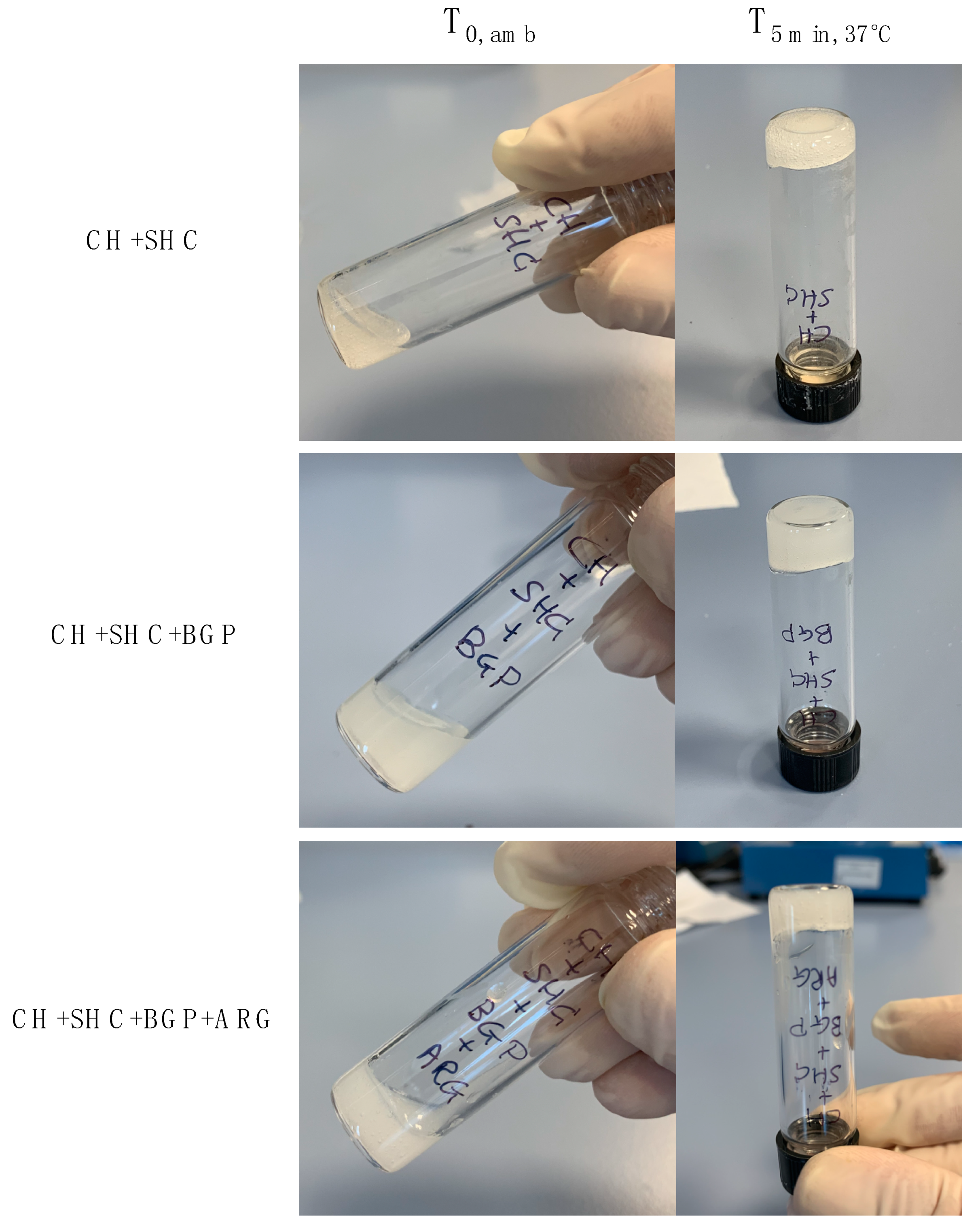
3.2. Hydrogel Injectability and Inversion Tube Tests

All samples were injectable immediately after mixing at RT through a 23G needle (see Video_S1). The thermoresponsive behavior was verified by the inversion tube test. All samples stopped to flow along the vial walls a few minutes after being placed at 37 °C, indicating a rapid gelation as soon as the temperature reached 37 °C (Figure 1). Moreover, when injected into PBS at 37 °C, all CH+GA solutions showed the sol-gel transition forming a stable wire. Instead, no gelation occurred when the CH+GA solutions were injected into PBS at 4 °C (Figure S1, Video_S2 and Video_S3). As shown by rheological tests (Supplementary Materials), the elastic contribution was predominant on the viscous one (material already at the gel state and not sol anymore) for all hydrogel formulations at both 21 and 37 °C. However, both storage modulus (G’) and loss modulus (G’’) are significantly higher when measured at 37 °C, confirming the thermoresponsive behavior observed in the inversion tube test.

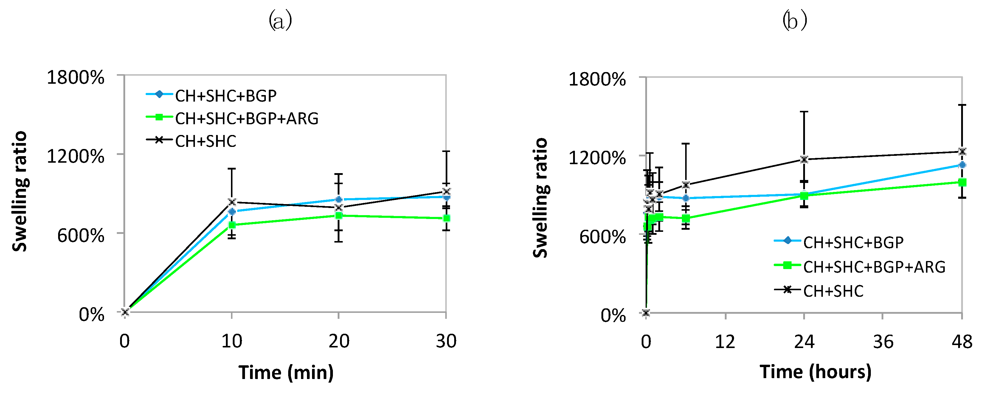
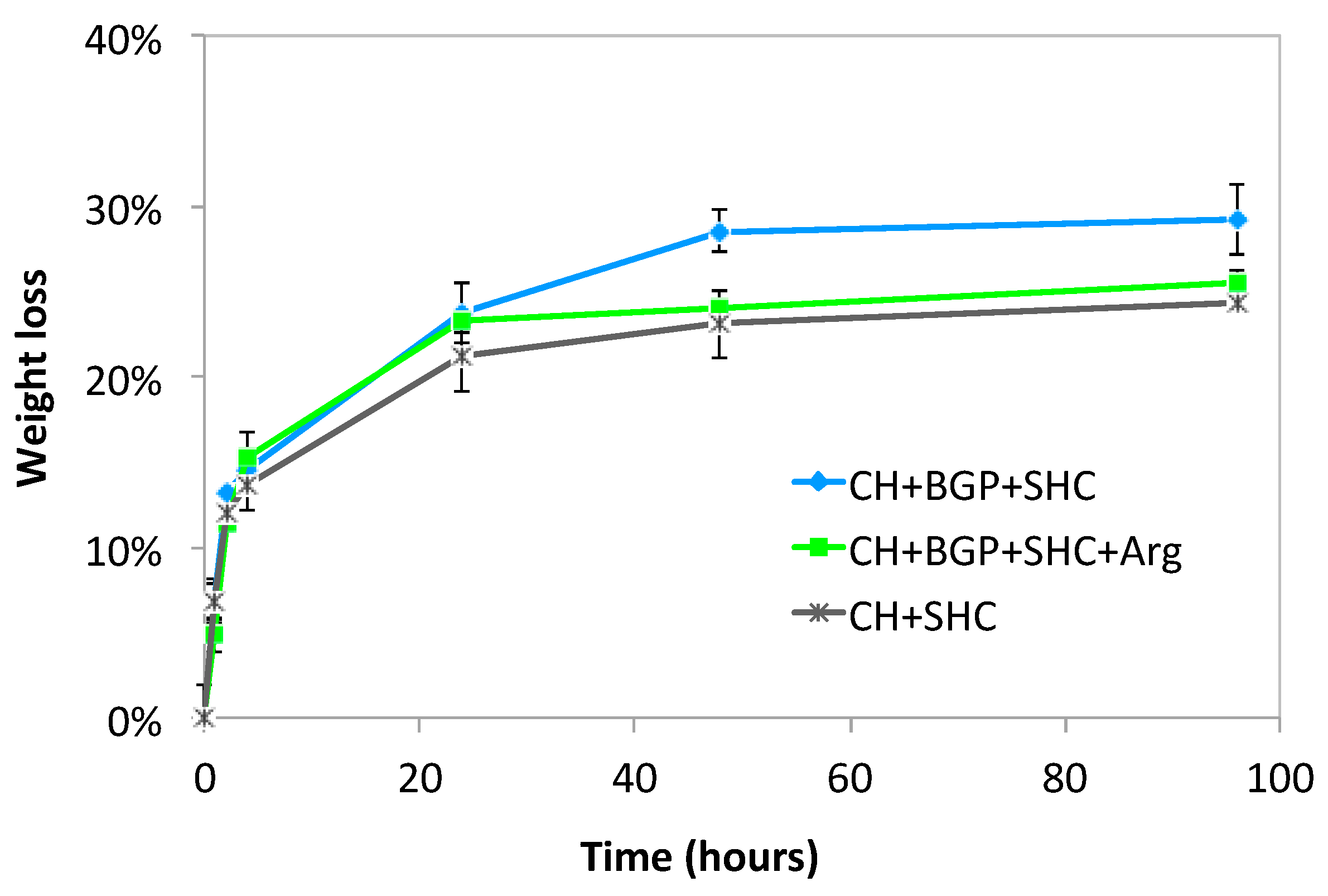
3.3. Swelling Ratio and Stability Tests

For each type of sample, three freeze-dried samples were weighted before and after hydration at different time points in order to investigate their swelling properties. The swelling tests showed that all hydrogels absorbed most of the PBS solution in the first 30 min (SR equal to 920 ± 302, 880 ± 92, 714 ± 92 for CH+SHC, CH+SHC+BGP, CH+SHC+BGP+ARG, respectively) and were able to retain the absorbed water without any release up to 48 h (Figure 2). The three hydrogel formulations have all very high SR values, although CH+SHC+BGP+ARG present a SR significantly lower suggesting a stronger interaction among the chitosan chains due to the presence of three GAs. Moreover, the higher standard deviation of CH+SHC samples might suggest a high inter-specimen variability.
The samples were weighted at different time points in order to determine their stability at the operating conditions. Hydrogels showed weight loss rapidly increasing in the first 6 h, which reached after 24 h a maximum value of 21.2% ± 1.5% and 23.2% ± 0.7% for CH+SHC and CH+SHC+BGP+ARG that remained almost constant up to 96 h in PBS at 37 °C. Samples with SHC+BGP without ARG presented weight loss after 24 h of 23.7% ± 1.7%, similar to the other two hydrogel formulations that however showed a small but significantly different increase in the weight loss (28.5 ± 1.2%) at 48 h, approximately constant throughout the rest of the test (p < 0.05) (Figure 3).

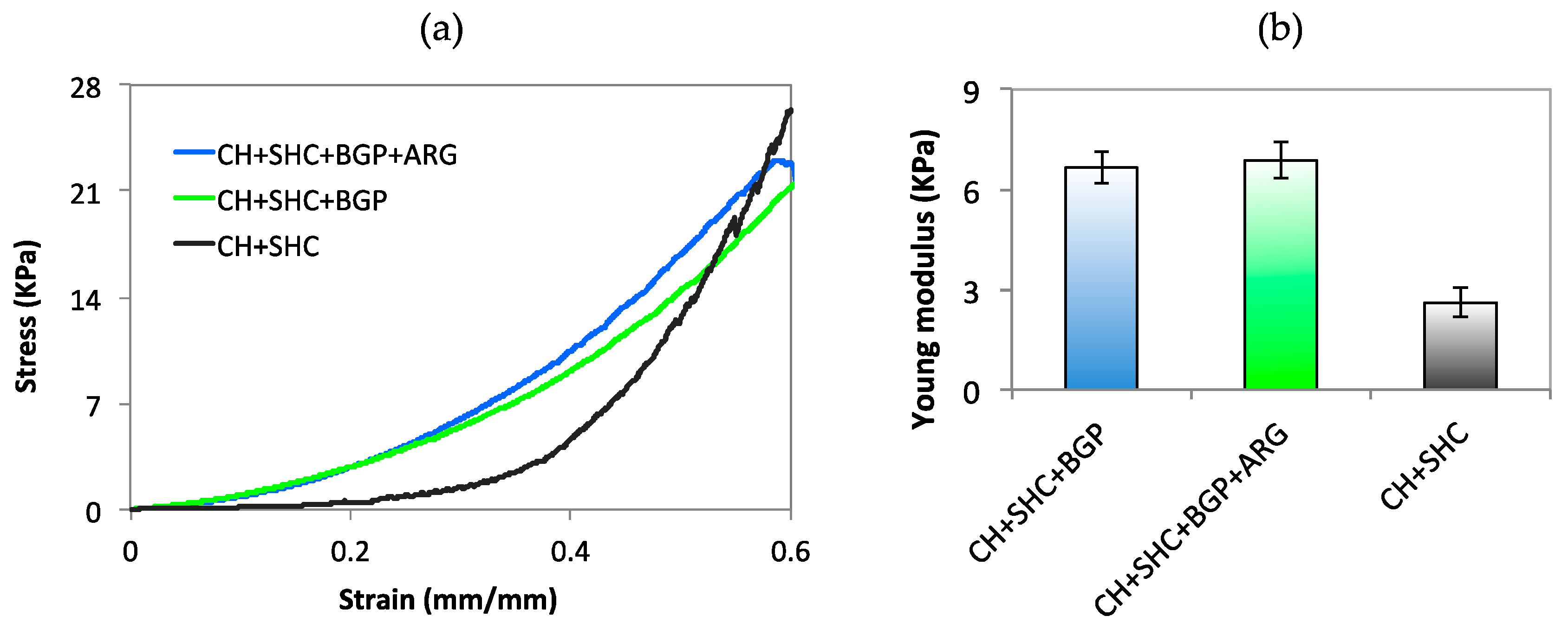
3.4. Compression Test

Unconfined compression tests were carried out to evaluate the stiffness of the hydrogel. The stress-strain curve of all the samples showed a non-linear elastic behavior without any sign of hydrogel fracture up to 50% of strain (Figure 4a). For all samples, the stiffness increased as the strain increased, showing the typical compressive stress-strain behavior of hydrogel materials [40]. However, the stress-strain curve of the CH+SHC sample resulted significantly lower (up to 0.55 of strain) than other two hydrogel formulations. The Young modulus, calculated as the slope of the compressive stress-strain curve at low strain values (0–5%), for CH+SHC+BGP and CH+SHC+BGP+ARG resulted equal to 6.65 ± 0.50 KPa and 6.87 ± 0.53 KPa respectively, significantly higher (p < 0.05) than Young modulus of CH+SHC, equal to 2.61 ± 0.43 KPa (Figure 4b). The compression test results indicated that the addition of BGP to CH+SHC system conferred more stiffness to chitosan hydrogels, suggesting that stronger interactions occur between chitosan chains in the presence of BGP salt. Addition of ARG did not induce any significant change in the hydrogel mechanical properties. Hydrogels showed a compressive stiffness in the range 2–10 KPa, range in which most of the biological soft tissues’ compressive stiffness falls [41].

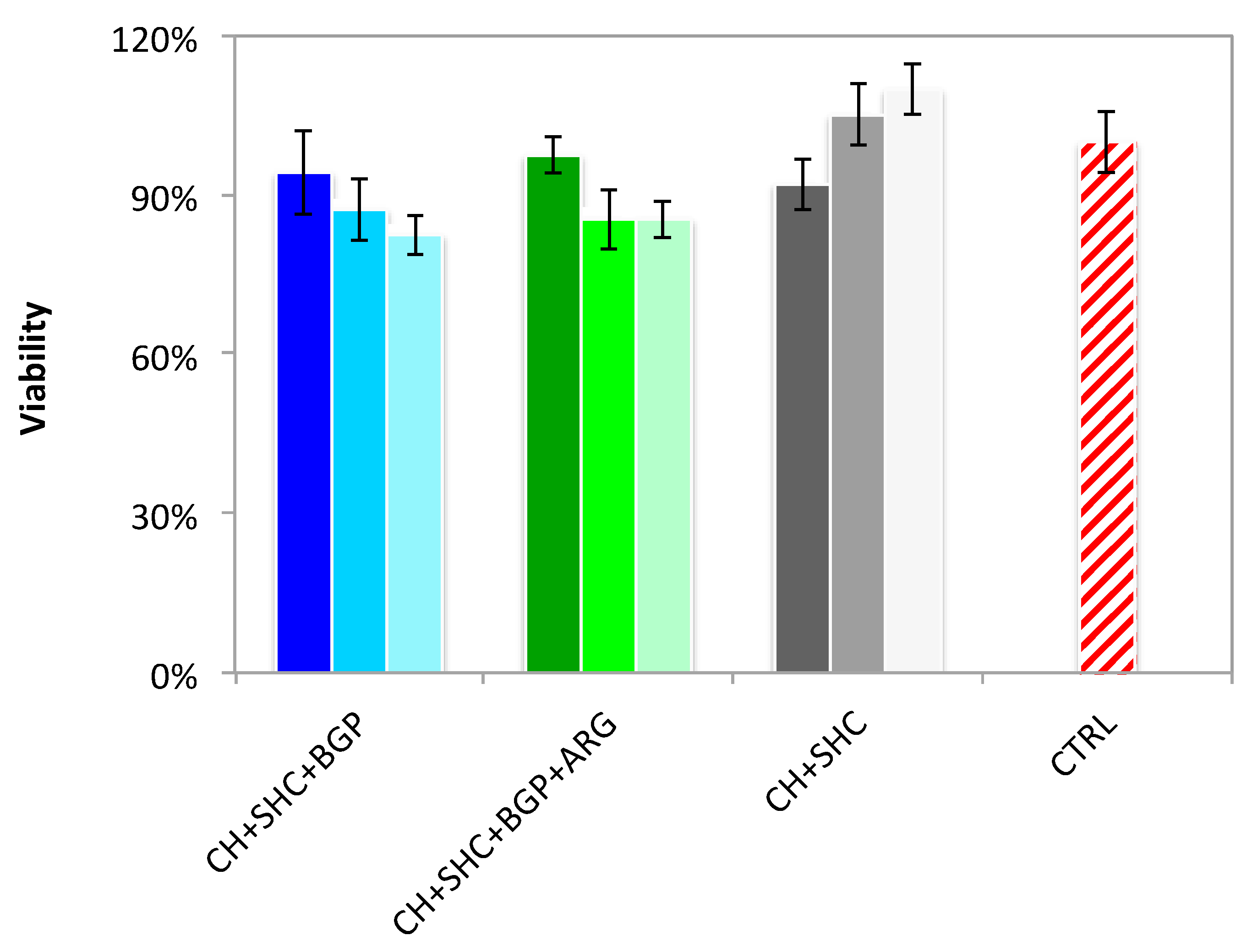
3.5. Cytotoxicity Assay: 3-(4,5-dimethylthiazol-2-yl)-2,5-diphenyltetrazolium bromide (MTT) Assay

Hydrogel biocompatibility was analyzed in SH-SY5Y cells by indirect toxicity test using MTT assay. Cell viability was greater than 80% compared to the control (unconditioned DMEM) for all the hydrogels formulations analyzed. These results indicate that the three different hydrogels did not release into the medium (up to 72 h) any compounds or agents that can significantly affect cell survival/growth and reveal the good biocompatibility properties of these hydrogels (Figure 5).

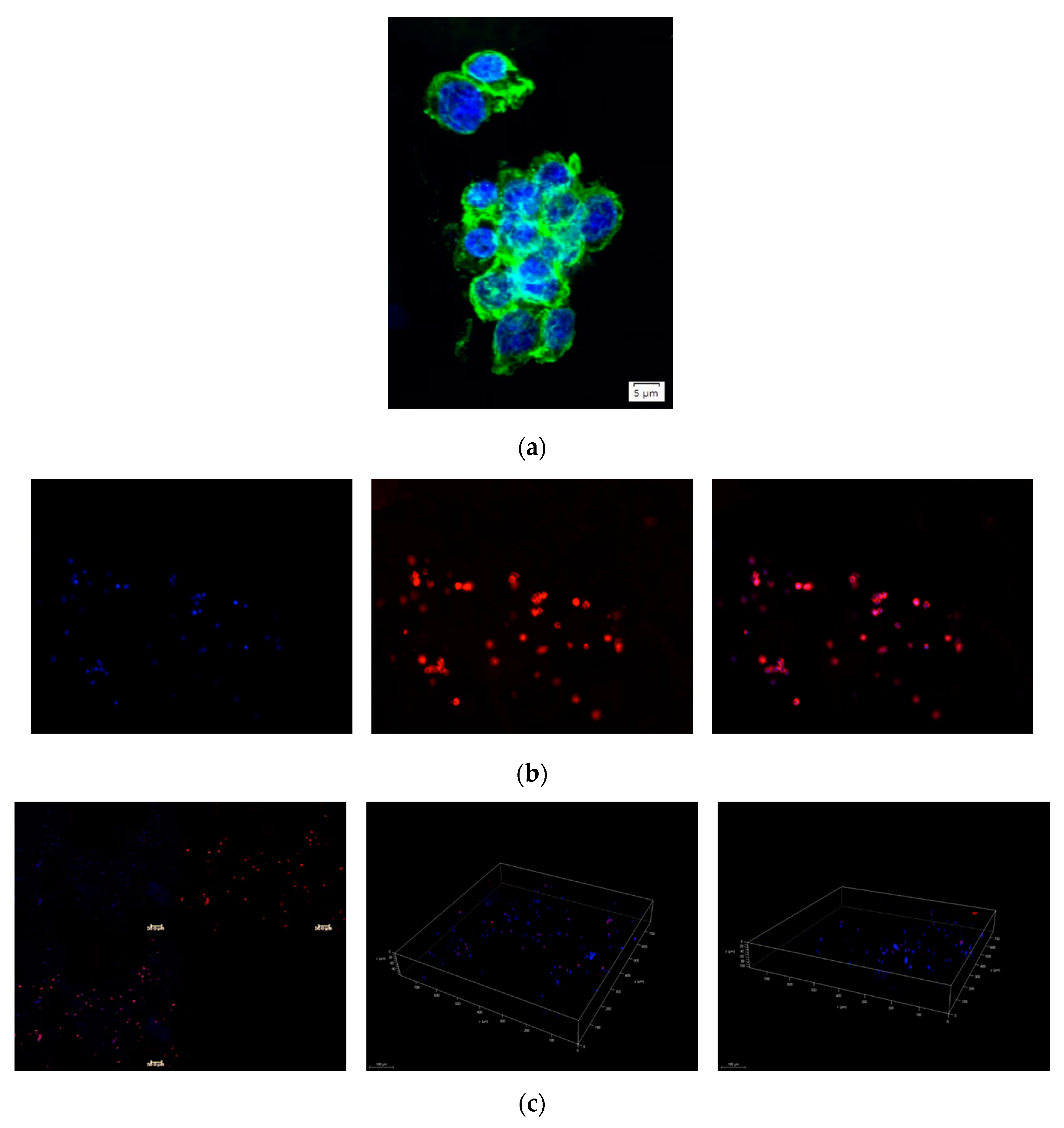
3.6. Analysis of Cell Distribution in Hydrogel by Dapi/Phalloidin Staining

To evaluate the morphology and distribution of SH-SY5Y cells throughout the hydrogels transversal and longitudinal sections of the CH+SHC+BGP hydrogel loaded with the cells were stained for actin-phalloidin, counterstained with DAPI (nuclei), and observed under a fluorescence microscope (Figure 6a,b). SH-SY5Y cells presented a rounded shape morphology (Figure 6a) and were found to be spread over the entire surface of all the slices analyzed (Figure 6b,c), indicating that the cell embedding method allowed the homogeneous distribution of the cells through the hydrogel.

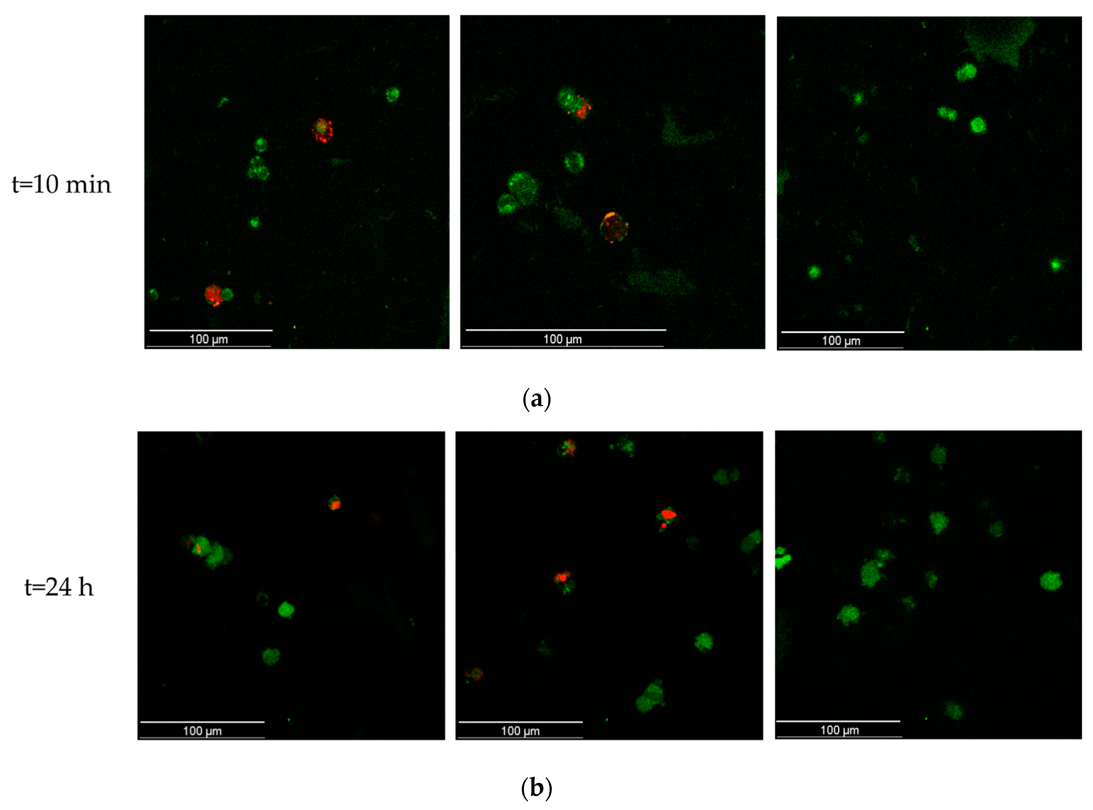
3.7. Cell-Embedded Hydrogel Viability through Live/Dead Staining

The CH+SHC+BGP hydrogel system was tested by live/dead assay and the results clearly indicate that the viability of SH-SY5Y was unaffected by the presence of hydrogel components (Figure 7). Very rare red nuclei, indicating dead cells, could be detected, while numerous metabolically active cells (green colored) were imaged.

4. Discussion

Although other studies reported the obtainment of thermoresponsive chitosan-based hydrogel by using BGP as gelling agent [21] and the addition of SHC to increase the mechanical properties of the CH+BGP system [31], here, we combined for the first time CH with SHC solution as GA and studied the influence of the BGP and ARG addition on the CH+SHC hydrogel properties. Hydrogel solutions flowing freely at RT were successfully obtained after mixing CH solution with SHC, BGP and ARG solutions. The hydrogel solutions turned into a solid state at 37 °C due to the decrease of chitosan chain polarity and the increase of BGP hydrophobic interactions with CH [42]. The hydrogel solutions could all be easily injected into a PBS solution at 37 °C. A sol-gel transition was observed as the temperature increased, showing the hydrogel thermo-responsiveness (see Videos S1, S2, S3 in SI). The inversion tube test results, in agreement with literature, indicated that the gel state of CH-SHC systems is reached after a few minutes of incubation at 37 °C [43]. The addition of an organic salt, BGP, and L-Arg, did not induce any change in the thermoresponsive behavior of the CH+SHC system.
One of the most important properties of hydrogels for 3D cell culture is their ability to incorporate large amounts of water, conventionally evaluated by measuring their swelling capability. The SR was almost equal for the different hydrogel formulations, with average values in the range 600–1000%, in perfect agreement with literature results [18], clearly indicating a superabsorbent behavior. As expected, the SR of the herein proposed hydrogel systems was lower than SR values reported for pure CH hydrogel, confirming that BGP, SHC and Arg interact with CH chain limiting CH swelling capability [40]. Moreover, though all the three hydrogels presented very high SR values, CH+SHC+BGP+ARG SR after 30 min was significantly lower, suggesting a stronger initial interaction among chitosan chains due to the presence of three GAs.
Degradation behavior is another key parameter for the design of 3D hydrogel systems, as it provides an indication of their short and long-term stability during cell culture. The tested hydrogel formulations presented a similar degradation profile, showing a faster weight loss percentage in the first 24 h, without any further increase up to 4 days. The maximum weight loss settled in the range of 25–30%, in agreement with other studies [44]. Samples with SHC+BGP without ARG, however, presented weight loss after 48 h of 28.5 ± 1.2% significantly higher than those of other two hydrogel formulations, indicating the possibility of tuning the degradation profile according to the need, by simply varying the GA solution components.
It is well known that the mechanical properties of cell microenvironment influence cell behavior and functions [45]. From adhesion to differentiation, the most relevant cellular events from early embryogenesis onwards are all, in some way, affected by tissue stiffness [46]. To evaluate the hydrogel bulk stiffness, compression tests have been performed and the results indicated that the addition of BGP induced an increase in the Young modulus in comparison to CH hydrogels with only SHC, likely due to stronger interactions among chitosan chains occurring in the presence of BGP. Conversely, the presence of ARG does not change the hydrogel mechanical properties in compression mode even if a stabilizing effect has been underlined by swelling test. Interestingly, the three hydrogel formulations provide different Young values in compression at low strain values according to the GA composition but always in the range of most of “soft” biological tissues, i.e., 1–10 KPa [41,42,43,44,45,46].
Preliminary biological tests have been performed to assess the hydrogel biocompatibility. The indirect test showed that all three hydrogels systems do not release any cytotoxic substance and are highly biocompatible. As a proof of concept, human neuroblastoma cells were encapsulated within the 3D hydrogel: after 24 h, the cells resulted metabolically active in a large number and spatially uniform within the hydrogel, confirming the hydrogel potential for cell encapsulation. Cell morphology was different from cells grown on a traditional culture plate. In particular, cells showed a more rounded shape and tended to form small clusters, probably because of the 3D configuration provide by hydrogel. Cells may need more time to stretch and extend actin filaments across the hydrogel structure [47].

5. Conclusions

In the present study, three injectable, thermoresponsive, biocompatible CH-based hydrogels were developed and characterized. The hydrogel sol-gel transition occurred at 37 °C and, at the gel state, the system presented a physiological pH compatible with cell encapsulation. The addition of BGP to SHC solutions conferred the CH-based hydrogel system with higher (i) stiffness in compression without any changes in the swelling ratio, (ii) degradation properties and (iii) biocompatibility. The introduction of ARG in the hydrogel system did not alter the overall properties of CH hydrogels, besides conferring a slightly smaller weigh loss percentage due to an increased stability in cell culture conditions. Further biological experiments will elucidate the role of ARG as metabolic boost in this system, as it was observed previously [38]. Overall, this study suggests that by varying the GA solution composition it may be possible to modulate the physical properties of CH-based hydrogel systems according to the target cell requirements. To the best of our knowledge, no studies have been reported on the bioencapsulation of SH-SY5Y using a thermosensitive CH+SHC+BGP+ARG injectable hydrogel as 3D cell culture system.

Supplementary Materials

The following are available online at https://0-www-mdpi-com.brum.beds.ac.uk/2076-3417/10/18/6550/s1, Figure S1, Figure S2, Video S1: CH+SHC+BGP injection at RT, Video S2: CH+SHC+BGP injection in PBS at 37 °C, Video S3: CH+SHC+BGP injection in PBS at 4 °C.

Author Contributions

Conceptualization, F.G., L.M. and A.P.; methodology, A.S.; A.S. hydrogel preparation, swelling and stability tests and cell encapsulation, A.S.; compression tests, result analysis supplementary videos and picture acquisition, F.G.; supervision of cell study, contribution to analyze the biological results, V.L.P., A.R. and A.Q; writing—original draft preparation, A.S. and F.G.; writing—review and editing, A.P. and L.M.; supervision, G.G., L.M. and A.Q.; funding acquisition, G.G. All authors have read and agreed to the published version of the manuscript.

Funding

This research was funded by Regione Puglia, Progetto FISR—C.N.R. “Tecnopolo di nanotecnologia e fotonica per la medicina di precisione”—CUP B83B17000010001, and “Tecnopolo per la medicina di Precisione, Regione Puglia”—CUP:B84I18000540002.

Conflicts of Interest

The authors declare no conflict of interest. The funders had no role in the design of the study; in the collection, analyses, or interpretation of data; in the writing of the manuscript, or in the decision to publish the results.

References

  1. Edmondson, R.; Broglie, J.J.; Adcock, A.F.; Yang, L. Three-Dimensional Cell Culture Systems and Their Applications in Drug Discovery and Cell-Based Biosensors. ASSAY Drug Dev. Technol. 2014, 12, 207–218. [Google Scholar] [CrossRef] [PubMed] [Green Version]
  2. Souza, G.R.; Molina, J.R.; Raphael, R.M.; Ozawa, M.G.; Stark, D.J.; Levin, C.S.; Bronk, L.; Ananta, J.S.; Mandelin, J.; Georgescu, M.-M.; et al. Three-dimensional tissue culture based on magnetic cell levitation. Nat. Nanotechnol. 2010, 5, 291–296. [Google Scholar] [CrossRef] [PubMed] [Green Version]
  3. Huang, Y.-T.; Lai, P.C.; Chiu, T.H. Overexpression of BDNF and TrkB in human bladder cancer specimens. Oncol. Rep. 2010, 24, 1265–1270. [Google Scholar] [CrossRef]
  4. Baharvand, H.; Hashemi, S.M.; Ashtiani, S.K.; Farrokhi, A. Differentiation of human embryonic stem cells into hepatocytes in 2D and 3D culture systems in vitro. Int. J. Dev. Biol. 2006, 50, 645–652. [Google Scholar] [CrossRef] [PubMed] [Green Version]
  5. Chang, T.T.; Hughes-Fulford, M. Monolayer and Spheroid Culture of Human Liver Hepatocellular Carcinoma Cell Line Cells Demonstrate Distinct Global Gene Expression Patterns and Functional Phenotypes. Tissue Eng. Part A 2009, 15, 559–567. [Google Scholar] [CrossRef] [PubMed]
  6. Härmä, V.; Virtanen, J.; Mäkelä, R.; Happonen, A.; Mpindi, J.-P.; Knuuttila, M.; Kohonen, P.; Lötjönen, J.; Kallioniemi, O.; Nees, M. A Comprehensive Panel of Three-Dimensional Models for Studies of Prostate Cancer Growth, Invasion and Drug Responses. PLoS ONE 2010, 5, e10431. [Google Scholar] [CrossRef] [PubMed]
  7. Mitra, M.; Mohanty, C.; Harilal, A.; Maheswari, U.K.; Sahoo, S.K.; Krishnakumar, S. A novel in vitro three-dimensional retinoblastoma model for evaluating chemotherapeutic drugs. Mol. Vis. 2012, 18, 1361–1378. [Google Scholar] [PubMed]
  8. Badylak, S.F.; Freytes, D.; Gilbert, T.W. Extracellular matrix as a biological scaffold material: Structure and function. Acta Biomater. 2009, 5, 1–13. [Google Scholar] [CrossRef]
  9. Hutmacher, D.W. Scaffolds in Tissue Engineering Bone and Cartilage. Biomater. Silver Jubil. Compend. 2000, 21, 175–189. [Google Scholar]
  10. Gasperini, L.; Mano, J.F.; Reis, R.L. Natural polymers for the microencapsulation of cells. J. R. Soc. Interface 2014, 11, 20140817. [Google Scholar] [CrossRef] [Green Version]
  11. Dutta, R.C.; Dutta, A.K. Cell-interactive 3D-scaffold; advances and applications. Biotechnol. Adv. 2009, 27, 334–339. [Google Scholar] [CrossRef] [PubMed]
  12. Hoffman, A.S. Hydrogels for biomedical applications. Adv. Drug Deliv. Rev. 2012, 64, 18–23. [Google Scholar] [CrossRef]
  13. Zhao, W.; Jin, X.; Cong, Y.; Liu, Y.; Fu, J. Degradable natural polymer hydrogels for articular cartilage tissue engineering. J. Chem. Technol. Biotechnol. 2012, 88, 327–339. [Google Scholar] [CrossRef]
  14. Chang, C.; Duan, B.; Cai, J.; Zhang, L. Superabsorbent hydrogels based on cellulose for smart swelling and controllable delivery. Eur. Polym. J. 2010, 46, 92–100. [Google Scholar] [CrossRef]
  15. Lee, J.; Cuddihy, M.J.; Kotov, N.A. Three-Dimensional Cell Culture Matrices: State of the Art. Tissue Eng. Part B Rev. 2008, 14, 61–86. [Google Scholar] [CrossRef] [PubMed] [Green Version]
  16. Buenger, D.; Topuz, F.; Groll, J. Hydrogels in sensing applications. Prog. Polym. Sci. 2012, 37, 1678–1719. [Google Scholar] [CrossRef]
  17. Klouda, L.; Mikos, A.G. Thermoresponsive hydrogels in biomedical applications. Eur. J. Pharm. Biopharm. 2008, 68, 34–45. [Google Scholar] [CrossRef] [Green Version]
  18. Slaughter, B.V.; Khurshid, S.S.; Fisher, O.Z.; Khademhosseini, A.; Peppas, N.A. Hydrogels in Regenerative Medicine. Adv. Mater. 2009, 21, 3307–3329. [Google Scholar] [CrossRef] [Green Version]
  19. Peppas, N.A.; Hilt, J.Z.; Khademhosseini, A.; Langer, R. Hydrogels in Biology and Medicine: From Molecular Principles to Bionanotechnology. Adv. Mater. 2006, 18, 1345–1360. [Google Scholar] [CrossRef]
  20. Thiele, J.; Ma, Y.; Bruekers, S.M.C.; Ma, S.; Huck, W.T.S. 25th Anniversary Article: Designer Hydrogels for Cell Cultures: A Materials Selection Guide. Adv. Mater. 2013, 26, 125–148. [Google Scholar] [CrossRef]
  21. Zhou, H.; Jiang, L.J.; Cao, P.P.; Li, J.B.; Chen, X. Glycerophosphate-based chitosan thermosensitive hydrogels and their biomedical applications. Carbohydr. Polym. 2015, 117, 524–536. [Google Scholar] [CrossRef] [PubMed]
  22. Gurski, L.A.; Jha, A.K.; Zhang, C.; Jia, X.; Farach-Carson, M.C. Hyaluronic acid-based hydrogels as 3D matrices for in vitro evaluation of chemotherapeutic drugs using poorly adherent prostate cancer cells. Biomaterials 2009, 30, 6076–6085. [Google Scholar] [CrossRef] [PubMed] [Green Version]
  23. Azab, A.K.; Orkin, B.; Doviner, V.; Nissan, A.; Klein, M.; Srebnik, M.; Rubinstein, A. Crosslinked chitosan implants as potential degradable devices for brachytherapy: In vitro and in vivo analysis. J. Control. Release 2006, 111, 281–289. [Google Scholar] [CrossRef] [PubMed]
  24. Ehrbar, M.; Djonov, V.; Schnell, C.; Tschanz, S.; Martiny-Baron, G.; Schenk, U.; Wood, J.; Burri, P.H.; Hubbell, J.A.; Zisch, A.H. Cell-Demanded Liberation of VEGF121From Fibrin Implants Induces Local and Controlled Blood Vessel Growth. Circ. Res. 2004, 94, 1124–1132. [Google Scholar] [CrossRef] [Green Version]
  25. Butcher, J.T.; Nerem, R.M. Porcine aortic valve interstitial cells in three-dimensional culture: Comparison of phenotype with aortic smooth muscle cells. J. Hear. valve Dis. 2004, 13, 478–486. [Google Scholar]
  26. Barralet, J.E.; Wang, L.; Lawson, M.; Triffitt, J.T.; Cooper, P.R.; Shelton, R.M. Comparison of bone marrow cell growth on 2D and 3D alginate hydrogels. J. Mater. Sci. Mater. Electron. 2005, 16, 515–519. [Google Scholar] [CrossRef] [Green Version]
  27. Bhattarai, N.; Gunn, J.; Zhang, M. Chitosan-based hydrogels for controlled, localized drug delivery. Adv. Drug Deliv. Rev. 2010, 62, 83–99. [Google Scholar] [CrossRef]
  28. Elieh-Ali-Komi, D.; Hamblin, M.R. Chitin and Chitosan: Production and Application of Versatile Biomedical Nanomaterials. Int. J. Adv. Res. 2016, 4, 411–427. [Google Scholar]
  29. Cho, J.; Heuzey, M.-C.; Bégin, A.; Carreau, P.J. Physical Gelation of Chitosan in the Presence of β-Glycerophosphate: The Effect of Temperature. Biomacromolecules 2005, 6, 3267–3275. [Google Scholar] [CrossRef]
  30. Huang, Z.; Yu, B.; Feng, Q.; Li, S. Modification of an Injectable Chitosan Scaffold by Blending with NaHCO3 to Improve Cytocompatibility. Polym. Polym. Compos. 2011, 19, 781–788. [Google Scholar] [CrossRef]
  31. Assaad, E.; Maire, M.; Lerouge, S. Injectable thermosensitive chitosan hydrogels with controlled gelation kinetics and enhanced mechanical resistance. Carbohydr. Polym. 2015, 130, 87–96. [Google Scholar] [CrossRef] [PubMed]
  32. Efron, D.T.; Kirk, S.J.; Regan, M.C.; Wasserkrug, H.L.; Barbul, A. Nitric oxide generation from L-arginine is required for optimal human peripheral blood lymphocyte DNA synthesis. Surgery 1991, 110, 327–334. [Google Scholar] [PubMed]
  33. Greene, J.M.; Feugang, J.M.; Pfeiffer, K.E.; Stokes, J.V.; Bowers, S.D.; Ryan, P.L. L-arginine enhances cell proliferation and reduces apoptosis in human endometrial RL95-2 cells. Reprod. Biol. Endocrinol. 2013, 11, 15. [Google Scholar] [CrossRef] [PubMed] [Green Version]
  34. Rackéf, K.; Warnken, M. L-Arginine Metabolic Pathways~!2009-11-28~!2010-03-03~!2010-05-04~! Open Nitric Oxide J. 2010, 2, 9–19. [Google Scholar] [CrossRef]
  35. Damodaran, V.B.; Bhatnagar, H.; Rubin, D.; Reynolds, M.M. Chapter 6—Nitric oxide donors in nerve regeneration. In Nitric Oxide Donors; Seabra, A.B., Ed.; Academic Press: Cambridge, MA, USA, 2017; pp. 141–168. [Google Scholar]
  36. Shi, H.P.; Wang, S.M.; Zhang, G.X.; Zhang, Y.J.; Barbul, A. Supplemental l-arginine enhances wound healing following trauma/hemorrhagic shock. Wound Repair Regen. 2007, 15, 66–70. [Google Scholar] [CrossRef]
  37. Izzo, D.; Palazzo, B.; Scalera, F.; Gullotta, F.; Lapesa, V.; Scialla, S.; Sannino, A.; Gervaso, F. Chitosan scaffolds for cartilage regeneration: Influence of different ionic crosslinkers on biomaterial properties. Int. J. Polym. Mater. 2018, 68, 936–945. [Google Scholar] [CrossRef]
  38. Scialla, S.; Barca, A.; Palazzo, B.; D’Amora, U.; Russo, T.; Gloria, A.; De Santis, R.; Verri, T.; Sannino, A.; Ambrosio, L.; et al. Bioactive chitosan-based scaffolds with improved properties induced by dextran-grafted nano-maghemite and l -arginine amino acid. J. Biomed. Mater. Res. Part A 2019, 107, 1244–1252. [Google Scholar] [CrossRef]
  39. Nitti, P.; Gallo, N.; Palazzo, B.; Sannino, A.; Polini, A.; Verri, T.; Barca, A.; Gervaso, F. Effect of l-Arginine treatment on the in vitro stability of electrospun aligned chitosan nanofiber mats. Polym. Test. 2020, 91, 106758. [Google Scholar] [CrossRef]
  40. Puppi, D.; Migone, C.; Morelli, A.; Bartoli, C.; Gazzarri, M.; Pasini, D.; Chiellini, F. Microstructured chitosan/poly(γ-glutamic acid) polyelectrolyte complex hydrogels by computer-aided wet-spinning for biomedical three-dimensional scaffolds. J. Bioact. Compat. Polym. 2016, 31, 531–549. [Google Scholar] [CrossRef]
  41. Vedadghavami, A.; Minooei, F.; Mohammadi, M.H.; Khetani, S.; Kolahchi, A.R.; Mashayekhan, S.; Sanati-Nezhad, A. Manufacturing of hydrogel biomaterials with controlled mechanical properties for tissue engineering applications. Acta Biomater. 2017, 62, 42–63. [Google Scholar] [CrossRef]
  42. Deng, A.; Kang, X.; Zhang, J.; Yang, Y.; Yang, S. Enhanced gelation of chitosan/β-sodium glycerophosphate thermosensitive hydrogel with sodium bicarbonate and biocompatibility evaluated. Mater. Sci. Eng. C 2017, 78, 1147–1154. [Google Scholar] [CrossRef] [PubMed]
  43. Ke, X.; Li, M.; Wang, X.; Liang, J.; Wang, X.; Wu, S.; Long, M.; Hu, C. An injectable chitosan/dextran/β -glycerophosphate hydrogel as cell delivery carrier for therapy of myocardial infarction. Carbohydr. Polym. 2020, 229, 115516. [Google Scholar] [CrossRef] [PubMed]
  44. Ceccaldi, C.; Assaad, E.; Hui, E.; Buccionyte, M.; Adoungotchodo, A.; Lerouge, S. Optimization of Injectable Thermosensitive Scaffolds with Enhanced Mechanical Properties for Cell Therapy. Macromol. Biosci. 2017, 17, 1600435. [Google Scholar] [CrossRef] [PubMed]
  45. Chen, J.; Irianto, J.; Inamdar, S.; Pravincumar, P.; Lee, D.A.; Bader, D.; Knight, M.M. Cell Mechanics, Structure, and Function Are Regulated by the Stiffness of the Three-Dimensional Microenvironment. Biophys. J. 2012, 103, 1188–1197. [Google Scholar] [CrossRef] [PubMed] [Green Version]
  46. Guimarães, C.F.; Gasperini, L.; Marques, A.P.; Reis, R.L. The stiffness of living tissues and its implications for tissue engineering. Nat. Rev. Mater. 2020, 5, 351–370. [Google Scholar] [CrossRef]
  47. Aubin, H.; Nichol, J.W.; Hutson, C.B.; Bae, H.; Sieminski, A.L.; Cropek, D.M.; Akhyari, P.; Khademhosseini, A. Directed 3D cell alignment and elongation in microengineered hydrogels. Biomaterials 2010, 31, 6941–6951. [Google Scholar] [CrossRef] [Green Version]
Figure 1. Inversion tube test: pictures of the three hydrogel formulations taken immediately after the mixing at RT and after 5 min of incubation at 37 °C.
Applsci 10 06550 g001
Figure 2. Swelling ratio of the three hydrogels: CH+SHC, CH+SHC+BGP, CH+SHC+BGP+ARG in the first 30 min (a) and up to 48 h (b).
Applsci 10 06550 g002
Figure 3. Stability of the three hydrogels CH+SHC, CH+SHC+BGP, CH+SHC+BGP+ARG up to 4 days.
Applsci 10 06550 g003
Figure 4. (a) Example of the stress-strain curves from the compression test performed on the three hydrogel formulations. (b) Young modulus of the three hydrogels CH+SHC, CH+SHC+BGP, CH+SHC+BGP+ARG (average + standard deviation, n = 3).
Applsci 10 06550 g004
Figure 5. Viability of SH-SY5Y cells exposed for 24 h to medium conditioned with the three hydrogels CH+SHC, CH+SHC+BGP, CH+SHC+BGP+ARG for 24 (darker color), 48 (medium color), 72 h (lighter color). Cell viability was measured using MTT assay and data was reported as average ± standard deviation (n = 3).
Applsci 10 06550 g005
Figure 6. (a) Morphology of SH-SY5Y cells growth in the CH+BGP+SHC hydrogel for 24 h. Cells were stained with FITC-phalloidin (green, actin filaments) and DAPI (blue, nuclei). Merged FITC/DAPI. Scale bar, 5 µm. (b) Low magnification images of taken after 24 h of SH-SY5Y embedded in CH+SHC+BGP hydrogel loaded with SH-SY5Y after 24 h of culture. Cells were stained with TRICT-phalloidin for actin filaments (red, left panel) and DAPI for nuclei (blue, middle panel). Right panel shows the merged image. Scale bar, 200 µm. (c) 3D reconstruction of cell distribution in the hydrogel (CH+BGP+SHC) sample obtained from confocal Z-stacks.
Applsci 10 06550 g006
Figure 7. Representative live/dead (green/red) images for SH-SY5Y cells encapsulated in CH+BGP+SHC hydrogel for 10 min (a) and 24 h (b). Scale bar, 100 µm.
Applsci 10 06550 g007
Table 1. Initial and final concentration of GA and CH solutions. pH values of GA solutions (before mixing) and hydrogels (after mixing).
GA Solution/HydrogelsInitial ConcentrationInitial pHFinal Concentration in HydrogelFinal pH
BGP0.5M8.60.2 M-
BGP0.75 M8.60.2 M-
BGP1.5 M8.60.2 M-
SHC0.375 M8.50.05 M-
Arg0.075 M5.50.01 M-
CH3%5.52%-
CH+SHC---7.4
CH+SHC+BGP---7.5
CH+SHC+BGP+Arg---7.2

