Article

Salivary Extracellular DNA and DNase Activity in Periodontitis

by
Barbora Konečná
1,
Alexandra Gaál Kovalčíková
2,
Alexandra Pančíková
3,
Bohuslav Novák
4,
Eva Kovaľová
5,
Peter Celec
1,6,7 and
Ľubomíra Tóthová
1,*
1
Institute of Molecular Biomedicine, Faculty of Medicine, Comenius University in Bratislava, 811 08 Bratislava, Slovakia
2
Department of Pediatrics, National Institute of Children’s Diseases and Faculty of Medicine, Comenius University in Bratislava, 831 01 Bratislava, Slovakia
3
School of Biological Sciences, University of Edinburgh, Edinburgh EH9 3FF, UK
4
Department of Stomatology and Maxillofacial Surgery, Faculty of Medicine, Comenius University, 811 08 Bratislava, Slovakia
5
Department of Maxillofacial Surgery and Dental Hygiene of J.A. Reiman Hospital in Prešov, 080 01 Prešov, Slovakia
6
Institute of Pathophysiology, Faculty of Medicine, Comenius University in Bratislava, 811 08 Bratislava, Slovakia
7
Department of Molecular Biology, Faculty of Natural Sciences, Comenius University in Bratislava, 841 04 Bratislava, Slovakia
*
Author to whom correspondence should be addressed.
Submission received: 2 October 2020 / Revised: 22 October 2020 / Accepted: 22 October 2020 / Published: 25 October 2020
(This article belongs to the Special Issue Salivary Biomarkers: Future Diagnostic and Clinical Utilities)

Abstract

:
Extracellular DNA (ecDNA) is a potential marker and predictor in several inflammatory diseases. Periodontitis, a chronic inflammatory disease, is associated with epithelial cell death and could lead to release of DNA. Our aim was to analyze salivary DNA concentration and deoxyribonuclease (DNase) activity in periodontitis patients. We hypothesized that salivary ecDNA will be higher than in controls and could serve as a marker of periodontitis severity. Samples of saliva were collected from 25 patients with chronic periodontitis and 29 age-matched controls. DNA was quantified fluorometrically in whole saliva, as well as in supernatants after centrifugation (depletion of cells at 1600× g) and in double-centrifuged supernatants (depletion of cell debris at 1600× g and 16,000× g). The subcellular origin of ecDNA was assessed using real-time PCR. In comparison to controls, patients with periodontitis had twofold higher salivary DNA (p < 0.01), higher mitochondrial DNA in centrifuged supernatants (p < 0.05) and lower nuclear ecDNA in double-centrifuged samples (p < 0.05). No correlations were found between salivary DNA and oral health status, but mitochondrial DNA positively correlated with papillary bleeding index in centrifuged samples. Salivary DNase activity was comparable between the groups. In conclusion, we proved that salivary DNA is higher in periodontitis. The source of the higher mitochondrial DNA in cell-free saliva and the causes of lower nuclear ecDNA remain to be elucidated. Further studies should focus on the role of mitochondrial DNA as a potential driver of inflammation in periodontitis.

1. Introduction

Extracellular DNA (ecDNA), i.e., DNA outside of the cells, was first discovered in the plasma of pregnant women [1] and cancer patients [2]. EcDNA could trigger inflammation and worsen the state of patients during pregnancy complications or cancer. In plasma, ecDNA has been studied also as a potential biomarker of sepsis [3], trauma [4], diabetes mellitus or organ transplantation [5]. It has been hypothesized that plasmatic ecDNA could be used as a personalized cancer therapy [6]. Even though usually plasma serves as a source of ecDNA, despite the common difficulties, such as high fragmentation and high variability in the yield, saliva might be of special interest in oral diseases, since it reflects the local oral conditions. Also, ecDNA has been studied in saliva of head and neck cancer patients, and it has been shown that mitochondrial DNA concentration is increased in saliva in these patients when compared to control healthy group [7]. All of the above indicates that body fluids DNA or ecDNA are not a disease-specific marker, however, it points towards pathogeneses of diseases, which is not fully understood.
Periodontitis is a chronic multifactorial inflammatory oral disease. It starts with bacterial plaque concentrating near the gingival part followed by gingivitis. The oral microbiome indeed plays a role in this initial stage [8]. Under normal circumstances, there is a fragile balance between microbes that act as the nonharmful commensals and the oral health status [9]. The dysbiosis consequence includes plaque build-up consisting of immunogenic substances in the subgingival crevice of teeth, which leads to the further activation of the immune system. The resulting inflammation and neutrophils activation, as a nonspecific reaction, damages the surrounding healthy tissue releasing several other immunogenic substances, triggering thus immune overreaction and circulus vitiosus. In susceptible individuals, the untreated inflammation can later outgrow into loss of connective tissue and decline in bone support (periodontitis) and eventually a tooth loss [10,11] with permanent inflammation of surrounding tissue. Recently, neutrophil extracellular traps as weblike structures containing DNA were already described as potential immunogenic agents in periodontitis, and they were hypothesized to be involved in the pathogenesis of this disease [12,13,14]. Neutrophils are activated during inflammation, directed towards the location of inflammation, in our case the oral cavity, and the fight against bacteria forming the traps. Neutrophil extracellular traps consist of histone proteins and DNA, therefore they are one of the various sources of ecDNA among others such as apoptosis or necrosis [15]. Salivary ecDNA obtained by double-centrifugation, as well as DNA in whole and centrifuged saliva, as potential triggers of inflammation in periodontitis, are worth testing as possible indicators of the oral status. As in plasma, there are also nucleases in saliva, such as deoxyribonuclease (DNase) that is responsible for degradation of ecDNA [16,17]. Therefore, we think that, together with DNA concentrations and origin analysis, it is also important to specify activity of DNase in saliva. We hypothesize that salivary DNA will be increased in patients with worsened oral health and DNase activity could be also altered. Therefore, in this study, we investigated whether DNA in whole saliva, in centrifuged and double-centrifuged samples differs between periodontitis patients and healthy controls and whether salivary DNase activity is affected.

2. Methods

2.1. Patients

Total number of participants was 54 of whom 25 patients were diagnosed with periodontitis (11 women and 14 men) and 29 were aged matched without periodontitis (18 women and 11 men). The presence of clinical attachment loss of at least 3 mm in two or more nonadjacent teeth was considered to be periodontitis along with radiographically confirmed alveolar bone loss. For an accurate diagnosis, the examination of the periodontal status of subjects was performed using several clinical markers. Gingival index (GI)—qualitative changes of gingival soft tissue—was determined according to Loe [18]. Briefly, a score of 0 represents no changes of gingival soft tissue; a score of 1 means slight inflammation with the mild change of color and discrete edema without bleeding on probing (BOP); and a score of 3 describes severe inflammation, redness, edema with ulcerations and the presence of spontaneous bleeding.
Papillary bleeding index (PBI)—the bleeding after cautiously probing the sulcus—was determined by PBI as described formerly by Newbrun [19]. A score of 0 means no bleeding; score 1 means one bleeding point few seconds after probing; a score of 2 means intermediate bleeding from several points and a score of 3 represents profuse bleeding along the gingival sulcus even after gentle probing. The bleeding on probing at the bottom of the pocket is defined as BOP. The BOP was assessed using a standardized probe (WHO probe 550B, LM Dental, Finland) as described previously [20]. The patients were evaluated for six areas for all teeth (midbuccal, midlingual, mesiolingual, distolingual, mesiobuccal, and distobuccal). To consider differences in the number of teeth, all indexes were divided by the total number of teeth. All measurements were performed by a single trained examiner to avoid interindividual differences. The clinical evaluation of patients can be seen in Table 1.
Prior to examination, all participants were asked not to eat, drink or brush their teeth 60 min before saliva sampling. All samples were collected at a dental clinic in Prešov, Slovakia. Sampling was performed in the morning (8:00–10:00) into sterile tubes. The whole unstimulated saliva samples were collected by passive drooling for 10 min and salivary flow was recorded. Whole saliva samples were aliquoted and stored; the rest of the whole saliva was centrifuged at 1600× g for 10 min to wash down the cell debris. The centrifuged supernatant was aliquoted and stored, followed by 16,000× g centrifugation of the rest of the supernatant for 10 min to obtain cellfree double-centrifuged supernatant [21]. Original saliva as well as samples after each centrifugation were measured by bicinchonic acid protein assay kit (Sigma-Aldrich, St. Louis, Missouri, United States) to determine the protein content and stored at −20 °C until further processes. Patients with a known extraoral systemic disease, taking any medications including vitamin supplements and smokers were excluded from this study. The study was approved by the Ethical Committee of the Department of Maxillofacial Surgery and Dental Hygiene of J.A. Reiman Hospital in Prešov. The clinical part of this study was performed according to the principles given in the Declaration of Helsinki. All participants signed written informed consent declarations.

2.2. DNA Isolation

DNA was isolated from 200 µL of all saliva samples using QIAamp DNA Blood Mini Kit (Qiagen, Hilden, Germany) according to the protocol of the manufacturer. Sixty µL of ultrapure water were used for the elution of DNA as the final step. DNA samples were stored at −20 °C until further analysis.

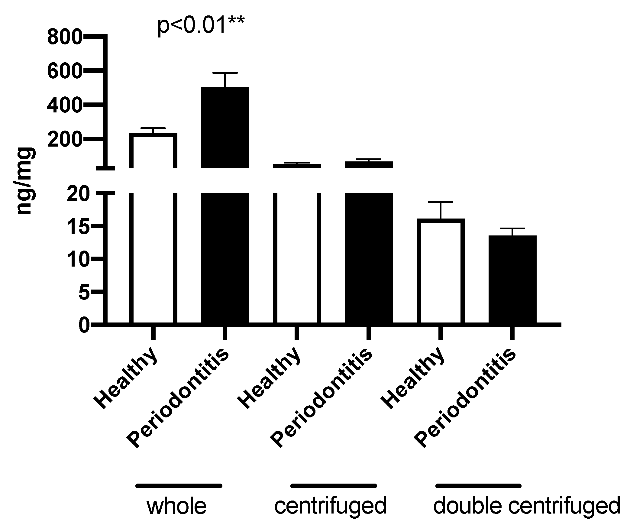
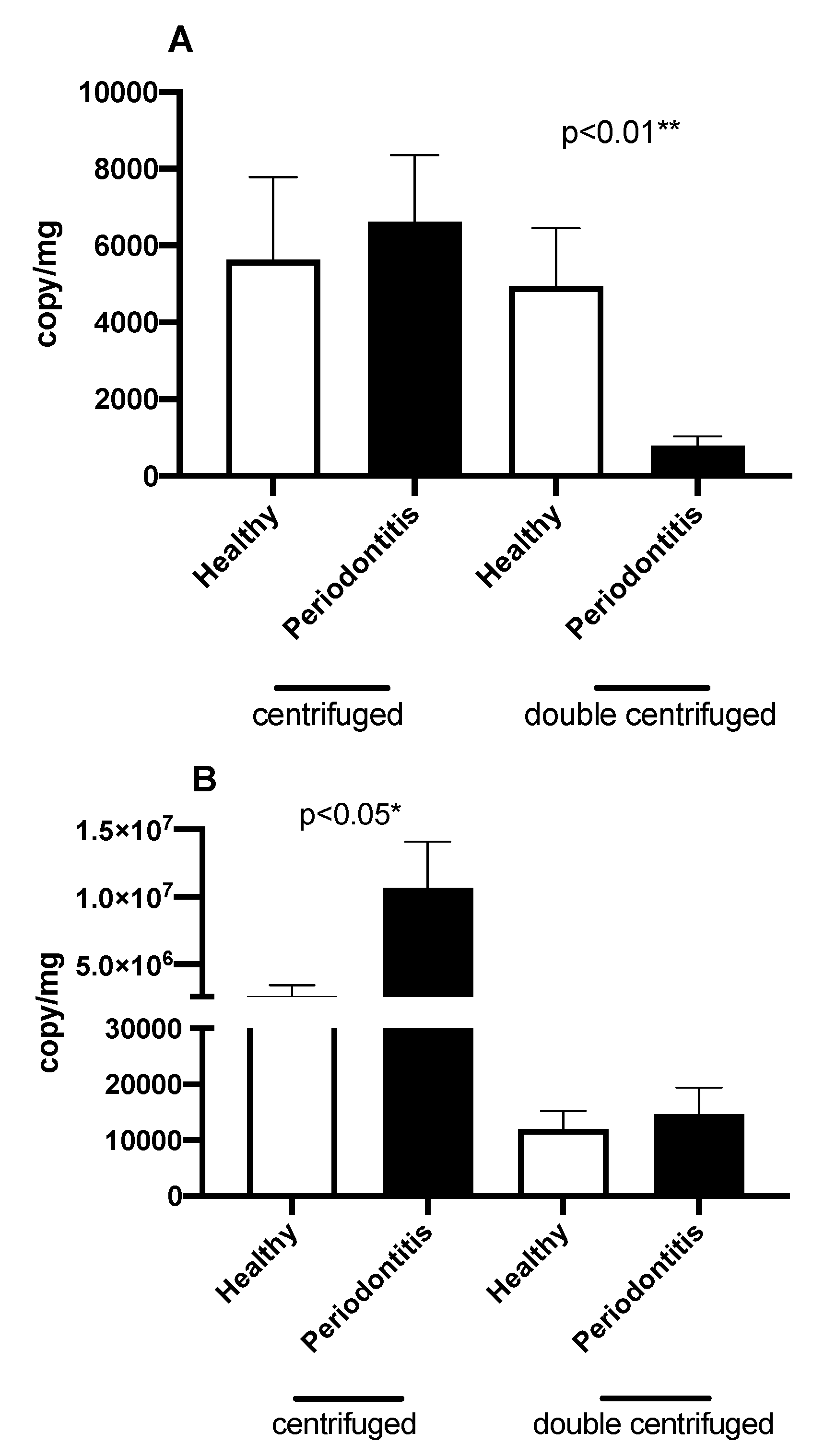
2.3. Quantification of ecDNA

Qubit Fluorometer and Qubit dsDNA HS Assay Kit (Invitrogen, Carlsbad, CA, USA) were used for the measurement of total cfDNA according to the protocol from the manufacturer. Real-time polymerase chain reaction (RT-PCR) was used for the quantification of nuclear DNA (ncDNA) and mitochondrial DNA (mtDNA) in saliva isolates. RT-PCR was performed on the Mastercycler realplex 4 (Eppendorf, Hamburg, Germany) using the SsoAdvanced Universal SYBR Green Supermix (Bio-Rad, Hercules, CA, USA) with the following program: one cycle of 3 min at 98 °C, followed by 40 cycles of 98 °C for 15 s for denaturation and annealing for 30 s at 51 °C for ncDNA and 47 °C for mtDNA and extension 30 s at 60 °C. Primers were designed for the amplification of human globin gene to quantify ncDNA (F: GCT TCT GAC ACA ACT GTG TTC, R: CAC CAA CTT CAT CCA CGT TCA) and D-loop to quantify mtDNA (F: CAT AAA AAC CCA ATC CAC ATC A, R: GAC GGG TGG CTT TGG AGT). All values were normalized to proteins.

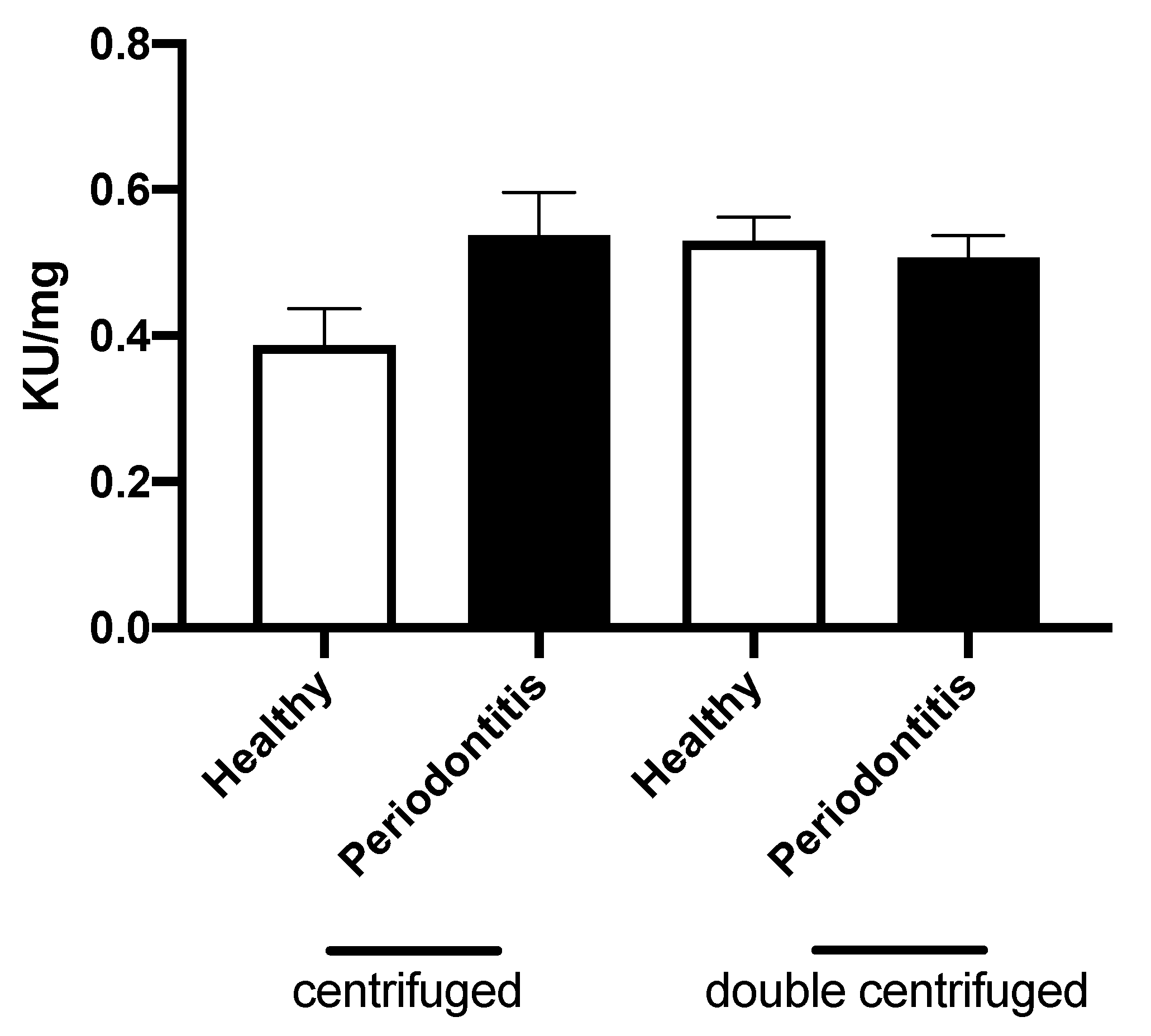
2.4. DNase Activity Measurement

DNase activity was measured by single radial enzyme diffusion (SRED) method using 1% agarose gel (20 mM Tris–HCl, pH 7.5, 2 mM MgCl2, 2 mM CaCl2), which contained DNA isolated from chicken liver (7 mL of 5 mg/mL DNA into 100 mL of gel). From each saliva sample, 1 µL was pipetted into the gel and incubated over night at 37 °C in the dark. Dilutions of DNase I (Qiagen, Hilden, Germany) of known concentrations were used for creating the calibration curve. The diameters of circles were measured by ImageJ software. The values were normalized to proteins.

3. Statistical Analysis

Analysis was performed using GraphPad Prism 6.01 (GraphPad Software, San Diego, CA, USA). To compare the measured parameters between healthy controls and patients with periodontitis, unpaired t-test was performed. Correlations were performed as nonparametric using Spearman correlation. Level α = 0.05 is considered as a limit level of significance.

4. Results

This study included 25 patients with periodontitis and 29 healthy controls. Patient characteristics are shown in Table 1. All patients were characterized using GI and PBI score as well as BOP expressed as a percentage. Individuals in the control group had no BOP and no sign of alveolar bone loss. Significantly higher values were observed in periodontitis patients in comparison to healthy controls according to GI, PBI and BOP (Table 1). No gender differences were observed in further analyses.
DNA concentration in whole saliva showed significantly higher values in periodontitis compared to healthy controls (504.6 ± 82.32 vs. 237.4 ± 26.66 ng/mg; p < 0.01) (Figure 1). Once the whole saliva was centrifuged, there were no differences observed in concentration of DNA between periodontitis patients and healthy controls (69.59 ± 13.37 vs. 54.22 ± 8.06 ng/mg; p = ns) (Figure 1). Similarly, in double-centrifuged samples, total ecDNA concentrations did not differ between the groups, periodontitis vs. healthy (13.54 ± 1.14 vs. 16.14 ± 2.54 ng/mg; p = ns; Figure 1). Determination of nuclear DNA in centrifuged samples showed no differences between groups, periodontitis vs. healthy (6629 ± 1727 vs. 5638 ± 2151 copy/mg; p = ns; Figure 2A). However mtDNA was significantly increased in periodontitis in comparison to healthy controls (10,656,989 ± 3,419,262 vs. 2,630,658 ± 809,983 copy/mg; p < 0.05; Figure 2B). In double-centrifuged samples, ncDNA concentration was significantly lower in the periodontitis group in comparison to the healthy group (788.8 ± 238.8 vs. 4947 ± 1508 copy/mg; p < 0.01) (Figure 2A), although mtDNA concentration was similar in both groups (14,608 ± 4809 vs. 11,957 ± 3296 copy/mg; p = ns; Figure 2B).
Correlation analysis revealed that there is no linear association between concentration of total DNA, mtDNA, ncDNA and health indexes of periodontitis patients (Table 2). Only significant and positive correlation was observed between PBI and mtDNA in centrifuged samples (r = 0.62; Table 2). No significant differences were found in DNase activity between periodontitis samples and healthy controls either in centrifuged samples (0.54 ± 0.06 vs. 0.39 ± 0.05 KU/mg; p = ns) or in double-centrifuged samples (0.51 ± 0.03 vs. 0.53 ± 0.03 KU/mg; p = ns; Figure 3).
No significant correlations were found beside the positive significant correlation between PBI and mtDNA in centrifuged samples (p < 0.05*).

5. Discussion

Saliva and its content can reflect health status. As the latest research shows, saliva contains various markers helpful for the diagnosis and monitoring of many disorders such as cancer [22,23], neurological diseases [24], renal diseases [25], inflammatory bowel disease [26] or cardiovascular diseases [27]. The health status of the teeth, gingiva or salivary glands is also reflected in saliva [28]. Markers of inflammation, such as cytokines and inflammatory mediators, are produced as a result of bacterial infection and were proved to be higher in saliva of patients with periodontitis in comparison to healthy controls [29].
Based on the above-mentioned studies, total ecDNA or ecDNA of different origin were expected to be increased in periodontitis and could serve as a possible biomarker of periodontitis severity. However, as is defined by Jiang et al. [21], ecDNA is obtained after 16,000× g centrifugation, although they used plasma, not saliva. In our previously published study [30], we tried to determine the total DNA in whole saliva as well as in salivary supernatant. We hypothesized that we have analyzed ecDNA once the fluid is centrifuged and depleted of cells. However, no differences were found either in ecDNA between healthy and chronic periodontitis, and similarly no differences were observed between genders. These results were possibly obtained due to technical issues such as insufficient centrifuging while analyzing ecDNA. Therefore, we decided to repeat this observational study, in which we obtained ecDNA appropriately prepared using two-step centrifugation, 1600× g followed by 16,000× g according to Jiang et al. [21]. It is assumed that, after 1600× g centrifugation, only cells are spun down. However, generally, when talking about ecDNA, 16,000× g centrifugation is needed in order to spin down the bigger particles and cell debris. Based on this, we decided to analyze DNA in all three fractions, i.e., in whole saliva, centrifuged saliva and double-centrifuged saliva. According to our results, total DNA was higher in patients with periodontitis in whole saliva perhaps due to increased cell debris as a result of bleeding and worsened oral health. Once the whole saliva was centrifuged, the differences in total DNA were not observed anymore. Also, when we quantified separately ncDNA and mtDNA, we could observe differences in mtDNA, but not in ncDNA. Increased concentration of mtDNA in periodontitis indicates the presence of mitochondria, which could potentiate the inflammation in oral cavity [31]. Mitochondria are more likely to be still intact, because after the double-centrifugation, the mtDNA concentrations are same in both groups. EcDNA of mitochondrial origin is the same in healthy controls as well as in periodontitis, however ecDNA of nuclear origin is increased in healthy controls, even though there are no differences observed in total ecDNA. This could happen due to higher fragmentation of ncDNA in periodontitis [30], since short fragments are not detectable by qPCR but are visible by spectrofluorometry. Moreover, fragmented DNA could further potentiate inflammatory status [21,32,33], and thus worsen the course of disease. This hypothesis should be clarified by further studies. Nevertheless, if this hypothesis is proven correct, the ecDNA might become another therapeutic target.
We expected that altered DNase activity could be potentially associated with periodontitis. There was not significant change in DNase activity in our samples, however studying the activity of this enzyme is still in process as it is still not clear if the DNase activity is related with ecDNA concentrations. Even though the preliminary experiments on DNase activity have been already published [34], it is also not clear whether differences exist among genders under physiological circumstances [35]. However, it is important to remark that not all ecDNA is similarly sensitive to DNase activity. It has been found that the microbiome might influence DNase activity [36]. The shift of commensals to more pathogenic bacteria in gingivitis and periodontitis was shown to affect the DNase activity in saliva. However, we did not search for microbiota difference in our patients, which is the limitation of this study.
The collection of samples could not be done without freezing in our conditions, which is a limitation of many studies. It was shown that freezing of serum and plasma samples causes release of free nucleic acids from large circulating vesicles [37]. However, the uniqueness of our study is that saliva samples were processed right after sampling without freezing and thawing. Only the original samples intended for DNase activity measurement were frozen. In our preliminary unpublished data, we showed that DNase activity could decrease with longer storage period. Therefore, we can only hypothesize, that the measurement of DNase activity in fresh salivary samples could reveal its higher activity. Nevertheless, we measured the DNase activity within the period of two months from the collection.
In conclusion, we proved that worse periodontal status in patients and the presence of inflammation is associated with increased total and mtDNA concentrations in different fractions of saliva. Further studies should focus on the role of mtDNA as a potential driver of inflammation in periodontitis. It is also important to focus on DNA fragmentation as it shows that ecDNA originating in the nucleus seems to be decreased in periodontitis. This approach could lead to new treatment targets related to DNA origin, metabolism and recognition by immune cells.

Author Contributions

Conceptualization, P.C., B.N., E.K., and Ľ.T.; methodology, A.G.K., B.K., and P.C.; formal analysis, P.C. and Ľ.T.; investigation, B.K., E.K., and A.P.; data curation, B.K., A.G.K. and A.P.; writing—original draft preparation, B.K. and Ľ.T.; writing—review and editing, A.G.K., A.P., E.K., B.N., and P.C.; supervision, P.C., Ľ.T.”, please turn to the CRediT taxonomy for the term explanation. All authors have read and agreed to the published version of the manuscript.

Funding

This work was supported by Ministry of Health of the Slovak Republic under the project registration numbers 2018/33-LFUK-7. And the APC was funded by Ministry of Health of the Slovak Republic under the project registration numbers 2018/33-LFUK-7.

Conflicts of Interest

The authors declare no conflict of interest.

References

  1. Lo, Y.D.; Corbetta, N.; Chamberlain, P.F.; Rai, V.; Sargent, I.L.; Redman, C.W.; Wainscoat, J.S. Presence of fetal DNA in maternal plasma and serum. Lancet 1997, 350, 485–487. [Google Scholar] [CrossRef]
  2. Chen, X.Q.; Stroun, M.; Magnenat, J.L.; Nicod, L.P.; Kurt, A.M.; Lyautey, J.; Lederrey, C.; Anker, P. Microsatellite alterations in plasma DNA of small cell lung cancer patients. Nat. Med. 1996, 2, 1033–1035. [Google Scholar] [CrossRef] [PubMed]
  3. Dwivedi, D.J.; Toltl, L.J.; Swystun, L.L.; Pogue, J.; Liaw, K.L.; Weitz, J.I.; Cook, D.J.; Fox-Robichaud, A.E.; Liaw, P.C.; Canadian Critical Care Translational Biology Group. Prognostic utility and characterization of cell-free DNA in patients with severe sepsis. Crit. Care. 2012, 16, R151. [Google Scholar] [CrossRef] [PubMed] [Green Version]
  4. Lo, Y.M.; Rainer, T.H.; Chan, L.Y.; Hjelm, N.M.; Cocks, R.A. Plasma DNA as a prognostic marker in trauma patients. Clin. Chem. 2000, 46, 319–323. [Google Scholar] [CrossRef] [Green Version]
  5. Butt, A.N.; Swaminathan, R. Overview of circulating nucleic acids in plasma/serum. Ann. N. Y. Acad. Sci. 2008, 1137, 236–242. [Google Scholar] [CrossRef] [PubMed]
  6. Oellerich, M.; Schütz, E.; Beck, J.; Kanzow, P.; Plowman, P.N.; Weiss, G.J.; Walson, P.D. Using circulating cell-free DNA to monitor personalized cancer therapy. Crit. Rev. Clin. Lab. Sci. 2017, 54, 205–218. [Google Scholar] [CrossRef]
  7. Jiang, W.W.; Masayesva, B.; Zahurak, M.; Carvalho, A.L.; Rosenbaum, E.; Mambo, E.; Zhou, S.; Minhas, K.; Benoit, N.; Westra, W.H.; et al. Increased mitochondrial DNA content in saliva associated with head and neck cancer. Clin. Cancer Res. 2005, 11, 2486–2491. [Google Scholar] [CrossRef] [Green Version]
  8. Kinane, D.F.; Stathopoulou, P.G.; Papapanou, P.N. Periodontal diseases. Nat. Rev. Dis. Primers. 2017, 3, 17038. [Google Scholar] [CrossRef]
  9. Nowicki, E.M.; Shroff, R.; Singleton, J.A.; Renaud, D.E.; Wallace, D.; Drury, J.; Zirnheld, J.; Colleti, B.; Ellington, A.D.; Lamont, R.J.; et al. Microbiota and Metatranscriptome Changes Accompanying the Onset of Gingivitis. MBio 2018, 9. [Google Scholar] [CrossRef] [Green Version]
  10. Hajishengallis, G. Periodontitis: From microbial immune subversion to systemic inflammation. Nat. Rev. Immunol. 2015, 15, 30–44. [Google Scholar] [CrossRef]
  11. Pihlstrom, B.L.; Michalowicz, B.S.; Johnson, N.W. Periodontal diseases. Lancet 2005, 366, 1809–1820. [Google Scholar] [CrossRef] [Green Version]
  12. Konecna, B.; Sysak, R.; Kacerovsky, M.; Celec, P.; Vlkova, B. Deoxyribonuclease activity in plasma of pregnant women and experimental animals. J. Matern. Fetal Neonatal Med. 2018, 31, 1807–1809. [Google Scholar] [CrossRef] [PubMed]
  13. White, P.; Sakellari, D.; Roberts, H.; Risafi, I.; Ling, M.; Cooper, P.; Milward, M.; Chapple, I. Peripheral blood neutrophil extracellular trap production and degradation in chronic periodontitis. J. Clin. Periodontol. 2016, 43, 1041–1049. [Google Scholar] [CrossRef] [PubMed]
  14. White, P.C.; Chicca, I.J.; Cooper, P.R.; Milward, M.R.; Chapple, I.L. Neutrophil Extracellular Traps in Periodontitis: A Web of Intrigue. J. Dent. Res. 2016, 95, 26–34. [Google Scholar] [CrossRef]
  15. Thierry, A.R.; El Messaoudi, S.; Gahan, P.B.; Anker, P.; Stroun, M. Origins, structures, and functions of circulating DNA in oncology. Cancer Metastasis Rev. 2016, 35, 347–376. [Google Scholar] [CrossRef] [Green Version]
  16. Lauková, L.; Bertolo, E.M.J.; Zelinková, M.; Borbélyová, V.; Conka, J.; Kovalcíková, A.G.; Domonkos, E.; Vlková, B.; Celec, P. Early Dynamics of Plasma DNA in a Mouse Model of Sepsis. Shock 2019, 52, 257–263. [Google Scholar] [CrossRef]
  17. Rathnayake, N.; Åkerman, S.; Klinge, B.; Lundegren, N.; Jansson, H.; Tryselius, Y.; Sorsa, T.; Gustafsson, A. Salivary biomarkers of oral health: A cross-sectional study. J. Clin. Periodontol. 2013, 40, 140–147. [Google Scholar] [CrossRef]
  18. Loe, H. The Gingival Index, the Plaque Index and the Retention Index Systems. J. Periodontol. 1967, 38, 610–616. [Google Scholar] [CrossRef]
  19. Newbrun, E. Indices to measure gingival bleeding. J. Periodontol. 1996, 67, 555–561. [Google Scholar] [CrossRef]
  20. Pyysalo, M.J.; Pyysalo, L.M.; Hiltunen, J.; Järnstedt, J.; Helminen, M.; Karhunen, P.J.; Pessi, T. The dental infections in patients undergoing preoperative dental examination before surgical treatment of saccular intracranial aneurysm. BMC Res. Notes. 2018, 11, 600. [Google Scholar] [CrossRef] [PubMed]
  21. Jiang, P.; Chan, C.W.; Chan, K.A.; Cheng, S.H.; Wong, J.; Wong, V.W.S.; Wong, G.L.; Chan, S.L.; Mok, T.S.; Chan, H.L.; et al. Lengthening and shortening of plasma DNA in hepatocellular carcinoma patients. Proc. Natl. Acad. Sci. USA 2015, 112, E1317–E1325. [Google Scholar] [CrossRef] [Green Version]
  22. Nair, S.; Tang, K.D.; Kenny, L.; Punyadeera, C. Salivary exosomes as potential biomarkers in cancer. Oral. Oncol. 2018, 84, 31–40. [Google Scholar] [CrossRef] [PubMed]
  23. Rapado-González, Ó.; Majem, B.; Muinelo-Romay, L.; Álvarez-Castro, A.; Santamaría, A.; Gil-Moreno, A.; López-López, R.; Suárez-Cunqueiro, M.M. Human salivary microRNAs in Cancer. J. Cancer 2018, 9, 638–649. [Google Scholar] [CrossRef] [PubMed] [Green Version]
  24. Farah, R.; Haraty, H.; Salame, Z.; Fares, Y.; Ojcius, D.M.; Said Sadier, N. Salivary biomarkers for the diagnosis and monitoring of neurological diseases. Biomed. J. 2018, 41, 63–87. [Google Scholar] [CrossRef]
  25. Kovalčíková, A.; Janšáková, K.; Gyurászová, M.; Podracká, L.; Šebeková, K.; Celec, P.; Tóthová, L. Salivary creatinine and urea are higher in an experimental model of acute but not chronic renal disease. PLoS ONE 2018, 13, e0200391. [Google Scholar] [CrossRef] [PubMed] [Green Version]
  26. Zheng, X.; Chen, F.; Zhang, Q.; Liu, Y.; You, P.; Sun, S.; Lin, J.; Chen, N. Salivary exosomal PSMA7: A promising biomarker of inflammatory bowel disease. Protein Cell 2017, 8, 686–695. [Google Scholar] [CrossRef] [Green Version]
  27. Abdul Rehman, S.; Khurshid, Z.; Hussain Niazi, F.; Naseem, M.; Al Waddani, H.; Sahibzada, H.A.; Sannam Khan, R. Role of Salivary Biomarkers in Detection of Cardiovascular Diseases (CVD). Proteomes 2017, 5, 21. [Google Scholar] [CrossRef]
  28. Yoshizawa, J.M.; Schafer, C.A.; Schafer, J.J.; Farrell, J.J.; Paster, B.J.; Wong, D.T. Salivary biomarkers: Toward future clinical and diagnostic utilities. Clin. Microbiol. Rev. 2013, 26, 781–791. [Google Scholar] [CrossRef] [Green Version]
  29. Ebersole, J.L.; Schuster, J.L.; Stevens, J.; Dawson, D.; Kryscio, R.J.; Lin, Y.; Thomas, M.V.; Miller, C.S. Patterns of salivary analytes provide diagnostic capacity for distinguishing chronic adult periodontitis from health. J. Clin. Immunol. 2013, 33, 271–279. [Google Scholar] [CrossRef]
  30. Baňasová, L.; Kamodyová, N.; Janšáková, K.; Tóthová, L.; Stanko, P.; Turňa, J.; Celec, P. Salivary DNA and markers of oxidative stress in patients with chronic periodontitis. Clin. Oral. Investig. 2015, 19, 201–207. [Google Scholar] [CrossRef]
  31. Missiroli, S.; Genovese, I.; Perrone, M.; Vezzani, B.; Vitto, V.A.M.; Giorgi, C. The Role of Mitochondria in Inflammation: From Cancer to Neurodegenerative Disorders. J. Clin. Med. 2020, 9, 740. [Google Scholar] [CrossRef] [Green Version]
  32. Mouliere, F.; Robert, B.; Peyrotte, E.A.; Del Rio, M.; Ychou, M.; Molina, F.; Gongora, C.; Thierry, A.R. High fragmentation characterizes tumour-derived circulating DNA. PLoS ONE 2011, 6, e23418. [Google Scholar] [CrossRef]
  33. Zhang, R.; Nakahira, K.; Guo, X.; Choi, A.M.; Gu, Z. Very Short Mitochondrial DNA Fragments and Heteroplasmy in Human Plasma. Sci. Rep. 2016, 6, 36097. [Google Scholar] [CrossRef] [PubMed] [Green Version]
  34. Malíčková, K.; Ďuricová, D.; Bortlík, M.; Hrušková, Z.; Svobodová, B.; Machková, N.; Komárek, V.; Fučíková, T.; Janatková, I.; Zima, T.; et al. Impaired deoxyribonuclease I activity in patients with inflammatory bowel diseases. Autoimmune Dis. 2011, 2011, 945861. [Google Scholar] [CrossRef] [Green Version]
  35. Janovicova, L.; Konecna, B.; Vokalova, L.; Laukova, L.; Vlkova, B.; Celec, P. Sex, Age, and Bodyweight as Determinants of Extracellular DNA in the Plasma of Mice: A Cross-Sectional Study. Int. J. Mol. Sci. 2019, 20, 4163. [Google Scholar] [CrossRef] [PubMed] [Green Version]
  36. Palmer, L.J.; Chapple, I.L.; Wright, H.J.; Roberts, A.; Cooper, P.R. Extracellular deoxyribonuclease production by periodontal bacteria. J. Periodontal Res. 2012, 47, 439–445. [Google Scholar] [CrossRef]
  37. Muller, L.; Hong, C.S.; Stolz, D.B.; Watkins, S.C.; Whiteside, T.L. Isolation of biologically-active exosomes from human plasma. J. Immunol. Methods 2014, 411, 55–65. [Google Scholar] [CrossRef] [PubMed] [Green Version]
Figure 1. Total DNA in whole saliva, centrifuged saliva (1600× g) and double-centrifuged saliva (1600× g + 16,000× g). Results are expressed as mean + SEM. Salivary DNA is higher in periodontitis in whole saliva (p < 0.01**). No differences were found in centrifuged and double-centrifuged samples between the healthy and periodontitis group.
Applsci 10 07490 g001
Figure 2. The effect of periodontitis on A: nuclear DNA (ncDNA). No differences were found in centrifuged samples. NcDNA was significantly lower in periodontitis in double-centrifuged samples (p < 0.01**). The effect of periodontitis on B: mitochondrial DNA (mtDNA). MtDNA was significantly higher in periodontitis in centrifuged samples. No differences were found in double-centrifuged samples. Results are expressed as mean + SEM.
Applsci 10 07490 g002
Figure 3. The effect of periodontitis on activity of DNase in centrifuged and double-centrifuged samples. Results are expressed as mean + SEM. No differences were found between healthy and periodontitis samples.
Applsci 10 07490 g003
Table 1. Oral health indexes of subjects.
Group
ParameterHealthy WomenWomen with PeriodontitisHealthy MenMen with PeriodontitisDisease Effect p Value
Age (years)43.6 ± 11.646.8 ± 9.540.7 ± 6.050.7 ± 10.8
GI (score)0.59 ±0.461.08 ± 0.350.82 ± 0.451.54 ± 0.66<0.001
PBI (score)0.36 ± 0.250.81 ± 0.470.83 ± 0.561.32 ± 0.48<0.001
BOP (%)0.00 ± 0.0022.5 ± 14.960.00 ± 0.0032.2 ± 21.26<0.001
Gingival index (GI)—0–3 as qualitative changes of gingival soft tissue; 0—normal gingiva, 1—mild inflammation, 2—moderate inflammation, 3—severe inflammation. Papilla bleeding index (PBI)—(0, 1, 2, 3)—for bleeding after cautiously probing to the sulcus; 0 means no bleeding; 1 means one bleeding point few seconds after probing; 2 means intermediate bleeding in few points; 3 profuse bleeding along gingival sulcus as a reaction on slightest. Bleeding on probing (BOP)—number of sites where bleeding to the total number of available sites, expressed as percentage. Data are expressed as mean ± SD.
Table 2. Correlation between oral health indexes and total, nuclear DNA (ncDNA), and mitochondrial DNA (mtDNA) in whole saliva, centrifuged (1600× g) and double-centrifuged (1600× g + 16,000× g) samples.
ParameterCentrifugedDouble Centrifuged
Total DNA
GI (score)r = 0.01r = −0.01
PBI (score)r = 0.24r = −0.08
BOP (%)r = −0.01r = −0.1
Nuclear DNA
GI (score)r = 0.16r = 0.25
PBI (score)r = 0.35r = 0.2
BOP (%)r = 0.03r = 0.28
Mitochondrial DNA
GI (score)r = 0.43r = -0.09
PBI (score)r = 0.62 *r = 0.33
BOP (%)r = −0.04r = -0.12
Publisher’s Note: MDPI stays neutral with regard to jurisdictional claims in published maps and institutional affiliations.

