Article

Screening of Antibacterial Activity, Antioxidant Activity, and Anticancer Activity of Euphorbia hirta Linn. Extracts

1
School of Biotechnology, International University–Vietnam National University, Ho Chi Minh City 70000, Vietnam
2
Research Center of Ginseng and Medicinal Materials Ho Chi Minh City 2, 41 Dinh Tien Hoang Street, District 1, Ho Chi Minh City 70000, Vietnam
3
Faculty of Chemistry, University of Science–Vietnam National University, Ho Chi Minh City 70000, Vietnam
*
Author to whom correspondence should be addressed.
Submission received: 1 September 2020 / Revised: 25 September 2020 / Accepted: 4 October 2020 / Published: 26 November 2020

Abstract

:

Featured Application

Study of Euphorbia hirta Linn. extracts provide technical insight for functional food and drug development.

Abstract

This study aimed to screen the anticancer and antioxidant potential and antimicrobial activity of methanol, petroleum ether, chloroform, ethyl acetate, butanol of Euphorbia hirta Linn. extracts (EH-Me, EH-PE, EH-Ch, EH-EA and EH-Bu, respectively). The results of 2,2-diphenyl-1-pycrylhydrazyl (DPPH) radical scavenging assay and lipid peroxidation inhibition assay showed that EH-EA was the strongest antioxidant (IC50 = 10.33 ± 0.01 µg/mL; IC50 = 1.48 ± 0.12 µg/mL, respectively) compared to all other extracts. In the antimicrobial activity of the extracts against eight strains of Gram-positive and Gram-negative bacteria using the agar disc diffusion method, we found the EH-EA to be the best antimicrobial agent. Anticancer activities of those extracts were examined by sulforhodamine B (SRB) in vitro cytotoxicity assay on two cancer cell lines, including lung cancer cells NCI-H460 and liver cancer cells Hep G2. EH-EA at concentration of 100 μg/mL has significant inhibitory activity the growth of lung cancer cells NCI-H460 and liver cancer cells Hep G2 compared to all other extracts. Our results suggest that E. hirta Linn. extracts possess significant biological activities, including antimicrobial, antioxidant, and moderate anticancer properties. Our results show that this plant could be a good source for natural antioxidants and a possible pharmaceutical supplement. Among five analyzed extracts, EH-EA extract has the strongest activities, and should be used to determine phytochemicals and mechanisms of these activities.

1. Introduction

Since ancient times, herbs have been confirmed for their biological ability to relief pain, prevent diseases and cure illnesses. Nowadays, scientists around the world have developed a research system for medicinal plants and their medicinal properties. Each plant species contains hundreds to thousands of different constituent chemicals that interact in complex ways in the bodies of animals and human [1,2,3]. In developing countries, according to estimates by the World Health Organization, about 80% of the population still depends on folk medicines made from plants for prevention and treatment [4]. Traditional medicine or folk medicine has been proven to be more affordable, clinically effective and has relatively less adverse effects than modern drugs [5,6,7]. Plant-derived secondary metabolites that are biosynthesized in plants are not only essential for plant survival, but are also extremely important for development, growth and reproduction and plant protection [8,9,10]. Secondary metabolites in plants include several groups of molecules, including steroids, alkaloids, phenolic, lignans, carbohydrates and glycosides, etc., which possess several biological activities. For example, phenolic compounds are one big group of secondary metabolites that are popularly distributed in plants, and possess a diversity of biological properties beneficial to humans, such as their antiallergic, anticancer, antimicrobial, anti-inflammatory, antidiabetic and antioxidant activities [11,12,13].
The antibacterial properties of plants or herbs have been researched popularly. Ocimum basilicum, Momordica charantia, Cinnamomum verum, Allium sativum, Curcuma longa, for example, exhibit antimicrobial properties against both Gram-positive and Gram-negative bacteria [14,15,16]. Much research has shown that these plants are rich in a diversity of bioactive compounds found in vitro to inhibit or kill microorganisms effectively [16].
Lipid peroxidation is an ambiguous event in all biological species. Currently, lipid peroxidation is considered as the main molecular mechanism involved in the oxidative damage to cell structures and in the toxicity process that leads to cell death [17]. Lipid peroxidation has been suggested to comprise of three distinct mechanisms: free radical-mediated oxidation, free radical-independent, nonenzymatic oxidation, and enzymatic oxidation [18,19]. Lipid peroxidation is a serious problem for human health. Plant-derived polyphenols have been demonstrated to have a variety of biological effects, due to their antioxidant activities in inhibition of lipid peroxidation in the body [20,21].
Cancer is the second highest cause of mortality globally. In particular, there were 9.6 million cancer-related deaths in 2018 [22,23]. The most common cancers include breast, lung, colorectal, etc. Traditional medicine and phytochemicals extracted from herbs have been increasingly noticed and applied as one of the useful treatments against cancer. A series of clinical studies in recent years have shown the positive effects of herbal drugs on survival, regulation of the immune system and improvement in the quality of life of cancer patients, when combining these herbal medicines with conventional treatments [24]. Diversity of phytochemicals such as phenolic compounds, terpenoids, lignans, tannins, alkaloids, etc., have been investigated from herbal sources that possess strongly effective antioxidant properties to inhibit cell proliferation and stimulate the immune system to enhance prevention or treatment of cancer diseases.
Euphorbia hirta Linn. (E. hirta) belongs to the Euphorbiaceae family, a group of small prostrate herbaceous annual weeds in Vietnam. This plant has slender stem of 30–50 cm height, yellow hair, and opposite spear-shaped leaves. It is abundant in open grasslands and is distributed in most Asian countries [25]. In Vietnam, E. hirta is commonly distributed in many provinces of the southern area (Figure 1). Many investigations have demonstrated that this plant has been widely used as traditional medicine to treat many diseases, including wound healing, gastrointestinal disorders, tumors, etc. Additionally, this herb was used as a traditional medicine in the treatment of diabetes for a long time [26,27,28,29].
E. hirta contains several secondary metabolites, including flavonoids, terpenoids, phenols, essential oil, etc. Many flavonoids have been identified in E. hirta including quercetin, quercitrin, quercitol and derivatives. Some terpenoids in E. hirta have been isolated and identified successfully, including triterpenoids, α-amyrin, β-amyrin, friedelin, teraxerol, cycloartenol, 24-methylene-cycloartenol, ingenol triacetate β- sitosterol, campestrol, stigmasterol, etc. [27,28]. The antibacterial activity of E. hirta has been discovered and proven by using many different extracts that showed the property against Shigella species, Escherichia coli, Staphylococcus aureus, Pseudomonas aeruginosa, and Bacillus subtilis [28]. The aqueous extract of E. hirta also showed antioxidant activity and a free radical scavenging capacity [27]. Moreover, E. hirta extracts have exhibited anticancer activity [28].
For purpose of evaluation of biological activities of Euphorbia hirta Linn., our study was carried out to evaluate the antibacterial activity, lipid peroxidation inhibitory activity and anticancer activity of methanol, petroleum ether, chloroform, ethyl acetate and butanol extracts of this herb.

2. Materials and Methods

2.1. Animals and Materials

2.1.1. Animals

Male Swiss albino mice (25 ± 2 g, aged 5–6 weeks), were supplied by the Institute of Vaccines and Medical Biological Products in Nha Trang City. This research was approved by the University of Science Animal Care and Use Committee (No. 248b/KHTN-ACUCUS). Each extract requires 2 mice, so the total number of mice required for the test is 10 mice. Before any experience, all animals were kept for 1 week under the same laboratory conditions of temperature (22 ± 2 °C), relative humidity (70 ± 4%), and a 12 h light/dark cycle, and received a nutritionally standard diet and tap water.

2.1.2. Chemicals and Reagents

The organic solvents that were used in extraction of E. hirta include absolute methanol (Me), petroleum ether (PE), chloroform (Ch), ethyl acetate (EA), and butanol (Bu) purchased from Chemsol Company, Vietnam. Sulforhodamine B, 1-diphenyl-2-picrylhydrazyl (DPPH) and [±]-6-hydroxy-2, 5, 7, 8-tetramethyl-chroman-2-carboxylic acid (Trolox) were purchased from Sigma-Aldrich Co. (USA). Thiobarbituric acid and ascorbic acid were purchased from Merck (Germany). Gallic acid, quercetin and Folin were purchased from Sigma-Aldrich Co. (USA). Dimethyl sulfoxide (DMSO) was purchased from Merck (Germany). All other chemicals were of analytical grade.

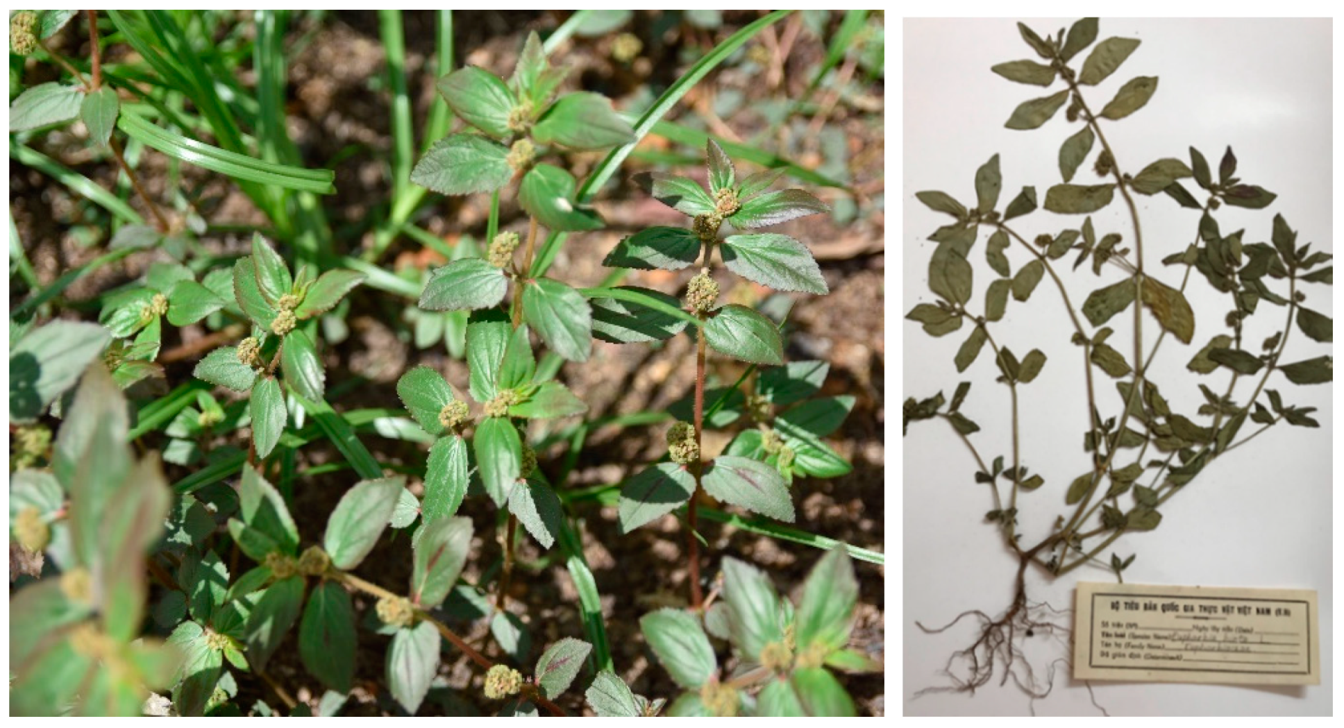
2.2. Plant Material Collection and Identification

E. hirta was identified by the Institute of Tropical Biology under the Vietnam Academy of Science and Technology. The fresh E. hirta plant was collected in Long Khanh City, Dong Nai Province, Vietnam in February 2015. The collected raw plant was cleaned with tap water to remove any type of contamination and then soaked in 70% ethanol solution to exterminate all enzymes. Next, cleaned material was naturally dried for 3 days under room temperature. Sunlight was strictly prevented from interacting with the preliminarily processed sample. Finally, the dried plant was ground into powder, kept in desiccator to avoid microbial attack before using it for extraction process. The total weight of the raw plant after preliminary preparation was approximately 8 kg.

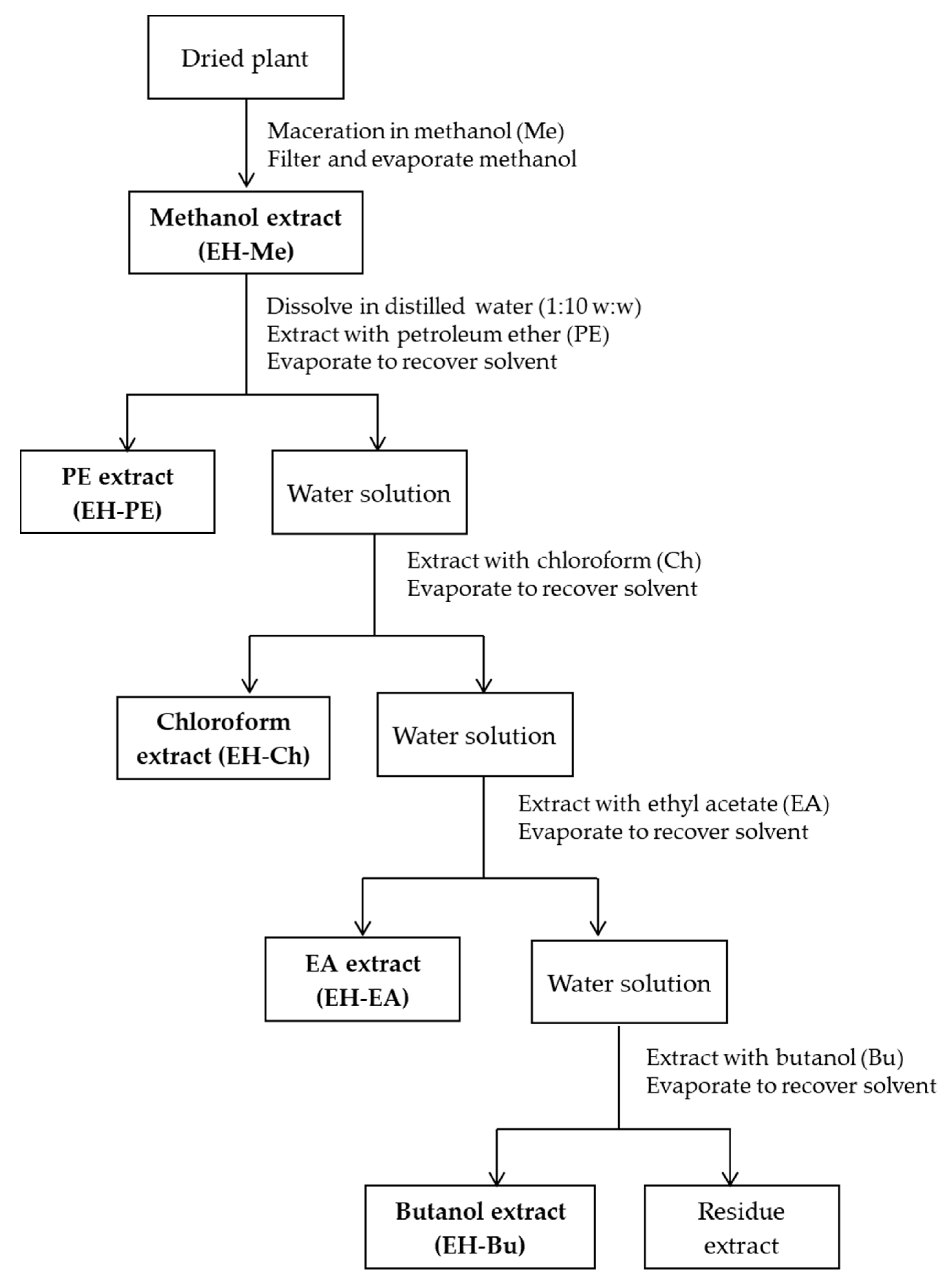
2.3. Preparation of E. hirta Extracts Using Maceration Method

The powdered plant (8 kg) was macerated with absolute methanol solvent by the ratio of 1:10 (w/w) within 7 days [30]. After 7 days, methanol extract solution was obtained and filtered through Whatman No.1 filter paper to remove ground and other impurities. Methanol extract solution from maceration was reclaimed methanol solvent by a rotary evaporator under high pressure to get methanol crude extraction. After yielding total crude extract, EH-Me extract was mixed with distilled water by ratio 1:10 (w/w) and was processed with PE, Ch, EA, and Bu solvents, respectively. The organic solvents were selected from low polarity to high polarity so as to harvest phytochemicals separately depending on the differences in their polarity. After each fractional extraction, the rotary evaporator was used to evaporate all solvents to form concentrated extracts (Figure 2). Before beginning the experiments about the biological properties of all extracts, the percentage of humidity of all extracts was determined to evaluate the moisture content of the samples. The moisture content was determined by moisture balance, type MOC-120H, no. D207302059. Five extract samples of E. hirta (5 mg) were weighed and dried in the oven at 105 °C until its weight was constant [31]. The results are expressed as mean ± standard error of mean (SEM) of three replicates.
The percentage of yield of extract was calculated as:
%   Yield   ( % Y ) = weight   of   dried   extract   ( g ) weight   of   sample   ( g ) × 100

2.4. Determination of Total Phenolic and Flavonoid Content of the Extracts from Euphorbia hirta Linn.

2.4.1. Determination of Total Phenolic Content (TPC)

The total phenols content of the extract was determined by the Folin–Ciocalteu method [32,33]. The concentration gradient of gallic acid was prepared as standard solution (0–50 µg/mL), and the calibration curve was established using gallic acid. The samples were diluted with DMSO as sample solutions, after they were made up to 10 mL by methanol. The 6 mL of distilled water and 100 μL of sample or standard solution and 0.5 mL Folin–Ciocalteu reagent were mixed for 5 min, followed by the addition of 1.5 mL of 20% sodium carbonate and 1.9 mL of distilled water. The mixture was placed for 120 min at room temperature. Every experiment was performed in triplicate. The absorbance of the mixture was measured at 758 nm using a UV spectrophotometer. Total phenols content of the samples was expressed as mg of gallic acid equivalents (GAE) per gram dried extract [33,34]. The experimental results are expressed as mean ± standard error of mean (SEM) of three replicates.

2.4.2. Determination of Total Flavonoid Content (TFC)

Total flavonoid content was determined by aluminum chloride colorimetric assay [33]. The concentration gradient of quercetin was prepared as standard solution (0–80 µg/mL), and the calibration curve was established using quercetin. The samples were diluted with DMSO as sample solutions, after they were made up to 50 mL by methanol. The 4 mL of distilled water and 1 mL of sample or standard solution, and 300 µL NaNO2 (5%) were added to a glass cuvette. Three hundred microliters of AlCl3 (10%) were added after 5 min and 2 mL NaOH (1 M) and 2.4 mL distilled water were added after 6 min. The mixture was placed for 10 min at room temperature. Every experiment was performed in triplicate. The absorbance of the mixture was measured at 510 nm using a UV spectrophotometer. Total flavonoid content of the samples was expressed as mg of quercetin equivalents (QE) per gram dry extract. The experimental results are expressed as mean ± standard error of mean (SEM) of three replicates.

2.5. In vitro Antibacterial Activity Using Disc Diffusion Method

The following standard bacterial strains were used in this study belonging to Gram-positive and Gram-negative species: Bacillus subtilis (B. subtilis), Escherichia coli ATCC 25,922 (E. coli), Staphylococcus aureus ATCC 25,923 (S. aureus), Pseudomonas aeruginosa ATCC 27,853 (P. aeruginosa), Streptococcus preumoniae ATCC 49,619 (S. preumoniae), Salmonella typhi Ty 2 (S. typhi), Vibrio cholearea AI 4450 (V. cholearea), Shigella flexneri NCDC 2747-71 (S. flexneri) and cultured on specialized media overnight. They were obtained from the laboratory of Ho Chi Minh City Pasteur Institute, Vietnam. Due to their ability to survive in harsh conditions and their multiple environmental habitats, these bacterial organisms including Gram-positive and Gram-negative are the main sources of severe infections in humans.
The antibacterial activity of E. hirta extracts was investigated using the disk diffusion method with some modifications [35,36,37]. The bacterial suspension phase in physiological saline reaches a concentration of McFarland (0.5%) (equivalent to 108 CFU/mL). Preparation of Mueller-Hinton (MH) medium plates were prepared containing 5% of sheep blood after being allowed to dry before inoculating bacteria. The bacterial suspension was spread evenly over the surface of MH agar. Thereafter, we conducted holes in the surface of wells, the wells were 20–30 mm apart, and had a diameter of 6 mm. The diluted samples at five different concentrations of 25, 50, 100, 200 and 400 µg/mL for five extracts in DMSO solvent were prepared to add to each well of every disc. All discs were incubated at 37 °C for 18–20 h. Each concentration was repeated 2 times, checking for bacterial growth. The negative control was the same volume of sterilized water. The diameter (in millimeters) of the inhibition zone was measured to estimate the antibacterial activity of E. hirta. The inhibition zone was defined as where bacteria cannot grow.

2.6. Study on Antioxidant Activity

2.6.1. DPPH radical scavenging assay

DPPH (1-diphenyl-2-picrylhydrazyl) is a stable free radical that has a maximum absorption of 515–517 nm (purple color). When an antioxidant (as a hydrogen donor) reacts with DPPH, DPPH is reduced to DPPH-H, and as a consequence the solution turns from purple to yellow. Therefore, the antioxidant activity of DPPH samples can be evaluated by the decrease in absorption at 515–517 nm. The potential scavenging abilities of the samples were assessed using ascorbic acid (Merck, Germany) as positive control [38].
Briefly, 0.5 mL of various concentrations of each sample (10, 50, 100, 250, 500, 750 and 1000 μg/mL) or ascorbic acid (0.5, 0.25, 0.1, 0.05, and 0.01 mM) was added into a tube containing 0.5 mL of 0.6 mM DPPH solution dissolved in methanol and the volume was made uniformly to 4 mL using methanol. The solution was mixed and then allowed to stand in the dark at room temperature for 30 min. Absorbance was taken at 515 nm using methanol as blank on UV-visible spectrometer. Then, 0.5 mL of DPPH was added to 3.5 mL of methanol and absorbance was taken for control reading. All analyses were run in triplicate [39].

2.6.2. Lipid Peroxidation Inhibition Assay (Malondialdehyde Assay)

Lipid peroxidation is a well-established mechanism of cellular injury in both plants and animals and is used as an indicator of oxidative stress in cells and tissues. The measurement of malondialdehyde (MDA) and 4-hydroxynonenal (4-HNE) has been used as an indicator of lipid peroxidation. MDA is one of the products of lipid peroxidation on cell membranes. The assay is based on the reaction of MDA with thiobarbituric acid (TBA), forming an MDA-TBA2 adduct that absorbs strongly at 532 nm. The potential lipid peroxidation (i.e., MDA formation) inhibition power of extract samples was measured using mouse brain homogenate as lipid rich substrate. Trolox, a water-soluble analog of vitamin E, was used as positive control [40].
After the sampling, the mouse brain tissue was immediately placed in ice-cold 5 mM phosphate buffer (pH 7.4) with the brain and phosphate buffer ratio of 1:10 (w/v). The tissue was then homogenized at 13,000 rpm in an ice-cold condition. Then, 0.1 mL of various concentrations of each sample or Trolox (10, 5, 1, 0.5, and 0.1 mM) was added to 0.5 mL of brain homogenate and the volume was made to reach 2 mL using 50 mM phosphate buffer. The mixture was then incubated at 37 °C for 15 min to allow lipid peroxidation and thus MDA production. The reaction was stopped with 1 mL of 10% trichloroacetic acid. The tube was then centrifuged at 10,000 rpm at 5 °C for 5 min. The supernatant (2 mL) was transferred into a different tube and then allowed to react with 0.8% TBA solution (1 mL) at 95 °C for 15 min. The control was prepared as above without the test sample. Absorbance of the mixture was measured at 532 nm against a blank solution without sample and TBA after the tubes reached room temperature.

2.6.3. Data Analysis

The percentage of antioxidant activity was calculated as [(∆A0 − ∆As)/∆A0] × 100, where ∆A0 was the absorbance of the control (∆A0 = Acontrol − Ablank), and ∆As was the absorbance of the extract/standard (∆As = Asample/standard − Ablank).
The extract concentration providing 50% inhibition (IC50—the half maximal inhibitory concentration) was obtained by interpolation from linear regression analysis.

2.7. Anticancer Activity Using Sulforhodamine B Assay

2.7.1. Cell Lines and Cell Culture

The NCI-H460 cell line was derived by A.F. Gazdar and associates in 1982 from the pleural fluid of a patient with large cell cancer of the lung. HepG2 hepatocellular carcinoma cell line was purchased from the American Tissue Culture Collection (ATCC; catalog no. HB-8065; Manassas, VA). NCI-H460 (HTB-177) and Hep G2 cells (HB-8065) were purchased from the American Type Culture Collection (Manassas, Rockville). Cells were cultured at 37 °C and 5% CO2 in Eagle’s minimal essential medium (EMEM) supplemented with 10% (v/v) FBS (Sigma), 2 mM L-glutamine (Sigma), 20 mM HEPES (Sigma), 0.025 μg/mL amphotericin B (Sigma), 100 IU/mL penicillin G (Sigma), and 100 μg/mL streptomycin (Sigma).

2.7.2. The Protocol of Toxicological Activity Experiment by Sulforhodamine B (SRB) Method

The assay was performed as previously described with some modifications [41]. Cells, seeded at a density of 10,000 cells/well (Hep G2) and 7,500 cells/well (NCI-H460) in 96-well plates, were cultured for 24 h before being incubated with samples at different concentrations for 48 h. Treated cells were fixed with cold 50% (w/v) trichloroacetic acid (Merck) solution for 1–3 h, washed, and stained with 0.2% (w/v) SRB (Sigma) for 20 min. After five washes with 1% acetic acid (Merck), protein-bound dye was solubilized in 10 mM Tris base solution (Promega). Optical density values were determined with a 96-well micro-titer plate reader (Synergy HT, Biotek Instruments) at the wavelengths of 492 nm and 620 nm.
After 24 h of culture, the cell population was incubated with the analyte at concentrations for 48 h. The total protein from the test cell was then fixed with cold 50% (w/v) trichloroacetic acid (Sigma) and stained with 0.2% sulforhodamine B solution (Sigma) for 20 min. After five washes with 1% acetic acid (Merck), protein-bound dye was solubilized in 10 mM Tris base solution (Promega). Results are read by ELISA reader at 492 nm and 620 nm. The experiments were repeated three times and the results are presented as mean ± standard deviations.
The percentage of growth inhibition (I%) was calculated according to the formula:
I% = (1 − [ODt/ODc] × 100) %, in which ODt and ODc are the optical density value of the test sample and the control sample, respectively. Camptothecin (Calbiochem) was used as a positive control.

2.8. Statistical Analysis

SPSS version 20 (statistical package for the social sciences) software (Schrödinger, LLC New York, NY, USA) was used for data analysis and all comparisons were made by using one-way ANOVA followed by Dunnett’s test. Values were expressed as means ± SEM (standard errors of means), for which the P-values less than 0.05, 0.01, and 0.001 were considered statistically significant.

3. Results

3.1. Plant Extraction

Five fractional extracts of E. hirta were obtained after the extraction process. They are EH-Me, EH-PE, EH-Ch, EH-EA, EH-Bu extracts. Detailed information about these extracts are mentioned in Table 1.

3.2. Determination of Total Phenolic and Flavonoid Contents

According to previous investigations, E. hirta extracts contain a large amount of phenolic and flavonoid components [10]. The total phenolic content of the extracts was quantified. The regression equation of the standard curve of gallic acid was y = 0.0113x + 0.02261 with R2 = 0.9958. The results show that the linear relationship was good in the detection ranges. In the present study, the total phenolic and flavonoid contents are shown in Table 2. The value of the phenolic content was 109.86 ± 1.38, 90.89 ± 1.45, 55.86 ± 0.66, 254.96 ± 10.05 and 70.90 ± 0.65 mg of GAE/g extract for EH-Me extract, EH-PE extract, EH-Ch extract, EH-EA extract, and EH-Bu extract, respectively. EH-EA had the highest total phenol content (254.96 ± 10.05 mg GAE/g extract), followed by EH-Me, EH-PE, EH-Bu, EH-Ch.
The total flavonoid content of the extracts was quantified. The regression equation of the standard curve of quercetin was y = 0.008x − 0.0447 with R2 = 0.9927. The results show that the linear relationship was good in the detection ranges (Table 2). The value of the total flavonoid content was 18.92 ± 1.33, 8.48 ± 1.16, 16.41 ± 1.44, 27.66 ± 0.73 and 12.43 ± 1.66 mg of quercetin/g extract for EH-Me extract, EH-PE extract, EH-Ch extract, EH-EA extract, and EH-Bu extract, respectively (Table 2). EH-EA had the highest total flavonoid content (27.66 ± 0.73 mg quercetin/g extract), followed by EH-Me, EH-Ch, EH-Bu, EH-PE.

3.3. In Vitro Antibacterial Activity Using Disc Diffusion Method

Five E. hirta extracts were investigated to evaluate their antibacterial activity against pathogenic bacteria including two strains of Gram-positive bacteria (S. pneumoniae, B. subtilis, and S. aureus) and three strains of Gram-negative bacteria (E. coli, S. typhi, S. flexneri, P. aeruginosa, and V. cholearea) using the agar disc diffusion method. The results for the antibacterial activity of the five E. hirta extracts are displayed in Table 3. The antibacterial activity of extracts stopped the growth of most of the colonies of bacteria by forming significant inhibition zones (inhibition zone 10 mm).
The results reveal that three extracts (EH-PE, EH-Ch, EH-EA) are potentially effective in suppressing bacterial growth with variable potency. EH-PE extract was effective against the growth of S. typhi and B. subtilis at concentrations of 25 mg/mL, 50 mg/mL, 100 mg/mL, 200 mg/mL, 400 mg/mL. With EH-Ch extract, at concentration of 400 mg/mL, there are inhibition zones on six pathogenic strains, including B. subtilis, S. aureus, S. preumoniae, S. typhi, V. cholearea, S. flexneri. In addition, EH-Ch extract exhibited an inhibitory effect against three pathogenic strains at concentration of 200 mg/mL including V. cholearea, B. subtilis, S. preumoniae. Moreover, at a concentration of 100 mg/mL, this extract was effective against Bacillus subtilis. With EH-EA extract, at concentration of 100 mg/mL, 200 mg/mL and 400 mg/mL, there are inhibition zones on four pathogenic strains, including S. typhi, B. subtilis, S. flexneri, V. cholearea. At a concentration of 400 mg/mL, EH-EA extract was able to inhibit S. aureus, S. preumoniae and at a concentration of 200 mg/mL, this extract exhibited an inhibitory effect against S. preumoniae.
For the remaining extracts, they have weak effectiveness in the inhibition of investigated bacteria. Both EH-Me extract and EH-Bu extract just exhibited significant antibacterial activities against two strains (V. cholerae and B. subtilis) at a high concentration of 400 mg/mL.

3.4. Study on Antioxidant Activity

3.4.1. DPPH Radical Scavenging Assay of E. hirta Extracts

As shown in Table 4, IC50 values of EH-Me, EH-PE, EH-Ch, EH-EA, EH-Bu were 17.26 ± 0.12 µg/mL, 122.86 ± 2.65 µg/mL, 92.48 ± 1.47 µg/mL, 10.33 ± 0.01 µg/mL, 55.54 ± 0.09 µg/mL, respectively in DPPH radical scavenging activity. According to IC50 values, it can be speculated that EH-EA showed the highest scavenging activity. However, DPPH scavenging activity of EH-EA was weaker than the positive control ascorbic acid (IC50 of 5.11 ± 0.02 µg/mL).

3.4.2. Lipid Peroxidation Inhibitory Activity of E. hirta Extracts

For lipid peroxidation inhibitory activity, IC50 values of EH-Me, EH-PE, EH-Ch, EH-EA, EH-Bu were 4.69 ± 0.13 µg/mL, 25.99 ± 0.09 µg/mL, 56.19 ± 0.98 µg/mL, 1.48 ± 0.12 µg/mL, 5.19 ± 0.02 µg/mL, respectively (Table 5). EH-EA extract appeared to be 18.74-fold as potent as Trolox, whereas EH-Me, EH-Bu, EH-PE were about 5.93, 5.36, 1.07-fold, respectively, as potent. EH-Ch extract was weaker than the positive control Trolox (IC50 of 27.85 ± 1.22 µg/mL).

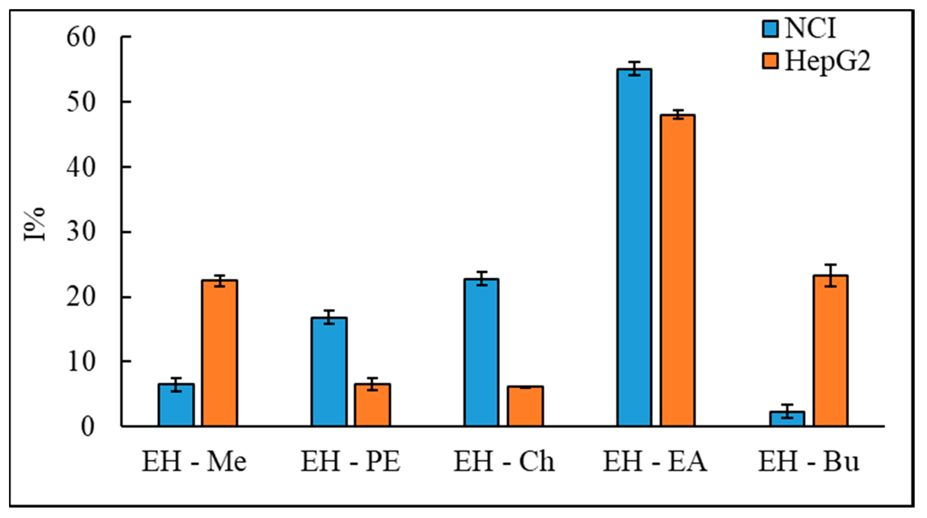
3.5. In vitro Screening for Cytotoxic Activity of E. hirta Linn.

The SRB assay (sulforhodamine B) is a simple and sensitive colorimetric method for determining the cytotoxicity of a substance. SRB is a negatively charged dye containing two sulfonic groups that bind static electricity to positively charged parts of protein under mild acidic conditions and dissociate under basic conditions. The amount of bound dye will reflect the total protein amount of the cell. In this experiment, cells were fixed, washed and stained with SRB. The SRB then binds to the dissolved cell protein to form a pink, transparent solution. The measured optical density of the solution correlates with the total protein amount or the number of cells. The change in cell count compared to controls reflects the cytotoxicity of the study substance. In the present study, at a constant concentration of 100 μg/mL, the cancer inhibitory activity on the growth of lung cancer cells NCI-H460 and liver cancer cells Hep G2 of five extracts were measured (Figure 3).

4. Discussion

The tendency to use traditional medicines in the treatment of many diseases has been increasing. Numerous studies have proven that plant-based medicines have not only demonstrated the ability to prevent and treat diseases effectively, but also limited side effects and adverse effects of modern medicines. Plants are rich in natural bioactive compounds, such as phenolics, flavonoids, and their derivatives. These compounds have obtained attention because of their bioactivities and physiological functions, including antioxidant, anti-allergic, anti-inflammatory, antimicrobial, and antidiabetic activities [42].
From methanol crude extract of E. hirta (EH-Me extract) containing multiple different groups of phytoconstituents, liquid–liquid extraction technique can be employed to separate many different fractions. As a result, five extracts of E. hirta were obtained based on their polarity by specific solvents including petroleum ether, chloroform, ethyl acetate and butanol extracts. Qualitative phytochemical analysis of five extracts of E. hirta was carried out to evaluate the bioactive compounds in each extract. The concentration of bioactive compounds in the plant extracts depends on the solvent polarity that was used in the extraction preparation [33,43]. The extraction method is the first step to separate phytochemicals from raw materials. According to the principle of extraction, solvent extraction is the most popularly used method. In this method, solvent polarity significantly affects the efficiency of extraction and constituents of phytochemicals. Depending on chemical nature, a variety of secondary metabolites are extracted in solvents of different polarity as no single solvent may reliably extract all the phytochemical and antioxidant compounds present in the plant material. The serial exhaustive extraction method involves the successive extraction with solvents of increasing polarity from non-polar (petroleum ether) to more polar solvent (water) to ensure the extraction of a wide range of compounds with different polarity. The ascending order of the polarization of solvents is petroleum ether < chloroform < ethyl acetate < butanol < methanol. Therefore, methanol was used to extract raw material. In relation to the solvent used, the highest concentrations of total phenolic and flavonoid contents were found in EH-EA extracts. EH-EA had the highest total phenol content (254.96 ± 10.05 mg GAE/g extract) and the highest total flavonoid contents (27.66 ± 0.73 mg quercetin/g extract). The results of the determination of total phenolic content and total flavonoid content of these extracts were almost compatible with previous studies [44]. From the above results, it can be predicted that EH-EA extract has better activity in biological activity trials. For the purpose of screening biological activities of these extracts, we investigated antibacterial, antioxidant, lipid peroxidation inhibitory and anticancer properties.
Five E. hirta extracts were investigated to evaluate their antibacterial activity against pathogenic bacteria including two strains of Gram-positive bacteria and three strains of Gram-negative bacteria using the agar disc diffusion method. The strains of bacteria selected for antibatesting cause diarrhea, acute diarrhea and pneumonia, etc., which are diseases with a high mortality rate. E. coli are a large and diverse group of aerobic bacteria in the environment and food, and either live in the digestive tract of humans or animals. E. coli have contributed to many important functions in human life such as stimulating immunity, improving the metabolization of sugars, and preventing the attack of other bacteria from entering the digestive system. However, they can cause diseases such as urinary tract infection, meningitis, etc. S. aureus is a major bacterial human pathogen that causes a wide variety of clinical manifestations. The results show that EH-Ch extract and EH-EA exhibited antibacterial inhibitory effect better than other extracts. Among them, EH-EA has the strongest antibacterial activity. However, all extracts almost have no inhibitory activity against E. coli, S. aureus and P. aeruginosa at most concentrations. The result of antimicrobial property of E. hirta extracts is almost compatible with previous studies [45,46,47,48,49,50].
DPPH free radical scavenging is one of the important mechanisms for the evaluation of the antioxidant activity of a sample. The effect of antioxidants on DPPH were predicted through their hydrogen donating ability of the sample [51]. Literature suggested that total polyphenols content and radical scavenging antioxidant activity are highly correlated. Radical scavenging activities are very important to prevent the deleterious role of free radicals in different diseases, including cancer [52,53,54]. To investigate the antioxidant activity of E. hirta extracts, DPPH radical scavenging assay of E. hirta extracts was carried out. The IC50 values of EH-Me, EH-PE, EH-Ch, EH-EA, EH-Bu were measured to estimate the potential scavenging abilities of the samples. Our results reveal that EH-Me and EH-EA extracts had stronger free radical scavenging activity than other extracts. This result could be explained by EH-Me and EH-EA extracts containing many phenolic compounds and flavonoids. According to IC50 values, it can be speculated that EH-EA showed the highest scavenging activity. This result shows that there is similarity to previous research results [55].
For lipid peroxidation inhibitory activity, IC50 values of EH-Me, EH-PE, EH-Ch, EH-EA, EH-Bu were also measured to evaluate antioxidant properties of E. hirta extracts. According to IC50 values given, EH-Me, EH-PE, EH-EA and EH-Bu extracts possess strong inhibitory properties against lipid oxidation. In particular, EH-EA extract appeared to be 18.74-fold as potent as Trolox, whereas EH-Me, EH-Bu, EH-PE were about 5.93, 5.36, 1.07-fold, respectively (Table 4). These experimental results can be explained by the high concentration of bioactive ingredients extracted from extracts using highly polar solvents. Taken together, these results indicate that E. hirta extracts may have therapeutic effects such as inhibiting lipid peroxidation and preventing radical damage. Moreover, previous studies have also reported that E. hirta extract had efficacy against selective cancer cell lines such as Hep-2, malignant melanoma, and squamous cell carcinoma [56,57]. In 2014, Neelesh Sharma et al. evaluated anticancer activities of E. hirta ethanolic extract [55]. The ethanolic extract showed selective anticancer activity at a concentration of 100 µg/mL (p < 0.05). Anticancer activity and cytotoxicity of silver nanoparticles tested against neuroblastoma cell (SH-SY5Y cells) and breast cancer (MCF-7 cell) was reported. That study proved that the ethanolic extract of E. hirta showed significant anticancer activity against neuroblastoma cell (SH-SY5Y cells) and breast cancer (MCF-7 cell) [58]. In the present investigation, in the in vitro screening for cytotoxic activity of E. hirta extracts, at a concentration of 100 μg/mL, EH-EA extract has significant inhibitory activity against the growth of lung cancer cells NCI-H460 and live cancer cells Hep G2, about 55% and 48%, respectively. Similar to the biological activities described above, EH-EA extract was also shown to be much more effective in inhibiting two line of cancer cells than the crude extract EH-Me and three remaining fractions. At the same concentration of 100 μg/mL, EH-Me, EH-PE, EH-Ch and EH-Bu did not show significant high cytotoxicity on NCI H460 and Hep G2. For the mentioned results, EH-EA extract may be rich in bioactive compounds, and these compounds exhibited significant antibacterial, antioxidant and anticancer activities.

5. Conclusions

Recent investigations have proven that secondary metabolites from natural resources containing bioactive components have a wide variety of biological properties. This study provides significant evidence about the biological activity of different extracts of E. hirta. These results indicate that E. hirta extract can be a good source for antioxidant, antimicrobial, and anticancer herbal-related drugs. From the above-mentioned results about biological activities, the EH-EA extract outperformed the EH-Me extract and the remaining extracts. Therefore, further studies to isolate and identify of bioactive compounds from EH-EA extract for in vitro and in vivo investigations of observed activities are highly recommended. In addition, it is necessary to elucidate the mechanisms of action of these extracts and bioactive compounds isolated from this plant at the cellular and molecular level to evaluate biological capacity of substances on specific therapeutic properties.

Author Contributions

Conceptualization, N.T., N.N. and L.L.; methodology, N.T., M.N. and K.P.L.; software, K.P.L. and M.N.; validation, N.T., M.N. and N.N.; formal analysis, N.T. and M.N.; investigation, N.T., N.N. and M.N.; resources, L.L. and Q.T.; data curation, N.N. and L.L.; writing—original draft preparation, N.T.; writing—review and editing, N.T., M.N. and L.L.; visualization, N.N.; supervision, L.L.; project administration, Q.T. and L.L. All authors have read and agreed to the published version of the manuscript.

Funding

This work was funded by Vietnam National University at Ho Chi Minh City under grant number GEN2019-28-01/HD-KHCN together with the ethical approval for the research content.

Conflicts of Interest

The authors declare no conflict of interest.

References

  1. Ji, H.-F.; Li, X.-J.; Zhang, H.-Y. Natural products and drug discovery. EMBO Rep. 2009, 10, 194–200. [Google Scholar] [CrossRef] [PubMed] [Green Version]
  2. Koparde, A.A.; Doijad, R.C.; Magdum, C.S. Natural Products in Drug Discovery. In Pharmacognosy—Medicinal Plants, 1st ed.; Perveen, S., Al-Taweel, A., Eds.; IntechOpen: London, UK, 2019; Available online: https://www.intechopen.com/books/pharmacognosy-medicinal-plants/natural-products-in-drug-discovery (accessed on 18 August 2020). [CrossRef] [Green Version]
  3. World Health Organisation. Traditional, Complementary and Integrative Medicine. Available online: https://www.who.int/health-topics/traditional-complementary-and-integrative-medicine (accessed on 19 August 2020).
  4. Ikegami, F.; Fujii, Y.; Ishihara, K.; Satoh, T. Toxicological aspects of Kampo medicines in clinical use. Chem. Biol. Interact. 2003, 145, 235–250. [Google Scholar] [CrossRef]
  5. Al-Rifai, A.; Aqel, A.; Al-Warhi, T.; Wabaidur, S.M.; Al-Othman, Z.A.; Badjah-Hadj-Ahmed, A.Y. Antibacterial, Antioxidant Activity of Ethanolic Plant Extracts of Some Convolvulus Species and Their DART-ToF-MS Profiling. Evid. Based Complement. Altern. Med. 2017, 2017, 1–9. [Google Scholar] [CrossRef] [Green Version]
  6. Wachtel-Galor, S.; Benzie, I.F.F. Herbal Medicine: An Introduction to Its History, Usage, Regulation, Current Trends, and Research Needs. In Herbal Medicine: Biomolecular and Clinical Aspects, 2nd ed.; Benzie, I.F.F., Wachtel-Galor, S., Eds.; CRC Press/Taylor & Francis: Boca Raton, FL, USA, 2011. Available online: https://0-www-ncbi-nlm-nih-gov.brum.beds.ac.uk/books/NBK92773/ (accessed on 1 August 2020).
  7. Prachayasittikul, S.; Buraparuangsang, P.; Worachartcheewan, A.; Isarankura-Na-Ayudhya, C.; Ruchirawat, S.; Prachayasittikul, V. Antimicrobial and Antioxidative Activities of Bioactive Constituents from Hydnophytum formicarum Jack. Molecules 2008, 13, 904–921. [Google Scholar] [CrossRef] [Green Version]
  8. Park, C.H.; Yeo, H.J.; Baskar, T.B.; Park, Y.E.; Park, J.S.; Lee, S.Y.; Park, S.U. In Vitro Antioxidant and Antimicrobial Properties of Flower, Leaf, and Stem Extracts of Korean Mint. Antioxidants 2019, 8, 75. [Google Scholar] [CrossRef] [Green Version]
  9. Duthie, G.G.; Duthie, S.J.; Kyle, J.A.M. Plant polyphenols in cancer and heart disease: implications as nutritional antioxidants. Nutr. Res. Rev. 2000, 13, 79–106. [Google Scholar] [CrossRef] [Green Version]
  10. Myburgh, K.H. Polyphenol Supplementation: Benefits for Exercise Performance or Oxidative Stress? Sports Med. 2014, 44, 57–70. [Google Scholar] [CrossRef] [Green Version]
  11. Douglas, C.J. Phenylpropanoid metabolism and lignin biosynthesis: from weeds to trees. Trends Plant Sci. 1996, 1, 171–178. [Google Scholar] [CrossRef]
  12. Islam, Z.; Hossain, T.; Hossen, F.; Mukharjee, S.K.; Sultana, N.; Paul, S.C. Evaluation of antioxidant and antibacterial activities of Crotalaria pallida stem extract. Clin. Phytosci. 2018, 4, 8. [Google Scholar] [CrossRef]
  13. Lalitha, T.P.; Jayanthi, P. Preliminary studies on phytochemicals and antimicrobial activity of solvent extracts of Eichhornia crassipes (Mart.) Solms. Asian J. Plant Sci. 2012, 2, 115–122. [Google Scholar]
  14. Elisha, I.L.; Botha, F.S.; McGaw, L.J.; Eloff, J.N. The antibacterial activity of extracts of nine plant species with good activity against Escherichia coli against five other bacteria and cytotoxicity of extracts. BMC Complement. Altern. Med. 2017, 17, 133. [Google Scholar] [CrossRef] [PubMed] [Green Version]
  15. Gonelimali, F.D.; Lin, J.; Miao, W.; Xuan, J.; Charles, F.; Chen, M.; Hatab, S.R. Antimicrobial Properties and Mechanism of Action of Some Plant Extracts Against Food Pathogens and Spoilage Microorganisms. Front. Microbiol. 2018, 9, 1639. [Google Scholar] [CrossRef] [PubMed]
  16. Cowan, M.M. Plant Products as Antimicrobial Agents. Clin. Microbiol. Rev. 1999, 12, 564–582. [Google Scholar] [CrossRef] [PubMed] [Green Version]
  17. Repetto, M.; Semprine, J.; Boveris, A. Lipid Peroxidation: Chemical Mechanism, Biological Implications and Analytical Determination. In Lipid Peroxidation; Catala, A., Ed.; IntechOpen: London, UK, 2012; Available online: https://www.intechopen.com/books/lipid-peroxidation/lipid-peroxidation-chemical-mechanism-biological-implications-and-analytical-determination (accessed on 15 August 2020). [CrossRef] [Green Version]
  18. Shichiri, M. The role of lipid peroxidation in neurological disorders. J. Clin. Biochem. Nutr. 2014, 54, 151–160. [Google Scholar] [CrossRef] [Green Version]
  19. Hall, E.D. The Contributing Role of Lipid Peroxidation and Protein Oxidation in the Course of CNS Injury Neurodegeneration and Neuroprotection: An Overview. In Brain Neurotrauma: Molecular, Neuropsychological, and Rehabilitation Aspects; Kobeissy, F.H., Ed.; CRC Press/Taylor & Francis: Boca Raton, FL, USA, 2015. Available online: https://0-www-ncbi-nlm-nih-gov.brum.beds.ac.uk/books/NBK299180/ (accessed on 15 August 2020).
  20. Halliwell, B.; Chirico, S. Lipid peroxidation: its mechanism, measurement, and significance. Am. J. Clin. Nutr. 1993, 57 (Suppl. 715S–725S), 715S–725S. [Google Scholar] [CrossRef] [Green Version]
  21. Fadlelmula, A.A.; Al-Ghamdi, A.Y.; Abdalla, M.O. In vitro Antioxidant Activity, Total Phenolic Content and Antimicrobial Activity of Coleus forskohlii Grown in Al-Baha, Saudi Arabia. Asian J. Chem. 2020, 32, 2033–2037. [Google Scholar] [CrossRef]
  22. World Health Organisation. Cancer. Available online: https://www.who.int/news-room/fact-sheets/detail/cancer (accessed on 19 August 2020).
  23. Bray, F.; Me, J.F.; Soerjomataram, I.; Siegel, R.L.; Torre, L.A.; Jemal, A. Global cancer statistics 2018: GLOBOCAN estimates of incidence and mortality worldwide for 36 cancers in 185 countries. CA Cancer J. Clin. 2018, 68, 394–424. [Google Scholar] [CrossRef] [Green Version]
  24. Xiang, Y.; Guo, Z.; Zhu, P.; Chen, J.; Huang, Y. Traditional Chinese medicine as a cancer treatment: Modern perspectives of ancient but advanced science. Cancer Med. 2019, 8, 1958–1975. [Google Scholar] [CrossRef]
  25. Kausar, J.; Muthumani, D.; Hedina, A.; Ramasamy, S.; Anand, V. Review of the phytochemical and pharmacological activities of Euphorbia hirta Linn. Pharmacogn. J. 2016, 8, 310–313. [Google Scholar] [CrossRef] [Green Version]
  26. Sharma, M.; Sheliya, M.A.; Begum, R.; Pillai, K.K.; Aeri, V.; Mir, S.R.; Ali, A.; Sharma, M. In vitro α-glucosidase and α-amylase inhibition by aqueous, hydroalcoholic, and alcoholic extract of Euphorbia hirta L. Drug Dev. Ther. 2016, 7, 26. [Google Scholar] [CrossRef]
  27. Sharma, N.K.; Dey, S.; Prasad, R. In vitro antioxidant potential evaluation of Euphorbia hirta L. Pharmacologyonline 2007, 1, 91–98. [Google Scholar]
  28. Kumar, S.; Malhotra, R.; Kumar, D. Euphorbia hirta: Its chemistry, traditional and medicinal uses, and pharmacological activities. Pharmacogn. Rev. 2010, 4, 58–61. [Google Scholar] [CrossRef] [Green Version]
  29. Tran, N.; Tran, M.; Truong, H.; Le, L. Spray-Drying Microencapsulation of High Concentration of Bioactive Compounds Fragments from Euphorbia hirta L. Extract and Their Effect on Diabetes Mellitus. Foods 2020, 9, 881. [Google Scholar] [CrossRef]
  30. Zhang, Q.-W.; Lin, L.-G.; Ye, W.-C. Techniques for extraction and isolation of natural products: a comprehensive review. Chin. Med. 2018, 13, 20. [Google Scholar] [CrossRef] [PubMed] [Green Version]
  31. Susantikarn, P.; Donlao, N. Optimization of green tea extracts spray drying as affected by temperature and maltodextrin content. Int. Food Res. J. 2016, 23, 1327–1331. [Google Scholar]
  32. Tambe, V.D.; Bhambar, S.R. Estimation of total phenol, tannin, alkaloid and flavonoid in Hibiscus tiliaceus Linn. wood extracts. Res. Rev. J. Pharm. Phytochem. 2014, 2, 41–47. [Google Scholar]
  33. Bunea, A.; Rugina, D.; Pintea, A.; Diaconeasa, Z.; Bunea, C.I.; Socaciu, C. Comparative Polyphenolic Content and Antioxidant Activities of Some Wild and Cultivated Blueberries from Romania. Not. Bot. Horti Agrobot. Cluj-Napoca 2011, 39, 70–76. [Google Scholar] [CrossRef] [Green Version]
  34. Bajalan, I.; Zand, M.; Goodarzi, M.; Darabi, M. Antioxidant activity and total phenolic and flavonoid content of the extract and chemical composition of the essential oil of Eremostachys laciniata collected from Zagros. Asian Pac. J. Trop. Biomed. 2017, 7, 144–146. [Google Scholar] [CrossRef]
  35. Guo, L.; Wang, Y.; Bi, X.; Duo, K.; Sun, Q.; Yun, X.; Zhang, Y.; Fei, P.; Han, J. Antimicrobial Activity and Mechanism of Action of the Amaranthus tricolor Crude Extract against Staphylococcus aureus and Potential Application in Cooked Meat. Foods 2020, 9, 359. [Google Scholar] [CrossRef] [PubMed] [Green Version]
  36. Aghraz, A.; Albergamo, A.; Benameur, Q.; Salvo, A.; Larhsini, M.; Markouk, M.; Gervasi, T.; Cicero, N. Polyphenols contents, heavy metals analysis and in vitro antibacterial activity of extracts from Cladanthus arabicus and Bubonium imbricatum of Moroccan Origin. Nat. Prod. Res. 2019, 34, 63–70. [Google Scholar] [CrossRef]
  37. Díaz, J.G.; Tuenter, E.; Arranz, J.C.E.; Maury, G.L.; Cos, P.; Pieters, L. Antimicrobial activity of leaf extracts and isolated constituents of Croton linearis. J. Ethnopharmacol. 2019, 236, 250–257. [Google Scholar] [CrossRef]
  38. Amarowicz, R.; Pegg, R.; Rahimi-Moghaddam, P.; Barl, B.; Weil, J. Free-radical scavenging capacity and antioxidant activity of selected plant species from the Canadian prairies. Food Chem. 2004, 84, 551–562. [Google Scholar] [CrossRef]
  39. Kedare, S.B.; Singh, R.P. Genesis and development of DPPH method of antioxidant assay. J. Food Sci. Technol. 2011, 48, 412–422. [Google Scholar] [CrossRef] [Green Version]
  40. Apak, R.; Özyürek, M.; Güçlü, K.; Çapanoğlu, E. Antioxidant Activity/Capacity Measurement. 1. Classification, Physicochemical Principles, Mechanisms, and Electron Transfer (ET)-Based Assays. J. Agric. Food Chem. 2016, 64, 997–1027. [Google Scholar] [CrossRef] [PubMed]
  41. Nguyen, M.-N.T.; Ho-Huynh, T.-D. Selective cytotoxicity of a Vietnamese traditional formula, Nam Dia long, against MCF-7 cells by synergistic effects. BMC Complement. Altern. Med. 2016, 16, 220. [Google Scholar] [CrossRef] [PubMed] [Green Version]
  42. Noreen, H.; Semmar, N.; Farman, M.; Mccullagh, J.S. Measurement of total phenolic content and antioxidant activity of aerial parts of medicinal plant Coronopus didymus. Asian Pac. J. Trop. Med. 2017, 10, 792–801. [Google Scholar] [CrossRef] [PubMed]
  43. Basma, A.A.; Zakaria, Z.; Latha, L.Y.; Sasidharan, S. Antioxidant activity and phytochemical screening of the methanol extracts of Euphorbia hirta L. Asian Pac. J. Trop. Med. 2011, 4, 386–390. [Google Scholar] [CrossRef]
  44. Loh, D.S.Y.; Er, H.M.; Chen, Y.S. Mutagenic and antimutagenic activities of aqueous and methanol extracts of Euphorbia hirta. J. Ethnopharmacol. 2009, 126, 406–414. [Google Scholar] [CrossRef]
  45. Singh, G.; Kumar, P. Phytochemical study and screening for antimicrobial activity of flavonoids of Euphorbia hirta. Int. J. Appl. Basic Med. Res. 2013, 3, 111–116. [Google Scholar] [CrossRef]
  46. Alisi, C.S.; Abanobi, S.E. Antimicrobial Properties of Euphorbia hyssopifolia and Euphorbia hirta against Pathogens Complicit in Wound, Typhoid and Urinary Tract Infections. Int. J. Trop. Dis. Health 2012, 2, 72–86. [Google Scholar] [CrossRef]
  47. Titilope, K.K.; Rashidat, E.A.; Christiana, O.C.; Kehinde, E.R.; Ombolaji, J.N.; Olajide, A.J. In-vitro antimicrobial activities of Euphorbia hirta against some clinical isolates. Agric. Biol. J. N. Am. 2012, 3, 169–174. [Google Scholar] [CrossRef]
  48. Perumal, S.; Mahmud, R.; Pillai, S.; Lee, W.C.; Ramanathan, S. Antimicrobial Activity and Cytotoxicity Evaluation of Euphorbia hirta (L.) Extracts from Malaysia. APCBEE Procedia 2012, 2, 80–85. [Google Scholar] [CrossRef] [Green Version]
  49. Abubakar, E.-M.M. Antibacterial activity of crude extracts of Euphorbia hirta against some bacteria associated with enteric infections. J. Med. Plants Res. 2009, 3, 498–505. [Google Scholar]
  50. Rajeh, M.A.B.; Zuraini, Z.; Sasidharan, S.; Latha, L.Y.; Amutha, S. Assessment of Euphorbia hirta L. Leaf, Flower, Stem and Root Extracts for Their Antibacterial and Antifungal Activity and Brine Shrimp Lethality. Molecules 2010, 15, 6008–6018. [Google Scholar] [CrossRef] [Green Version]
  51. Baumann, J.; Wurm, G.; Bruchhausen, F.V. Hemmung der Prostaglandinsynthetase durch Flavonoide und Phenolderivate im Vergleich mit deren O2--Radikalfänger-eigenschaften. [Prostaglandin synthetase inhibition by flavonoids and phenolic compounds in relation to their O2-scavenging properties (author’s transl)]. Arch. Pharm. Pharm. Med. Chem. 1980, 313, 330–337. (In German) [Google Scholar] [CrossRef]
  52. Lobo, V.; Patil, A.; Phatak, A.; Chandra, N. Free radicals, antioxidants and functional foods: Impact on human health. Pharmacogn. Rev. 2010, 4, 118–126. [Google Scholar] [CrossRef] [PubMed] [Green Version]
  53. Pham-Huy, L.A.; He, H.; Pham-Huy, C. Free Radicals, Antioxidants in Disease and Health. Int. J. Biomed. Sci. IJBS 2008, 4, 89–96. [Google Scholar] [PubMed]
  54. Rahman, M.; Islam, B.; Biswas, M.; Alam, A.H.M.K. In vitro antioxidant and free radical scavenging activity of different parts of Tabebuia pallida growing in Bangladesh. BMC Res. Notes 2015, 8, 621. [Google Scholar] [CrossRef] [PubMed] [Green Version]
  55. Sharma, N.; Samarakoon, K.W.; Gyawali, R.; Park, Y.-H.; Lee, S.J.; Oh, S.J.; Lee, T.H.; Jeong, D.K. Evaluation of the Antioxidant, Anti-Inflammatory, and Anticancer Activities of Euphorbia hirta Ethanolic Extract. Molecules 2014, 19, 14567–14581. [Google Scholar] [CrossRef] [Green Version]
  56. Sidambaram, R.R.; Dinesh, M.G.; Jayalakshmi, E.T. An in vitro study of cytotoxic activity of Euphorbia hirta on hep2 cells of human epithelioma of larynx. Int. J. Pharm. Pharm. Sci. 2011, 3, 101–103. [Google Scholar]
  57. Ping, K.Y.; Darahb, I.; Chenc, Y.; Sasidharan, S.; Dharmaraj, S. Cytotoxicity and genotoxicity assessment of Euphorbia hirta in MCF-7 cell line model using comet assay. Asian Pac. J. Trop. Biomed. 2013, 3, 692–696. [Google Scholar] [CrossRef] [Green Version]
  58. Selvam, P.; Vijayakumar, T.; Wadhwani, A.; Muthulakshmi, L. Bioreduction of silver nanoparticles from aerial parts of Euphorbia hirta L. (EH-ET) and its potent anticancer activities against neuroblastoma cell lines. Indian J. Biochem. Biophys. 2019, 56, 132–136. [Google Scholar]
Figure 1. Euphorbia hirta Linn.
Applsci 10 08408 g001
Figure 2. The chart indicates the process of solvent extraction.
Applsci 10 08408 g002
Figure 3. Cytotoxicity of E. hirta L. on NCI H460 and Hep G2 cells.
Applsci 10 08408 g003
Table 1. Weight and yield of extract after liquid–liquid extract.
Name of ExtractWeight (g)Yield (%)Humidity (%)
Dried plant8000-8.01 ± 0.38
EH-Me90011.2515.05 ± 0.05
EH-PE3003.756.01 ± 0.29
EH-Ch90.113.03 ± 0.12
EH-EA921.155.31 ± 0.21
EH-Bu1171.4617.44 ± 0.33
Total141817.725-
Table 2. Total phenolic and flavonoid contents in E. hirta extracts. Each value is the average of three analyses ± standard error of mean (n = 3). Values with different superscripts in the columns are significantly different based on one-way ANOVA followed by Dunnett’s test.
SamplesTotal Phenolic Content
(mg GAE/g Dried Extract)
Total Flavonoid Content
(mg QE/g Dried Extract)
EH-Me109.86 ± 1.38 a18.92 ± 1.33 f
EH-PE90.89 ± 1.45 b8.48 ± 1.16 g
EH-Ch55.86 ± 0.66 c16.41 ± 1.44 h
EH-EA254.96 ± 10.05 d27.66 ± 0.73 i
EH-Bu70.90 ± 0.65 e12.43 ± 1.66 j
Table 3. Antibacterial activity of E. hirta Linn extracts by disc diffusion method. - No inhibition zone observed.
Tested BacteriumEH-Me
(mg/mL)
EH-PE
(mg/mL)
EH-Ch
(mg/mL)
EH-EA
(mg/mL)
EH-Bu
(mg/mL)
ConcentrationDIZConcentrationDIZConcentrationDIZConcentrationDIZConcentrationDIZ
B. subtilis--2001040011----
E. coli---------
S. aureus----------
P. aeruginosa----------
S. preumoniae------40012--
S. typhi------20011--
V. cholearea40011--200112001340010
S. flexneri------40013--
Table 4. 2,2-diphenyl-1-pycrylhydrazyl (DPPH) radical scavenging activity of extracts.
SamplesIC50 (µg/mL)
EH-Me17.26 ± 0.12
EH-PE122.86 ± 2.65
EH-Ch92.48 ± 1.47
EH-EA10.33 ± 0.01
EH-Bu55.54 ± 0.09
Ascorbic acid5.11 ± 0.02
Table 5. Lipid peroxidation inhibitory activity of extracts.
SamplesIC50 (µg/mL)
EH-Me4.69 ± 0.13
EH-PE25.99 ± 0.09
EH-Ch56.19 ± 0.98
EH-EA1.48 ± 0.12
EH-Bu5.19 ± 0.02
Trolox27.85 ± 1.22
Publisher’s Note: MDPI stays neutral with regard to jurisdictional claims in published maps and institutional affiliations.

