Article

A New Approach to Set the Absolute Midsagittal Plane of the Mandible Using a Similarity Index in Skeletal Class III Patients with Facial Asymmetry

1
Department of Orthodontics, Institute of Craniofacial Deformity, Yonsei University College of Dentistry, Seoul 03722, Korea
2
Department of Oral and Maxillofacial Radiology, Institute of Translational Dental Sciences, School of Dentistry, Pusan National University, Yangsan 50612, Korea
3
BK21 FOUR Project, Yonsei University College of Dentistry, Seoul 03722, Korea
*
Author to whom correspondence should be addressed.
These authors contributed equally to this article.
Submission received: 27 October 2020 / Revised: 25 November 2020 / Accepted: 27 November 2020 / Published: 29 November 2020
(This article belongs to the Special Issue Applied Sciences in Dentistry)

Abstract

:
This study sought to test the feasibility of a newly developed plane called computed modified absolute mandibular midsagittal plane (cmAMP) based on the similarity index (SI) for evaluating the stereoscopical symmetry of the mandible by comparison with other proposed midsagittal planes. This study involved 29 adult patients (15 men, 14 women; average age, 23.1 ± 6.9 years) with skeletal Class III facial asymmetry who underwent bimaxillary orthognathic surgery. Using cone-beam computed tomography images taken before and 1 year after surgery, cmAMP with the highest SI value between the two anterior segments of the hemi-mandible was set by a computer algorithm. Results show that the SI using cmAMP had the highest value (0.83 ± 0.04) before surgery compared to the other midsagittal planes, and was not significantly different from the SI (0.80 ± 0.05) using a facial midsagittal plane (MSP) after surgery. The distance (1.15 ± 0.74 mm) and angle (2.02 ± 0.82°) between MSP and cmAMP after surgery were significantly smaller than those between MSP and other midsagittal planes. In conclusion, the cmAMP plane best matches the two anterior segments of hemi-mandible symmetrically and is the closest to MSP after orthognathic surgery in skeletal Class III patients with facial asymmetry.

1. Introduction

Facial asymmetry is one of the main reasons for patients to undergo orthognathic surgery [1], especially among Asians, where asymmetry between the right and left hemi-mandibles with mandibular prognathism is common. Currently, bimaxillary surgery with mandibular setback procedures, such as the sagittal split ramus osteotomy (SSRO) or intraoral vertical ramus osteotomy (IVRO), is the main approach to correct this asymmetry. Traditionally, SSRO has been preferred over IVRO in asymmetric surgery due to rapid postoperative recovery and reliable stability through rigid fixation. However, if a large amount of mandibular setback is required for the improvement of facial asymmetry in Asians, IVRO has the advantage of reducing the possibility of injury to the inferior alveolar nerve and less adversely affecting temporomandibular disorders [2,3].
Recent advances in the field of dentistry allow clinicians to harness the power of 3D imaging, using cone-beam computed tomography (CBCT) among other imaging modalities, to precisely diagnose the patient’s skeletal deformity, set up a pre- and post-surgical orthodontic treatment plan, and simulate orthognathic surgery [4,5,6,7]. Nevertheless, to date, when planning bimaxillary surgery for a patient with mandibular asymmetry using 3D CBCT images, the goal is merely to position a menton (Me) located below the mandibular symphysis on the facial midsagittal plane (MSP) as the lateral deviation of the chin is the most characteristic feature of facial asymmetry [8,9]. However, Me can only be defined as a single point during the two-dimensional (2D) lateral and posteroanterior cephalometric analyses. If the only Me is overlapped with MSP as a treatment objective to correct mandibular asymmetry, then the morphological asymmetry between the two hemi-mandibles likely remain after surgery, especially when asymmetry exists primarily in the mandibular body. In other words, when only considering one Me for predicting surgical results, it may not be possible to capture the exact shape of the mandible and to evaluate stereoscopic asymmetry in the 3D space.
To evaluate mandible symmetry in the 3D space, a plane that allows superimposition of the left and right hemi-mandibles is required for using a mirroring technique [10,11]. With the availability of such a plane, the goal of mandibular surgery for correction of a patient’s facial asymmetry would thus be to match it with the MSP to the greatest extent possible by moving the anterior mandibular segment in front of the osteotomy line during the IVRO or SSRO procedure.
Many studies have aimed to establish this setting plane as a midsagittal plane of the mandible. For example, You et al. [12,13] proposed a midsagittal plane of the mandible comprising of the line connecting the Me, B point, and the middle point between genial tubercles (G). Fang et al. [14,15] evaluated the structural symmetry of the mandible by using an optimal symmetry plane created by computing the best pairing of the bony voxels on the two hemi-mandibles. Lin et al. [16] measured the residual mandible asymmetry following the menton point correction to the facial sagittal plane by mirroring one side of the hemi-mandible onto the other, and concluded that the symphysis and para-symphysis, and the body and angle of the hemi-mandible exhibited transversal and vertical discrepancies that contributed to mandible asymmetry. However, these studies do not intuitively show the difference in the 3D structure of the two hemi-mandibles by comparing them mainly using two-dimensional (2D) measurement methods such as distance and angle. In our previous study [17], the similarity index (SI), which indicates the ratio of the overlapping volume to the whole mandibular volume when the two hemi-mandibles are superimposed using the mirrored images created by the setting plane, allowed the intuitive assessment of 3D mandibular asymmetry.
Therefore, this study aimed to test the feasibility of using a newly developed plane called the computed modified absolute mandibular midsagittal plane (cmAMP) based on the SI for evaluating the mandible 3D symmetry. We hypothesized that when mirroring two anterior mandibular segments in front of the cutting line for IVRO surgery using various midsagittal planes, cmAMP would be the most precise midsagittal plane with the highest SI value. In addition, cmAMP would also be the closest plane to the MSP after bimaxillary surgery in patients with skeletal Class III malocclusion and facial asymmetry.

2. Materials and Methods

2.1. Study Design and Patients

Twenty-nine adult patients diagnosed with skeletal Class III facial asymmetry were selected for this study. All patients received bimaxillary orthognathic surgery (Le Fort I osteotomy for the maxilla and bilaterally IVRO for the mandible) preceded by pre-surgical orthodontic treatment from March 2010 to January 2019 at the Yonsei University Dental Hospital, Seoul, Republic of Korea. The inclusion criteria were as follows: (1) patients 18 years of age or older who were confirmed to have no further growth through the analyses of hand-wrist and lateral cephalometric radiographs at 6 months intervals; (2) diagnosis of skeletal Class III malocclusion with the angle of the lines connecting point A, the nasion, and point B smaller than 0° on the 2D lateral cephalometric image before surgery; (3) Me deviation larger than 4 mm, as measured by the perpendicular distance from the Me to MSP on 2D posteroanterior cephalometric images before surgery [17]; and (4) there were 3D CBCT images available before surgery and 1 year after surgery. The exclusion criteria for this study were as follows: (1) history of orthodontic treatment or orthognathic surgery; (2) single-jaw surgery or pre-orthodontic orthognathic bimaxillary surgery, or bimaxillary surgery with genioplasty; (3) craniofacial deformities that can affect the growth of the maxilla or mandible such as cleft lip and palate and hemifacial microsomia; (4) history of maxillofacial trauma; and (5) inappropriate CBCT images due to motion blurring artifacts.
This study was approved by the institutional review board of Yonsei University Dental Hospital (2-2018-0063) and follows the Declaration of Helsinki. All patients agreed with the study’s procedures by providing written informed consent before the orthodontic treatment.

2.2. Surgical and Orthodontic Treatment

Bimaxillary orthognathic surgery was performed between March 2010 and January 2019 by two surgeons at Yonsei University Dental Hospital, Seoul, Republic of Korea. For the IVRO operations, a conventional double-slide osteotomy was performed. A round oscillating saw was used to simplify the angle at the ante-lingular prominence and was directed anterosuperiorly towards the sigmoid notch [7]. Maxillomandibular fixation with an occlusal splint appliance was carried out for 2 weeks after surgery. All patients were instructed to perform, for another 2 weeks, elastic traction with active physiotherapy, which included exercising mouth opening and lateral excursion of jaw movement. Post-surgical orthodontic treatment was then initiated and implemented for 6 months to 1 year [18].

2.3. Data Acquisition, 3D Landmark Determination, and Image Reorientation

CBCT data were acquired using the Alphard3030 (Alphard Roentgen Ind., Ltd., Kyoto, Japan) at 80 kVp and 10 mA and with a 200 × 200 mm field of view at two different times, namely before and 1 year after surgery. The voxel size was 0.39 mm. The CBCT images were converted to DICOM 3.0 files and stored on a Windows-10 based workstation (Intel Core i7-4770, 32 GB). Using the medical studio of Invivo 6 (Anatomage, San Jose, CA, USA), eight 3D anatomical landmarks were selected in accordance with the procedure used in previous studies (Table 1) [12,13,17].
The images were reoriented using two reference planes: the MSP, passing through the nasion (N) and the sella (S), and perpendicular to the Frankfort horizontal plane (FHP). FHP passed through the center of the bilateral porions (Po) and the left orbitale (Or), according to the Frankfort Craniometric Agreement of August 1882, among a variety of options in this study.
After configuration of the two reference planes and hard tissue landmarks via the Invivo 6 software as described above, the coordinates of 3D landmarks and the binarization thresholds of bone (320–520 HU) were saved in an Excel file format. A custom program using MATLAB 2019b (MathWorks, Natick, MA, USA) was used for successive image processing for all methods in this study [17,19].

2.4. Morphological Operation of Mandible

Separate images of the mandible were binarized and a morphological operation was performed for surface area and volume calculation. In each binarized image, the holes were filled using morphological closing (dilation followed by erosion) for accurate measurements, based on the method used in previous studies [17,20]. In this article, a sphere structuring element with a radius of 10 pixels was used for morphological closure.

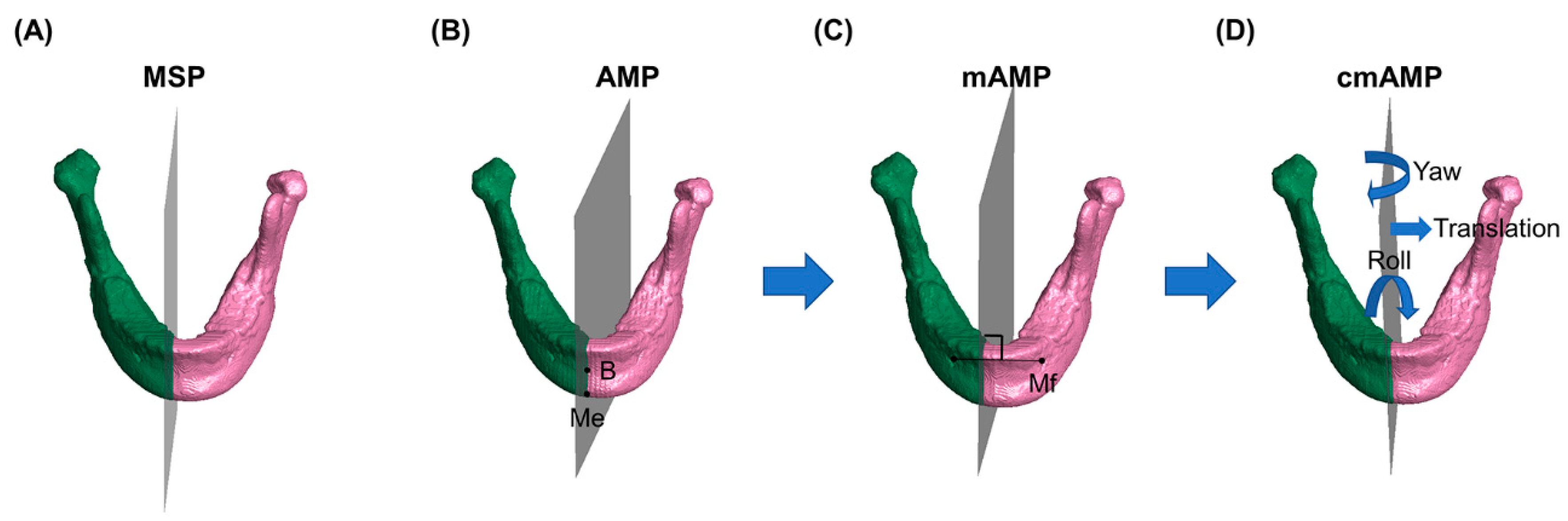
2.5. AMP, mAMP, cmAMP Assessments

The AMP was obtained by setting up a plane containing Me, B, and G. The modified absolute mandibular midsagittal plane (mAMP) was set up as the plane that passed through the center point of the bilateral mental foramen and was simultaneously vertical to the line connecting those two points (Figure 1). The cmAMP was established at the point with the highest SI value while controlling for the roll, yaw, and translation (x-axis) using the center point of the bilateral mental foramen as the center of rotation. The grid search method was used for seeking the highest SI value for the anterior segment of the hemi-mandible.

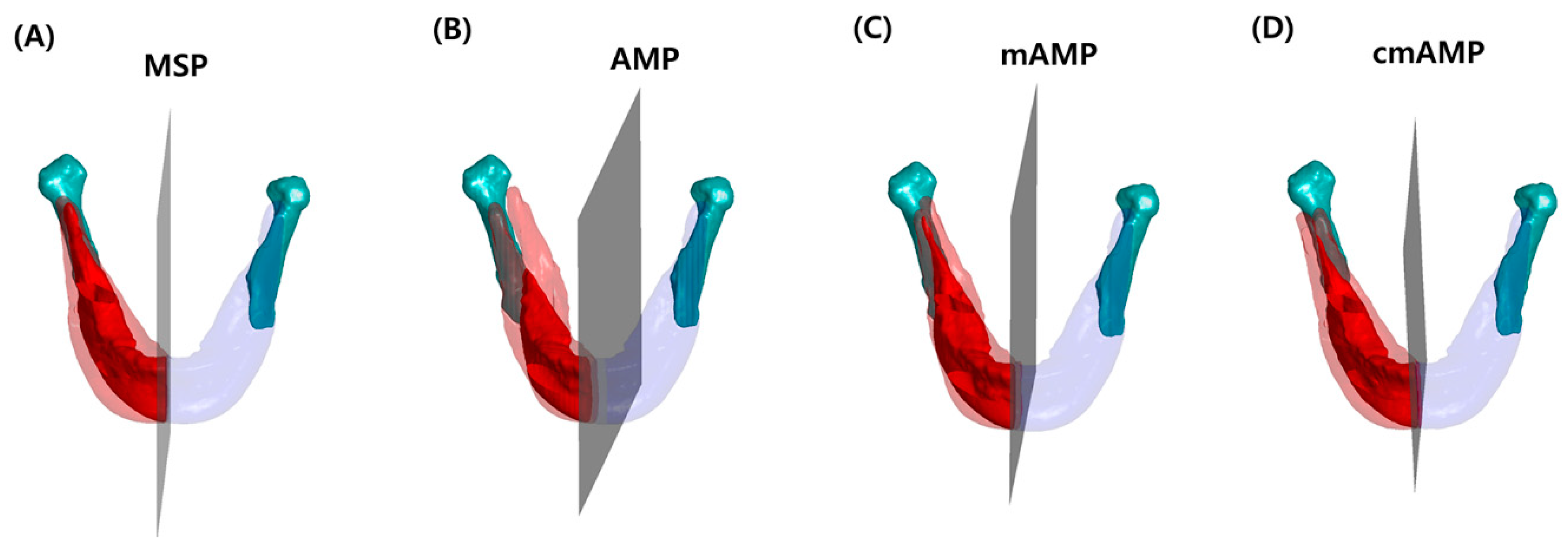
2.6. Non-Overlapping Surface, Volume, and SI with Each Mandibular Midsagittal Plane

To divide the anterior and posterior mandibular segments, the IVRO surgical procedure was simulated using the conventional osteotomy line from the angle at the ante-lingular prominence to the sigmoid notch (Figure 2). The non-overlapping surface, volume, and SI were calculated with each mandibular midsagittal plane in the anterior segment before and 1 year after surgery. The surface area was calculated using the distance around the boundary multiplied by the square of voxel size. The volume was calculated using the number of voxels in each image multiplied by the cube of the voxel size. The SI, which intuitively evaluated mandibular symmetry by expressing it as a value ranging from 0 to 1 and was calculated with one part of the hemi-mandible overlapped onto the other as the reference plane, was estimated according to the equation outlined in our previous study [17].

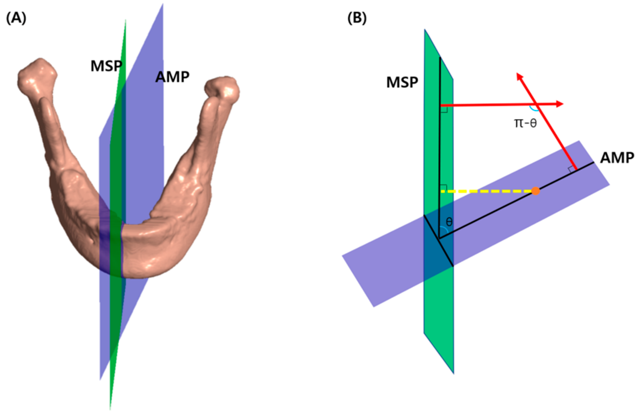
2.7. Angle and Distance between MSP and Each Midsagittal Plane of the Mandible

Before and 1 year after surgery, the angle between the two planes was obtained by calculating the angle between the normal vector of the two planes (Figure 3).
From the three landmarks that determine each midsagittal plane of the mandible, the normal vector of each plane was obtained using Equation (1), and the angle between the planes was calculated using Equation (2).
A normal vector n of the plane passing through three points p 1 ,   p 2 ,   p 3 is computed by
n = ( p 2 p 1 ) × ( p 3 p 1 )
And the angle θ of two planes is computed by
θ = cos 1 ( n 1 n 2 n 1 n 2 )
where n 1 ,   n 2 are normal vectors of the two planes and denotes the Euclidean norm as shown in Figure 3.
The distance between the two planes was obtained by calculating the distance between MSP and the center of Me, B, and G in AMP, the center of the bilateral mental foramen in mAMP, and the center of rotation (during grid search) of the bilateral mental foramen in cmAMP, respectively.

2.8. Reliability

A single observer measured the anatomical landmarks in all patients twice every two weeks. The estimated intra-examiner correlation coefficient of these landmarks ranged from 0.994 to 0.999.

2.9. Statistical Analysis

All statistics were analyzed using SPSS software for Windows, version 22.0 (IBM Corp., Armonk, NY, USA). The sample size was calculated based on a previous study [17]. For detection of the statistical significance of the change in SI before and 1 year after surgery, G*Power 3 (Dusseldorf, Germany) was used with a significance level of p < 0.05, a power of 80%, and an effect size of 0.6; the calculated sample size was 24.
The normality of the data was confirmed by the Shapiro-Wilk test. A one-way analysis of variance (ANOVA) test followed by Tukey’s test was used to compare the differences in the non-overlapping surface, volume, and SI for each hemi-mandible anterior segment using each midsagittal plane before and 1 year after surgery. One-way ANOVA was also used to compare the distance and angle between MSP and each mandibular midsagittal plane at each time point. A paired t-test was used to analyze the change in the non-overlapping surface, volume, and SI based on MSP before and 1 year after surgery. A p value of less than 0.05 was considered to be statistically significant.

3. Results

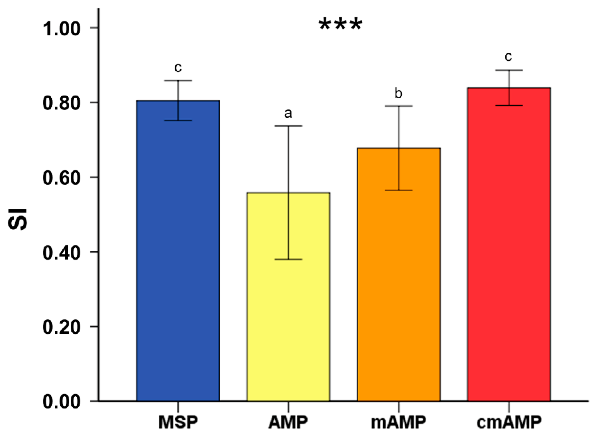
3.1. Non-Overlapping Surface, Volume, and SI Using Mirroring Technique Based on Each Mandibular Midsagittal Plane Prior to Surgery

This study included 29 patients (15 men, 14 women; average age, 23.1 ± 6.9 years). The non-overlapping surface (3543.14 ± 734.13 mm2) and volume (5088.85 ± 2280.33 mm3) of the right anterior segment of the hemi-mandible based on cmAMP before surgery were significantly smaller than those with MSP, AMP, and mAMP as the reference plane (p < 0.001) (Table 2). A similar trend was observed in the hemi-mandible left anterior segment. On the other hand, the SI with cmAMP showed a significantly higher value than that when using the other planes as the reference, reaching 0.83 ± 0.04. These results suggested that cmAMP is a plane that can divide the anterior segment of the mandible most symmetrically left and right.

3.2. Differences in Non-Overlapping Surface, Volume, and SI Based on the MSP before and after Surgery

The patient groups studied had larger values for the non-overlapping surface and volume on the left than on the right (Table 2 and Table 3). This suggests that the Me deviated to the left with respect to the midsagittal reference plane for mirroring. This is consistent with the results of a Japanese study [21]; it reported that in skeletal class III patients, Me was mainly biased to the left. With the MSP as the reference plane, the non-overlapping surface and volume of the hemi-mandible right anterior segment decreased significantly from 4347.03 ± 775.29 and 7942.44 ± 2790.26 mm3 before surgery to 3899.65 ± 842.71 and 6036.33 ± 2357.24 mm3 after surgery, respectively (Table 3). A similar trend was observed in the left anterior segment of the hemi-mandible. In contrast with the results for the non-overlapping surface and volume, the SI significantly increased from 0.74 ± 0.07 before surgery to 0.80 ± 0.05 1 year after surgery. The method of measuring the non-overlapping surface area and volume has been widely used as a conventional method in other studies to evaluate the asymmetry of the mandible. It is possible that the symmetry of the anterior segment of the mandible improved 1 year after surgery through the tendency of the non-overlapping surface and volume to decrease after surgery compared to before surgery; however, it is still difficult to discern the amount of improvement, both quantitatively and intuitively. However, when using a color-coded map for SI expression, the improvement in the symmetry of the anterior segment of the mandible can be easily and intuitively observed, as the numerical values improve from 0.74 to 0.8.

3.3. Distance and Angle between MSP and Each Mandibular Midsagittal Plane before and after Surgery

The distance (1.77 ± 1.19 mm) and angle (2.87 ± 1.49°) between the MSP and cmAMP before surgery were significantly smaller than those with MSP, AMP, and mAMP as the reference plane (p = 0.001) (Table 4). This distance and angle between the MSP and cmAMP after surgery decreased to 1.15 ± 0.74 mm and 2.02 ± 0.82°, respectively. These values were also significantly smaller than those between MSP and AMP and between MSP and mAMP. As shown in Figure 3, the smaller the angle and distance difference between the two planes in the three-dimensional space, the closer and more consistent are the two planes. Since the difference in the angle and distance between the cmAMP and the postoperative MSP is the smallest, which is thought to reflect an improvement in the asymmetry of the anterior segment of the hemi-mandible, it indicates that that cmAMP is the closest plane to the MSP 1 year after surgery.

3.4. Comparison of SI between MSP 1 Year after Surgery and Each Mandibular Midsagittal Plane before Surgery

Upon analysis using the one-way ANOVA followed by Tukey’s test, the SI before surgery, which was 0.55 ± 0.17 using AMP and 0.67 ± 0.11 using mAMP, showed a statistically significant difference with the SI using MSP 1 year after surgery. This finding indicates that, in the patient with mandibular asymmetry, if the pre-surgical AMP or mAMP is assumed to be the midsagittal plane of the mandible and these mandible anterior segment planes move in a direction overlapping with the MSP, then the mandible symmetry may not be greatly recovered (p < 0.001) (Figure 4). In other words, if pre-surgical AMP and mAMP are considered midsagittal planes of the mandible and postoperative MSP matches while planning and during the surgery, other additive surgeries, including cortical osteotomy, genioplasty, or bone grafting, will be required to obtain an SI value as high as the SI value calculated based on MSP 1 year after surgery. In contrast, the SI calculated using MSP as the reference plane 1 year after surgery was 0.80 ± 0.05, had no significant difference with the SI calculated using cmAMP as the reference plane before surgery, which was 0.83 ± 0.04. This indicates that the symmetry of the mandible anterior segment, as calculated based on cmAMP before surgery, does not significantly differ from the symmetry of the mandible anterior segment, as calculated based on postsurgical MSP. This implies that the symmetry of the mandible can be maximized after surgery only when surgery is performed in the direction where the cmAMP overlaps the MSP.

4. Discussion

Bimaxillary orthognathic surgery should be an optimal treatment option for patients with mandibular prognathism and asymmetry. However, to date, mandibular surgery has mainly been performed in a manner where the maxilla is repositioned and then adjusted accordingly [21]. For many years, and even to this day, the main objective of the surgical treatment for facial asymmetry has conventionally been to coincide the Me, which is a designated anatomical landmark in the symphysis area, with the postsurgical MSP, despite the recent developments for diagnosis, surgical plans, and simulations using 3D technologies [8,9,22].
Diagnostic and surgical methods for facial asymmetry face few clinical problems when the direction of the menton deviations, as measured as the difference in length between the left and right ramus, and the canting direction of the maxillary occlusal plane, coincide [9]. However, if there is an asymmetry in the body shape of the left and right mandibles, residual asymmetry often remains after surgery [9]. In other words, facial asymmetry mainly due to the right and left hemi-mandibular body may remain or be inadvertently increased after orthognathic surgery. This is not only because post-surgery changes in the soft tissue and adaptation to the skeletal structures are hard to anticipate precisely, but also because structural analysis of the hemi-mandible is not carefully considered [17,23]. You et al. [13] reported that condylar and body units both affected the asymmetrical differences in the mandible. Therefore, to reflect the morphological difference between the left and right hemi-mandible bodies, a 3D stereoscopic image mirroring technique is required. Furthermore, to optimize such mirroring, an optimal mandibular reference plane other than Me is needed.
In a typical IVRO surgical procedure, the proximal segment containing the condylar region is overlapped with the distal segment of the mandible by cortex-to-cortex contact after the conventional double-slide osteotomy line is defined from the angle at the ante-lingular prominence to the sigmoid notch. To focus on the main contributing components of residual mandible asymmetry after IVRO surgery, the anterior segment of the hemi-mandible was sectioned, apart from the distal segment along the osteotomy line for IVRO surgery, and chosen for comparative analysis in this study.
When thoroughly assessing studies that evaluated or analyzed the stereoscopic structure of the mandible in the 3D space using either mirroring or segmentation, it was very important to set up the reference plane which could distinguish the right and left hemi-mandibles [10,11,12,13,14,16,17]. In 2019, Kwon et al. [17] proposed the new concept of SI, which intuitively assessed the mandible asymmetry on 3D images by using the mirroring technique with AMP as the internal reference plane of the mandible. AMP is the plane connecting Me, B, and G. According to the literal definitions of these anatomical landmarks, B is the midpoint of greatest concavity on the anterior border of the symphysis and Me is the most inferior midpoint on the symphysis. Rather than the exact configuration that is usually used and defined as the landmark in 2D lateral and posteroanterior cephalogram analysis, here the words “greatest” and “most” refer to the highest possibility. Since the mirroring technique is very sensitive to slight alterations of the plane formed by the coordinates of these points in the 3D space, it is crucial to reduce empirical errors and bias as well as to increase reproducibility. Furthermore, the genial tubercle is an anatomical structure at the inner surface of the mandible symphysial region that shows great morphological variation among individuals, with possibly (1) two superior and two inferior tubercles; (2) two superior and one inferior tubercle; (3) the same as in (2), but with more thickened tubercles; (4) a common spine; (5) the absence of genial tubercles; and (6) only one superior pair of tubercles [24]. Ultimately, it is very difficult to define the unified point of genial tubercle and to preserve the reproducibility of that point, whatever the chosen configuration of reference. To evaluate the mandibular asymmetry, the midsagittal reference plane can be accurately calculated symmetrically only if there is a bilateral landmark, not a single landmark point, such as the aforementioned Me, B point, and genial tubercle. In this regard, the mental foramen represents the bilaterally important landmark in the mandible that allows dividing it into the body and chin parts, and it is easier to identify fully based on appearance on 3D CBCT images with good visibility [25,26]. The asymmetric position of each mental foramen is not important since it acts as a boundary in which mAMP can be set, and helps cmAMP be set by a computer algorithm on this boundary. When the mAMP, which passes through the midpoint of the two bilateral mental foramen and is simultaneously vertical to the line connecting these two points, was used as the reference plane, the non-overlapping surface of the right anterior segment of the hemi-mandible (4423.69 ± 777.71 mm2) and the distance between the MSP and mAMP before surgery (3.16 ± 2.07 mm) showed no significant difference with those with AMP (4786.67 ± 1029.46 mm2 and 3.64 ± 2.22 mm, respectively) as the reference plane (Table 2 and Table 4). Also, the SI value based on the mAMP before surgery (0.67 ± 0.11) was lower than that based on the MSP (0.74 ± 0.07) (Figure 4). This indicates that mAMP is an absolute midsagittal plane of the mandible that represents an inappropriate alternative to AMP.
While continuously using the mental foramen as the anatomical landmark, the cmAMP with the highest SI value, on which the center point of bilateral mental foramen was taken as the center of rotation for controlling the roll, yaw, and translation, was newly set using the algorithmic grid search method of a computer algorithm. The results in this study showed that the distance and angle between MSP and cmAMP were significantly smaller after surgery and that SI calculated using cmAMP as the reference plane before surgery had no significant difference with, or was even greater than, the SI based on postsurgical MSP. Hence, cmAMP is the only absolute midsagittal plane of the mandible anterior segment that may be fit into MSP for correcting facial asymmetry, and that may reduce the possibility of morphological asymmetry of the mandibular body after surgery.
Several studies have attempted to establish the median plane as the reference plane with the help of computer-aided and computer-designed algorithm programs. In particular, Fang et al. [14,15] introduced a new approach for establishing the landmark-free voxel-based median plane of the mandible, called the optimal symmetry plane, with the aid of a computer algorithm. Our concept of processing the cmAMP based on the SI with a computer algorithm was similar to Fang’s concept of generating the new median plane by calculating the paired voxels out of the whole matching image. However, in addition to revealing transverse differences as in Fang’s study, we were able to represent stereoscopic structural discrepancies between the right and left segments of the hemi-mandible in 3D space, such as in volume, through numbers and color-coded maps. Also, the intuitive analysis of differences in the 3D shape of the hemi-mandible was feasible, avoiding the additional step of contour analysis for the evaluation of facial asymmetry as described in Fang’s consecutive study.
Since cmAMP is the inner median plane of the hard tissue of the mandible and does not involve any soft tissue or dental problem, orthognathic surgery without pre-surgical orthodontics (surgery-first approach or pre-orthodontic orthognathic surgery) may be ideal for matching cmAMP to MSP as closely as possible during surgery [2,7]. When cmAMP is applied to conventional orthognathic surgery with pre-surgical orthodontic treatment, the postsurgical occlusal relationship between the upper and lower teeth should be considered and adjusted compromisingly with overlapping cmAMP in the direction of MSP. Moreover, the need for any auxiliary surgeries like bone grafting, cortical osteotomy, and genioplasty could be reduced if cmAMP was used during surgical planning and simulation because mandible symmetry could be maximized by using cmAMP. Should additive surgery be required, it would be possible to create surgical guides through images expressed in volume discrepancies in color-codes by grafting cmAMP to commercially available 3D software.
Among several factors that cause mandibular asymmetry, the different growth rates of the condylar and body units independently occurring are known to be the main etiologic cause for the asymmetry [27,28,29]. In this study, only the anterior segment divided by the osteotomy line was compared and analyzed in cases that received orthognathic surgery with IVRO only. If the cause of the mandibular asymmetry is the morphological difference of the condyle or posterior part of the ramus, it is possible to evaluate the asymmetry of the entire mandibular structure stereoscopically, as shown in Figure 1 and Figure 2, such that the technique we introduced here can be applied to each case. In this study, since the anterior part can be easily improved through orthognathic surgery, we focused on the recovery of the anterior part’s similarity index before and after surgery. Not only do patients with severe facial asymmetry want corrective orthognathic surgery, but patients with minor or even inexistent asymmetry now also want to receive bimaxillary surgery for improving facial aesthetics. Thus, future studies are needed to investigate whether this new approach of establishing cmAMP may be applied to patients with minor asymmetry who were planning to undergo bimaxillary orthognathic surgery. Conversely, in the case of severe asymmetric patients with a congenital deformity, if the landmark of bilateral structures, such as the mental foramen used in this study, are well maintained and detected, we can apply cmAMP to diagnose patients, help to plan surgery, and evaluate the results after surgery. However, in growing patients, even if they perform orthognathic surgery with cmAMP, the left and right structures can be changed through continuous growth. The fact that this study, as such, was only conducted among adults remains a limitation. In addition, asymmetric patients may need care throughout their lifetime.
Furthermore, to date, most studies on the changes in soft tissue following bimaxillary surgical correction of facial asymmetry remain inconclusive [5,30,31]. Since soft tissue changes are difficult to predict with simple mathematical formulas or calculations, we have been forced to focus on improving the most predictable hard tissue in our study. However, even though it remains difficult to accurately predict the change in soft tissue according to the change in hard tissue through orthognathic surgery using CBCT images, the amount of change in the volume of the soft tissue of the corresponding area according to the change of hard tissue volume is intuitively calculated and evaluated by applying the technique we introduced. If measured, the predictability of soft tissue changes can be improved. To maximize the similarity index of soft tissue after surgery through the application of cmAMP in future studies, it is necessary to reflect the various factors (soft tissue tension, gender, age, weight, vertical skeletal pattern, etc.) of each patient that affect the prediction of soft tissue in the algorithm rather than using simple measurement points [32].
Finally, following the necessary further development of our software, additional research will be required for the clinical applicability of using cmAMP as the median sagittal plane of the mandible anterior segments to diagnose patients, make a surgical plan, perform surgery, and evaluate results in cooperation with oral and maxillofacial surgeons.

5. Conclusions

A novel method to set up the midsagittal plane of the mandible anterior segments called cmAMP was developed by using the SI to evaluate the stereoscopic symmetry of the mandible. cmAMP was the most precise midsagittal plane with the highest SI value, that is, it offered the best matching between the right and left segments of the hemi-mandible as divided by this plane. Moreover, cmAMP was also the closest plane to the MSP after bimaxillary orthognathic surgery in patients with skeletal Class III malocclusion and facial asymmetry.

Author Contributions

Conceptualization, J.J.H. and S.-H.C.; methodology, W.-J.H., J.J.H., and S.-H.C.; validation, J.J.H. and S.-H.C.; formal analysis, S.-H.C.; resources, Y.-H.J., B.-H.C., K.-J.L., and H.-S.Y.; data curation, J.-J.H. and S.-H.C.; writing—original draft preparation, W.-J.H.; writing—review and editing, J.-J.H. and S.-H.C.; visualization, J.J.H.; supervision, J.J.H. and S.-H.C.; project administration, J.J.H. and S.-H.C.; funding acquisition, J.J.H. and S.-H.C. All authors have read and agreed to the published version of the manuscript.

Funding

This research was supported by a grant from the Korea Health Technology R&D Project through the Korea Health Industry Development Institute (KHIDI), funded by the Ministry of Health & Welfare, Republic of Korea (grant number: HI20C0611).

Conflicts of Interest

The authors declare no conflict of interest.

References

  1. Severt, T.R.; Proffit, W.R. The prevalence of facial asymmetry in the dentofacial deformities population at the University of North Carolina. Int. J. Adult Orthodon. Orthognath. Surg. 1997, 12, 171–176. [Google Scholar] [PubMed]
  2. Choi, S.H.; Yoo, H.J.; Lee, J.Y.; Jung, Y.S.; Choi, J.W.; Lee, K.J. Stability of pre-orthodontic orthognathic surgery depending on mandibular surgical techniques: SSRO vs IVRO. J. Craniomaxillofac. Surg. 2016, 44, 1209–1215. [Google Scholar] [CrossRef] [PubMed]
  3. Al-Gunaid, T.; Yamaki, M.; Takagi, R.; Saito, I. Soft and hard tissue changes after bimaxillary surgery in Japanese class III asymmetric patients. J. Orthod. Sci. 2012, 1, 69–76. [Google Scholar] [CrossRef] [PubMed] [Green Version]
  4. Kim, S.J.; Lee, K.J.; Yu, H.S.; Jung, Y.S.; Baik, H.S. Three-dimensional effect of pitch, roll, and yaw rotations on maxillomandibular complex movement. J. Craniomaxillofac. Surg. 2015, 43, 264–273. [Google Scholar] [CrossRef] [PubMed]
  5. Lo, L.J.; Weng, J.L.; Ho, C.T.; Lin, H.H. Three-dimensional region-based study on the relationship between soft and hard tissue changes after orthognathic surgery in patients with prognathism. PLoS ONE 2018, 13, e0200589. [Google Scholar] [CrossRef] [Green Version]
  6. Ludlow, J.B.; Gubler, M.; Cevidanes, L.; Mol, A. Precision of cephalometric landmark identification: Cone-beam computed tomography vs conventional cephalometric views. Am. J. Orthod. Dentofacial Orthop. 2009, 136, 312. [Google Scholar] [CrossRef]
  7. Choi, S.H.; Hwang, C.J.; Baik, H.S.; Jung, Y.S.; Lee, K.J. Stability of Pre-Orthodontic Orthognathic Surgery Using Intraoral Vertical Ramus Osteotomy Versus Conventional Treatment. J. Oral Maxillofac. Surg. 2016, 74, 610–619. [Google Scholar] [CrossRef]
  8. Chen, Y.J.; Yao, C.C.; Chang, Z.C.; Lai, H.H.; Yeh, K.J.; Kok, S.H. Characterization of facial asymmetry in skeletal Class III malocclusion and its implications for treatment. Int. J. Oral Maxillofac. Surg. 2019, 48, 1533–1541. [Google Scholar] [CrossRef]
  9. Kwon, S.M.; Baik, H.S.; Jung, H.D.; Jang, W.; Choi, Y.J. Diagnosis and Surgical Outcomes of Facial Asymmetry According to the Occlusal Cant and Menton Deviation. J. Oral Maxillofac. Surg. 2019, 77, 1261–1275. [Google Scholar] [CrossRef]
  10. Davies, J.C.; Chan, H.H.L.; Jozaghi, Y.; Goldstein, D.P.; Irish, J.C. Analysis of simulated mandibular reconstruction using a segmental mirroring technique. J. Craniomaxillofac. Surg. 2019, 47, 468–472. [Google Scholar] [CrossRef]
  11. Leonardi, R.; Muraglie, S.; Bennici, O.; Cavallini, C.; Spampinato, C. Three-dimensional analysis of mandibular functional units in adult patients with unilateral posterior crossbite: A cone beam study with the use of mirroring and surface-to-surface matching techniques. Angle Orthod. 2019, 89, 590–596. [Google Scholar] [CrossRef] [PubMed] [Green Version]
  12. You, K.H.; Kim, K.H.; Lee, K.J.; Baik, H.S. Three-dimensional computed tomography analysis of mandibular morphology in patients with facial asymmetry and mandibular retrognathism. Am. J. Orthod. Dentofacial. Orthop. 2018, 153, 685–691. [Google Scholar] [CrossRef] [PubMed]
  13. You, K.H.; Lee, K.J.; Lee, S.H.; Baik, H.S. Three-dimensional computed tomography analysis of mandibular morphology in patients with facial asymmetry and mandibular prognathism. Am. J. Orthod. Dentofacial. Orthop. 2010, 138, 540. [Google Scholar] [CrossRef]
  14. Fang, J.J.; Tu, Y.H.; Wong, T.Y.; Liu, J.K.; Zhang, Y.X.; Leong, I.F.; Chen, K.C. Evaluation of mandibular contour in patients with significant facial asymmetry. Int. J. Oral Maxillofac. Surg. 2016, 45, 922–931. [Google Scholar] [CrossRef]
  15. Wong, T.Y.; Liu, J.K.; Fang, J.J.; Wu, T.C.; Tu, Y.H. Use of the matching optimal symmetry plane method in planning surgical correction of facial asymmetry—A preliminary report of 20 patients. J. Oral. Maxillofac. Surg. 2014, 72, 1180.e1–1180.e13. [Google Scholar] [CrossRef]
  16. Lin, H.; Zhu, P.; Lin, Q.; Huang, X.; Xu, Y.; Yang, X. Comprehensive Analysis of Mandibular Residual Asymmetry after Bilateral Sagittal Split Ramus Osteotomy Correction of Menton Point Deviation. PLoS ONE 2016, 11, e0161601. [Google Scholar] [CrossRef] [PubMed]
  17. Kwon, S.M.; Hwang, J.J.; Jung, Y.H.; Cho, B.H.; Lee, K.J.; Hwang, C.J.; Choi, S.H. Similarity index for intuitive assessment of three-dimensional facial asymmetry. Sci. Rep. 2019, 9, 10959. [Google Scholar] [CrossRef] [Green Version]
  18. Jung, H.D.; Jung, Y.S.; Park, J.H.; Park, H.S. Recovery pattern of mandibular movement by active physical therapy after bilateral transoral vertical ramus osteotomy. J. Oral Maxillofac. Surg. 2012, 70, e431–437. [Google Scholar] [CrossRef]
  19. Sun, L.; Lee, K.M. Three-Dimensional Evaluation of the Postsurgical Stability of Mandibular Setback With the Surgery-First Approach: Comparison Between Patients With Symmetry and Asymmetry. J. Oral Maxillofac. Surg. 2019, 77, 1469-e1. [Google Scholar] [CrossRef]
  20. Hwang, J.J.; Han, S.S.; Lee, C.; Jung, Y.H. Automatic analysis algorithm for acquiring standard dental and mandibular shape data using cone-beam computed tomography. Sci. Rep. 2018, 8, 13516. [Google Scholar] [CrossRef]
  21. Haraguchi, S.; Takada, K.; Yasuda, Y. Facial asymmetry in subjects with skeletal Class III deformity. Angle Orthod. 2002, 72, 28–35. [Google Scholar] [PubMed]
  22. Wu, T.Y.; Denadai, R.; Lin, H.H.; Ho, C.T.; Lo, L.J. The outcome of skeletofacial reconstruction with mandibular rotation for management of asymmetric skeletal class III deformity: A three-dimensional computer-assisted investigation. Sci. Rep. 2019, 9, 13337. [Google Scholar] [CrossRef] [PubMed] [Green Version]
  23. Vittert, L.; Katina, S.; Ayoub, A.; Khambay, B.; Bowman, A.W. Assessing the outcome of orthognathic surgery by three-dimensional soft tissue analysis. Int. J. Oral. Maxillofac. Surg. 2018, 47, 1587–1595. [Google Scholar] [CrossRef] [PubMed] [Green Version]
  24. Greyling, L.M.; Le Grange, F.; Meiring, J.H. Mandibular spine: A case report. Clin. Anat. 1997, 10, 416–418. [Google Scholar] [CrossRef]
  25. Park, W.; Kim, B.C.; Yu, H.S.; Yi, C.K.; Lee, S.H. Architectural characteristics of the normal and deformity mandible revealed by three-dimensional functional unit analysis. Clin. Oral Investig. 2010, 14, 691–698. [Google Scholar] [CrossRef]
  26. Parnia, F.; Moslehifard, E.; Hafezeqoran, A.; Mahboub, F.; Mojaver-Kahnamoui, H. Characteristics of anatomical landmarks in the mandibular interforaminal region: A cone-beam computed tomography study. Med. Oral Patol. Oral Cir. Bucal. 2012, 17, e420–e425. [Google Scholar] [CrossRef] [Green Version]
  27. Bruce, R.A.; Hayward, J.R. Condylar hyperplasia and mandibular asymmetry: A review. J. Oral Surg. 1968, 26, 281–290. [Google Scholar]
  28. Bjork, A. Variations in the growth pattern of the human mandible: Longitudinal radiographic study by the implant method. J. Dent. Res. 1963, 42, 400–411. [Google Scholar] [CrossRef]
  29. Moss, M.L.; Rankow, R.M. The role of the functional matrix in mandibular growth. Angle Orthod. 1968, 38, 95–103. [Google Scholar]
  30. Olate, S.; Zaror, C.; Blythe, J.N.; Mommaerts, M.Y. A systematic review of soft-to-hard tissue ratios in orthognathic surgery. Part III: Double jaw surgery procedures. J. Craniomaxillofac. Surg. 2016, 44, 1599–1606. [Google Scholar] [CrossRef]
  31. Olate, S.; Zaror, C.; Mommaerts, M.Y. A systematic review of soft-to-hard tissue ratios in orthognathic surgery. Part IV: 3D analysis–Is there evidence? J. Craniomaxillofac. Surg. 2017, 45, 1278–1286. [Google Scholar] [CrossRef] [PubMed]
  32. Choi, S.H.; Lee, H.; Hwang, J.J.; Jung, H.D.; Hwang, C.J.; Cha, J.Y. Differences in soft-tissue thickness changes after bimaxillary surgery between patients with vertically high angle and normal angle. Am. J. Orthod. Dentofacial. Orthop. 2020. [Google Scholar] [CrossRef] [PubMed]
Figure 1. Setting up each mandibular midsagittal plane: (A) facial midsagittal plane (MSP); (B) absolute mandibular midsagittal plane (AMP); (C) modified absolute mandibular midsagittal plane (mAMP); (D) computed modified absolute mandibular midsagittal plane (cmAMP). Me, menton; B, supramentale; Mf, mental foramen.
Applsci 10 08550 g001
Figure 2. Segmentation of the hemi-mandible into anterior and posterior parts. The hemi-mandible was divided into anterior and posterior segments using a conventional osteotomy line for the intraoral vertical ramus osteotomy (IVRO) operation. The opaque section indicates overlapping volume and the translucent section indicates the non-overlapping volume, using each mandibular midsagittal plane in the anterior segment. (A) facial midsagittal plane (MSP); (B) absolute mandibular midsagittal plane (AMP); (C) modified absolute mandibular midsagittal plane (mAMP); and (D) computed modified absolute mandibular midsagittal plane (cmAMP).
Applsci 10 08550 g002
Figure 3. Procedure for calculating the distance and angle between the facial midsagittal plane (MSP) and absolute mandibular midsagittal plane (AMP). (A) MSP and AMP; (B) The angle formed by two normal vectors of MSP and AMP is the same as the angle between the two planes. The distance between MSP and the center (orange dot) of Me, B, and G in the absolute mandibular midsagittal plane (AMP) is shown with the yellow dotted line.
Applsci 10 08550 g003
Figure 4. Comparison of the similarity index (SI) between the facial midsagittal plane (MSP) 1 year after surgery and each mandibular midsagittal plane before surgery. MSP, facial midsagittal plane; AMP, absolute mandibular midsagittal plane; mAMP, modified absolute mandibular midsagittal plane; cmAMP, computed modified absolute mandibular midsagittal plane. The p values were calculated using one-way ANOVA. Different letters indicate significant differences between groups. *** p < 0.001.
Applsci 10 08550 g004
Table 1. Anatomical landmarks.
LandmarkDefinition
N (nasion)Midpoint of nasofrontal suture
S (sella)Center of sella turcica
Or (orbitale)Most inferior point of the lower margin of the orbit
Po (porion)Most superior point of the external auditory meatus
Me (menton)Most inferior midpoint on the symphysis
B (supramental)Midpoint of greatest concavity on the anterior border of the symphysis
G (genial tubercle)Midpoint on genial tubercle
Mf (mental foramen)Midpoint of mental foramen
Table 2. Non-overlapping surface, volume, and similarity index (SI) using a mirroring technique based on each plane before surgery.
MSPAMPmAMPcmAMPp-Value
MeanSDMeanSDMeanSDMeanSD
Right segment
Non-overlapping surface (mm2)4347.03 b775.294786.67 b1029.464423.69 b777.713543.14 a734.13<0.001
Non-overlapping volume (mm3)7942.44 ab2790.2614,175.40 c6718.9110,382.64 b5019.155088.85 a2280.33<0.001
Left segment
Non-overlapping surface (mm2)4567.87 a1047.015044.53 b1117.224603.61 a949.714112.34 a990.930.01
Non overlapping volume (mm3)8212.43 ab3045.5314,093.04 c6689.5310,641.74 b4926.485423.47 a2467.45<0.001
SI0.74 b0.070.55 a0.170.67 b0.110.83 c0.04<0.001
MSP, facial midsagittal plane; AMP, absolute mandibular midsagittal plane; mAMP, modified absolute mandibular midsagittal plane; cmAMP, computed modified absolute mandibular midsagittal plane. p values were calculated by one-way ANOVA. Different letters indicate significant differences between groups.
Table 3. Differences in the non-overlapping surface, volume, and similarity index (SI) using the mirroring technique based on the MSP before and 1 year after surgery.
Before SurgeryAfter SurgeryDifferencep-Value
MeanSDMeanSDMeanSD
Right segment
Non-overlapping surface (mm2)4347.03775.293899.65842.71447.37684.560.001
Non-overlapping volume (mm3)7942.442790.266036.332357.241906.112567.01<0.001
Left segment
Non-overlapping surface (mm2)4567.871047.014240.631008.61327.23623.120.009
Non-overlapping volume (mm3)8212.433045.536469.822703.881742.602944.470.004
SI0.740.070.800.05−0.050.07<0.001
MSP, facial midsagittal plane; SD, standard deviation. p values were calculated by paired t-test.
Table 4. Distance and angle between MSP and each plane before and after surgery.
Between MSP andAMPBetween MSP and mAMPBetween MSP and cmAMPp-Value
MeanSDMeanSDMeanSD
Before surgery
Distance (mm)3.64 b2.223.16 b2.071.77 a1.190.001
Angle (degree)5.21 b3.613.98 ab2.352.87 a1.490.005
After surgery
Distance (mm)2.46 b1.842.34 b1.651.15 a0.740.002
Angle (degree)4.91 b3.152.90 a1.632.02 a0.82<0.001
MSP, facial midsagittal plane; AMP, absolute mandibular midsagittal plane; mAMP, modified absolute mandibular midsagittal plane; cmAMP, computed modified absolute mandibular midsagittal plane. p values were calculated by one-way ANOVA. Different letters indicate significant differences between groups.
Publisher’s Note: MDPI stays neutral with regard to jurisdictional claims in published maps and institutional affiliations.

