Article

The Isolation of a Novel Streptomyces sp. CJ13 from a Traditional Irish Folk Medicine Alkaline Grassland Soil that Inhibits Multiresistant Pathogens and Yeasts

1
School of Biomedical Sciences, Ulster University, Coleraine, Northern Ireland BT52 1SA, UK
2
Department of Biotechnology, University of Diyala, Baqubah 32001, Iraq
3
Department of Biology, College of Science, Princess Nourah bint Abdulrahman University, P.O. Box 84428, Riyadh 11671, Saudi Arabia
4
Institute of Biological, Environmental & Rural Sciences (IBERS), Aberystwyth University, Gogerddan, Ab-erystwyth, Wales SY23 3EE, UK
5
Instituto de Biologia, Rua Outeiro de São João Batista, s/nº Campus do Valonguinho, Universidade Federal Fluminense, Niterói 24210-130, Brazil
6
Institute of Life Science, Medical School, Swansea University, Swansea, Wales SA2 8PP, UK
7
School of Pharmacy and Pharmaceutical Sciences, Ulster University, Coleraine, Northern Ireland BT52 1SA, UK
*
Author to whom correspondence should be addressed.
Submission received: 29 November 2020 / Revised: 21 December 2020 / Accepted: 23 December 2020 / Published: 27 December 2020
(This article belongs to the Section Applied Microbiology)

Abstract

:
The World Health Organization recently stated that new sources of antibiotics are urgently required to stem the global spread of antibiotic resistance, especially in multiresistant Gram-negative bacteria. Although it was thought that many of the original sources of antibiotics were exhausted, innovative research has revealed promising new sources of antibiotic discovery in traditional medicine associated with Streptomyces. In this work we investigated the potential of a specific limestone grassland soil, associated with Irish folk medicine, as a new source of antimicrobial discovery. Using selective enrichment and isolation techniques on a limestone grassland soil sample obtained from Boho, West Fermanagh, we isolated Streptomyces sp. CJ13. This bacterium inhibited the growth of a broad range of pathogens in vitro including Gram positive Staphylococcus aureus (MRSA 43300) and Gram negative multiresistant Pseudomonas aeruginosa (PA01), as well as the anaerobic bacteria Propionibacterium acnes and the yeast Starmerella bombicola. Genome sequencing and phylogenetic analysis revealed Streptomyces sp. CJ13 to be closely related to an unclassified Streptomyces sp. MJM1172, Streptomyces sp. Mg1 and two species known as Streptomyces sp. ICC1 and ICC4 from a karst region in British Columbia. The closest type species to Streptomyces sp. CJ13 was Streptomyces lavendulae subspecies lavendulae. Analysis of Streptomyces sp. CJ13 whole genome sequence using the secondary metabolite prediction tool antiSMASH revealed similarities to several antibiotic gene synthesis clusters including salinichelin, mediomycin A, weishanmycin, combamide, heat stable antifungal factor and SAL-2242. These results demonstrate the potential of this alkaline grassland soil as a new resource for the discovery of a broad range of antimicrobial compounds including those effective against multiresistant Gram negative bacteria.

1. Introduction

It is almost taken for granted that the days of incurable diseases and the uncontrollable spread of infections are a thing of the past. However, the rising tide of multiresistant pathogens serves as a wake-up call to researchers that new sources of antimicrobial compounds are an urgent priority [1,2]. The world health organisation (WHO) has described the current shortage of new antibiotics as a threat to the containment of drug resistant infections, stating that it is insufficient to tackle the challenge of antimicrobial resistance.
There have been several attempts to revive the antibiotic discovery pipeline after the lull of the past few decades by using automated processes. This mainly took the form of combinatorial chemistry which synthesized large repositories of small compounds derived from antibiotic core structures and screened these against an array of pathogenic bacterial antigens [3,4]. The investment in this technology did not produce as many lead compounds as expected, perhaps due to the limited number of core structures. Although chemical modification of existing antibiotic core units is the current failsafe of antimicrobial chemotherapy [5], major investment in antibiotic discoveries was abandoned by major drug companies mainly due to high cost and limited returns scenarios [4]. As a consequence, most of the new antibiotic discoveries are now in the hands of small to medium enterprises (SMEs) that have fewer resources [2].
The discovery of new antibiotics is now heavily reliant on innovative solutions. Some researchers have returned to isolation of antimicrobial-producing organisms from the natural environment as it offers the prospect of increased chemical diversity of core antibiotic structures [6]. This has included an examination of traditional medicine as a new source of lead compounds [7,8]. One of the inspirations for research into traditional medicine may have come from Geoffrey Cordell, who devised a series of systematic searches of plant metabolites for anticancer medicines. This included an ethnomedical approach, which evaluated and highlighted the importance of written or historical evidence from traditional medicinal practice [9]. Additionally, there have been a number of reports of epiphytic and endophytic species of Streptomyces associated with traditional medicine plants from many continents [10,11]. Even more interesting is the discovery of traditional medicine soils [11]. Unlike the antimicrobial discoveries during the peak of post war antibiotic discovery, traditional medical soils are quite specific in their character and location. These have been reported for the Red Soil of Jordan, [12] and glacial clay from British Columbia, Canada [13,14].
Although traditional medicines are quite well known for Chinese and African traditions, many European traditions have almost disappeared or are the preserve of rural backwaters. Some of the last vestiges of these traditions can be found on the furthest fringes of Western Europe on the island of Ireland [15]. Although some of this traditional medicine has been partially documented [16,17], many of the traditional cures were closely guarded secrets passed down through generations that have now almost faded from memory. However, these cures are still associated with specific habitats which enables us to unravel some of their common characteristics. The area of the West Fermanagh Scarplands is one such area. It constitutes the majority of limestone grassland in Northern Ireland, a consequence of the weathering of the underlying limestone bedrock which forms a thin layer of slightly alkaline soil. The area is well known for its caves, limestone pavement and upland bog, which provide unique habitats for a large diversity of flora. The area is also known for traditional medicines or “cures”, some of which are publicly known and others are secrets passed down through generations. According to anecdotal evidence, this soil has been used for millennia for treatment of various ailments including skin infections, ring worm and digestive problems. In previous research we isolated Streptomyces sp. myrophorea from this very specific area of upland grassland that had inhibitory activity against several of the most common hospital multiresistant (ESKAPE) pathogens [18].
However, respecting the strong sensitivities around this specific soil, which remains within consecrated ground, we feel this cannot be considered as an exploitable resource for scientists in any normal sense. Instead, we returned to an adjacent upland alkaline grassland region which is still associated with traditional medicine approximately a kilometer from our original discovery to examine whether the same type of soil could be a new resource for antimicrobial discovery.

2. Materials and Methods

2.1. Soil Samples

Soil sampling permissions were granted by a local landowner in Boho, County Fermanagh, Northern Ireland, at a distance of a few kilometers from a previous examination [18].

2.2. Soil pH Measurement

Triplicate samples were used to measure the soil pH using a Jenway standard digital 3510 pH meter. Approximately 10 g of field soil was dissolved with 20 mL of water, shaken gently to form a slurry, incubated for 30 min and measured [19].

2.3. Microorganisms

S. aureus MRSA (ATCC 43300), S. aureus (MSSA), Pseudomonas aeruginosa PA01, Starmerella bombicola, Bacillus subtilis (ATCC 6633), Escherichia coli (ATCC 25922) and Propionibacterium acne (DSM 1897) were kind gifts from Prof. Ibrahim Banat (Ulster University).

2.4. Soil Sample Pretreatment

Soil samples were air dried in sterile laminar flow cabinet. Soil samples were then incubated for one hour at 60 °C, dry heat. A small fraction of this soil was removed and dissolved in 1 mL of sterile water. This was mixed on a rotary shaker for 30 min before removing 10 µL aliquots and inoculating onto one-fifth nutrient strength ISP2 containing 0.03% chlorhexidine gluconate (to discourage fungi) and original mineral extract from soil. These Petri dishes were then incubated for 2 h at room temperature followed by 2 weeks at 8 °C.

2.5. Media

The core media used in these experiments were plain “base” agar, ISP-2 agar, TSA, Muller Hinton and Sabouraud dextrose agar (Oxoid, Thermo Fisher Scientific Inc, Basingstoke, Hampshire, UK). Sterilized washes of original soil were also used in some media as a mineral supplement. These were prepared by taking a small portion of the original soil from the isolation area, mixing with sterile water (5 g per 20 mL w/v) and vortexed for several minutes (to supplement with essential minerals that might not be found in laboratory media). This was centrifuged in small aliquots at 14,000× g. It was then passed through a 0.2 µm filter. Five mL of this wash may be added to 200 mL of agar.

2.6. Antibiotic Tests

Antimicrobial tests were based on a modification of an ultrasensitive assay developed by Lehrer [20] and a Streptomyces overlay method [21]. Briefly, several wells were cored from sterilized base agar without nutrients once it had cooled. These were filled with either antibiotic solutions or agar cores from Streptomyces cultivation (killed by inverting Streptomyces agar plate over 4 mL chloroform in a watch glass for 1 h). Once the antibiotics had absorbed into the test wells and surrounding agar, several drops of cooled base agar were added to seal the wells that contained liquid (lest there was any spillover once the top layer of agar was added) and to hold the Streptomyces agar plugs in position. Test bacteria were added to 15 mL of hand warm (no more than 42 °C) Muller Hinton agar. This vessel was rotated to mix the organisms evenly and poured over the base agar. In cases where the organisms were not amenable to this method, such as S. bombicola, the organisms were streaked over the surface of the agar using a sterile swab. In cases where test pathogen growth was weak on Muller Hinton agar, this was substituted by TSA in the case of S. aureus or Sabouraud agar in the case of S. bombicola. Plates were incubated overnight at 28 °C. The presence of a clear zone in the test pathogen indicated inhibitory activity. The procedure was similar for anaerobic organisms such as Propionibacterium acnes except that the final incubation conditions were 24 h in an anaerobic cabinet.

2.7. DNA Extraction

A single colony of Streptomyces was mixed in 200 μL sterile PBS and inoculated into 25 mL of sterile ISP2 media. The broth was shaken at 28 °C for two days. A subsample was taken to determine the purity of the bacteria. The rest of the sample was pelleted for 10 min at 4000× g. The pellet was suspended in 500 µL of the cryopreservant from a barcoded bead tube supplied by MicrobesNG. The whole genome sequencing was performed by Microbes NG (Units 1–2 First Floor, The BioHub, Birmingham Research Park, UK).

2.8. Sequencing Protocol

Three beads from the cryopreservant tube were washed with extraction buffer containing lysozyme (or lysostaphin for Staphylococcus sp.) and RNase A, incubated for 25 min at 37 °C. Proteinase K and RNaseA were added and incubated for 5 min at 65 °C. Genomic DNA was purified using an equal volume of SPRI beads and resuspended in EB buffer. DNA was quantified in triplicate using the Quantit dsDNA HS assay in an Eppendorf AF2200 plate reader. Genomic DNA libraries were prepared using Nextera XT Library Prep Kit (Illumina, San Diego, CA, USA) following the manufacturer’s protocol with the following modifications: two nanograms of DNA instead of one were used as input, and PCR elongation time was increased to 1 min from 30 s. DNA quantification and library preparation were carried out on a Hamilton Microlab STAR automated liquid handling system. Pooled libraries were quantified using the Kapa Biosystems Library Quantification Kit for Illumina on a Roche light cycler 96 qPCR machine. Libraries were sequenced on the Illumina HiSeq using a 250 bp paired end protocol. Reads were adapter-trimmed using Trimmomatic 0.30 with a sliding window quality cut off of Q15 [22]. De novo assembly was performed on samples using SPAdes version 3.7 [23] and contigs were annotated using Prokka 1.11 [24].

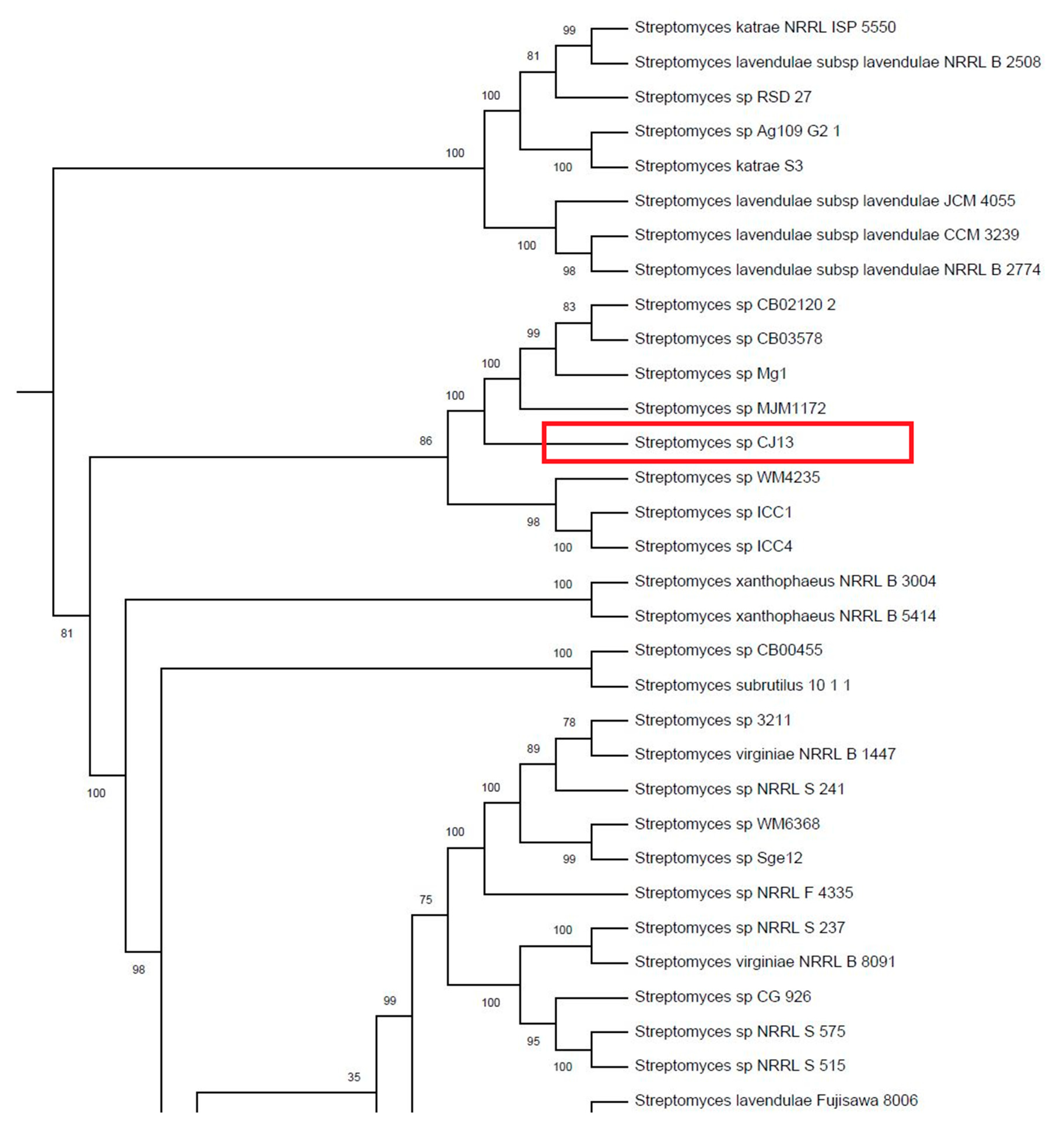
2.9. Phylogenetic Tree

Whole genome sequences (WGS) for all Streptomycetes were retrieved autonomously from GenBank using a WGET script. Genomes were annotated in Prokka [24] and the resulting translated CoDing Sequence (CDS) files (.faa files) were used to generate a multilocus, concatenated alignment of 400 proteins in PhyloPhlan [25]. Proteins used for the alignment were shown to be conserved among bacterial genera [25]. A maximum-likelihood phylogeny was reconstructed in FastTree [26] and rendered in iTOL [27]. The robustness of the phylogeny was assessed using 1000 bootstrap replications. Tree was outgroup-rooted along the lineage leading to Frankia.

2.10. In Silico Secondary Metabolite Analysis

Genome sequences were analysed using subsystem technology (RAST) version 4.0 (http://rast.nmpdr.org) and SEED [28]. Similarities to known antibiotic gene clusters and other secondary metabolites were predicted by antiSMASH [29,30].

2.11. Accession Numbers

The whole genome of Streptomyces sp. CJ13 was deposited at DDBJ/ENA/GenBank under the accession JAAZTW000000000. The version described in this paper is version JAAZTW010000000, BioProject PRJNA433829 BioSample SAMN14520815.

3. Results

3.1. Characterisation of Streptomyces sp. Isolate CJ13

A limestone grassland soil sample was examined from an area classified as calcareous grassland/acid flush mosaic. Soil pH was measured at an average of pH 7.3 (over four samples), which is a neutral to slightly alkaline soil. The Streptomyces sp. isolate CJ13 was selectively cultivated from the soil sample using base agar with a mineral rinse of the original soil sample. Although other Streptomyces were cultivated, colonies of Streptomyces sp. CJ13 had the greatest inhibitory activity in provisional tests. The sporing colonies of this Streptomyces were subcultured onto ISP-2 media (also with soil mineral rinse) and labelled as Streptomyces sp. isolate CJ13.
Morphologically, Streptomyces sp. CJ13 appeared as hard raised dark colonies with light purple or white spores on ISP-2 agar (Figure 1). In most cases, the colonies produced a diffusible brown pigment but in other variants this colour was weak or even absent. The original isolate produced two varieties of colonies depending on age and cultivation conditions characterised by either white or purple spores (Figure 1). Later whole genome sequencing confirmed the species belonged to the Streptomyces genus through a comparison with 930 other species of Streptomyces.
Microscopically, hyphae of Streptomyces sp. CJ13 appeared as thin strands with short branches occasionally bunched together into thicker fibers (Figure 2).
A list of these characteristics is available in Table 1.

3.2. Antibiotic Tests

3.2.1. Growth Inhibition by Streptomyces sp. CJ13

Streptomyces sp. CJ13 was cultivated on ISP2-agar. Agar cores were removed from these plates and tested against bacteria and fungi. Tests were performed a minimum of three times. In vitro tests show that Streptomyces sp. CJ13 inhibited the growth of S. aureus (MRSA), P. aeruginosa, B. subtilis, P. acnes and S. bombicola, but had no effect on the growth of E. coli (Figure 3). This antibiotic activity varied depending on the pigmentation of the Streptomyces. Highly pigmented variants of Streptomyces sp. CJ13 (d) showed more inhibitory activity against Gram negative bacteria such as Pseudomonas, as well as the yeast S. bombicola. Lighter coloured Streptomyces (l) had more effect on Gram positive organisms (Figure 3 and Table 2).

3.2.2. Stability of Inhibitory Factor from Streptomyces sp. CJ13

We also examined the effects of heat and storage on the stability of the inhibitory substances present in the growth media of Streptomyces sp. CJ13. Antimicrobial tests showed that the agar on which strains of Streptomyces sp. CJ13 were grown was still inhibitory even after 11 months of storage at 4–10 °C. However, this activity could be reduced by as much as 40% after 11 months storage. We were unable to measure any inhibitory activity in liquid cultures. The heat stability of the inhibitory component of Streptomyces sp. CJ13 present in agar that had been stored for 2 months between 4 and 10 °C was tested by heating agar cores to 100 °C in a heat block for five minutes with 300 µL sterile water (to improve heat transfer) in an Eppendorph tube, rapidly cooling on ice and transferring into a standard inhibition test. These results showed that inhibitory activity was still detectable even after exposure to these high temperatures (Table 3).

3.3. Whole Genome Sequencing of Streptomyces CJ13

The whole genome sequence of Streptomyces sp. CJ13 was characterised using rapid annotation subsystem technology (RAST), [28] (Figure 4, Table 4).

3.4. Phylogeny of Streptomyces sp. CJ13

Streptomyces sp. CJ13 sits within a well-supported clade closely related to Streptomyces sp. MJM1172 (Figure 5). The sister clade below Streptomyces sp. CJ13 is composed of Streptomyces sp. ICC1 and ICC4, isolated from a cave in British Columbia, Canada, a similar karst environment to Boho, West Fermanagh, where Streptomyces sp. CJ13 was isolated [31,32]. These isolates are related to the clade containing S. lavendulae subspecies lavendulae (Waksman et Curtis, 1916) [33], which match several phenotypic characteristics of our isolate.

3.5. Streptomyces sp. CJ13 Secondary Metabolite Prediction

In order to predict the potential synthesis of antibiotics by Streptomyces sp., CJ13, we analysed its whole genome sequence using antiSMASH (antibiotics and secondary metabolite analysis shell), which predicts the potential for synthesis of secondary metabolite clusters [29]. Some of the secondary metabolites that we identified are common to many species of Streptomyces, including: spore pigment, melanin, siderophores, alkylresorcinol (phenolic lipid, also contact allergen) and hopene (condenses membranes and decreases permeability) [34,35,36].
Most importantly antiSMASH predicted similarities from the whole genome sequence of Streptomyces sp. CJ13 to known antibiotic gene clusters, including salinichelins [37], antifungal factor [38], mediomycin A [39], weishanmycin [40], combamide (macrolactam) [41] and SAL-2242 [42] (Table 5).

3.6. In Silico Prediction of Streptomyces sp. CJ13 Resistance Genes

It is thought that antibiotic resistance (AMR) coevolved with antibiotic production in Streptomyces [43]. We therefore listed resistance clusters from Streptomyces sp. CJ13 (Table 6). We also noted similarities to metal resistance genes since these are also linked to antimicrobial resistance [44] (Table 6).

4. Discussion

There have been several attempts over the last few decades to replenish the antibiotic discovery pipeline. This has become more urgent given the rise in reports of antimicrobial resistance [45]. Rather than repeat older methods of antibiotic discovery, researchers have tried new innovations. One such strand is the examination of traditional medicines for antimicrobial producing bacteria or antimicrobials themselves. This follows from successful generation of lead compounds in the field of cancer [9].
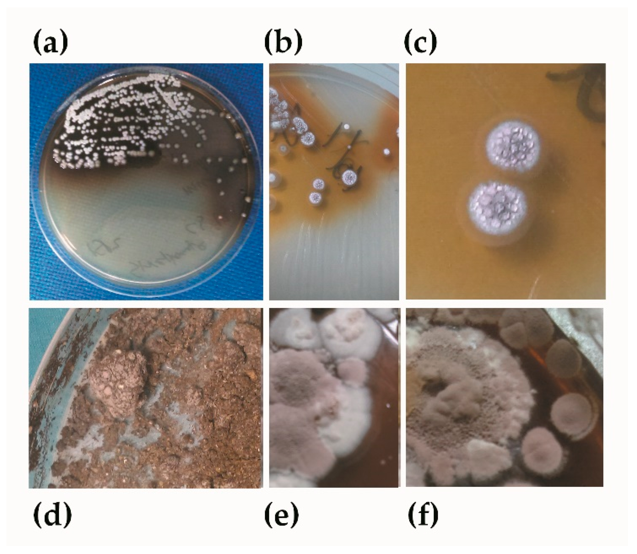
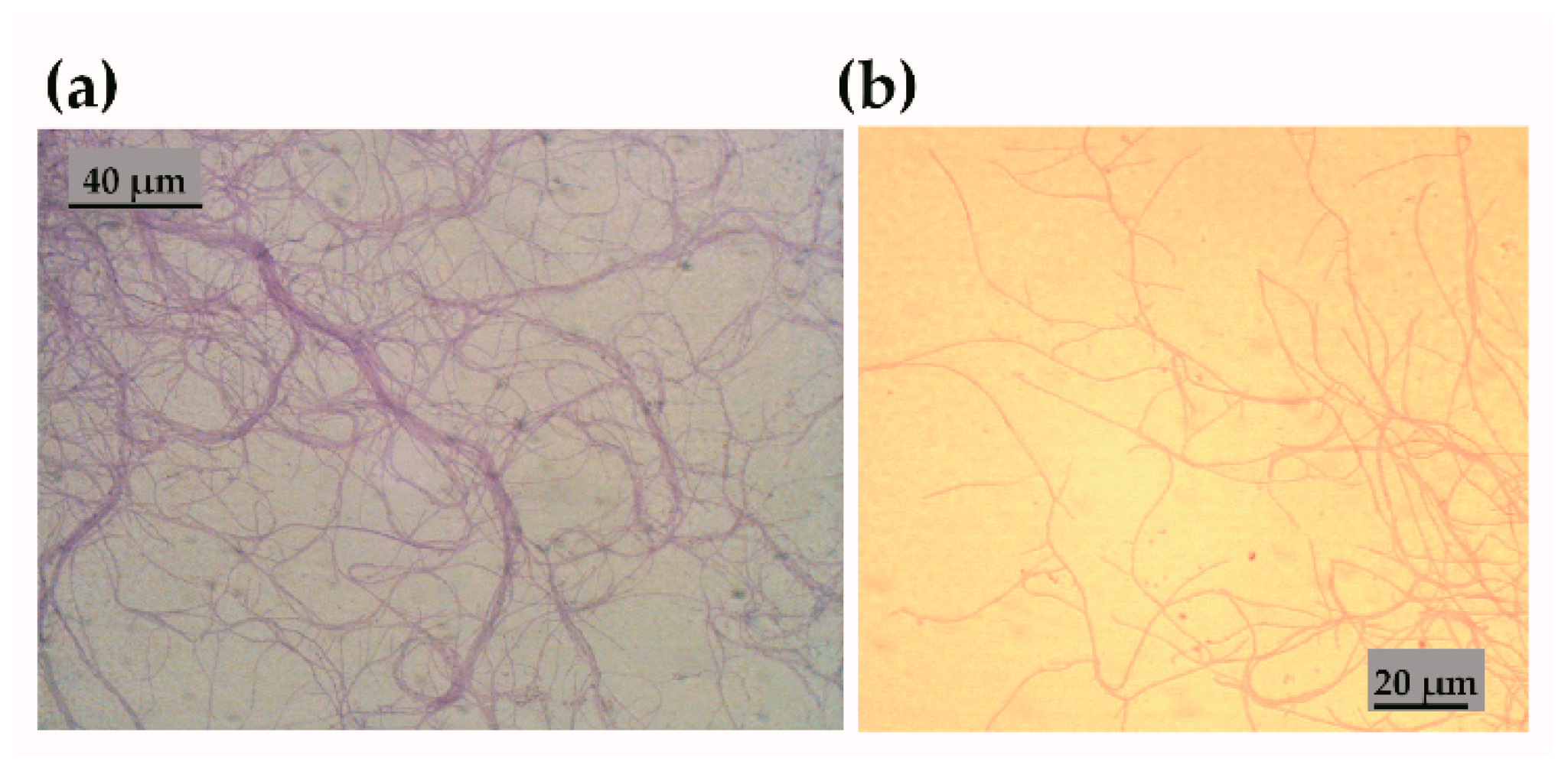
In this manuscript, we isolated a species of Streptomyces (Streptomyces sp. CJ13) from an alkaline grassland soil traditionally known for its healing properties. This had a broad range of inhibitory activity against multiresistant bacteria, anaerobes and fungi. There are many antibiotic producing species of Streptomyces associated with traditional medicines, both as endophytes and epiphytes [10,11]. Many of the discoveries during the golden age of antibiotics also came from soils. The difference between the older types of antimicrobial surveys in soils and those that have been recently isolated from traditional medicine is their specific soil character and location. For instance, Kisameet clay is based on a glacial clay deposit found in British Columbia that inhibits ESKAPE pathogens [13,14]. Similarly, traditional soil discoveries in Jordan focus on red soil that inhibits various pathogens [12]. Whilst our experiments were based on alkaline grassland soil, we did not test the whole soil for antimicrobial activity but rather tried to selectively isolate Streptomyces from the soil. Other studies have shown that karst regions, i.e. regions where there is dissolution of the rocks, especially limestone, support the growth of many Streptomyces [46,47,48].
By coincidence, some of the closest phylogenetic matches to Streptomyces sp. CJ13 were Streptomyces sp. ICC 1 and ICC4 originating from the Iron Curtain Cave in Chilliwack, British Columbia [32]. Both the West Fermanagh Scarplands and Iron Curtain Cave are regions of karst and share other botanical species including Spiranthes romanzoffiana (Irish lady’s-tresses) and Sisyrinchium bermudiana (blue-eyed grass). Additionally, both areas also have indigenous soil-based traditional medicines: in British Columbia from Kisameet Bay and in Fermanagh from the Boho area [13,18]. Streptomyces sp. CJ13 also shares phylogenetic similarities with the larger group of S. lavendulae. This group was originally described as Actinomyces lavendulae by Waksman and Henrici (1916), although relative distinctions between Actinomycetes had not been formalised at that time [33].
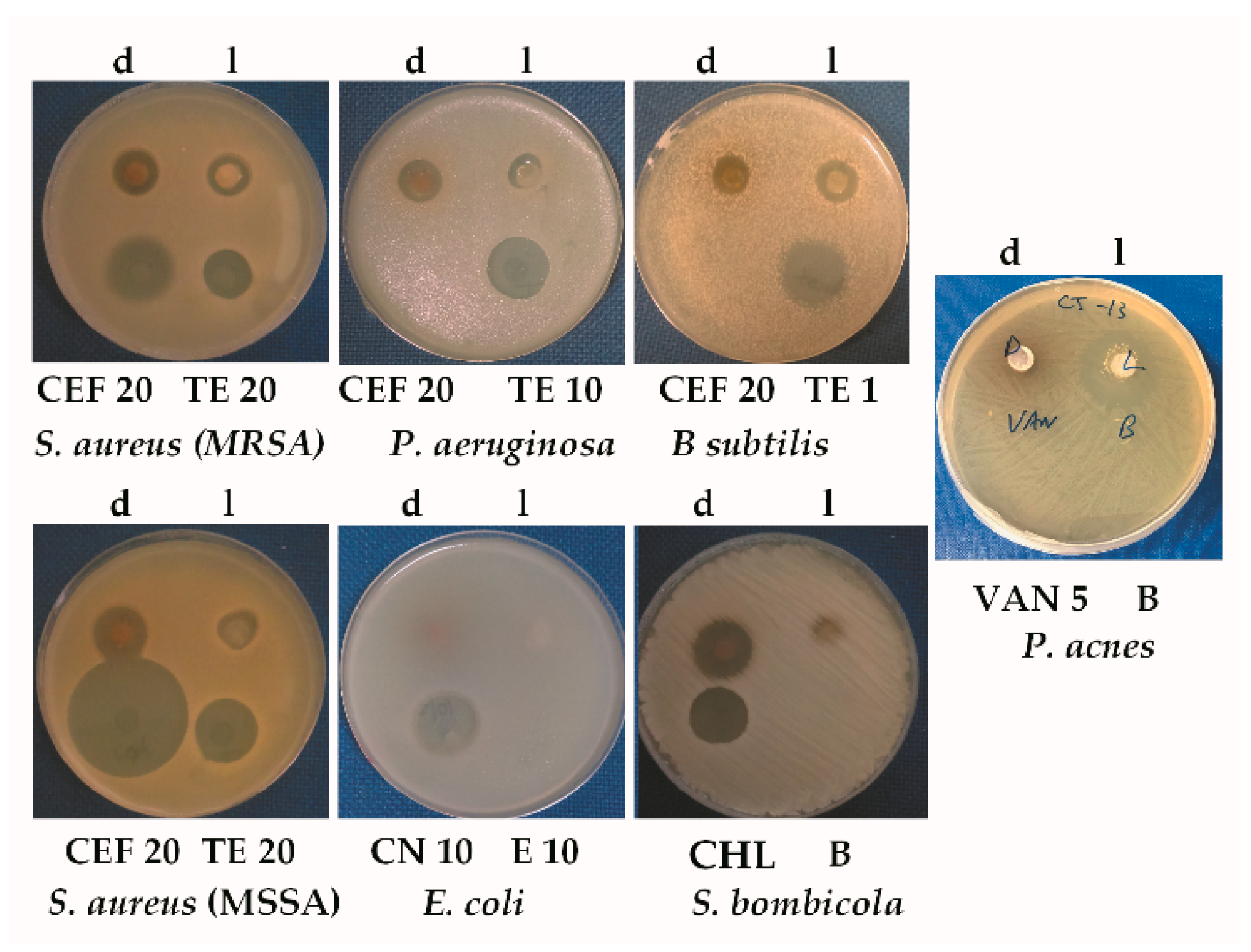
One of the world’s most pressing health issues is treating multidrug resistant Gram-negative bacteria. These can cause serious infections such as pneumoniae and urinary tract infections, which are often difficult to treat [49]. Fortunately, there are many more antibiotic discoveries for regulatory approval than in the previous decades, but still not enough to meet projected patient needs [2]. The inhibitory activity of Streptomyces sp. CJ13 covered a surprisingly broad array of organisms, including Gram-negative, multiresistant P. aeruginosa PA01.
The expression of antimicrobial activity varied between pigmentation types in our experiments. This phenomenon was also noted in the Iron Curtain Cave (ICC) strains from British Columbia [32]. To maintain a consistently high level of antibiotic activity, the ICC group grew their isolates on minimal media at low temperatures over an extended period of time [32]. This suggests that more antibiotics were induced by stressful growth conditions. Streptomyces are known to have silent antibiotic gene synthesis clusters stimulated during stress, microbial competition or interspecies communication [50,51,52,53]. In contrast to the ICC strains of British Columbia, Streptomyces sp. CJ13 grew quite rapidly (in a number of days) and also expressed antibiotic activity over that same time period. To maintain optimal antibiotic production, we followed a regime similar to the ICC group using very Spartan media. However, most of our antimicrobial experiments were derived from stocks of Streptomyces sp. CJ13, which had been frozen at −80 °C in glycerol.
The phylogenetically related group S. lavendulae produce many antimicrobial substances, including ileumycin, mitomycin C, eurymycin, glomecidin, saframycin, lavendamycin, angucycline, chlorocarcin, mimosamycin, streptothricin and streptomycin [33,54,55,56,57]. Additionally, secondary metabolite prediction software showed similarities to the antibiotic gene synthesis clusters for salinichelins, mediomycin A, weishanmycin, combamide (macrolactam), heat stable antifungal factor and SAL-2242. However, preliminary studies of antimicrobial Streptomyces sp. CJ13 extracts by mass spectrometry failed to find any identities with compounds in the Streptomyces or small molecule databases (results not shown). This raises the possibility that the inhibitory activity detected in Streptomyces sp. CJ13 could be from new antimicrobials. Further rounds of purification and analysis by mass spectrometry and NMR might reveal the identity of these compounds.

5. Conclusions

The discovery of new sources of antimicrobial compounds is still seen as a critical priority to contain the global spread of multidrug resistant diseases. We have isolated Streptomyces CJ13 from a soil associated with traditional Irish medicine that inhibits the growth of a broad range of organisms, including multiresistant Gram-negative bacteria. We are certain that this isolate represents a small fraction of the potential discoveries from this soil, which could serve as a new resource for future antibiotic discovery.

Supplementary Materials

The following are available online at https://0-www-mdpi-com.brum.beds.ac.uk/2076-3417/11/1/173/s1, Figure S1. Phylogenic comparison of Streptomyces sp. isolate CJ13 with 930 other Streptomycetes. Numbers at nodes are bootstrap values of 1000 pseudoreplicates—these are percentages.

Author Contributions

Conceptualization, G.A.Q. and I.M.B.; data curation, A.M.A., M.T.S. and P.D.F.; formal analysis, G.A.Q., A.M.A., N.K.A., D.C., S.A.A., M.T.S. and P.D.F.; funding acquisition, G.A.Q., N.K.A., H.A.B. and M.M.T.; investigation, G.A.Q., D.C., S.A.A., M.T.S., H.C.C. and M.M.T.; methodology, G.A.Q., A.M.A., H.C.C., P.D.F., H.A.B. and M.M.T.; resources, D.C. and I.M.B.; software, A.M.A., N.K.A. and M.T.S.; supervision, G.A.Q. and I.M.B.; validation, M.T.S., H.A.B. and M.M.T.; visualization, G.A.Q.; writing—original draft, G.A.Q., A.M.A., N.K.A., D.C., S.A.A., M.T.S., H.C.C., P.D.F., H.A.B. and I.M.B.; writing—review and editing, G.A.Q., A.M.A., N.K.A., D.C., S.A.A., M.T.S., H.C.C., P.D.F., H.A.B., M.M.T. and I.M.B. All authors have read and agreed to the published version of the manuscript.

Funding

This research received no external funding.

Institutional Review Board Statement

Not applicable.

Informed Consent Statement

Not applicable.

Data Availability Statement

All datasets generated or analysed for this study are included in the manuscript and the Supplementary materials. The data for the whole genome of Streptomyces sp. CJ13 is deposited at DDBJ/ENA/GenBank under the accession JAAZTW000000000. The version described in this paper is version JAAZTW010000000, BioProject PRJNA433829 BioSample SAMN14520815.

Acknowledgments

We are extremely grateful for the technical assistance of Daniel McShane from Ulster University and the provision of local knowledge and assistance from M. McGurn, B. Quinn and F. McHugh.

Conflicts of Interest

The authors declare no conflict of interest.

References

  1. Capozzi, C.; Maurici, M.; Pana, A. Antimicrobial resistance: It is a global crisis, “a slow tsunami”. Igiene e Sanita Pubblica 2019, 75, 429–450. [Google Scholar]
  2. Paulin, S.; Beyer, P. 2019 Antibacterial Agents in Clinical Development: An Analysis of the Antibacterial Clinical Development Pipeline; World Health Organization: Geneva, Switzerland, 2019. [Google Scholar]
  3. Liu, R.; Li, X.; Lam, K.S. Combinatorial chemistry in drug discovery. Curr. Opin. Chem. Biol. 2017, 38, 117–126. [Google Scholar] [CrossRef]
  4. Ribeiro da Cunha, B.; Fonseca, L.P.; Calado, C.R.C. Antibiotic Discovery: Where Have We Come from, Where Do We Go? Antibiotics 2019, 8, 45. [Google Scholar] [CrossRef] [PubMed] [Green Version]
  5. Wang, C.-H.; Hsieh, Y.-H.; Powers, Z.M.; Kao, C.-Y. Defeating Antibiotic-Resistant Bacteria: Exploring Alternative Therapies for a Post-Antibiotic Era. Int. J. Mol. Sci. 2020, 21, 1061. [Google Scholar] [CrossRef] [PubMed] [Green Version]
  6. Rateb, M.E.; Ebel, R.; Jaspars, M. Natural product diversity of actinobacteria in the Atacama Desert. Antonie Van Leeuwenhoek 2018, 111, 1467–1477. [Google Scholar] [CrossRef] [PubMed] [Green Version]
  7. Anand, U.; Jacobo-Herrera, N.; Altemimi, A.; Lakhssassi, N. A Comprehensive Review on Medicinal Plants as Antimicrobial Therapeutics: Potential Avenues of Biocompatible Drug Discovery. Metabolites 2019, 9, 258. [Google Scholar] [CrossRef] [Green Version]
  8. Dettweiler, M.; Lyles, J.T.; Nelson, K.; Dale, B.; Reddinger, R.M.; Zurawski, D.V.; Quave, C.L. American Civil War plant medicines inhibit growth, biofilm formation, and quorum sensing by multidrug-resistant bacteria. Sci. Rep. 2019, 9, 7692. [Google Scholar] [CrossRef] [Green Version]
  9. Cordell, G.A.; Farnsworth, N.R.; Beecher, C.W.W.; Doel Soejarto, D.; Kinghorn, A.D.; Pezzuto, J.M.; Wall, M.E.; Wani, M.C.; Cobb, R.R.; O’Neill, M.J.; et al. Novel strategies for the discovery of plant-derived anticancer agents. In Anticancer Drug Discovery and Development: Natural Products and New Molecular Models. Developments in Oncology; Valeriote, F.A., Baker, L.H., Eds.; Springer: Boston, MA, USA, 1994; ISBN 978-1-4613-6118-3. [Google Scholar]
  10. Liu, M.; Abdel-Mageed, W.M.; Ren, B.; He, W.; Huang, P.; Li, X.; Bolla, K.; Guo, H.; Chen, C.; Song, F.; et al. Endophytic Streptomyces sp. Y3111 from traditional Chinese medicine produced antitubercular pluramycins. Appl. Microbiol. Biotechnol. 2014, 98, 1077–1085. [Google Scholar] [CrossRef]
  11. Quinn, G.A.; Banat, A.M.; Abdelhameed, A.M.; Banat, I.M. Streptomyces from traditional medicine: Sources of new innovations in antibiotic discovery. J. Med. Microbiol. 2020, 69, 1040–1048. [Google Scholar] [CrossRef]
  12. Falkinham, J.O., 3rd; Wall, T.E.; Tanner, J.R.; Tawaha, K.; Alali, F.Q.; Li, C.; Oberlies, N.H. Proliferation of antibiotic-producing bacteria and concomitant antibiotic production as the basis for the antibiotic activity of Jordan’s red soils. Appl. Environ. Microbiol. 2009, 75, 2735–2741. [Google Scholar] [CrossRef] [Green Version]
  13. Behroozian, S.; Svensson, S.L.; Davies, J. Kisameet Clay Exhibits Potent Antibacterial Activity against the ESKAPE Pathogens. mBio 2016, 7, e01842-15. [Google Scholar] [CrossRef] [PubMed] [Green Version]
  14. Svensson, S.L.; Behroozian, S.; Xu, W.; Surette, M.G.; Li, L.; Davies, J. Kisameet Glacial Clay: An Unexpected Source of Bacterial Diversity. mBio 2017, 8, e00590-17. [Google Scholar] [CrossRef] [PubMed] [Green Version]
  15. Foley, R. Indigenous narratives of health: (re)placing folk-medicine within Irish health histories. J. Med. Humanit. 2015, 36, 5–18. [Google Scholar] [CrossRef] [PubMed] [Green Version]
  16. Shannon, F.; Sasse, A.; Sheridan, H.; Heinrich, M. Are identities oral? Understanding ethnobotanical knowledge after Irish independence (1937–1939). J. Ethnobiol. Ethnomed. 2017, 13, 65. [Google Scholar] [CrossRef] [Green Version]
  17. Koay, A.; Shannon, F.; Sasse, A.; Heinrich, M.; Sheridan, H. Exploring the Irish National Folklore Ethnography Database (Dúchas) for Open Data Research on Traditional Medicine Use in Post-Famine Ireland: An Early Example of Citizen Science. Front. Pharmacol. 2020, 11, 1670. [Google Scholar] [CrossRef]
  18. Terra, L.; Dyson, P.J.; Hitchings, M.D.; Thomas, L.; Abdelhameed, A.; Banat, I.M.; Gazze, S.A.; Vujaklija, D.; Facey, P.D.; Francis, L.W.; et al. A novel alkaliphilic Streptomyces inhibits ESKAPE pathogens. Front. Microbiol. 2018, 9, 2458. [Google Scholar] [CrossRef]
  19. McLean, E.O. Soil pH and line requirements. In Methods of Soil Analysis, 2nd ed.; Part 2. Chemical and Microbiological Properties; American Society of Agronomy and Soil Sciences Society of America: Madison, WI, USA, 1982. [Google Scholar]
  20. Lehrer, R.I.; Rosenman, M.; Harwig, S.S.; Jackson, R.; Eisenhauer, P. Ultrasensitive Assays for Endogenous Antimicrobial Polypeptides. J. Immunol. Methods 1991, 137, 167–173. [Google Scholar] [CrossRef]
  21. Nkanga, E.; Hagedorn, C. Detection of antibiotic-producing Streptomyces inhabiting forest soils. Antimicrob. Agents Chemother. 1978, 14, 51–59. [Google Scholar] [CrossRef] [Green Version]
  22. Bolger, A.M.; Lohse, M.; Usadel, B. Trimmomatic: A flexible trimmer for Illumina sequence data. Bioinforma. Oxf. Engl. 2014, 30, 2114–2120. [Google Scholar] [CrossRef] [Green Version]
  23. Bankevich, A.; Nurk, S.; Antipov, D.; Gurevich, A.A.; Dvorkin, M.; Kulikov, A.S.; Lesin, V.M.; Nikolenko, S.I.; Pham, S.; Prjibelski, A.D.; et al. SPAdes: A new genome assembly algorithm and its applications to single-cell sequencing. J. Comput. Biol. J. Comput. Mol. Cell Biol. 2012, 19, 455–477. [Google Scholar] [CrossRef] [Green Version]
  24. Seemann, T. Prokka: Rapid prokaryotic genome annotation. Bioinforma. Oxf. Engl. 2014, 30, 2068–2069. [Google Scholar] [CrossRef] [PubMed]
  25. Segata, N.; Börnigen, D.; Morgan, X.C.; Huttenhower, C. PhyloPhlAn is a new method for improved phylogenetic and taxonomic placement of microbes. Nat. Commun. 2013, 4, 2304. [Google Scholar] [CrossRef] [PubMed]
  26. Price, M.N.; Dehal, P.S.; Arkin, A.P. FastTree 2—Approximately maximum-likelihood trees for large alignments. PLoS ONE 2010, 5, e9490. [Google Scholar] [CrossRef] [PubMed]
  27. Letunic, I.; Bork, P. Interactive tree of life (iTOL) v3: An online tool for the display and annotation of phylogenetic and other trees. Nucleic Acids Res. 2016, 44, W242–W245. [Google Scholar] [CrossRef] [PubMed]
  28. Aziz, R.K.; Bartels, D.; Best, A.A.; DeJongh, M.; Disz, T.; Edwards, R.A.; Formsma, K.; Gerdes, S.; Glass, E.M.; Kubal, M.; et al. The RAST Server: Rapid Annotations Using Subsystems Technology. BMC Genom. 2008, 9, 75. [Google Scholar] [CrossRef] [PubMed] [Green Version]
  29. Medema, M.H.; Blin, K.; Cimermancic, P.; de Jager, V.; Zakrzewski, P.; Fischbach, M.A.; Weber, T.; Takano, E.; Breitling, R. antiSMASH: Rapid identification, annotation and analysis of secondary metabolite biosynthesis gene clusters in bacterial and fungal genome sequences. Nucleic Acids Res. 2011, 39, W339–W346. [Google Scholar] [CrossRef]
  30. Blin, K.; Shaw, S.; Steinke, K.; Villebro, R.; Ziemert, N.; Lee, S.Y.; Medema, M.H.; Weber, T. antiSMASH 5.0: Updates to the secondary metabolite genome mining pipeline. Nucleic Acids Res. 2019, 47, W81–W87. [Google Scholar] [CrossRef] [Green Version]
  31. Ghosh, S.; Paine, E.; Wall, R.; Kam, G.; Lauriente, T.; Sa-ngarmangkang, P.-C.; Horne, D.; Cheeptham, N. In Situ Cultured Bacterial Diversity from Iron Curtain Cave, Chilliwack, British Columbia, Canada. Diversity 2017, 9, 36. [Google Scholar] [CrossRef] [Green Version]
  32. Gosse, J.T.; Ghosh, S.; Sproule, A.; Overy, D.; Cheeptham, N.; Boddy, C.N. Whole Genome Sequencing and Metabolomic Study of Cave Streptomyces Isolates ICC1 and ICC4. Front. Microbiol. 2019, 10, 1020. [Google Scholar] [CrossRef]
  33. Waksman, S.A.; Harris, D.; Lechevalier, M. Studies on Streptomyces lavendulae. J. Bacteriol. 1951, 62, 149–161. [Google Scholar] [CrossRef] [Green Version]
  34. Nicault, M.; Tidjani, A.-R.; Gauthier, A.; Dumarcay, S.; Gelhaye, E.; Bontemps, C.; Leblond, P. Mining the Biosynthetic Potential for Specialized Metabolism of a Streptomyces Soil Community. Antibiotics 2020, 9, 271. [Google Scholar] [CrossRef] [PubMed]
  35. Santana-Molina, C.; Rivas-Marin, E.; Rojas, A.M.; Devos, D.P. Origin and Evolution of Polycyclic Triterpene Synthesis. Mol. Biol. Evol. 2020, 37, 1925–1941. [Google Scholar] [CrossRef] [PubMed] [Green Version]
  36. Terra, L.; Dyson, P.; Ratcliffe, N.; Castro, H.C.; Vicente, A.C.P. Biotechnological Potential of Streptomyces Siderophores as New Antibiotics. Curr. Med. Chem. 2020. [Google Scholar] [CrossRef] [PubMed]
  37. Letzel, A.-C.; Li, J.; Amos, G.C.A.; Millán-Aguiñaga, N.; Ginigini, J.; Abdelmohsen, U.R.; Gaudêncio, S.P.; Ziemert, N.; Moore, B.S.; Jensen, P.R. Genomic insights into specialized metabolism in the marine actinomycete Salinispora. Environ. Microbiol. 2017, 19, 3660–3673. [Google Scholar] [CrossRef] [PubMed]
  38. Yu, F.; Zaleta-Rivera, K.; Zhu, X.; Huffman, J.; Millet, J.C.; Harris, S.D.; Yuen, G.; Li, X.-C.; Du, L. Structure and biosynthesis of heat-stable antifungal factor (HSAF), a broad-spectrum antimycotic with a novel mode of action. Antimicrob. Agents Chemother. 2007, 51, 64–72. [Google Scholar] [CrossRef] [PubMed] [Green Version]
  39. Hong, H.; Samborskyy, M.; Usachova, K.; Schnatz, K.; Leadlay, P.F. Sulfation and amidinohydrolysis in the biosynthesis of giant linear polyenes. Beilstein J. Org. Chem. 2017, 13, 2408–2415. [Google Scholar] [CrossRef] [Green Version]
  40. Pan, G.; Xu, Z.; Guo, Z.; Ming Ma, H.; Yang, D.; Zhou, H.; Gansemans, Y.; Zhu, X.; Huang, Y.; Zhao, L.-X.; et al. Discovery of the leinamycin family of natural products by mining actinobacterial genomes. Proc. Natl. Acad. Sci. USA 2017, 114, E11131–E11140. [Google Scholar] [CrossRef] [Green Version]
  41. Cao, S.; Blodgett, J.A.V.; Clardy, J. Targeted Discovery of Polycyclic Tetramate Macrolactams from an Environmental Streptomyces Strain. Org. Lett. 2010, 12, 4652–4654. [Google Scholar] [CrossRef] [Green Version]
  42. Kersten, R.D.; Yang, Y.-L.; Xu, Y.; Cimermancic, P.; Nam, S.-J.; Fenical, W.; Fischbach, M.A.; Moore, B.S.; Dorrestein, P.C. A mass spectrometry-guided genome mining approach for natural product peptidogenomics. Nat. Chem. Biol. 2011, 7, 794–802. [Google Scholar] [CrossRef] [Green Version]
  43. Waglechner, N.; McArthur, A.G.; Wright, G.D. Phylogenetic reconciliation reveals the natural history of glycopeptide antibiotic biosynthesis and resistance. Nat. Microbiol. 2019, 4, 1862–1871. [Google Scholar] [CrossRef]
  44. Chen, J.; Li, J.; Zhang, H.; Shi, W.; Liu, Y. Bacterial Heavy-Metal and Antibiotic Resistance Genes in a Copper Tailing Dam Area in Northern China. Front. Microbiol. 2019, 10, 1916. [Google Scholar] [CrossRef] [PubMed] [Green Version]
  45. Hu, X.-Y.; Logue, M.; Robinson, N. Antimicrobial resistance is a global problem—A UK perspective. Eur. J. Integr. Med. 2020, 36, 101136. [Google Scholar] [CrossRef] [PubMed]
  46. Yucel, S.; Yamac, M. Selection of Streptomyces isolates from Turkish karstic caves against antibiotic resistant microorganisms. Pak. J. Pharm. Sci. 2010, 23, 1–6. [Google Scholar] [PubMed]
  47. Maciejewska, M.; Adam, D.; Martinet, L.; Naome, A.; Calusinska, M.; Delfosse, P.; Carnol, M.; Barton, H.A.; Hayette, M.-P.; Smargiasso, N.; et al. A Phenotypic and Genotypic Analysis of the Antimicrobial Potential of Cultivable Streptomyces Isolated from Cave Moonmilk Deposits. Front. Microbiol. 2016, 7, 1455. [Google Scholar] [CrossRef] [Green Version]
  48. Rangseekaew, P.; Pathom-aree, W. Cave Actinobacteria as Producers of Bioactive Metabolites. Front. Microbiol. 2019, 10, 387. [Google Scholar] [CrossRef] [Green Version]
  49. Butler, M.S.; Paterson, D.L. Antibiotics in the clinical pipeline in October 2019. J. Antibiot. 2020, 73, 329–364. [Google Scholar] [CrossRef]
  50. Chiang, Y.-M.; Chang, S.-L.; Oakley, B.R.; Wang, C.C. Recent advances in awakening silent biosynthetic gene clusters and linking orphan clusters to natural products in microorganisms. Omics 2011, 15, 137–143. [Google Scholar] [CrossRef] [Green Version]
  51. Reen, F.J.; Romano, S.; Dobson, A.D.W.; O’Gara, F. The Sound of Silence: Activating Silent Biosynthetic Gene Clusters in Marine Microorganisms. Mar. Drugs 2015, 13, 4754–4783. [Google Scholar] [CrossRef] [Green Version]
  52. Okada, B.K.; Seyedsayamdost, M.R. Antibiotic dialogues: Induction of silent biosynthetic gene clusters by exogenous small molecules. FEMS Microbiol. Rev. 2017, 41, 19–33. [Google Scholar] [CrossRef] [Green Version]
  53. Pishchany, G.; Mevers, E.; Ndousse-Fetter, S.; Horvath, D.J.; Paludo, C.R.; Silva-Junior, E.A.; Koren, S.; Skaar, E.P.; Clardy, J.; Kolter, R. Amycomicin is a potent and specific antibiotic discovered with a targeted interaction screen. Proc. Natl. Acad. Sci. USA 2018, 115, 10124. [Google Scholar] [CrossRef] [Green Version]
  54. Groupe, V.; Frankel, J.W.; Leche-Valier, M.P.; Waksman, S.A. Antiviral properties of ehrlichin, an antibiotic produced by Streptomyces lavendulae. J. Immunol. 1951, 67, 471–482. [Google Scholar] [PubMed]
  55. Arai, T.; Yazawa, K.; Mikami, Y. Isolation and characterization of satellite antibiotics, mimosamycin and chlorocarcins from Streptomyces lavendulae, streptothricin source. J. Antibiot. 1976, 29, 398–407. [Google Scholar] [CrossRef] [PubMed] [Green Version]
  56. Balitz, D.M.; Bush, J.A.; Bradner, W.T.; Doyle, T.W.; O’Herron, F.A.; Nettleton, D.E. Isolation of lavendamycin, a new antibiotic from Streptomyces lavendulae. J. Antibiot. 1982, 35, 259–265. [Google Scholar] [CrossRef] [PubMed] [Green Version]
  57. Matulova, M.; Feckova, L.; Novakova, R.; Mingyar, E.; Csolleiova, D.; Zduriencikova, M.; Sedlak, J.; Patoprsty, V.; Sasinkova, V.; Uhliarikova, I.; et al. A Structural Analysis of the Angucycline-Like Antibiotic Auricin from Streptomyces lavendulae Subsp. Lavendulae CCM 3239 Revealed Its High Similarity to Griseusins. Antibiotics 2019, 8, 102. [Google Scholar] [CrossRef] [PubMed] [Green Version]
Figure 1. Streptomyces sp. isolate CJ13 on (a) ISP-2 agar, (b,c) base agar + soil mineral rinse, (d) sterile soil from sample site and (e,f) older cultures with white or purple spores.
Applsci 11 00173 g001
Figure 2. Light microscopy of Streptomyces sp. isolate CJ13 (Gram stain) with long, twisted, branched hyphae. Scale bar (a) 40 µm, (b) 20 µm.
Applsci 11 00173 g002
Figure 3. Inhibitory effects of Streptomyces sp. isolate CJ13 dark (d) and light (l) variants on S. aureus (MRSA 43300), P. aeruginosa, B. subtilis, S. aureus (MSSA), E. coli, P. acne and S. bombicola. Control antibiotics (µg): cephalothin (CEP), teichoplanin (TE), gentimicin (CN), erythromycin (E), chlorhexidine (CHL 50 µg), vancomycin (VAN) and blank (B). Tests performed a minimum of three times.
Applsci 11 00173 g003
Figure 4. Subsystem analysis of Streptomyces sp. isolate CJ13 by RAST.
Applsci 11 00173 g004
Figure 5. Phylogenetic comparison of Streptomyces sp. isolate CJ13 with 930 other species of Streptomycetes. Numbers at nodes are bootstrap values of 1000 pseudoreplicates (percentages). Full phylogenetic comparison version available in Supplementary Materials Figures S1 and S2.
Applsci 11 00173 g005
Table 1. General features of Streptomyces sp. CJ13.
Classification
Domain:Bacteria
Phylum: Actinobacteria
Class:Actinobacteria
Order:Actinomycetales
Family:Streptomycetaceae
Genus:Streptomyces
Species: Streptomyces sp. isolate CJ_13
Gram stain:Positive
Motility:Negative
Pigmentation: Brown
Cell shape:Branched hyphae
Oxygen:Aerobic, anaerobic tolerant
Spores: Purple/white
pH in ISP2 broth6.85 to 7.85
Table 2. Zone of inhibition (ZOI) in mm produced by agar cores from Streptomyces sp. isolate CJ13 dark and light variants on S. aureus (MRSA 43300), P. aeruginosa, B. subtilis, S. aureus (MSSA), E. coli, P. acne and S. bombicola. Concentration of antibiotics in µg: cephalothin (CEP), teichoplanin (TE), gentimicin (CN), erythromycin (E), chlorhexidine (CHL 50 µg), vancomycin (VAN) and blank (B). Tests performed a minimum of three times.
AntimicrobialMicroorganisms
MSSAS. bombicolaB. subtilisE. coliP. aeruginosaMRSA
CJ13 Dark13181101314
CJ13 Light 126001013
CEF 2033-0-018
TE 2018----15
TE 10----18-
TE 1--24---
CN 10na--17--
E 10na--0--
CHL 50na-----
Table 3. Effect of temperature on antimicrobial activity of agar cores from Streptomyces sp. isolate CJ13. Agar from Streptomyces sp. CJ13 were incubated against cultures of S. aureus (MRSA 43300), P. aeruginosa (PA01) and S. bombicola. Zone of inhibition (ZOI) measured in mm. Antibiotic controls: cephalothin (CEP) 20 µg, teichoplanin (TE) 20 µg and chlorhexadine (CHL 50 µg). Tests performed a minimum of three times.
Scheme 20.ZOI at 20 °CZOI at 100 °CCEPCHLTE
MRSA16 918-17
P. aeruginosa129--16
S. bombicola22 16-18-
Table 4. Features of Streptomyces sp. CJ13 genome.
Attribute.Streptomyces sp. CJ13
Genome size9,230,829 bp
GC Content71.9%
N5022,457
L50114
Number of Contigs901
Number of Subsystems314
Total genes9260
Number of RNAs86
Table 5. Summary of antiSMASH (antibiotics and secondary metabolite analysis shell) 4.1.0 secondary metabolite prediction of Streptomyces sp. strain CJ13 (values above 40% gene similarity).
TypeAntibiotic Gene Cluster Similarity
NRPSSalinichelins 53%
NRPSAntifungal factor50%
T1PKSMediomycin A 46%
NRPSWeishanmycin 45%
T1PKS, NRPSCombamide (macrolactam) 44%
LanthipeptideSAL-224244%
Table 6. Antibiotic and metal resistance genes identified from WGS (whole genome sequences) of Streptomyces sp. CJ13 (predicted by Rapid Annotation using Subsystem Technology (RAST)).
Biological Role.Protein Name
Aminoglycoside adenylyltransferasesAminoglycoside N6’-acetyltransferase (EC 2.3.1.82)
Resistance to fluoroquinolonesDNA gyrase subunit B (EC 5.99.1.3)
Resistance to fluoroquinolonesDNA gyrase subunit A (EC 5.99.1.3)
Beta-lactamaseBeta-lactamase
Copper homeostasisMulticopper oxidase
Copper homeostasisCopper resistance protein
Copper homeostasisCopper-translocating P-type ATPase (EC 3.6.3.4)
Copper homeostasisCopper resistance protein
Copper homeostasisMultidrug resistance transporter,
Copper homeostasis: copper toleranceCytoplasmic copper homeostasis protein
Magnesium and cobalt toleranceMagnesium and cobalt efflux protein
Cobalt–zinc–cadmium resistanceCobalt–zinc–cadmium resistance protein
Cobalt–zinc–cadmium resistanceTranscriptional regulator
Mercuric reductaseFAD-dependent NAD(P)-disulphide oxidoreductase
Publisher’s Note: MDPI stays neutral with regard to jurisdictional claims in published maps and institutional affiliations.

