
Tilia sp. Seed Oil—Composition, Antioxidant Activity and Potential Use


Department of Pharmaceutical Biology, Faculty of Pharmacy, University of Ljubljana, Aškerčeva 7, 1000 Ljubljana, Slovenia
*
Author to whom correspondence should be addressed.
Appl. Sci. 2021, 11(11), 4932; https://0-doi-org.brum.beds.ac.uk/10.3390/app11114932
Submission received: 7 May 2021
/
Revised: 24 May 2021
/
Accepted: 25 May 2021
/
Published: 27 May 2021
(This article belongs to the Special Issue Plants: From Farm to Food and Biomedical Applications)
Abstract
:Research on new, untapped seed oil sources is receiving increased attention. In this study, 18 different seed samples of Tilia cordata and Tilia platyphyllos from various locations in Slovenia were collected and oil was extracted. The compositions of triglyceride fatty acids and unsaponifiable compounds were determined using GC-MS, while antioxidant activity was evaluated using the DPPH method. The oil content in the seeds varied significantly, from 9.1% to 21.7%. Linoleic acid (50–60%) was found to be the predominant fatty acid, followed by oleic acid (18–22%) and palmitic acid (8–9%). Characteristic cyclopropene fatty acids (sterculic, dihydrosterculic and malvalic acids) were present in the average range of 4–8.4%. Antioxidant activity ranged from 8.9% to 65.5%, and was higher, on average, for T. platyphyllos. Higher antioxidant activity was closely correlated with higher γ-tocopherol contents. Statistically significant correlations were confirmed between antioxidant activity and γ-tocopherol, between Δ-tocopherol and phytol, between stigmasterol and β-sitosterol and between squalene and malvalic acid. Tilia oil may be of great interest for cosmetic and dermal preparations. It is, however, not considered a good source of dietary fatty acids due to the undesired, significant content of omega-6 fatty acids.
1. Introduction
Tilia, commonly referred to as the linden or lime tree, is native to areas with a temperate climate in the Northern Hemisphere, where it is widespread and considered a tree of great economic and cultural importance. Besides its use in forestry, the timber trade and the paper industry, herbal substances from Tilia are also essential raw materials for the pharmaceutical and cosmetics industries.
Tilia flowers are approved as a category of traditional herbal medicinal products used for the relief of symptoms of the common cold and mental stress [1]. Tilia leaves have less significant therapeutic value [2], but have been well-researched in terms of environmental aspects [3,4,5,6,7]. Finally, Tilia fruits or seeds are a source of vegetable oil with a specific triglyceride composition characterised by the predominant linoleic acid (content of approximately 50%) and cyclopropene fatty acids (content of approximately 10%) [8,9]. However, limited data are available in terms of Tilia seed oil yield, composition and variability, and potential biological properties.
Detailed insight into Tilia seed oil chemistry is a prerequisite for the evaluation of its economic potential in the pharmaceutical and cosmetics industries where vegetable oils are widely used, for example, as active pharmaceutical ingredients, cosmetically active ingredients, ingredients of the lipid phase or other excipients, and extraction solvents. Chemically, vegetable oils are a mixture of esters composed of glycerol and fatty acids (i.e., triglycerides), and unsaponifiable compounds. Increased interest has been noted recently in the research of their dermal effects, which include hydrating, emollient, antimicrobial, antioxidative and anti-inflammatory activities [10,11].
Based on a lack of information, the aim of our study was to determine the compositions of fatty acid in triglycerides and of unsaponifiable matter, and the antioxidant activity of Tilia cordata and Tilia platyphyllos seed oils.
2. Materials and Methods
2.1. Reagents and Materials
The following reagents were used: hexane (Panreac), isopropanol (Carlo Erba Reagents, Chaussée du Vexin, France), dichloromethane (Panreac), methanol (Gram Mol, Zagreb, Croatia), sodium hydroxide (Riedel-de Haën, Honeywell, Charlotte, NC, USA), 14% boron trifluoride (BF3) in methanol (Sigma-Aldrich, Merck, Darmstadt, Germany), purified water (Faculty of Pharmacy, University of Ljubljana, Ljubljana, Slovenia), mixture of methyl esters of fatty acids F.A.M.E. Mix C4-C24 (Supelco, Merck, Darmstadt, Germany), mixture of n-alkanes C7–C33 (Restec, Bellefonte, PA, USA), potassium hydroxide (Sigma-Aldrich, Merck, Darmstadt, Germany), cholesterol (Sigma-Aldrich, Merck, Darmstadt, Germany), sodium chloride (Carlo Erba Reagents, Chaussée du Vexin, France), ethanol 96% (Carlo Erba Reagents, Chaussée du Vexin, France), phenolphthalein (Riedel-de Haën, Honeywell, Charlotte, NC, USA), diethyl ether (Sigma-Aldrich, Merck, Darmstadt, Germany), n-heptane (Carlo Erba Reagents, Chaussée du Vexin, France), HMDS+TMCS+pyridine (3:1:9) (Sylon HTP, Supelco, Merck, Darmstadt, Germany), diphenylpicrylhydrazyl radical (Fluka, Honeywell, Charlotte, NC, USA), gallic acid (Fluka, Honeywell, Charlotte, NC, USA) and 2-butanone (Merck, Darmstadt, Germany).
2.2. Plant Materials
Samples of the fruits of T. cordata and T. platyphyllos were collected at various locations throughout Slovenia (Figure 1). All samples were collected in autumn (October), in the phase of full ripeness, air-dried and stored in a dry, dark, cool place until the oil was extracted. All analyses were performed within four months of collection.
2.3. Oil Extraction
Fruit husks were removed by hand, and the seeds were separated and ground into powder. Hexane (herbal drug to solvent ratio = 1:3) was added to the seed powder in a glass conical flask, and ultrasound-assisted extraction was performed for 10 min. The extraction mixture was then poured into plastic tubes and centrifuged at 1500 rpm for 3 min. The supernatant was filtered and hexane was evaporated in a rotary evaporator. The whole extraction process was repeated three more times. The oils obtained from all four extractions were combined and refrigerated in an inert atmosphere to prevent oxidative degradation until the analyses were performed.
2.4. In Situ Derivatisation of Fatty Acids and Determination of the Fatty Acid Composition
Prior to analysis, fatty acids were converted to methyl esters, i.e., derivatives with a lower boiling point. Derivatisation also increases resolution and reduces tailing [12]. A total of 10 mg of an oil sample was weighed into a microcentrifuge tube, and 10 μL of dichloromethane and 200 μL of 0.5 M NaOH in MeOH were added. The reaction mixture was heated in a water bath at 90 °C for 10 min and cooled. A total of 200 μL of 14% BF3 in methanol was then added. The microcentrifuge tube was again placed in a water bath for 10 min. After in-situ derivatisation, fatty acid esters were extracted. A total of 200 μL of distilled water and 1 mL of hexane were added, and the mixture was shaken vigorously by hand for 1 min, and then left until the aqueous and organic phases separated. The upper, organic phase was transferred to a vial for GC-MS analysis.
2.5. Extraction, Derivatisation and Identification of Unsaponifiable Matter
Extraction was carried out according to the procedure of the European Pharmacopoeia 8.0 [13]. Cholesterol was used as an internal standard. Prior to GC-MS analysis, the unsaponifiable compounds were converted into silyl derivatives that are less polar, more volatile and more temperature-resistant, resulting in more symmetrical and flatter peaks. In addition, silyl reagents are compatible with most detection systems [14]. A total of 5 mg of each sample was placed in a 1.5 mL microcentrifuge tube. A total of 50 μL of the Sylon reagent (a mixture of HMDS, TMCS and pyridine) was added. The reaction mixture was stirred well and then centrifuged at 13,000 rpm for 5 min. The mixture was then heated in a water bath at 60 °C for 30 min and left to cool. A total of 950 μL of heptane was added, mixed again and centrifuged as before. Supernatant was transferred to a vial for GC-MS analyses.
2.6. Gas Chromatography Coupled with Mass Spectrometry
GC-MS analyses were performed using a GCMS-QP2010 Ultra system (Shimadzu Corporation, Kyoto, Japan), and NIST11 and FFNSC2 data libraries (Shimadzu Corporation, Kyoto, Japan). The GC system was equipped with a capillary column Rtx-1 F&F 100% dimethylpolysiloxane (length of 30 m, internal diameter of 0.25 mm and film thickness of 0.25 µm; Restek, Bellefonte, PA, USA). The carrier gas was helium at a constant linear velocity of 1 mL/min. The mass spectrometer ionisation energy was 70 eV, ion source temperature 200 °C and detector voltage 1 kV. The injection volume of samples was 1 μL and the split ratio was 1:100.
In the analysis of fatty acid composition, the injection port was set to 250 °C and the interface temperature was 280 °C. The filament was turned on at 2 min and a 2.5-min solvent delay was programmed when the samples were injected. A full scan was recorded in the mass range of 35–500 m/z with a scanning frequency of 5 Hz. The temperature programme began at 160 °C and increased to 250 °C at 3 °C/min (total analysis time was 30 min).
In the analysis of unsaponifiable matter, the injection port was set to 300 °C and the interface temperature was 300 °C. The filament was turned on at 2.5 min and a 3 min solvent delay was programmed when the samples were injected. A full scan was recorded in the mass range of 50–600 m/z with a scanning frequency of 5 Hz. The temperature programme began at 200 °C, increased to 270 °C at 5 °C/min and continued at 270 °C for 56 min (total analysis time was 70 min).
2.7. Antioxidant Activity
Antioxidant activity was evaluated using a DPPH (2,2-diphenyl-1-picrylhydrazyl) assay. DPPH was dissolved in 2-butanone:methanol (1:1, v/v) at a concentration of 0.04 mg of DPPH/mL. A solution of 10 mg of gallic acid in 10 mL of solvent was used as a positive control. Solvent was used as a blank control. Oil samples were prepared by dissolving 5 mg of oil in 1 mL of solvent. A total of 40 µL of each oil sample and control was mixed with 1 mL of the DPPH solution. After 30 min of incubation in the dark, radical scavenging activity was determined spectrophotometrically at 517 nm. Three measurements were made for each sample and the average absorbance was calculated. Antioxidant activity was calculated from the average absorbances as follows:
DPPH radical scavenging assay (%) = (Ablank − Atest)/(A blank − Amcontrol) × 100
2.8. Statistical Analysis
A Microsoft Excel 2013 (Microsoft Corporation, Washington, DC, USA) t-test was used to determine statistically significant differences between T. cordata and T. platyphyllos samples. Pearson correlation (Microsoft Excel 2013 (Microsoft Corporation, Washington, DC, USA)) was used to determine statistically significant correlations between parameters. Values with p ≤ 0.05 were considered significant.
3. Results and Discussion
3.1. Oil Content
Optimisation of extraction was performed using different solvents (hexane and hexane:isopropanol = 3:2), time of extraction and consecutive extractions (data not shown). Most of the lipids were extracted using the final method described in Section 2.2.
The content of oil obtained from our samples varies between 9.1% and 21.7% (Table 1). Differences are likely to be random or dependent on environmental factors (e.g., geographical location, soil and weather conditions). The results are comparable with those found in the study by Afandiyeva et al. [9], where a yield of 12.9% was obtained for T. caucasica. In contrast, the content for Tilia cordata referenced in [15] was higher (27.9%), while Kusmenoglu et al. reported a lower content of 5.6%, 8.12% and 6.16% in T. argentea, T. platyphyllos and T. rubra, respectively, growing in Turkey [16]. In our study, the average content in T. platyphyllos seeds was found to be higher (19.2%) than in T. cordata seeds (14.1%).
In terms of the oil content in relation to traditional, commercially important seeds used for the industrial production of linoleic acid-based vegetable oils, Tilia seeds are not a very rich source. For example, sunflower seeds contain 50% oil and pumpkin seeds contain between 40% and 50% oil. Tilia seeds are more similar to soy (20%) and corn seeds (15%) [17,18].
3.2. Fatty Acid Composition
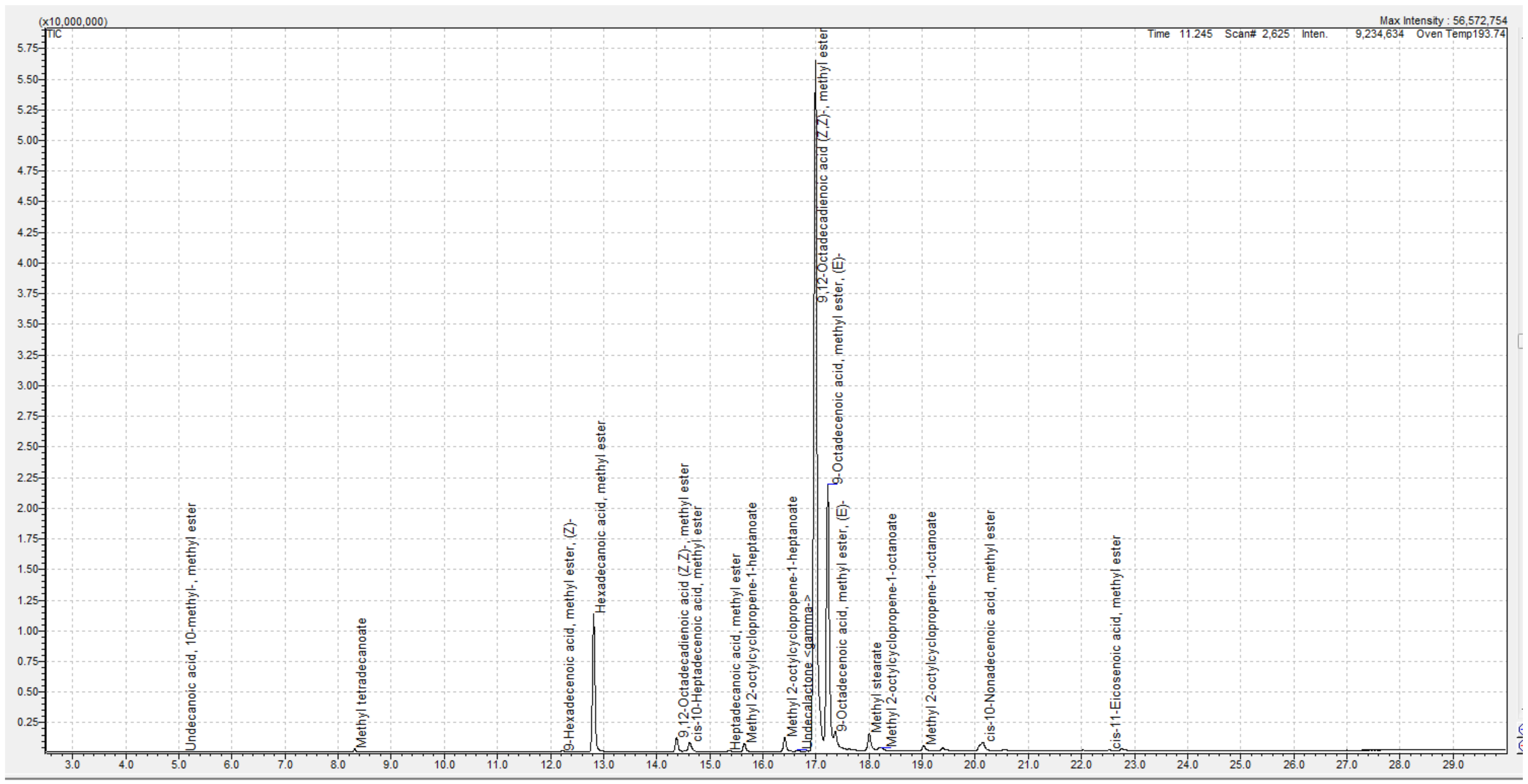
The fatty acids detected in our samples are shown in Table 2, while a typical GC-MS chromatogram of a Tilia oil sample is presented in Figure 2. The predominant fatty acid was linoleic acid with an average content of 53.3% in T. cordata and 59.6% in T. platyphyllos, followed by oleic acid (18.2% and 22.2%, respectively) and palmitic acid (8.6% for both species). The results are in line with those of Dowd et al. [8], which are presented in Table 2 as T. cordata or T. platyphyllos samples from the USA. However, Kusmenoglu et al. identified palmitic acid as the predominant fatty acid in T. platyphyllos triglycerides [16].
Characteristic of Tilia seed oil is the presence of cyclopropene fatty acids, e.g., sterculic, dihydrosterculic and malvalic acids (Figure 3), which were first identified in T. platyphyllos seed oil in 1966 [19]. Most abundant in our samples were malvalic acid (3.1% in T. cordata and 1.7% in T. platyphyllos) and sterculic acid (4.7% in Tilia cordata and 0.9% in T. platyphyllos) (Table 2). The difference in malvalic acid content was statistically significantly higher in T. cordata oil. The average total content of cyclopropene fatty acids was 8.4% in T. cordata and significantly lower (4.0%) in T. platyphyllos seed oil (Table 2), which is in line with the results of the study conducted by Dowd et al. [8].
Tilia seed oil was found to contain an average of 82.9% (T. cordata) and 87.3% (T. platyphyllos) unsaturated fatty acids, and 10.7% and 11.7% saturated fatty acids, respectively (Table 2). Short chain fatty acids were not found in Tilia seed oil [8,9], which corresponds to our data. According to Dowd et al. [8], the calculated fatty acid ratios of omega-6 to omega-3 in T. platyphyllos and T. cordata oils are 72 and 58, respectively, which are much lower than the ratio in sunflower (131) and pumpkin (257) oils [17]. We were not able to calculate this ratio, as no omega-3 fatty acid was detected in our samples.
The biological effects of the predominant linoleic and oleic acids have been researched extensively in fields such as cosmetology and dermatology [10,20], while scarce data are available about cyclopropene fatty acids. New research has opened important areas of potential medicinal use. In a recent study [21], sterculic acid was reviewed for therapeutic activities in pathologies such as metabolic syndrome, parasitic diseases and age-related macular degeneration, expressed through the anti-inflammatory activity and inhibition of stearoyl-CoA desaturase 1. The protective effect against metabolic syndrome on a fructose-induced rat model was confirmed using sterculic acid-dominant (64.2% fatty acids) seed oil extracted from Sterculia apetala [22].
3.3. Unsaponifiable Matter
A typical GC-MS chromatogram showing the composition of unsaponifiable matter from Tilia seed oil is presented in Figure 4. In our study, phytol, squalene, tocopherol, stigmasterol and β-sitosterol were detected. Concentrations of individual unsaponifiable substances vary significantly between the samples (Table 2). Generally, the most abundant are phytosterols and phytol. Squalene content was significantly higher in T. cordata unsaponifiable matter.
3.4. Antioxidant Activity
Vegetable oils are known for their antioxidant activity, which derives from unsaturated fatty acids and, particularly, from the compounds of the unsaponifiable matter [23,24]. In our study, a DPPH method was used for the determination of antioxidant activity, as it is one of the most widely used methods due to its rapid, simple and cost-effective implementation [25,26]. The antioxidant activity for individual Tilia seed oil samples was found to be highly variable, in the range of 8.9% to 65.5% (Table 2). The average antioxidant activity of T. platyphyllos and T. cordata oils were 47.3% and 38.3%, respectively.
A statistically significant correlation was confirmed between antioxidant activity and γ-tocopherol content, i.e., a higher antioxidant activity was found in the case of a higher content of γ-tocopherol. The significance of differences was not confirmed for antioxidant activity and higher contents of phytosterols, phytol and squalene, which are generally known as molecules that protect fatty acids from oxidative degradation. Statistically significant correlations were confirmed between antioxidant activity and γ-tocopherol, between Δ-tocopherol and phytol, between stigmasterol and β-sitosterol, and between squalene and malvalic acid.
3.5. Potentials for Tilia Seed oil Use
In terms of nutritional value, Tilia seed oil may not be considered a beneficial dietary source of fatty acids, as the oil predominantly contains an omega-6 linoleic acid and no omega-3 fatty acids. In addition, Tilia seed oil is not rich in phytosterols when compared, for example, to corn oil containing 809–1557 mg phytosterols per 100 g or sunflower oil containing 374–725 mg phytosterols per 100 g. Corn oil and original, low-oleic sunflower oil are linoleic acid-based oils. In terms of the phytosterol content, palm oil with 71–117 mg phytosterols per 100 g is similar to Tilia seed oil [27].
However, the high content of linoleic acid and the presence of phytosterols and squalene as the main compounds of unsaponifiable matter make the potential of Tilia seed oil of great importance in cosmetics. Linoleic acid was shown to activate a specific receptor, i.e., the peroxisome proliferator-activated receptor alpha (PPAR-α), in keratinocytes, which is involved in the regulation of keratinocyte proliferation, inflammation and the maintenance of skin homeostasis. Linoleic acid contributes to reduced transepidermal water loss and thus improves the skin’s hydration [10,28]. Squalene and tocopherols have antioxidant properties and protect the skin against lipid peroxidation [17]. Research shows that phytosterols protect the skin from aging. In vitro human keratinocyte assays have demonstrated that phytosterols inhibit the UV-induced expression of the matrix metalloproteinase-1 (MMP-1) enzyme that is responsible for the proteolysis of collagen degradation and the reduction of COL1A1 and COL1A2 genes responsible for collagen synthesis. Reduced phytosterols in the skin may therefore lead to increased susceptibility of the skin to UV damage. In addition, phytosterols act as emollients [17].
4. Conclusions
Following trends of sustainability, widely-present and industrially important plants, including the Tilia tree, represent an interesting area for research into their untapped commercial values. Taking this into account, plant materials that contain vegetable oils show great potential, as the oils have been increasingly studied in recent years due to their beneficial dermal, cosmetic and/or dietary effects. Tilia seed oil may be of particular interest to the fields of cosmetology and dermatology, as it is rich in linoleic acid and is a unique source of rare cyclopropene fatty acids. In addition, based on results presented in this article, it is reasonable to expect beneficial skin effects also deriving from the antioxidant activity of the oil.
Author Contributions
Conceptualization, N.K.G.; methodology, N.K.G.; validation, N.K.G. and N.P.; formal analysis, N.K.G. and N.P.; investigation, N.K.G. and N.P.; resources, N.K.G. and N.P.; data curation, N.K.G. and N.P.; writing—original draft preparation, N.P.; writing—review and editing, N.K.G.; visualization, N.P.; supervision, N.K.G.; project administration, N.K.G.; funding acquisition, N.K.G. All authors have read and agreed to the published version of the manuscript.
Funding
This research was funded by the Slovenian Research Agency, grant number P1-0208.
Informed Consent Statement
Not applicable.
Acknowledgments
The skillful laboratory assistance of Andreja Levpušček is gratefully acknowledged.
Conflicts of Interest
Both authors declare no conflict of interest.
References
- Tiliae Flos|European Medicines Agency. Available online: https://www.ema.europa.eu/en/medicines/herbal/tiliae-flos (accessed on 11 November 2020).
- Toker, G.; Küpeli, E.; Memisoǧlu, M.; Yesilada, E. Flavonoids with antinociceptive and anti-inflammatory activities from the leaves of Tilia argentea (silver linden). J. Ethnopharmacol. 2004. [Google Scholar] [CrossRef] [PubMed]
- Moser, A.; Rötzer, T.; Pauleit, S.; Pretzsch, H. Structure and ecosystem services of small-leaved lime (Tilia cordata Mill.) and black locust (Robinia pseudoacacia L.) in urban environments. Urban For. Urban Green. 2015, 14, 1110–1121. [Google Scholar] [CrossRef]
- Day, S.D.; Amateis, R.L. Predicting canopy and trunk cross-sectional area of silver linden (Tilia tomentosa) in confined planting cutouts. Urban For. Urban Green. 2011, 10, 317–322. [Google Scholar] [CrossRef]
- Aničić, M.; Spasić, T.; Tomašević, M.; Rajšić, S.; Tasić, M. Trace elements accumulation and temporal trends in leaves of urban deciduous trees (Aesculus hippocastanum and Tilia spp.). Ecol. Indic. 2011, 11, 824–830. [Google Scholar] [CrossRef]
- Serbula, S.M.; Kalinovic, T.S.; Ilic, A.A.; Kalinovic, J.V.; Steharnik, M.M. Assessment of Airborne Heavy Metal Pollution Using Pinus spp. and Tilia spp. Aerosol Air Qual. Res. 2013, 13, 563–573. [Google Scholar] [CrossRef] [Green Version]
- Kunneman, B.P.; Albers, M.R. Linden Trees (Tilia spp.). InTrees III; Springer: Berlin/Heidelberg, Germany, 1991; pp. 152–163. [Google Scholar]
- Dowd, M.K.; Farve, M.C. Fatty acid composition of Tilia spp. seed oils. Grasas y Aceites 2013, 64, 243–249. [Google Scholar] [CrossRef]
- Afandiyeva, S.E.; Novruzov, E.N. Fatty acid composition and organoleptic characteristics of Tilia caucasica Rupr. Fruit Oil. 2020, 3, 31–37. [Google Scholar]
- Poljšak, N.; Kreft, S.; Kočevar Glavač, N. Vegetable butters and oils in skin wound healing: Scientific evidence for new opportunities in dermatology. Phyther. Res. 2019, 34. [Google Scholar] [CrossRef] [PubMed]
- Poljšak, N.; Kočevar Glavač, N. Dermal Effects of Unsaponifiable Compounds: The Overlooked Perspective of Vegetable Butters and Oils. J. Cosmet. Sci. 2021, 72, 215–228. [Google Scholar]
- Eder, K. Gas chromatographic analysis of fatty acid methyl esters. J. Chromatogr. B Biomed. Sci. Appl. 1995, 671, 113–131. [Google Scholar] [CrossRef]
- Council of Europe. The European Pharmacopoeia 8th (Ph.Eur.), 8th ed.; Council of Europe: Strasbourg, France, 2014. [Google Scholar]
- Orata, F. Derivatization Reactions and Reagents for Gas Chromatography Analysis. In Advanced Gas Chromatography-Progress in Agricultural, Biomedical and Industrial Applications; IntechOpen: London, UK, 2012. [Google Scholar]
- Fontanel, D. Unsaponifiable Matter in Plant Seed Oils; Springer Berlin Heidelberg: Berlin/Heidelberg, Germany, 2013. [Google Scholar]
- Kusmenoglu, S.; Toker, G. Fatty acid composition of Tilia fruit oils. Acta Pharm. Turc. 1998, 40, 121–123. [Google Scholar]
- Modern Cosmetics, Ingredients of Natural Origin, A Scientific View, Volume 1; Janeš, D.; Kočevar Glavač, N. (Eds.) Širimo dobro besedo d.o.o.: Velenje, Slovenia, 2018. [Google Scholar]
- De Andrade, E.T.; Pinto, L.; da Silva, I.M.; Guimaraes, R.; Piamba Tulcan, O.E.; de Andrade, D.O. Facilities for Obtaining Soybean Oil in Small Plants. In Soybean-Bio-Active Compounds; IntechOpen: London, UK, 2013. [Google Scholar]
- Raju, P.K.; Reiser, R. Gas-liquid chromatographic analysis of cyclopropene fatty acids. Lipids 1966, 1, 10–15. [Google Scholar] [CrossRef] [PubMed]
- Vaughn, A.R.; Clark, A.K.; Sivamani, R.K.; Shi, V.Y. Natural Oils for Skin-Barrier Repair: Ancient Compounds Now Backed by Modern Science. Am. J. Clin. Dermatol. 2018, 19, 103–117. [Google Scholar] [CrossRef] [PubMed]
- Peláez, R.; Pariente, A.; Pérez-Sala, Á.; Larráyoz, I.M. Sterculic Acid: The Mechanisms of Action beyond Stearoyl-CoA Desaturase Inhibition and Therapeutic Opportunities in Human Diseases. Cells 2020, 9, 140. [Google Scholar] [CrossRef] [PubMed] [Green Version]
- Ramírez-Higuera, A.; Peña-Montes, C.; Herrera-Meza, S.; Mendoza-López, R.; Valerio-Alfaro, G.; Oliart-Ros, R.M. Preventive Action of Sterculic Oil on Metabolic Syndrome Development on a Fructose-Induced Rat Model. J. Med. Food 2020, 23, 305–311. [Google Scholar] [CrossRef] [PubMed] [Green Version]
- Liu, R.; Lu, M.; Zhang, T.; Zhang, Z.; Jin, Q.; Chang, M.; Wang, X. Evaluation of the Antioxidant Properties of Micronutrients in Different Vegetable Oils. Eur. J. Lipid Sci. Technol. 2020, 122, 1900079. [Google Scholar] [CrossRef]
- Dhavamani, S.; Poorna Chandra Rao, Y.; Lokesh, B.R. Total antioxidant activity of selected vegetable oils and their influence on total antioxidant values in vivo: A photochemiluminescence based analysis. Food Chem. 2014, 164, 551–555. [Google Scholar] [CrossRef]
- Pyrzynska, K.; Pȩkal, A. Application of free radical diphenylpicrylhydrazyl (DPPH) to estimate the antioxidant capacity of food samples. Anal. Methods 2013, 5, 4288–4295. [Google Scholar] [CrossRef]
- Kedare, S.B.; Singh, R.P. Genesis and development of DPPH method of antioxidant assay. J. Food Sci. Technol. 2011, 48, 412–422. [Google Scholar] [CrossRef] [Green Version]
- Brufau, G.; Canela, M.A.; Rafecas, M. Phytosterols: Physiologic and metabolic aspects related to cholesterol-lowering properties. Nutr. Res. 2008, 217, 217–225. [Google Scholar] [CrossRef]
- Danby, S.G.; AlEnezi, T.; Sultan, A.; Lavender, T.; Chittock, J.; Brown, K.; Cork, M.J. Effect of olive and sunflower seed oil on the adult skin barrier: Implications for neonatal skin care. Pediatr. Dermatol. 2013, 30, 42–50. [Google Scholar] [CrossRef] [PubMed]
Figure 1.
Map of locations where Tilia seed samples were collected. Tilia cordata samples are marked with circles and Tilia platyphyllos samples are marked with triangles.
Figure 1.
Map of locations where Tilia seed samples were collected. Tilia cordata samples are marked with circles and Tilia platyphyllos samples are marked with triangles.

Figure 2.
GC-MS chromatogram of a Tilia oil sample after derivatisation, showing the fatty acid composition.
Figure 2.
GC-MS chromatogram of a Tilia oil sample after derivatisation, showing the fatty acid composition.

Figure 3.
Chemical structures of sterculic, dihydrosterculic and malvalic acids.

Figure 4.
GC-MS chromatogram of unsaponifiable matter from Tilia seed oil after the derivatisation.


Table 1.
Oil content, oil colour, antioxidant activity, and content and composition of unsaponifiable matter. Unsaponifiable matter denoted with “/” was not determined due to limited amounts of oil samples. Unsaponifiable matter denoted with “<LOQ” was below limit of quantification. Tilia cordata samples are marked with circles and Tilia platyphyllos samples are marked with triangles.
| Sample | Oil Content (%) | Oil Colour | Antioxidant Activity (%) | Unsaponifiable Substance (mg/100 g Total Unsaponifiable Matter) | |||||
|---|---|---|---|---|---|---|---|---|---|
| Phytol | Squalene | Δ-Tocopherol | γ-Tocopherol | Stigmasterol | β-Sitosterol | ||||
| 1 ● | 9.3 | brown | 18.0 | / | / | / | / | / | / |
| 2 ● | 15.1 | yellow | 54.8 | / | / | / | / | / | / |
| 3 ● | 12.5 | yellow | 15.6 | / | / | / | / | / | / |
| 4 ● | 21.7 | yellow | 62.7 | / | / | / | / | / | / |
| 5 ● | 14.9 | yellow | 52.5 | 81.5 | 17.6 | 4.0 | 13.3 | 0.7 | 54.4 |
| 6 ● | 9.1 | brown | 8.9 | / | / | / | / | / | / |
| 7 ● | 14.7 | yellow | 42.0 | 78.1 | 17.1 | 4.9 | 13.3 | 11.8 | 99.8 |
| 8 ● | 15.6 | yellow | 51.9 | / | / | / | / | / | / |
| 9 ▲ | 15.7 | yellow | 58.2 | / | / | / | / | / | / |
| 10 ▲ | 19.4 | yellow | 48.8 | / | / | / | / | / | / |
| 11 ▲ | 21.2 | yellow | 31.7 | 60.5 | 13.9 | <LOQ | 6.9 | <LOQ | 50.7 |
| 12 ▲ | 21.0 | yellow | 52.6 | 64.3 | 12.3 | 4.2 | 14.6 | 2.4 | 84.7 |
| 13 ▲ | 17.4 | yellow | 41.5 | / | / | / | / | / | / |
| 14 ▲ | 17.9 | yellow | 48.3 | 81.1 | 13.4 | 4.5 | 14.2 | 10.6 | 105.3 |
| 15 ▲ | 19.1 | yellow | 46.3 | 49.6 | 10.2 | <LOQ | 10.7 | 1.0 | 45.0 |
| 16 ▲ | 19.3 | yellow | 65.5 | 104.9 | 13.0 | 11.5 | 15.7 | 11.2 | 73.2 |
| 17 ▲ | 20.9 | yellow | 50.9 | / | / | / | / | / | / |
| 18 ▲ | 20.3 | yellow | 29.2 | 57.7 | 15.6 | <LOQ | <LOQ | <LOQ | 47.4 |
Table 2.
Content of fatty acids in Tilia seed oil in Slovenian samples compared with samples from the USA [8].
Table 2.
Content of fatty acids in Tilia seed oil in Slovenian samples compared with samples from the USA [8].
| Lipid Number | Systematic Name (IUPAC) | Common Name | Average FA Content (%) in Tilia cordata Samples (USA) [8] | Average FA Content (%) in Tilia cordata Samples (Slovenia) | Average FA Content (%) in Tilia platyphyllos Samples (USA) [8] | Average FA Content (%) in Tilia platyphyllos Samples (Slovenia) |
|---|---|---|---|---|---|---|
| C14:0 | Tetradecanoic acid | Myristic acid | 0.21 0.18–0.24) | 0.14 (0.10–0.20) | 0.21 (0.18–0.23) | 0.14 (0.06–0.18) |
| C16:0 | Hexadecanoic acid | Palmitic acid | 9.17 (8.32–9.86) | 8.59 (7.58–9.22) | 8.31 (8.13–8.49) | 8.55 (7.93–9.21) |
| C16:1 (Δ9) | (9 Z)-9-hexadecenoic acid | Palmitoleic acid | 0.22 (0.19–0.24) | 0.13 (0.10–0.16) | 0.12 (0.11–0.12) | 0.07 (0.05–0.10) |
| C17:1 (Δ10) | (10 Z)-10-heptadecenoic acid | / | / | 0.74 (0.49–1.02) | / | 0.68 (0.51–0.91) |
| C17:1(Δ8) | 8-heptadecenoic acid | / | 0.76 (0.73–0.81) | / | 0.78 (0.69–0.86) | / |
| C17:2 (Δ8,11) | 8, 11-heptadienoic acid | / | 1.1 (1.05–1.14) | / | 1.25 | / |
| C18:0 | Octadecanoic acid | Stearic acid | 1.38 (1.13–1.61) | 1.38 (1.08–1.70) | 1.80 (1.45–2.15) | 1.55 (1.26–1.81) |
| C18:1 | (9 E)-9-octadecanoic acid | Elaidic acid | / | 2.73 (2.29–3.19) | / | 2.18 (1.87–2.55) |
| C18:1 | (11 E)-11-octadecanoic acid | Vaccenic acid | 1.36 (1.29–1.42) | - | 0.83 (0.76–0.89) | 0,02 (0–0.22) |
| C18:1 (Δ7) | (7 Z)-7-(2-octyl-1-cyclopropenyl)heptanoic acid | cis-Malvalic acid | 8.27 (6.43–9.74) | 3.10 (1.82–3.64) | 5.58 (5.50–5.65) | 1.65 (1.13–2.34) |
| C18:1 (Δ9) | (9 Z)-9-octadecanoic acid | Oleic acid | 22.0 (20.9–22.2) | 18.22 (14.87–19.83) | 19.5 (18.0–21.1) | 22.17 (19.23–23.79) |
| C18:2 (Δ9,12) | (9 Z, 12 Z)-9, 12-octadecanoic acid | Linoleic acid | 48.4 (45.9–51.3) | 53.31 (41.22–59.86) | 56.5 (54.8–58.1) | 59.63 (56.66–62.32) |
| C18:3 (Δ9,12,15) | (9 Z, 12 Z, 15 Z)-9, 12, 15-octadecatrienoic acid | α-Linolenic acid | 0.84 (0.72–0.92) | / | 0.78 (0.72–0.84) | / |
| C19:0 | 8-(2-octylcyclopropyl)octanoic acid | Dihydrosterculic acid | 0.63 (0.42–0.80) | 0.62 (0–1.64) | 0.78 (0.68–0.87) | 1.46 (1.03–2.02 |
| C19:1(Δ8) | (8 E)-8-(2-Octyl-1-cyclopropenyl)octanoic acid | cis-Sterculic acid | 4.91 (4.39–5.67) | 4.71 (1.56–10.98) | 2.89 (2.84–2.93) | 0.89 (0.36–1.82) |
| Other saturated FAs | 0.29 (0.23–0.33) | / | 0.27 | / | ||
| Other unsaturated FAs | 0.22 (0.18–0.27) | / | 0.27 (0.25–0.29) | / | ||
| Average total cyclopropene FAs | 13.81 | 8.43 | 9.25 | 4.0 | ||
| Average total saturated FAs | 11.68 | 10.73 | 11.37 | 11.7 | ||
| Average total unsaturated FAs | 88.08 | 82.94 | 88.5 | 87.29 | ||
| Sum of all identified FAs | 99.76 | 93.67 | 99.87 | 98.99 | ||
Publisher’s Note: MDPI stays neutral with regard to jurisdictional claims in published maps and institutional affiliations. |
© 2021 by the authors. Licensee MDPI, Basel, Switzerland. This article is an open access article distributed under the terms and conditions of the Creative Commons Attribution (CC BY) license (https://creativecommons.org/licenses/by/4.0/).
Share and Cite
MDPI and ACS Style
Poljšak, N.; Kočevar Glavač, N. Tilia sp. Seed Oil—Composition, Antioxidant Activity and Potential Use. Appl. Sci. 2021, 11, 4932. https://0-doi-org.brum.beds.ac.uk/10.3390/app11114932
AMA Style
Poljšak N, Kočevar Glavač N. Tilia sp. Seed Oil—Composition, Antioxidant Activity and Potential Use. Applied Sciences. 2021; 11(11):4932. https://0-doi-org.brum.beds.ac.uk/10.3390/app11114932
Chicago/Turabian StylePoljšak, Nina, and Nina Kočevar Glavač. 2021. "Tilia sp. Seed Oil—Composition, Antioxidant Activity and Potential Use" Applied Sciences 11, no. 11: 4932. https://0-doi-org.brum.beds.ac.uk/10.3390/app11114932
Note that from the first issue of 2016, this journal uses article numbers instead of page numbers. See further details here.