Article

Comparative Evaluation of Apoptosis Induction Using Needles, Bark, and Pollen Extracts and Essential Oils of Pinus eldarica in Lung Cancer Cells

1
Drug Applied Research Center, Student Research Committee, Tabriz University of Medical Sciences, Tabriz P.O. Box 51656-65811, Iran
2
Biotechnology Research Center, Tabriz University of Medical Sciences, Tabriz P.O. Box 51656-65811, Iran
3
Faculty Member of Academic Center for Education, Culture and Research (ACECR), Tabriz P.O. Box 51656-65811, Iran
4
Pharmaceutical Analysis Research Center, Tabriz University of Medical Sciences, Tabriz P.O. Box 51656-65811, Iran
5
Drug Applied Research Center, Tabriz University of Medical Sciences, Tabriz P.O. Box 51656-65811, Iran
6
School of Pharmacy, Sungkyunkwan University, Suwon 16419, Korea
7
School of Chemical Engineering, Sungkyunkwan University, Suwon 16419, Korea
*
Authors to whom correspondence should be addressed.
Submission received: 8 April 2021 / Revised: 8 June 2021 / Accepted: 9 June 2021 / Published: 22 June 2021

Abstract

:
Lung cancer is one of the leading causes of cancer-related mortality worldwide. Although effective clinical drugs for treating advanced stages are available, interest in alternative herbal medicines has gained momentum. Herbal extracts are potent antioxidants that reportedly inhibit the growth of various cancer cell lines. In the present study, we investigated the effects of essential oils and hexane, methanolic, and aqueous extracts, obtained from various parts (bark, needles, and pollen) of Pinus eldarica against human lung cancer (A549) cells. First, the DPPH radical scavenging activities of P. eldarica extracts and essential oils were examined, which revealed that methanolic extracts presented higher antioxidant activity than the other extracts and essential oils. Next, A549 cells were exposed to various concentrations of the extracts and essential oils for 48 h. P. eldarica extracts/essential oil-treated lung cancer cells demonstrated a significant decrease in cell proliferation, along with an induction of apoptotic cell death, particularly, the pollen hexane extract, bark essential oil, and methanolic needle extract showed superior results, with IC50 values of 31.7, 17.9, and 0.3 μg/mL, respectively. In the cell cycle analysis, treatment of A549 cells with the methanolic needle and pollen hexane extracts led to apoptosis and accumulation of cells in the sub-G1 phase. Further, exposure to the bark essential oil and methanolic needle extract decreased the cell population in the G2/M phase. Notably, treatment with the pollen hexane extract, bark essential oil, and methanolic needle extract resulted in caspase-3 activation, poly (ADP-ribose) polymerase cleavage, Bcl-2 downregulation, and Bax and p53 regulation in A549 cells. Furthermore, these extracts and essential oils decreased the migration, and colony formation of A549 cells. These findings provide experimental evidence for a new therapeutic effect of P. eldarica against human lung cancer and suggest P. eldarica as a potential chemopreventive natural resource for developing novel cancer therapeutics.

1. Introduction

Cancer is a term that encompasses a large family of diseases in which abnormal cells undergo uncontrolled division and can spread to other tissues through the blood and lymphatic systems. Cancer is the second leading cause of death worldwide and was reportedly responsible for 9.6 million deaths in 2018 [1]. Among these, lung cancer is the most commonly diagnosed cancer (11.6% of all cases), ranking first in terms of reported cancer deaths (18.4% of all cancer deaths) [1]. Throughout the history of human life, natural products derived from living organisms such as plants have been utilized as a primary source of medicinal agents [2]. Plants provide an extensive reservoir of therapeutic agents, representing a significant structural variety and remarkable chemical entities in modern medicine. Numerous studies have also reported that natural products derived from plants possess anticancer properties [3].
Gymnosperms are seed-bearing vascular plants, including cycads, ginkgo, yews, and conifers, in which the ovules or seeds are not enclosed within the ovary. Pinostrobin, pycnogenol, enzogenol, and pinocembrin are important anticancer agents of gymnosperm origin. Paclitaxel (taxol), isolated from the bark of yew trees (Taxus sp.), is reportedly cytotoxic to human prostate, ovarian, breast, and lung cancer cells [4,5]. The Pinus genus is the largest in the family Pinaceae [6]. These coniferous, evergreen, and typically monoecious trees are cultivated in most areas of the Northern Hemisphere [7]. Pinus eldarica, also named Tehran pine, is native to the South Caucasus between Europe and Asia and has been extensively grown in Iran. In traditional medicine, different parts of this plant (bark, needles, and nuts) have been used for the treatment of several conditions, including skin wounds, bronchial asthma, dermatitis, and skin irritation, and have reportedly shown antibacterial, antimutagenic, antitumor, anti-inflammatory, and glucose-lowering activities [8,9,10,11]. Essential oils and extracts from pine needles and bark are rich in terpenoids such as caryophyllene, limonene, α-pinene, and phenolic acids [12,13,14]. Pine extracts act as antioxidants by reacting with free radicals, thus protecting DNA against oxidative damage and apoptosis induced by hydroxyl radicals [15,16,17]. Phytochemical evidence has revealed that pine extracts contain catechin and taxifolin, two polyphenolic compounds that afford a potent anticancer product, Pycnogenol® [8,18]. Apoptosis plays an important role in modulating immunological competency, development, and homeostasis through changes in cell morphology, chromatin condensation, DNA fragmentation, caspase-3 activation, Bax and p53 elevation, Bcl-2 reduction, and poly (ADP-ribose) polymerase (PARP) cleavage [19]. For instance, a pinecone extract of Pinus koraiensis has revealed a beneficial effect in cancer prevention by inducing apoptosis in lung cancer cells through caspase-3 activation [20]. Therefore, induction of apoptosis in cancer cells with plant-derived products can be employed as an effective strategy against tumor development.
Several studies have reported the pharmacological properties and chemical diversity of P. eldarica extracts; however, the antineoplastic effects of P. eldarica against human lung cancer cells and their underlying mechanisms of action are yet to be reported. In the present study, we aimed to evaluate the antioxidant, cytotoxic, and proapoptotic effects of hexane, methanolic, and aqueous extracts, and essential oils, of various parts (bark, needles, and pollen) of P. eldarica on lung cancer (A549) cells, with the possible elucidation of cellular and molecular mechanisms. The results of this study could help pave the way for developing novel cancer therapeutic agents, opening new horizons for the application of natural products in lung cancer therapy.

2. Results

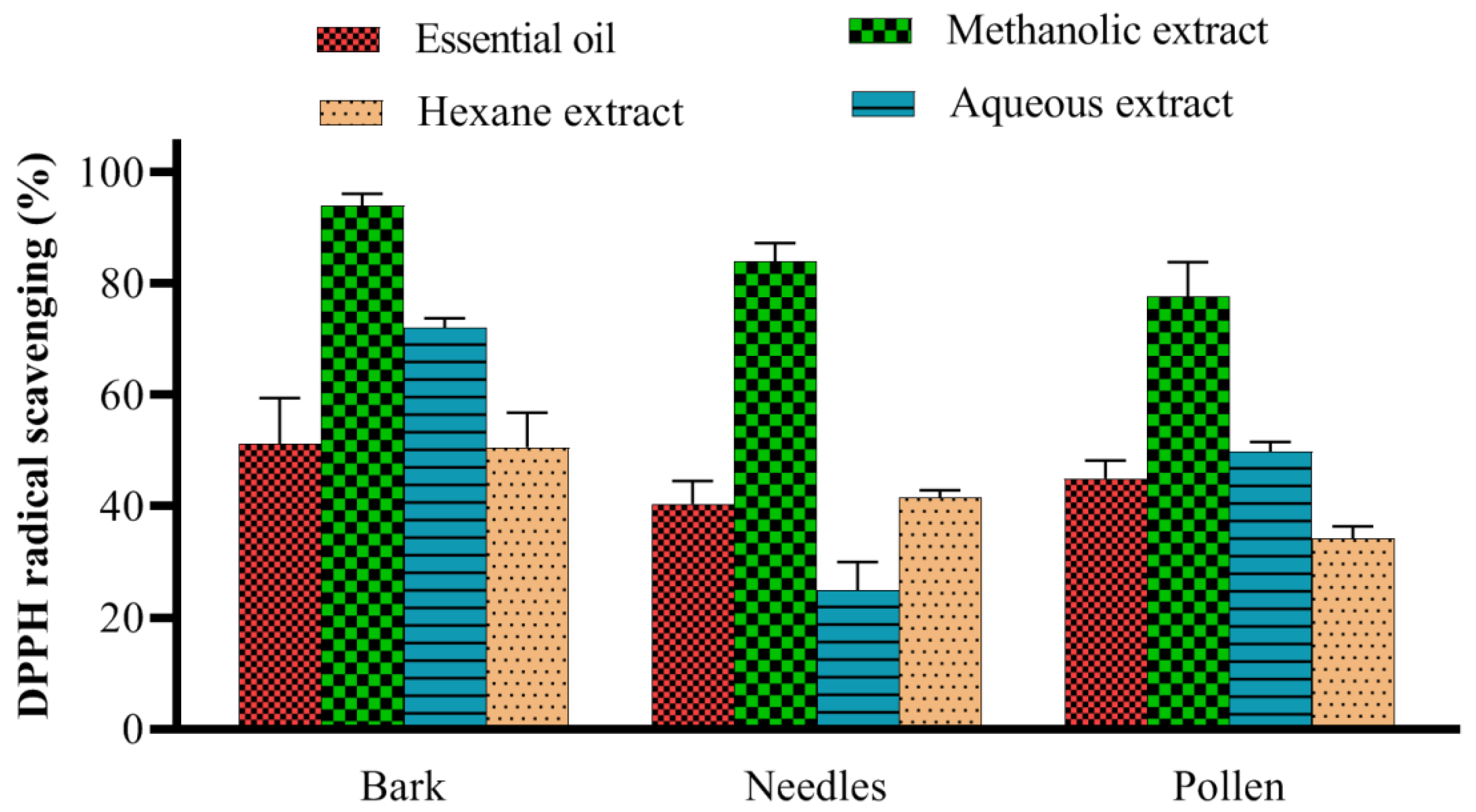
2.1. DPPH Radical Scavenging Assay

The 2,2-diphenyl-1-picrylhydrazyl (DPPH) radical scavenging assay was performed to determine the antioxidant activity of extracts and essential oils obtained from the various parts (bark, needle, and pollen) of P. eldarica (Figure 1). In the DPPH assay, a higher DPPH radical scavenging value indicates a higher antioxidant activity. The DPPH radical scavenging activity of methanolic extracts in all groups were significantly higher than that of other extracts and essential oils (p < 0.01). Among the active methanolic extracts, the methanolic bark extract (mean value 93.9%) demonstrated significantly higher antioxidant activity (p < 0.01) than the methanolic pollen extract (mean value 77.57%) and no significant difference with methanolic needle extract (mean value 83.90%).

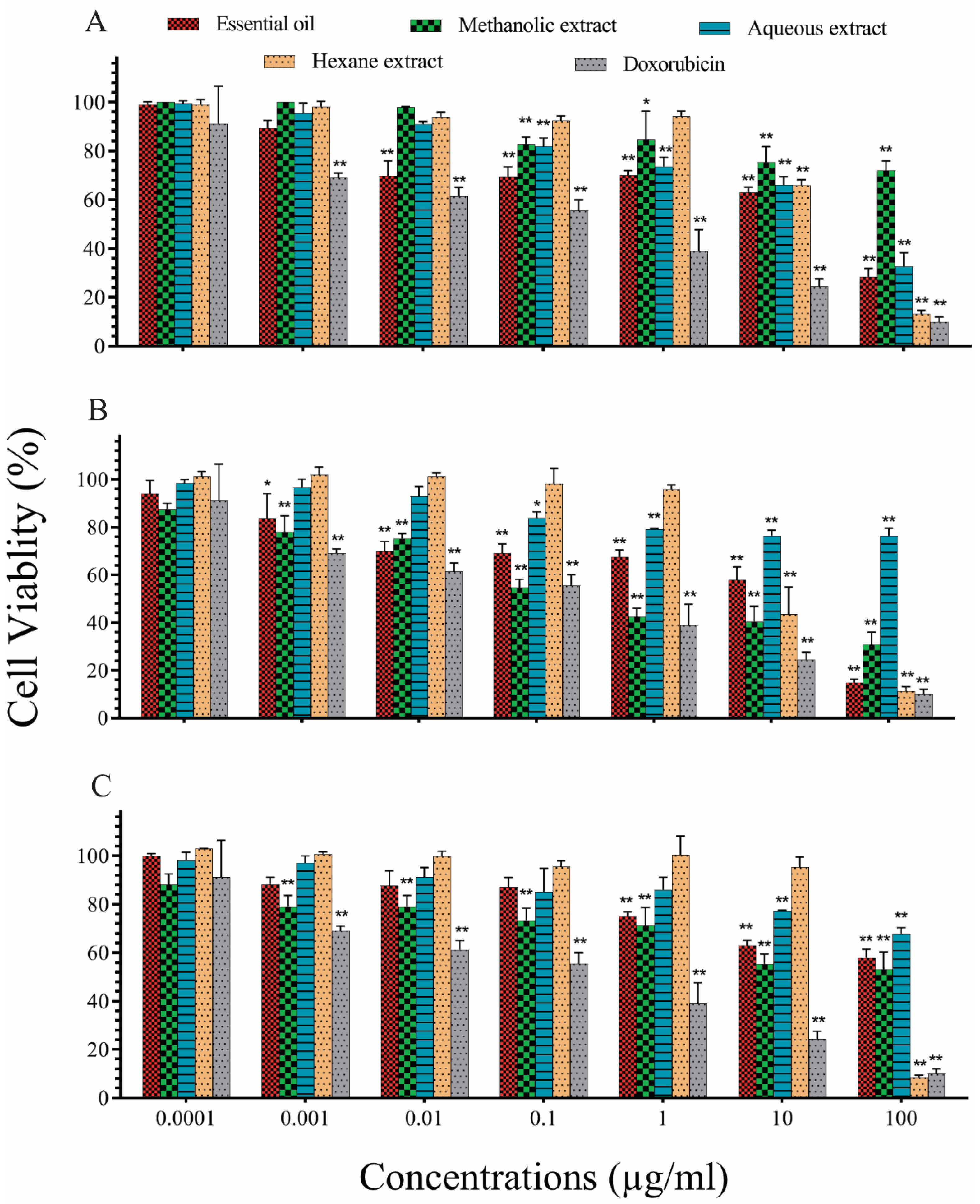
2.2. In Vitro Evaluation of Cytotoxic Effects of P. eldarica Extracts and Essential Oils on A549 Lung Cancer Cells

Cytotoxicity of P. eldarica extracts and essential oils on A549 lung cancer cells was measured using the 3-(4,5-dimethylthiazol-2-yl)-2,5-diphenyltetrazolium bromide (MTT) assay. We observed >80% cell death in A549 cells treated with 100 μg/mL of all hexane extracts, the methanolic needle extract, and needle essential oil (Figure 2). The half-maximal inhibitory concentration (IC50) of the methanolic needle extract at 0.3 μg/mL revealed its high toxicity, comparable with that of doxorubicin (DOX; a positive control). Among pollen fractions, the hexane extract with an IC50 value of 31.7 μg/mL was the most toxic; among bark fractions, the essential oil was the most toxic, with an IC50 value of 17.9 μg/mL (Table 1). Based on these findings, the methanolic needle extract, hexane pollen extract, and bark essential oil were selected for further investigation.

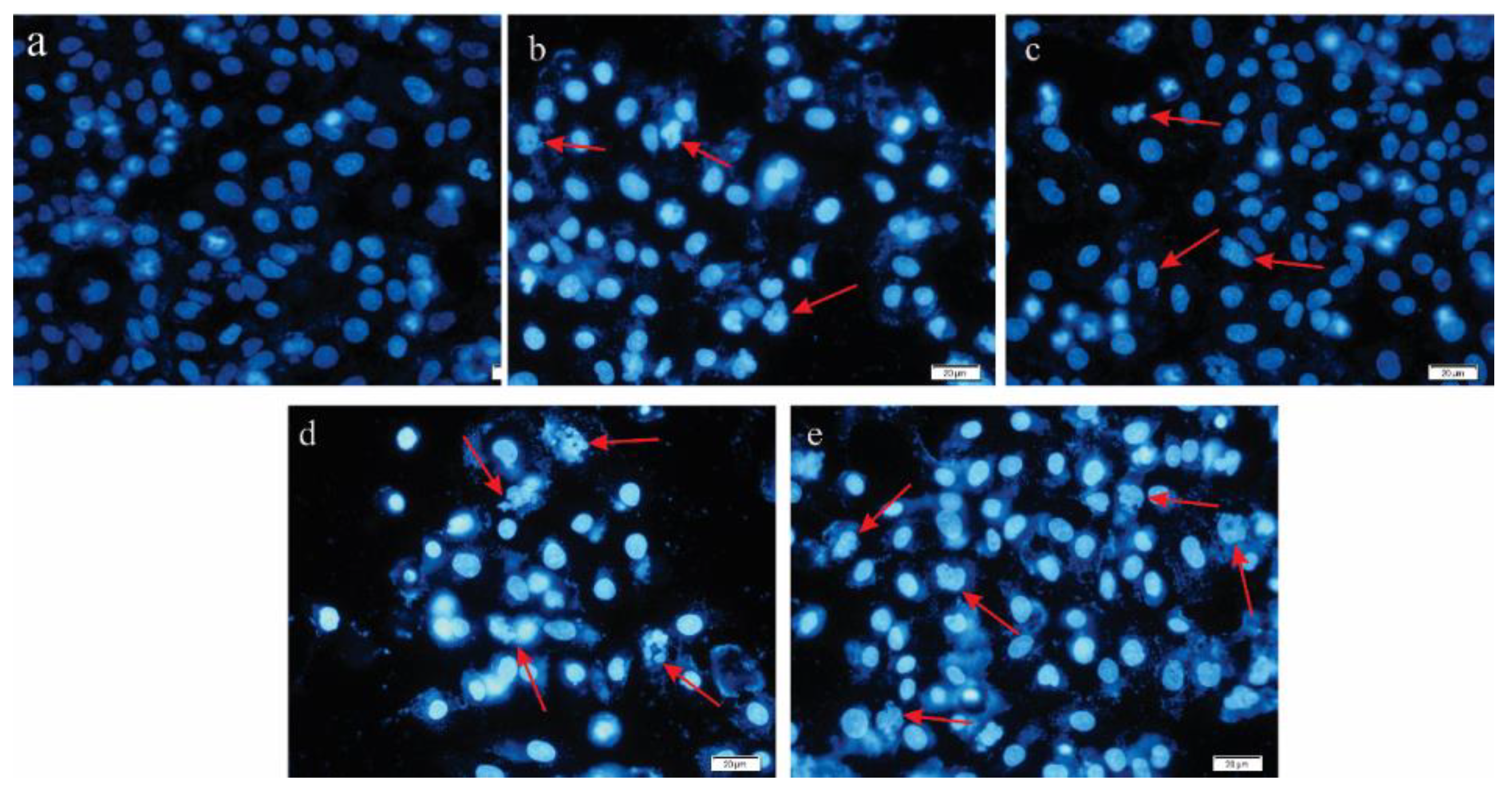
2.3. Evaluating Apoptosis Induction by DAPI Staining

4′,6-Diamidino-2-phenylindole (DAPI) staining was performed to evaluate apoptosis induction in the A549 cell line treated with P. eldarica extracts, essential oils, and DOX (as a positive control), based on IC50 values. The degree of nuclear fragmentation, as an indicator of apoptotic cells, is shown in Figure 3. Compared with untreated control cells, the treated A549 cells revealed increased nuclear fragmentation based on the morphology assessment. A large number of apoptotic cells with high DNA fragmentation were observed following treatment with the methanolic needle extract (Figure 3d). To show the apoptotic cell population, apoptosis analysis was performed by flow cytometry in the next step.
Table 1. IC50 values for different extracts (hexane, methanolic, aqueous extracts, and essential oils) prepared from P. eldarica. Data are expressed as mean ± standard deviation (SD), representative of three independent experiments.
IC50 (µg/mL) aEssential OilMethanolic ExtractsAqueous ExtractsHexane Extracts
Pollen88.9 ± 3649.4 ± 24174.5 ± 46.731.7 ± 7.2
Needles10.3 ± 4.10.3 ± 0.15262.4 ± 43.878.7 ± 0.9
Bark17.9 ± 7.2209 ± 6425.1 ± 6.818.0 ± 1.5
a The IC50 value represents the concentration of each compound that inhibit the growth of A549 cells by 50%.

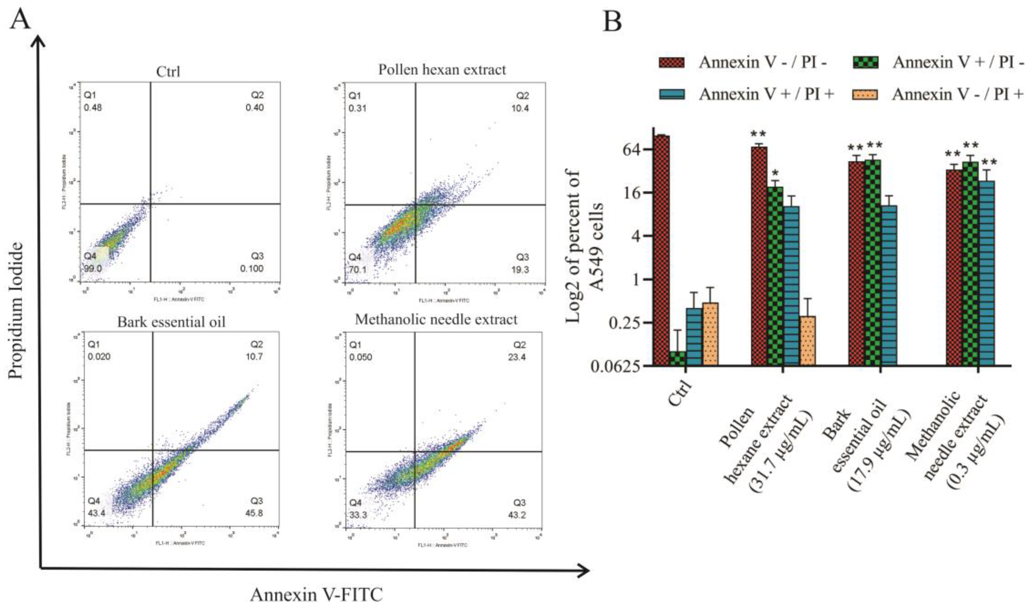
2.4. Analysis of Apoptosis Using Flow Cytometry

A549 cells were exposed to IC50 concentrations of the hexane pollen extract, bark essential oil, and methanolic needle extract for 48 h. Then, to investigate the rate of apoptosis and necrosis, both treated and untreated control cells were stained with Annexin V/propidium iodide (PI) and analyzed through flow cytometry (Figure 4). Quantitative results of Annexin V/PI revealed increased amount of late apoptotic cells 23.4% and early apoptotic cells 43.2% in the methanolic needle extract-treated group compared to control (p < 0.01). The hexane pollen extract-treated group showed considerably more viable cells (70.1%) than the methanolic needle extract- and bark essential oil-treated groups, where viable cells were reduced to 33.3% and 43.4%, respectively (p < 0.05).

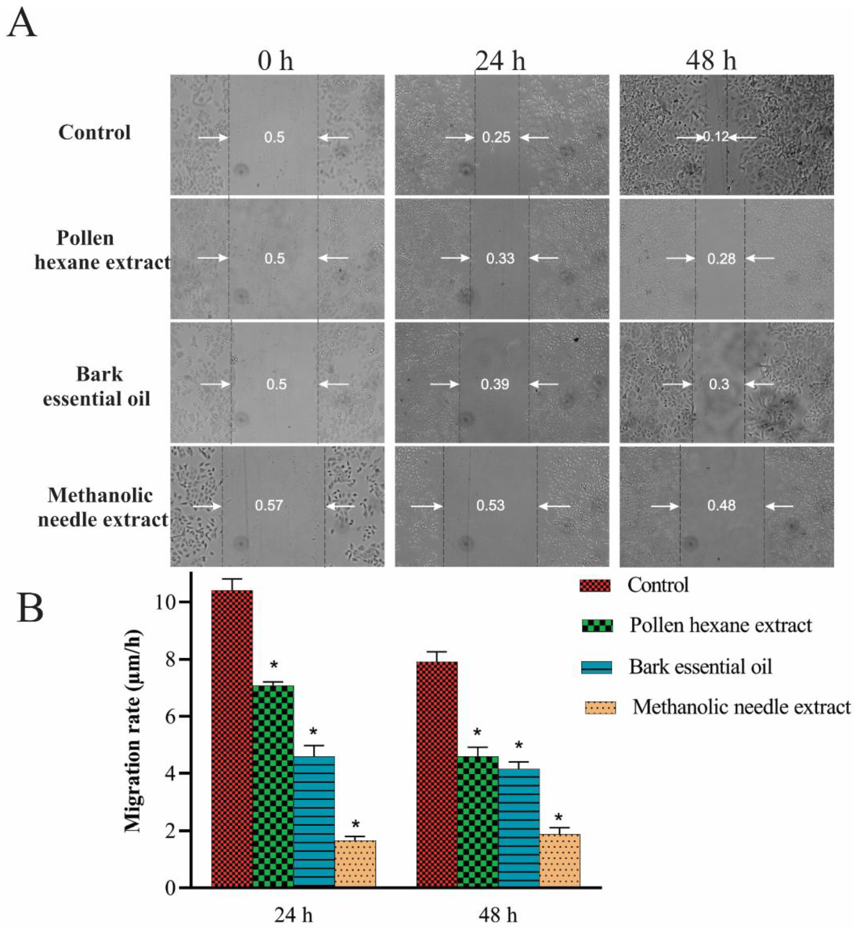
2.5. Scratch Migration Assay

A scratch migration assay was performed to observe the anticancer effects of P. eldarica extracts and essential oil on the migration of A549 cells. Wound areas in A549 cells after 24 and 48 h of treatment with the hexane pollen extract (31.7 µg/mL), bark essential oil (17.9 µg/mL), methanolic needle extract (0.3 µg/mL), and the control group (no treatment) were measured (Figure 5A). The cells incubated with 0.3 µg/mL of methanolic needle extract inhibited cell migration across the initial scratch zone. As shown in the migration rate chart (Figure 5B), after 24 and 48 h, the wound area in the pollen hexane extract- and bark essential oil-treated cells was greater than that of cells incubated with the control medium, indicating that these extracts decreased cell migration when compared with the control group (p < 0.05).

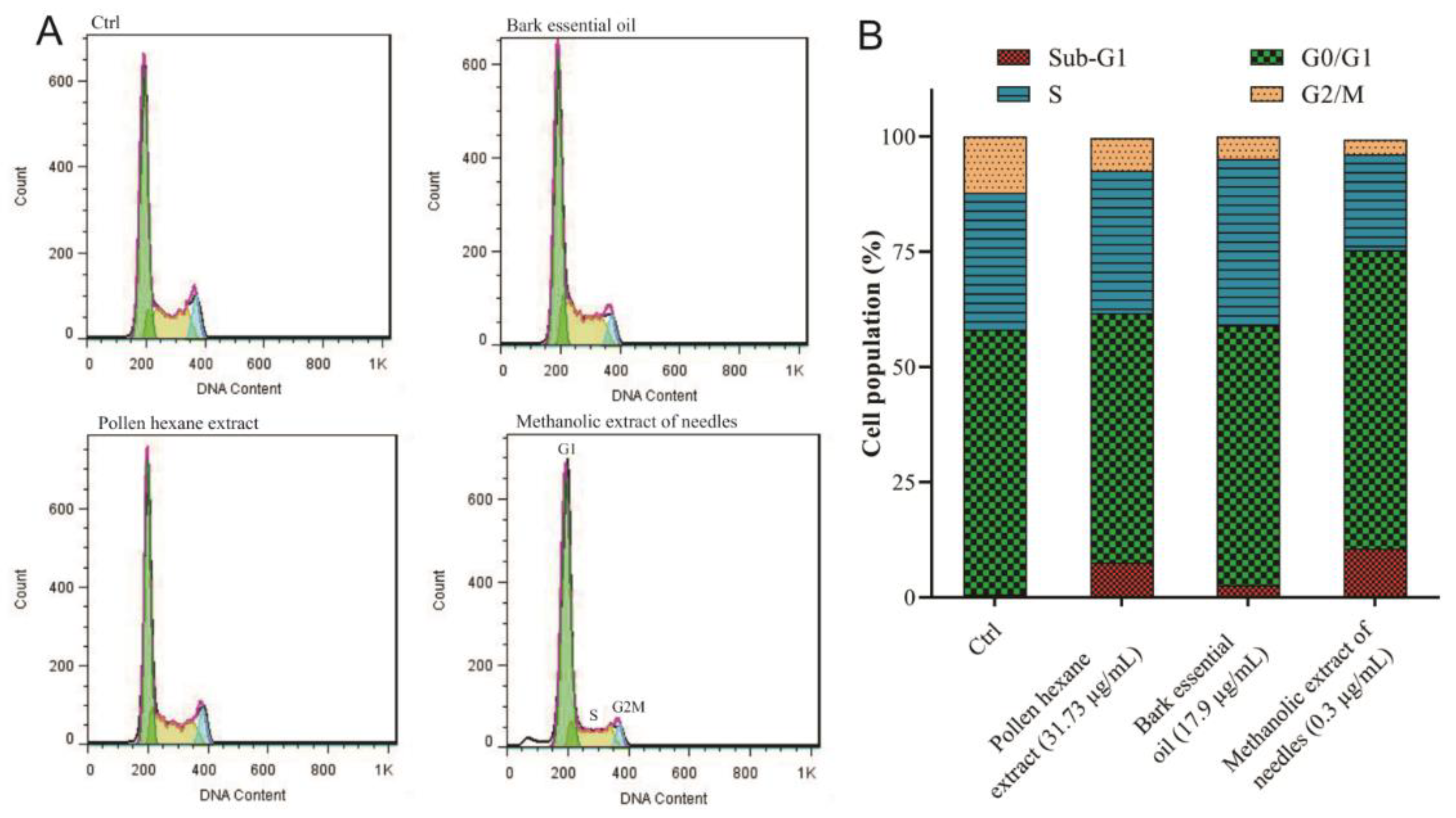
2.6. Cell Cycle Analysis and Apoptosis Evaluation

Treatment of A549 cells for 48 h with the methanolic needle extract and pollen hexane extract resulted in apoptosis induction and cell accumulation in the sub-G1 phase, which were comparable with those observed in the control group (Figure 6A,B). Exposure of cancer cells to the bark essential oil and methanolic needle extract decreased the cell population in the G2/M phase.

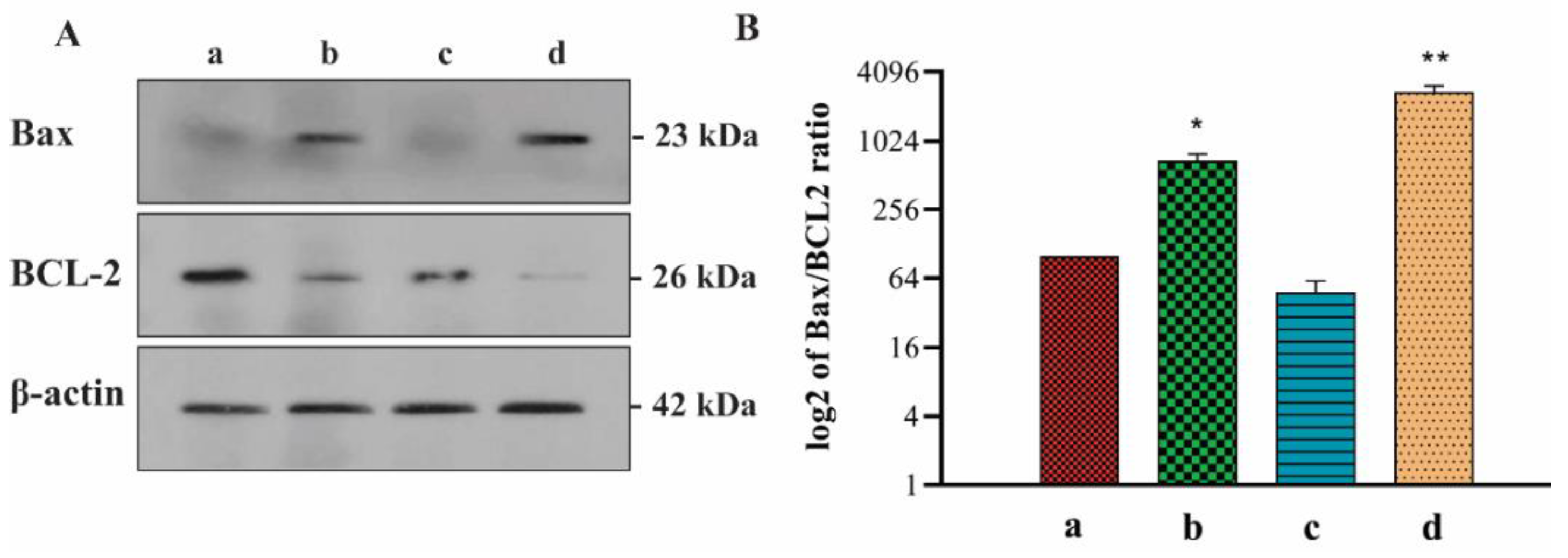
2.7. Western Blot Analysis: Effects of P. eldarica Extracts and Essential Oil on Bax/Bcl 2 Ratio

Next, to investigate the effects of P. eldarica extracts and essential oil on the Bcl-2 family in A549 cells, the expression of Bcl-2 (antiapoptotic) and Bax (proapoptotic) proteins was detected via Western blot analysis, and the Bax/Bcl-2 ratio was determined (Figure 7). A549 cells were treated with the IC50 concentration of the pollen hexane extract (31.7 µg/mL), bark essential oil (17.9 µg/mL), methanolic needle extract (0.3 µg/mL), and control group without treatment for 48 h, revealing a decrease in Bcl-2 and increase in Bax protein expression levels (Figure 7A). The Bax/Bcl-2 ratio (Figure 7B) increased by 6.8- and 27.2-times in the pollen hexane extract- and methanolic needle extract-treated cells, respectively, when compared with that in the control cells.

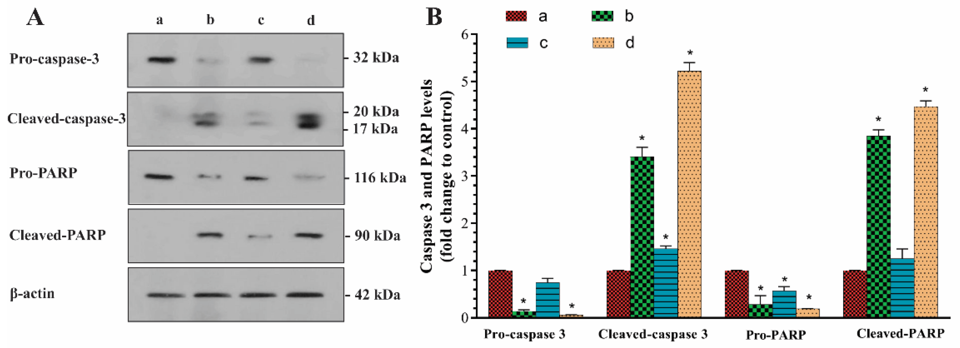
2.8. Western Blot Analysis: Effects of P. eldarica Extracts and Essential Oil on Caspase-3 Activation and PARP Cleavage

Treatment of A549 cells with IC50 concentrations of P. eldarica extracts and essential oil for 48 h significantly increased caspase-3 cleavage (p < 0.05) (Figure 8). PARP is the most crucial protein cleaved after caspase-3 activation. The treatment of cells with 0.3 µg/mL of the methanolic needle extract and 31.7 µg/mL of the pollen hexane extract induced PARP cleavage, compared with those observed in the control group (p < 0.05).

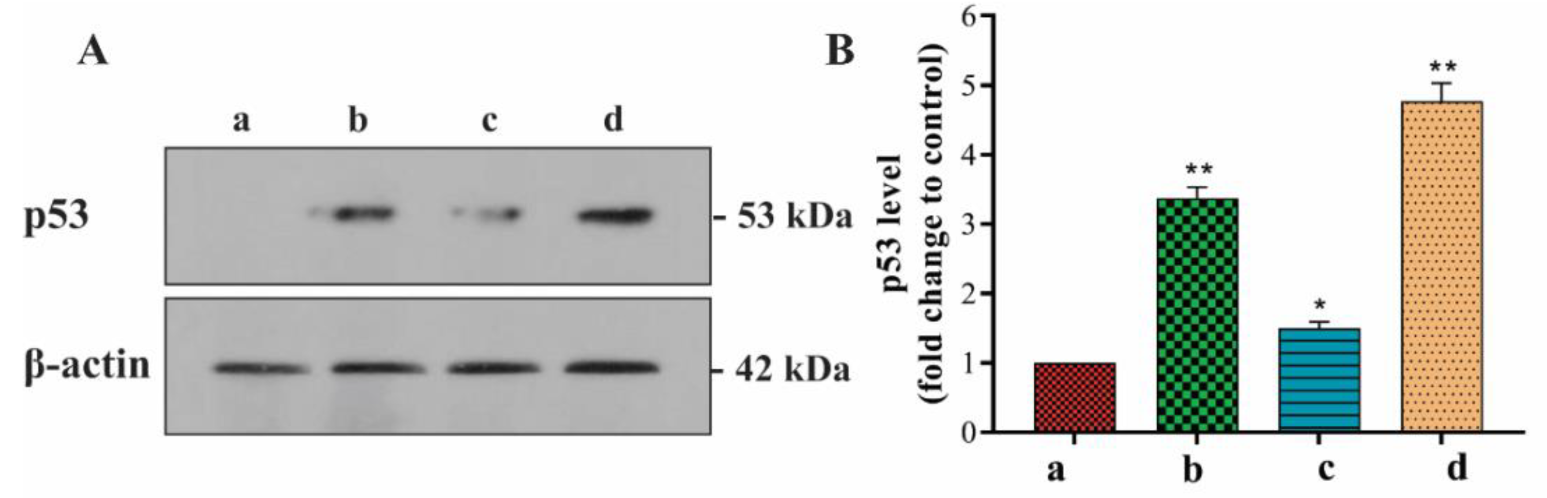
2.9. Western Blot Analysis: Effects of P. eldarica Extracts and Essential Oil on p53

Western blot analysis (Figure 9) revealed that protein expression levels of p53 were significantly higher in groups treated with the pollen hexane extract (31.7 µg/mL), bark essential oil (17.9 µg/mL), and methanolic needle extract (0.3 µg/mL) than in the control group.

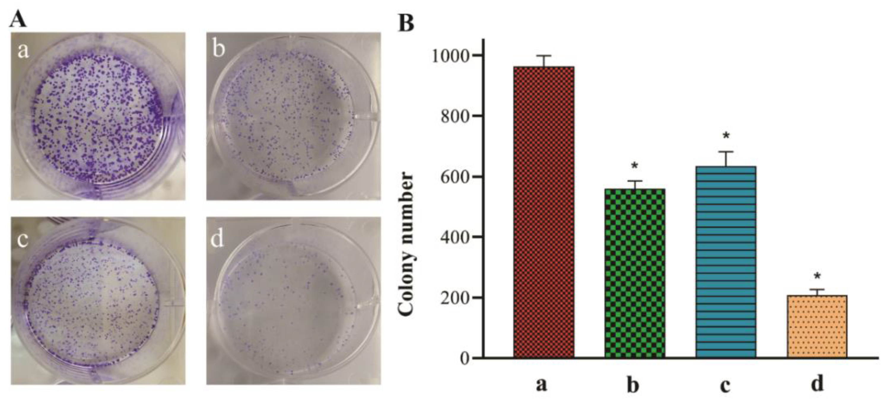
2.10. Colony Formation Assay

We investigated the colony formation ability of A549 cells in the presence or absence of P. eldarica extracts and essential oil for 14 days. The colony numbers were significantly reduced after treatment with IC50 concentrations of the pollen hexane extract, bark essential oil, and methanolic needle extract (Figure 10A). Compared with those in the control group, the colony numbers in the methanolic needle extract-treated group were reduced by over 80% (Figure 10B).

3. Discussion

P. eldarica needles and bark are rich in polyphenols, tannins, and terpenoids. Essential oils and extracts of pine needles and bark have various physiological and pharmacological activities. Extracts and essential oils of P. eldarica are known to possess antioxidant, anti-inflammatory, anticancer, and apoptotic properties, correlating with their effects on cyclooxygenase activity, nitric oxide synthesis, and apoptotic protein regulation [16,21,22]. Previous studies show that needles essential oil of P. eldarica was rich in D-germacrene and main component in the bark was limonene. P. eldarica pollen essential oil was rich in α-pinene [12,23]. Iravani et al. demonstrated that bark extract of P. eldarica had high amounts of polyphenolic compounds such as catechin, ferulic acid, and taxifolin [18].
In the study of Sarvmeili et al., MTT-based cytotoxic effects of essential oils and hydroalcoholic extracts of P. eldarica leaf and bark were evaluated on MCF-7 and HeLa cells [24]. Since this plant is native to Iran, it was necessary to conduct this more extensive, confirm the initial data, and identify molecular pathways. Therefore, we sequentially extracted P. eldarica from hydrophobic to hydrophilic compounds by hexane, methanol, and water extracts and essential oils from three parts of P. eldarica. Then, the most common lung cancer cell line, A549 was chosen and cellular and molecular mechanisms of extracts and essential oils were evaluated.
The results showed that the scavenging effects of methanolic extracts were higher than those of the other extracts and essential oils. These results indicate that methanolic extracts possess considerably higher antioxidant activities, and among them, the methanolic bark and needle extracts demonstrated high DPPH radical scavenging activity (93.9% and 83.90%, respectively). The hydroalcoholic extract of P. massoniana bark had the DPPH radical scavenging activity in the study of Yu et al. [25]. According to Dudonne et al., these extracts are potential natural antioxidant sources [26].
Patient-derived cancer cell lines could be used for the study of the effects of drugs on cancer cells. Scientists use different methods, such as monolayer in vitro assays, 3D cultures, bioreactors, or in vivo evaluations with immunocompromised animals [27,28]. In the present study, we used a monolayer A549 cell line to evaluate the biological effects of essential oils and extracts. For this purpose, the cytotoxic effects of essential oils and hexane, methanolic, and aqueous extracts of P. eldarica bark, needles, and pollen were evaluated using the A549 lung cancer cell line. Based on the MTT assay results, the cell mortality rate in all groups increased in a dose-dependent manner, and the methanolic needle extract, with an IC50 value of 0.3 µg/mL, presented the highest cytotoxicity when compared with other groups. After 48 h of treatment with the pollen essential oil and extracts group, the pollen hexane extract (IC50 = 31.7 µg/mL) was found to be more cytotoxic than the other employed treatments. Our findings, with those observed in previous reports on the effects of pine essential oils and extracts on cancer cell lines, revealing time- and dose-dependent effects. Reportedly, the cytotoxic effect of P. massoniana bark hydroalcoholic extract on a human breast cancer cell line was demonstrated at a higher IC50 value than 32.5 μg/mL [25]. Compared to our results, the IC50 values of P. eldarica bark essential oil and hexane, aqueous, and methanolic extracts were 17.9, 18.0, 25.1, and 209 µg/mL, respectively. P. eldarica essential oil and extracts have been shown to prevent the growth of several cancer cell lines, with the essential oil selectively inhibiting the growth of human breast cancer MCF-7 and HeLa cells, presenting IC50 values of 0.032 and 0.038 µg/mL, respectively [24].
Herein, we observed that the pollen hexane extract, bark essential oil, and methanolic needle extract of P. eldarica significantly suppressed the migration of lung cancer cells at 24 and 48 h. The migration rates of experimental groups were calculated, with the methanolic needle extract revealing the lowest rate (1.87 µm/h in 48 h). Recent studies have shown that pine extracts can inhibit lung cancer cell migration by reducing the expression of matrix metalloproteinases [29,30]. Matrix metalloproteinases are proteolytic enzymes that are secreted from tumor cells, involved in tumor metastasis by degrading the extracellular matrix. The result of the colony formation assay showed that colony numbers in treated groups decreased significantly compared to the control group (p < 0.01). Some studies suggest that natural products can inhibit colony formation by the change in tumor suppressor genes expression [31].
We explored the anticancer mechanisms of P. eldarica extracts and essential oils on A549 lung cancer cells. Apoptosis is a crucial pathway for reducing mutated or growing cancer cells [32]. Reportedly, some natural products have been shown to inhibit the growth of cancer cell lines by inducing apoptosis [33,34]. To evaluate the effects of P. eldarica extracts and essential oils on apoptosis, several experiments, including DAPI staining, Annexin V/PI staining, and cell cycle analysis, was performed. Morphological changes during apoptosis were observed by DAPI staining under a fluorescence microscope. This occurs by nucleic acid denaturation in the cytoplasm. DAPI binds to DNA and forms a complex that emits bluish-white fluorescence by ultraviolet light exciting, so small amounts of DNA can be visualized. Condensed and fragmented chromatin, as specific properties of apoptotic cells, were detected in all extract- and essential oil-treated groups, with a large number of apoptotic cells observed in the methanolic needle extract-treated group containing 0.3 µg/mL of extract; these findings were in agreement with our results obtained following the Annexin V/PI test. In the Annexin V/PI assay, the apoptotic cell population of A549 cells was increased from 0.5% in the control group to 66.6%, 56.5%, and 29.7% in the methanolic needle extract-, bark essential oil-, and pollen hexane extract-treated groups, respectively. The methanolic needle extract-treated cells showed the highest population of apoptotic cells when compared with the other two groups. Thus, we conclude that this group showed suitable efficacy as a potential therapeutic option.
In the cell cycle analysis, A549 cells treated with the methanolic needle extract (0.3 µg/mL) and pollen hexane extract (31.7 µg/mL) revealed cell accumulation in the sub-G1 phase, and the percentage of cells in the S phase was reduced in the methanolic needle extract-treated group. Ma et al. have investigated the cytotoxic effects of P. massoniana extract on HeLa cells, demonstrating that cell cycle arrest occurs in the sub-G1 phase [35]; this was in agreement with our results. Sub-G1 cells were non-apoptotic and viable. Some cells frequently returned and then proceeded to mitosis (hypodiploid and hyperdiploid cells), before arrested in the subsequent cell-cycle interphase. Arresting cells in sub-G1 inhibit cells entering mitosis [36]. Therefore, methanolic needle extract and pollen hexane extract caused post-mitotic arrest in proceeding cell cycles. This process was p53-dependent because when cells divided, p53 arrested the cell cycle in G1 [36]. Furthermore, we observed increased p53 protein expression levels in experimental groups when compared with those in the control group. p53 is a known tumor suppressor gene [37]. In several types of cancers, p53 is damaged owing to mutations, and p53 protein levels are decreased, leading to malignant tumors [38,39].
The cell cycle analysis showed that the accumulation of A549 cells in the S phase was observed after 48 h treatment with the bark essential oil (17.9 µg/mL). Cell cycle arrest at the S phase may result from different mechanisms of action, for example, interfering with cell cycle progression and apoptosis induction, probably by reducing Bcl-2 protein expression. Mitochondria play a critical role in apoptosis, releasing proapoptotic proteins from their membranes into the cytosol. Bcl-2 family proteins are essential mitochondrial regulators of apoptosis [40]. The primary proapoptotic member of these proteins is Bax, which can initiate the release of cytochrome c and subsequent activation of caspase-3 and caspase-9. Bcl-2 acts as an antiapoptotic protein. Bcl-2 inhibits cytochrome c translocation and inhibits caspase-3 activation and apoptosis initiation [19]. High Bax protein expression levels correlate with low Bcl-2 expression levels, which would stimulate apoptosis. The ratio of Bax/Bcl2 expression levels is a diagnostic indicator of cancer [41]. In the present study, Western blot analysis was performed to analyze changes in protein expression of Bcl-2 and Bax in the pollen hexane extract, bark essential oil, methanolic needle extract, and control groups. Based on our Western blot analysis results, the ratio of Bax/Bcl-2 expression levels, as a predictor of apoptosis, increased 6.8- and 27.2-times in the pollen hexane extract- (p < 0.05) and methanolic needle extract-treated groups (p < 0.01) when compared with the control, respectively. In recent studies, pine needle essential oil reportedly downregulated Bcl-2 protein expression and induced apoptosis in oral squamous cell carcinoma [42]. This was in agreement with the results of the present study, which showed that the methanolic needle extract induced apoptosis at a concentration of 0.3 µg/mL by altering Bcl-2 and Bax protein expression in A549 cells more efficiently than other evaluated extracts.
In normal cells, caspases exist in the procaspase form with specific molecular weights. However, upon stimulation of cells with an apoptogenic agent, caspases are activated via proteolytic cleavage. After activation, caspases can cleave other proteins, such as PARP, leading to apoptosis [43]. A previous report has reported the ability of caspases to induce apoptosis in cancer cells following treatment with natural plant-derived extracts [20]. For instance, pine cone water extract reportedly activates caspase-3 and induces apoptosis in a lung cancer cell line [20]. We observed elevated levels of cleaved caspase-3 and cleaved PARP in A549 cells treated with IC50 concentrations of the pollen hexane extract, bark essential oil, and methanolic needle extract. These results suggest that activation of the caspase pathway is essential for inducing apoptosis in A549 cells following treatment with P. eldarica extracts.
Our results showed that P. eldarica extracts mainly, methanolic needle extract, can prevent and treat cancer by various mechanisms. Cell cycle arrest, inhibition of cancer cell migration, and proliferation were some of the anticancer effects. Induction of apoptosis by downregulating of antiapoptotic gene products, such as Bcl-2, and stimulation of apoptotic protein expression such as Bax, caspases, and p53 lead to apoptosis, which was confirmed with the Annexin V/PI test.

4. Materials and Methods

4.1. Materials

Dimethyl sulfoxide (DMSO, 99.9%), Roswell Park Memorial Institute 1640 growth medium (RPMI-1640), trypsin, fetal bovine serum (FBS), penicillin/streptomycin, MTT, DPPH, 4′,6-diamidino-2-phenylindole (DAPI), radioimmunoprecipitation assay (RIPA) buffer, Triton X-100, PI, and 96% ethanol were purchased from Sigma Aldrich Co. (St. Louis, MO, USA). DOX was purchased from Stadapharm GmbH (Laichingen, Germany). The Annexin V/PI apoptosis detection kit was purchased from Exbio Co. (Exbio, Czech Republic). Antibodies against Bcl-2 (sc-492), Bax (sc-7480), caspase-3 (sc-136219), p53 (sc-126), β-actin (sc-47778), and PARP (sc-8007) antibodies were purchased from Santa Cruz Biotechnology, Inc. (Santa Cruz, CA). Phosphate-buffered saline (PBS), methanol, and n-hexane were purchased from Merck (Darmstadt, Germany). Polyvinylidene difluoride (PVDF) membranes were purchased from Roche (Germany). The chemiluminescence detection kit was purchased from Pierce (Rockford, IL, USA).

4.2. Plant Material

P. eldarica needles, pollen, and bark were collected in May and June 2018 from Tabriz district in Iran and identified (No.4036) by the Herbarium of Pharmacy Faculty, Tabriz University of Medical Sciences. All air-dried specimens were sequentially extracted in the order of hexane, methanol, and water by using the Soxhlet apparatus. Then, the extracts were concentrated using a rotary evaporator (Heidolph, Germany), dried using a freeze dryer (Christ Alpha1-4, Germany), and stored at −20 °C until use. For essential oil preparation, 100 g of P. eldarica needles, bark, and pollen powders were hydro-distilled in a Clevenger device separately. The resulting essential oil was dried using anhydrous sodium sulfate and stored in a sealed vial at 4 °C until further analysis. Different concentrations of the extracts and essential oil (0.001, 0.01, 0.1, 1, 10, and 100 μg/mL) were prepared. DMSO was used as the solvent to prepare the stock solution (max 1% v/v).

4.3. DPPH Radical Scavenging Assay

The DPPH radical scavenging activity was determined by adding 1 mL of the DPPH ethanolic solution (0.1 mM) to 1 mL of each extract (1 mg/mL), shaken, and incubated at 37 °C for 30 min in the dark. Then, the absorbance of each extract was measured at 517 nm by ultraviolet–visible (UV–Vis) spectrophotometry (Ultrospec 2000, Pharmacia Biotech, Amersham, UK) [44]. DPPH ethanolic solution was used as a control, and 70% ethanol was used as a blank. The radical scavenging activity as a percentage of DPPH discoloration was calculated using the following equation:
DPPH   activity   ( % )   = UV   Absorbance   of   extract UV   Absorbance   of   control   × 100 .

4.4. Cell Culture

The A549 cell line was obtained from the Pasteur Institute (Tehran, Iran). Cells were grown in RPMI-1640 supplemented with 10% FBS and 1% penicillin/streptomycin. The media was sterilized using 0.22-μm filters and stored at 4 °C before use. Cells were grown in RPMI-1640 medium at 37 °C in a 5% CO2 incubator.

4.5. MTT-Based Cytotoxicity Assay

The cytotoxicity of the essential oil and different extracts of P. eldarica against the A549 cell line was determined by employing a rapid colorimetric assay using MTT [45,46,47,48,49]. In this assay, soluble MTT is metabolized by the mitochondrial enzyme activity of viable cells into an insoluble formazan dye product. Subsequently, formazan was dissolved in DMSO and measured spectrophotometrically at 540 nm. Briefly, 180 μL of a cell suspension (5 × 10⁴ cells/mL of medium) was seeded in 96-well microplates and incubated for 24 h (37 °C, 5% CO2 humidified air). Then, prepared concentrations of essential oil and extracts ranging from 0 to 100 µg/mL were added to each well. The plates were incubated for another 48 h under the same conditions. DOX and 1% DMSO were used as positive and negative controls, respectively. To assess cell survival, 20 µL of MTT solution (5 mg/mL in PBS) was added to each well, and the plates were incubated at 37 °C for 3 h. Then, the old media containing MTT was removed and 150 µL DMSO was added to each well and pipetted to dissolve the formed formazan crystals. Absorbance was measured using an ELISA plate reader (Awareness Technology, Inc., Palm City, FL, USA) at 540 nm. Each extract concentration was repeated three times, and cell survival in the negative control was assumed to be 100%. The IC50 value was calculated using GraphPad Prism software version 6.0.

4.6. DAPI Staining

DAPI staining was performed to evaluate nuclei fragmentation. A549 cells (2 × 10⁶ cells/well) were seeded into 6-well plates. After incubation, cultured lung cancer cells at 70% confluence were treated with IC50 concentrations of extracts and essential oil for 48 h. Next, the cells were washed with PBS, fixed with paraformaldehyde (4%) for 20 min and permeabilized with 0.15% Triton X-100 for 15 min. Subsequently, the cells were stained in the dark with 0.1% DAPI for 15 min, washed with PBS, and observed by fluorescence microscopy (Olympus, Seoul, Korea) [50].

4.7. Annexin V/PI Flow Cytometry

A549 cells (2 × 10⁶ cells/well) were seeded in 6-well plates and incubated in RPMI-1640 medium for 48 h. Subsequently, the IC50 concentration of the extracts and essential oil was added to the wells and incubated for 48 h. Then, the cells were trypsinized and washed with cold PBS, supernatants were removed, and 100 μL binding buffer was added at ice-cold temperature. Finally, lung cancer cells were stained with Pl and Annexin V-fluorescein isothiocyanate (FITC) according to the manufacturer’s instructions. A FACSCalibur flow cytometer (BD Biosciences, San Jose, CA, USA) was used to analyze apoptosis rate. The data were analyzed using the FlowJo software package (Treestar, Inc., San Carlos, CA, USA) [51].

4.8. Scratch Migration Assay

To examine the migration of A549 cells in two-dimensional monolayers, cells were seeded in 6-well plates and incubated in RPMI-1640 medium. After the cells reach to confluent monolayer (at least 70% confluency), a thin linear scratch “wound” was created using a 200 µL pipette tip. The monolayers were then washed with PBS to remove detached cells and treated with IC50 concentrations of extracts and essential oil; images were captured at 0, 24, and 48 h after treatment with a digital camera connected to a bright-field microscope. Wound width was calculated as the distance between two edges of the scratch using CorelDRAW software [52]. The migration rate was calculated using the following equation:
Migration   rate   ( µ m / h )   = initial   wound   width   ( µ m ) final   wound   width ( µ m ) duration   of   migration   ( h )

4.9. Cell Cycle Analysis

A549 cells (3 × 10⁵ cells/mL) were seeded in 6-well plates. After 24 h, cells were treated with IC50 concentrations of extracts and essential oil. Cells were trypsinized after 48 h and fixed in ice-cold 70% ethanol at 4 °C for 2 h. A549 cells were centrifuged and washed with PBS, treated with 1 mL PI master mix containing 950 µL PBS, 40 µL PI, and 10 µL RNase A to stain cellular DNA, incubated at room temperature for 30 min, and analyzed using a FACSCalibur flow cytometer (BD Biosciences, San Jose, CA, USA) [50].

4.10. Western Blot Analysis

Expression of Bcl-2, Bax, caspase-3, p53, and PARP in lung cancer cells was analyzed by Western blot analysis, as described previously [33,34,53]. A549 cells were seeded in 6-well plates. On reaching the appropriate confluency, the cells were treated with IC50 concentration of extracts and essential oil for 48 h. Next, the cells were trypsinized and washed with ice-cold PBS. For cell lysis, RIPA buffer (1% NP-40, 1% sodium deoxycholate, 0.1% SDS, 1 mM EDTA, 150 mM NaCl, and 50 mM Tris-HCl, pH 7.8) containing a protease inhibitor cocktail was used. After centrifugation at 12,000× g for 10 min at 4 °C, the supernatant containing proteins was collected and stored at −80 °C. Proteins were separated by SDS-polyacrylamide gel electrophoresis and electro-transferred to polyvinylidene fluoride membranes. Membranes were incubated in Tris-buffered saline containing 5% non-fat dry milk for 2 h to block nonspecific binding. Membranes were incubated overnight at 4 °C with anti Bcl-2 (1:500, sc-492), Bax (1:500, sc-7480), caspase-3 (1:500, sc-136219), p53 (1:500, sc-126), β-actin (1:500, sc-47778), and PARP (1:500, sc-8007) antibodies for 16 h. Finally, membranes were incubated with secondary antibody (anti-rabbit; 1:1000) for 75 min in the antibody buffer (5% BSA, 1X TBS, 0.1% Tween-20). For visualization of the blots, a chemiluminescence (ECL) detection kit was used just before autoradiography. Band intensities were quantified by densitometry using the ImageJ software (U.S. National Institutes of Health, Bethesda, MD, USA).

4.11. Colony Formation Assay

The colony formation assay was performed as previously described [54]. A549 cells were seeded (1000 cells/well) in 6-well plates in triplicate in RPMI-1640 medium. After 24 h, the medium was removed and replaced with the IC50 concentration of respective extracts and essential oil. The group with no added treatment was considered as the control. After 14 days, the colonies were stained with 0.5% crystal violet solution containing 25% methanol for 15 min at 37 °C and washed with excess dye. The colony numbers were counted using the ImageJ software.

4.12. Statistical Analysis

The data were processed using GraphPad Prism version 6.0 (GraphPad Software, Inc., La Jolla, CA, USA) for averages and standard deviation (SD) by performing one-way and two-way analyses of variance. Data are presented as mean values ± SD, and p-values less than 0.05 were considered significant.

5. Conclusions

In the present study, extracts and essential oils of various parts of P. eldarica revealed robust antioxidant activity. Especially, the pollen hexane extract, bark essential oil, and methanolic needle extract of P. eldarica inhibited the proliferation and cell cycle progression in A549 lung cancer cells, mainly through Bcl-2 downregulation, stimulation of p53 expression, and induction of apoptosis in a caspase-dependent manner. Furthermore, the pollen hexane extract, bark essential oil, and methanolic needle extract suppressed the migration, and colony formation of lung cancer cells. Therefore, our findings suggest that P. eldarica extracts and essential oils, especially the methanolic extract of P. eldarica needles (the most active extract), can be potentially developed as chemopreventive natural resources for cancer prevention and treatment, with further investigations warranted to determine the main components responsible for anticancer activities.

Author Contributions

Conceptualization, H.H. and K.H.K.; Formal Analysis, T.G., S.A., E.I., A.D., S.F., J.-H.H. and C.P.; Investigation, T.G., S.A., A.D. and S.F.; Resources, H.H.; Writing—Original Draft Preparation, T.G., H.H. and K.H.K.; Writing—Review and Editing, T.G. and K.H.K.; Project Administration, H.H. and K.H.K.; Funding Acquisition, H.H. and K.H.K. All authors have read and agreed to the published version of the manuscript.

Funding

This study was supported financially by the Drug Applied Research Center, Tabriz University of Medical Science, Tabriz, Iran (Grant No. 58972), and the Iran National Science Foundation (Grant No. 97008952). This work was also supported by a National Research Foundation of Korea (NRF) grant funded by the Korean government (MSIT) (grant numbers 2019R1A5A2027340 and 2021R1A2C2007937).

Institutional Review Board Statement

Not applicable.

Informed Consent Statement

Not applicable.

Data Availability Statement

Not applicable.

Conflicts of Interest

The authors declare no conflict of interest.

References

  1. Bray, F.; Ferlay, J.; Soerjomataram, I.; Siegel, R.L.; Torre, L.A.; Jemal, A. Global cancer statistics 2018: GLOBOCAN estimates of incidence and mortality worldwide for 36 cancers in 185 countries. CA Cancer J. Clin. 2018, 68, 394–424. [Google Scholar] [CrossRef] [Green Version]
  2. Gurib-Fakim, A. Medicinal plants: Traditions of yesterday and drugs of tomorrow. Mol. Asp. Med. 2006, 27, 1–93. [Google Scholar] [CrossRef]
  3. Kumar, M.A.S.; Nair, M.; Hema, P.S.; Mohan, J.; Santhoshkumar, T.R. Pinocembrin triggers Bax-dependent mitochondrial apoptosis in colon cancer cells. Mol. Carcinog. 2007, 46, 231–241. [Google Scholar] [CrossRef]
  4. Kim, J.H.; Yoon, E.K.; Chung, H.J.; Park, S.Y.; Hong, K.M.; Lee, C.H.; Lee, Y.S.; Choi, K.; Yang, Y.; Kim, K.; et al. P53 acetylation enhances Taxol-induced apoptosis in human cancer cells. Apoptosis 2013, 18, 110–120. [Google Scholar] [CrossRef]
  5. Sun, N.K.; Huang, S.L.; Chang, T.C.; Chao, C.C.K. TLR4 and NFκB signaling is critical for taxol resistance in ovarian carcinoma cells. J. Cell. Physiol. 2018, 233, 2489–2501. [Google Scholar] [CrossRef]
  6. Ran, J.H.; Shen, T.T.; Wu, H.; Gong, X.; Wang, X.Q. Phylogeny and evolutionary history of Pinaceae updated by transcriptomic analysis. Mol. Phylogenet. Evol. 2018, 129, 106–116. [Google Scholar] [CrossRef] [PubMed]
  7. Stevenson, D.W. Gymnosperms. In The Evolution of Plant Form; John Wiley & Sons: Hoboken, NJ, USA, 2012; Volume 45, pp. 141–161. [Google Scholar]
  8. Kaushik, D.; Kumar, A.; Kaushik, P.; Rana, A.C. Analgesic and anti-inflammatory activity of pinus roxburghii sarg. Adv. Pharmacol. Sci. 2012, 2012, 245431. [Google Scholar] [CrossRef] [PubMed] [Green Version]
  9. Parihar, P.; Parihar, L.; Bohra, A. Antibacterial activity of extracts of Pinus roxburghii sarg. Bangladesh J. Bot. 2006, 35, 85–86. [Google Scholar]
  10. Huseini, F.H.; Mehrzadi, S.; Ghaznavi, H.; Tajallizadehkhoob, Y.; Fakhrzadeh, H. Effects of pinus eldarica Medw. nut extract on blood glucose and cholesterol levels in hypercholesterolemic alloxan-induced diabetic rats. J. Med. Plants 2013, 12, 68–74. [Google Scholar]
  11. Kwak, C.S.; Moon, S.C.; Lee, M.S. Antioxidant, antimutagenic, and antitumor effects of pine needles (Pinus densiflora). Nutr. Cancer 2006, 56, 162–171. [Google Scholar] [CrossRef]
  12. Ghaffari, T.; Kafil, H.S.; Asnaashari, S.; Farajnia, S.; Delazar, A.; Baek, S.C.; Hamishehkar, H.; Kim, K.H. Chemical Composition and Antimicrobial Activity of Essential Oils from the Aerial Parts of Pinus eldarica Grown in Northwestern Iran. Molecules 2019, 24, 3203. [Google Scholar] [CrossRef] [Green Version]
  13. Lee, Y.; Park, J.H.; Park, E. Effect of Sulgidduk containing pine needle juice on lipid metabolism in high fat-cholesterol diet induced dyslipidemic rats. J. Nutr. Health 2019, 51, 6–16. [Google Scholar] [CrossRef] [Green Version]
  14. Rohdewald, P. A review of the French maritime pine bark extract (Pycnogenol®), a herbal medication with a diverse clinical pharmacology. Int. J. Clin. Pharmacol. Ther. 2002, 40, 158–168. [Google Scholar] [CrossRef]
  15. Guri, A.; Kefalas, P.; Roussis, V. Antioxidant potential of six pine species. Phytother. Res. 2006, 20, 263–266. [Google Scholar] [CrossRef] [PubMed]
  16. Kim, J.S. Evaluation of in vitro antioxidant activity of the water extract obtained from dried pine needle (pinus densiflora). Prev. Nutr. Food Sci. 2018, 23, 134–143. [Google Scholar] [CrossRef] [PubMed]
  17. Jeong, J.B.; Seo, E.W.; Jeong, H.J. Effect of extracts from pine needle against oxidative DNA damage and apoptosis induced by hydroxyl radical via antioxidant activity. Food Chem. Toxicol. 2009, 47, 2135–2141. [Google Scholar] [CrossRef] [PubMed]
  18. Iravani, S.; Zolfaghari, B. Phytochemical analysis of Pinus eldarica bark. Res. Pharm. Sci. 2014, 9, 243–250. [Google Scholar]
  19. Borner, C. The Bcl-2 protein family: Sensors and checkpoints for life-or-death decisions. Mol. Immunol. 2003, 39, 615–647. [Google Scholar] [CrossRef]
  20. Lee, T.K.; Roh, H.S.; Yu, J.S.; Baek, J.; Lee, S.; Ra, M.; Kim, S.Y.; Baek, K.H.; Kim, K.H. Pinecone of Pinus koraiensis Inducing Apoptosis in Human Lung Cancer Cells by Activating Caspase-3 and its Chemical Constituents. Chem. Biodivers. 2017, 14. [Google Scholar] [CrossRef]
  21. Li, K.; Li, Q.; Li, J.; Gao, D.; Zhang, T.; Han, Z.; Zheng, F. Effect of procyanidins from Pinus koraiensis bark on growth inhibition and expression of PCNA and TNF-α in mice with U14 cervical cancer. Therapy 2007, 4, 685–690. [Google Scholar] [CrossRef]
  22. Kang, H.; Lim, J.W.; Kim, H. Inhibitory effect of Korean Red Ginseng extract on DNA damage response and apoptosis in Helicobacter pylori-infected gastric epithelial cells. J. Ginseng Res. 2020, 44, 79–85. [Google Scholar] [CrossRef] [PubMed]
  23. Sharifan, A.; Etebari, M.; Zolfaghari, B.; Aliomrani, M. Investigating the effects of bark extract and volatile oil of Pinus eldarica against cisplatin-induced genotoxicity on HUVECs cell line. Toxicol. Res. 2021, 10, 223–231. [Google Scholar] [CrossRef] [PubMed]
  24. Sarvmeili, N.; Jafarian-Dehkordi, A.; Zolfaghari, B. Cytotoxic effects of Pinus eldarica essential oil and extracts on HeLa and MCF-7 cell lines. Res. Pharm. Sci. 2016, 11, 476–483. [Google Scholar] [CrossRef] [Green Version]
  25. Yu, L.; Zhao, M.; Wang, J.s.; Cui, C.; Yang, B.; Jiang, Y.; Zhao, Q. Antioxidant, immunomodulatory and anti-breast cancer activities of phenolic extract from pine (Pinus massoniana Lamb) bark. Innov. Food Sci. Emerg. Technol. 2008, 9, 122–128. [Google Scholar] [CrossRef]
  26. Dudonné, S.; Vitrac, X.; Coutiére, P.; Woillez, M.; Mérillon, J.M. Comparative study of antioxidant properties and total phenolic content of 30 plant extracts of industrial interest using DPPH, ABTS, FRAP, SOD, and ORAC assays. J. Agric. Food Chem. 2009, 57, 1768–1774. [Google Scholar] [CrossRef]
  27. Miserocchi, G.; Mercatali, L.; Liverani, C.; De Vita, A.; Spadazzi, C.; Pieri, F.; Bongiovanni, A.; Recine, F.; Amadori, D.; Ibrahim, T. Management and potentialities of primary cancer cultures in preclinical and translational studies. J. Transl. Med. 2017, 15, 1–16. [Google Scholar] [CrossRef] [Green Version]
  28. Xu, R.; Zhou, X.; Wang, S.; Trinkle, C. Tumor organoid models in precision medicine and investigating cancer-stromal interactions. Pharmacol. Ther. 2020, 218, 107668. [Google Scholar] [CrossRef] [PubMed]
  29. Mao, P.; Zhang, E.; Chen, Y.; Liu, L.; Rong, D.; Liu, Q.; Li, W. Pinus massoniana bark extract inhibits migration of the lung cancer A549 cell line. Oncol. Lett. 2017, 13, 1019–1023. [Google Scholar] [CrossRef] [Green Version]
  30. Liu, J.; Bai, J.; Jiang, G.; Li, X.; Wang, J.; Wu, D.; Owusu, L.; Zhang, E.; Li, W. Anti-tumor effect of Pinus massoniana bark proanthocyanidins on ovarian cancer through induction of cell apoptosis and inhibition of cell migration. PLoS ONE 2015, 10, e0142157. [Google Scholar] [CrossRef]
  31. Nair, H.K.; Rao, K.V.; Aalinkeel, R.; Mahajan, S.; Chawda, R.; Schwartz, S.A. Inhibition of prostate cancer cell colony formation by the flavonoid quercetin correlates with modulation of specific regulatory genes. Clin. Diagn. Lab. Immunol. 2004, 11, 63–69. [Google Scholar] [CrossRef] [PubMed] [Green Version]
  32. Jalilzadeh, N.; Samadi, N.; Salehi, R.; Dehghan, G.; Iranshahi, M.; Dadpour, M.R.; Hamishehkar, H. Novel nano-vehicle for delivery and efficiency of anticancer auraptene against colon cancer cells. Sci. Rep. 2020, 10, 1606. [Google Scholar] [CrossRef] [Green Version]
  33. Lee, D.; Lee, Y.H.; Lee, K.H.; Lee, B.S.; Alishir, A.; Ko, Y.-J.; Kang, K.S.; Kim, K.H. Aviculin isolated from Lespedeza cuneata induce apoptosis in breast cancer cells through mitochondria-mediated caspase activation pathway. Molecules 2020, 25, 1708. [Google Scholar] [CrossRef] [Green Version]
  34. Lee, D.; Lee, S.R.; Kang, K.S.; Ko, Y.; Pang, C.; Yamabe, N.; Kim, K.H. Betulinic acid suppresses ovarian cancer cell proliferation through induction of apoptosis. Biomolecules 2019, 9, 257. [Google Scholar] [CrossRef] [PubMed] [Green Version]
  35. Ma, H.; Lai, F.; Xie, H.; Wang, J.; Hongbin, W. Involvement of the Bcl-2 family members in pinus massoniana bark extract induced apoptosis in HeLa cells. Phytother. Res. 2008, 22, 1472–1476. [Google Scholar] [CrossRef] [PubMed]
  36. Demidenko, Z.; Kalurupalle, S.; Hanko, C.; Lim, C.; Broude, E.; Blagosklonny, M. Mechanism of G1-like arrest by low concentrations of paclitaxel: Next cell cycle p53-dependent arrest with sub G1 DNA content mediated by prolonged mitosis. Oncogene 2008, 27, 4402–4410. [Google Scholar] [CrossRef] [PubMed] [Green Version]
  37. Pflaum, J.; Schlosser, S.; Müller, M. P53 family and cellular stress responses in cancer. Front. Oncol. 2014, 4, 285. [Google Scholar] [CrossRef]
  38. Cadwell, C.; Zambetti, G.P. The effects of wild-type p53 tumor suppressor activity and mutant p53 gain-of-function on cell growth. Gene 2001, 277, 15–30. [Google Scholar] [CrossRef]
  39. Lu, Z.; Xiao, Y.; Liu, X.; Zhang, Z.; Xiao, F.; Bi, Y. Matrine reduces the proliferation of A549 cells via the p53/p21/PCNA/eIF4E signaling pathway. Mol. Med. Rep. 2017, 15, 2415–2422. [Google Scholar] [CrossRef] [Green Version]
  40. Festjens, N.; Van Gurp, M.; Van Loo, G.; Saelens, X.; Vandenabeele, P. Bcl-2 family members as sentinels of cellular integrity and role of mitochondrial intermembrane space proteins in apoptotic cell death. Acta Haematol. 2004, 111, 7–27. [Google Scholar] [CrossRef]
  41. Giotakis, A.I.; Kontos, C.K.; Manolopoulos, L.D.; Sismanis, A.; Konstadoulakis, M.M.; Scorilas, A. High BAX/BCL2 mRNA ratio predicts favorable prognosis in laryngeal squamous cell carcinoma, particularly in patients with negative lymph nodes at the time of diagnosis. Clin. Biochem. 2016, 49, 890–896. [Google Scholar] [CrossRef]
  42. Jo, J.R.; Park, J.S.; Park, Y.K.; Chae, Y.Z.; Lee, G.H.; Park, G.Y.; Jang, B.C. Pinus densiflora leaf essential oil induces apoptosis via ROS generation and activation of caspases in YD-8 human oral cancer cells. Int. J. Oncol. 2012, 40, 1238–1245. [Google Scholar] [CrossRef] [PubMed] [Green Version]
  43. Lazebnik, Y.A.; Kaufmann, S.H.; Desnoyers, S.; Poirier, G.G.; Earnshaw, W.C. Cleavage of poly(ADP-ribose) polymerase by a proteinase with properties like ICE. Nature 1994, 371, 346–347. [Google Scholar] [CrossRef] [PubMed]
  44. Wu, H.C.; Chen, H.M.; Shiau, C.Y. Free amino acids and peptides as related to antioxidant properties in protein hydrolysates of mackerel (Scomber austriasicus). Food Res. Int. 2003, 36, 949–957. [Google Scholar] [CrossRef]
  45. Molavi, O.; Torkzaban, F.; Jafari, S.; Asnaashari, S.; Asgharian, P. Chemical compositions and anti-proliferative activity of the aerial parts and rhizomes of squirting cucumber, Cucurbitaceae. Jundishapur J. Nat. Pharm. Prod. 2020, 15. [Google Scholar] [CrossRef]
  46. Lee, S.; Ryoo, R.; Choi, J.H.; Kim, J.-H.; Kim, S.-H.; Kim, K.H. Trichothecene and tremulane sesquiterpenes from a hallucinogenic mushroom Gymnopilus junonius and their cytotoxicity. Arch. Pharmacal. Res. 2020, 43, 214–223. [Google Scholar] [CrossRef]
  47. Trinh, T.A.; Park, E.-J.; Lee, D.; Song, J.H.; Lee, H.L.; Kim, K.H.; Kim, Y.; Jung, K.; Kang, K.S.; Yoo, J.-E. Estrogenic activity of sanguiin H-6 through activation of estrogen receptor α coactivator-binding site. Nat. Prod. Sci. 2019, 25, 28–33. [Google Scholar] [CrossRef]
  48. Lee, S.R.; Kang, H.; Yoo, M.J.; Yu, J.S.; Lee, S.; Yi, S.A.; Beemelmanns, C.; Lee, J.; Kim, K.H. Anti-adipogenic pregnane steroid from a Hydractinia-associated fungus, Cladosporium sphaerospermum SW67. Nat. Prod. Sci. 2020, 26, 230–235. [Google Scholar]
  49. Lee, K.; Seo, Y.-J.; Song, J.-H.; Chei, S.; Lee, B.-Y. Ginsenoside Rg1 promotes browning by inducing UCP1 expression and mitochondrial activity in 3T3-L1 and subcutaneous white adipocytes. J. Ginseng Res. 2019, 43, 589–599. [Google Scholar] [CrossRef]
  50. Sabzichi, M.; Ramezani, M.; Mohammadian, J.; Ghorbani, M.; Mardomi, A.; Najafipour, F.; Mehdizadeh, A. The synergistic impact of quinacrine on cell cycle and anti-invasiveness behaviors of doxorubicin in MDA-MB-231 breast cancer cells. Process Biochem. 2019, 81, 175–181. [Google Scholar] [CrossRef]
  51. Mahmoudi, S.; Ghorbani, M.; Sabzichi, M.; Ramezani, F.; Hamishehkar, H.; Samadi, N. Targeted hyaluronic acid-based lipid nanoparticle for apigenin delivery to induce Nrf2-dependent apoptosis in lung cancer cells. J. Drug Deliv. Sci. Technol. 2019, 49, 268–276. [Google Scholar] [CrossRef]
  52. Grada, A.; Otero-Vinas, M.; Prieto-Castrillo, F.; Obagi, Z.; Falanga, V. Research Techniques Made Simple: Analysis of Collective Cell Migration Using the Wound Healing Assay. J. Investig. Dermatol. 2017, 137, e11–e16. [Google Scholar] [CrossRef] [PubMed] [Green Version]
  53. Yousefi, H.; Karimi, P.; Alihemmati, A.; Reza Alipour, M.; Habibi, P.; Ahmadiasl, N. Therapeutic potential of genistein in ovariectomy-induced pancreatic injury in diabetic rats: The regulation of mapk pathway and apoptosis. Iran. J. Basic Med. Sci. 2017, 20, 1009–1015. [Google Scholar] [PubMed]
  54. Alimova, I.N.; Liu, B.; Fan, Z.; Edgerton, S.M.; Dillon, T.; Lind, S.E.; Thor, A.D. Metformin inhibits breast cancer cell growth, colony formation and induces cell cycle arrest in vitro. Cell Cycle 2009, 8, 909–915. [Google Scholar] [CrossRef] [PubMed] [Green Version]
Figure 1. DPPH radical scavenging (%) of the four different extracts (hexane, methanolic, aqueous extracts, and essential oils (each 1 mg/mL)) prepared from the Pinus eldarica bark, needles, and pollen. Data are expressed as mean ± standard deviation (SD), representative of three independent experiments.
Applsci 11 05763 g001
Figure 2. Cytotoxic activity of the four different fractions (hexane, methanolic, aqueous extracts, and essential oils) prepared from Pinus eldarica (A: Bark, B: Needles, and C: Pollen) against the A549 cell line after 48 h of treatment with various concentrations of essential oil, and methanolic, aqueous, and hexane extracts. Data are expressed as mean ± standard deviation (SD), representative of three independent experiments. * p < 0.05 and ** p < 0.01 vs. control.
Applsci 11 05763 g002
Figure 3. Fluorescent microscopy of DAPI-stained A549 cells (magnification, ×100). Untreated control A549 (a), DAPI-stained A549 cells after 48 h of treatment with pollen hexane extract (31.7 µg/mL) (b), bark essential oil (17.9 µg/mL) (c), methanolic needle extract (0.3 µg/mL) (d), and doxorubicin (e) (the arrows indicate apoptotic nuclei).
Applsci 11 05763 g003
Figure 4. (A) Annexin V/propidium iodide (PI) staining was used to identify viable cells: Q4 (Annexin V−, PI−), early apoptotic cells: Q3 (Annexin V+, PI−), late apoptotic: Q2 (Annexin V+, PI+), and necrotic cells: Q1 (Annexin V−, PI+). The apoptotic effects of extracts and essential oil were determined by flow cytometry after 48 h in A549 cells, with untreated cells used as a control and cells treated with IC50 concentrations of pollen hexane extract, bark essential oil, and methanolic needle extract; (B) Quantitative results of apoptotic effects evaluated by the Annexin V/FITC assay. Data are expressed as mean ± standard deviation (SD), representative of three independent experiments. * p < 0.05 and ** p < 0.01 vs. control.
Applsci 11 05763 g004
Figure 5. (A) Inhibition of A549 cell migration by Pinus eldarica extracts after 0, 24, and 48 h of treatment with bark essential oil (17.9 µg/mL), pollen hexane extract (31.7 µg/mL), and methanolic needle extract (0.3 µg/mL). The distance between the two edges of the wound is shown in millimeters; (B) Migration rate (µm/h) of three extracts. Data are expressed as mean ± standard deviation (SD), representative of three independent experiments. * p < 0.05 vs. control.
Applsci 11 05763 g005
Figure 6. (A) Analysis of the cell cycle by flow cytometry in A549 cells after 48 h of treatment with bark essential oil (17.9 µg/mL), pollen hexane extract (31.7 µg/mL), and methanolic needle extract (0.3 µg/mL); (B) Cell cycle quantitative results.
Applsci 11 05763 g006
Figure 7. (A) Descriptive images of western blot analysis of Bax and Bcl-2 expression relative to β-actin as an internal control; (B) Bax/Bcl-2 ratio in A549 cell line treated with IC50 concentrations of P. eldarica extracts and essential oil for 48 h. a. Control group, b. Pollen hexane extract-treated group (31.7 µg/mL), c. Bark essential oil-treated group (17.9 µg/mL), d. Methanolic needle extract-treated group (0.3 µg/mL). Data are expressed as mean ± standard deviation (SD), representative of three independent experiments. * p < 0.05 and ** p < 0.01 vs. control.
Applsci 11 05763 g007
Figure 8. (A) Descriptive images of Western blot analysis of caspase-3 and PARP proteins; (B) Caspase-3 and PARP protein levels (fold change to control) in A549 cells treated with IC50 concentration of Pinus eldarica extracts and essential oil for 48 h. a. Control group, b. pollen hexane extract-treated group (31.7 µg/mL), c. bark essential oil-treated group (17.9 µg/mL), and d. methanolic needle extract-treated group (0.3 µg/mL). PARP, poly (ADP) ribose polymerase. Data are expressed as mean ± standard deviation (SD), representative of three independent experiments. * p < 0.05 vs. control.
Applsci 11 05763 g008
Figure 9. (A) Descriptive images of western blot analysis of p53 expression relative to β-actin as an internal control; (B) p53 protein level (fold change to control) in A549 cells treated with IC50 concentration of P. eldarica extracts and essential oil for 48 h. a. Control group, b. pollen hexane extract-treated group (31.7 µg/mL), c. bark essential oil-treated group (17.9 µg/mL), and d. methanolic needle extract-treated group (0.3 µg/mL). Data are expressed as mean ± standard deviation (SD), representative of three independent experiments. * p < 0.05 and ** p < 0.01 vs. control.
Applsci 11 05763 g009
Figure 10. (A) Representative images of colony formation test in A549 cells with IC50 concentrations of P. eldarica extracts and essential oil for 14 days; (B) Mean value of colony numbers showing inhibition in treated groups. a. Control group, b. pollen hexane extract-treated group (31.7 µg/mL), c. bark essential oil-treated group (17.9 µg/mL), and d. methanolic needle extract-treated group (0.3 µg/mL). Data are expressed as mean ± standard deviation (SD), representative of three independent experiments. * p < 0.01 vs. control.
Applsci 11 05763 g010
Publisher’s Note: MDPI stays neutral with regard to jurisdictional claims in published maps and institutional affiliations.

