
Titanium Nitride Plating Reduces Nickel Ion Release from Orthodontic Wire
by
 ,
,
Arata Ito
, 
Hideki Kitaura
*,
Haruki Sugisawa
,
Takahiro Noguchi
,
Fumitoshi Ohori
and
Itaru Mizoguchi
Division of Orthodontics and Dentofacial Orthopedics, Graduate School of Dentistry, Tohoku University, 4-1 Seiryo-machi, Aoba-ku, Sendai 980-8575, Japan
*
Author to whom correspondence should be addressed.
Appl. Sci. 2021, 11(20), 9745; https://0-doi-org.brum.beds.ac.uk/10.3390/app11209745
Submission received: 6 September 2021
/
Revised: 10 October 2021
/
Accepted: 14 October 2021
/
Published: 19 October 2021
(This article belongs to the Special Issue Dental Materials: Latest Advances and Prospects)
Abstract
:The leaching of metal ions from orthodontic appliances is a problem for their use in patients with metal allergies. Despite the development of a number of non-metal orthodontic appliances, including brackets, non-metal wires are not yet available. Therefore, it is necessary to modify the surfaces of orthodontic wires to prevent the leaching of metal ions into the oral environment for use in such patients. This study was performed to examine whether plating of orthodontic wire with titanium nitride (TiN), which does not impair its mechanical properties, could prevent the leaching of metal ions from the wire on immersion in acid. To investigate the acid corrosion resistance of the wire, the amount of metal ions eluted from the wire immersed in acid was measured by using inductively coupled plasma mass spectrometry (ICP-MS) and the dimethylglyoxime (DMG) test, the properties of the wire surface were examined by stereomicroscopy and scanning electron microscopy, and the surface roughness was measured using a surface roughness tester. The results indicated that TiN plating of orthodontic wire significantly suppressed the elution of metal ions on immersion in acid.
1. Introduction
The number of patients with metal allergies has increased in recent years [1,2]. The metals used in dentistry are considered to be among the factors responsible for metal allergies. Many orthodontic materials also contain such metals associated with allergic reactions, and therefore care is required in the orthodontic treatment of patients with metal allergies [3,4,5]. Some metals commonly used in dental practice, including nickel (Ni), cobalt (Co), and chromium (Cr), are known allergens for patients with metal-allergic disease [6,7]. These metals are also present in various orthodontic materials [8]. Several studies have shown that Ni is the most common allergen associated with metal-allergic disease [2,9,10]. Previously, we evaluated the surface elements of orthodontic materials by X-ray fluorescence spectroscopy, and found that Ni was present in almost all the orthodontic metal materials [8]. Metal ions can be eluted from orthodontic materials due to the electrochemical corrosion caused by saliva, electrolytes in food debris, and acids produced by bacteria [11]. Therefore, orthodontic patients can be exposed to Ni from orthodontic appliances. In fact, Fors et al. [12] reported significantly elevated Ni levels in the dental plaque of patients with orthodontic appliances than in that of non-orthodontic patients.
Despite the development of many non-metal orthodontic appliances, including brackets, non-metal wires are still not available [5]. Therefore, it is necessary to improve orthodontic wires to achieve high corrosion resistance. Plating can be used to provide increased surface hardness, wear resistance, corrosion resistance, low friction, and improved esthetics, as well as to improve interactions with biological and material substrates [13]. Physical vapor deposition (PVD) ion plating is a particularly suitable plating method to produce thin films on substrates. TiN layers fabricated by ion plating have demonstrated favorable characteristics in terms of corrosion resistance [14], wear resistance [15], hardness [16], and low friction [17], and may therefore be useful for the modification of the surfaces of orthodontic wires.
We described the mechanical and frictional properties of TiN-coated wires previously [17], but the details of corrosion resistance have yet to be elucidated. We focused on two acids as potential sources of metal ion leaching from orthodontic wires in the oral cavity. First, as fixed orthodontic appliances induce the retention of bacterial plaque, leading to an increase in levels of caries-inducing bacteria in the oral cavity, such as Lactobacillus spp. and Streptococcus mutans [18], and bacteria corrode orthodontic appliances [19], we focused on lactic acid, which is the main metabolite that causes low pH [20]. We also focused on gastric acid, mainly hydrochloric acid (HCl), which enters the oral cavity due to gastroesophageal reflux disease (GERD), which has a high incidence, of 10–20%, in adults [21]. In addition to acid erosion of teeth and dissolution of dental restorations [22], gastric acid has been reported to be involved in the elution of metal ions from orthodontic wires [23].
In the present study, we evaluated the corrosion resistance of TiN-plated stainless steel (SS) and nickel titanium (NiTi) orthodontic arch wire immersed in acids by measuring the concentrations of released metal ions, by stereomicroscopic and scanning electron microscopic observation of the wire surface, and by surface roughness measurement.
2. Materials and Methods
2.1. TiN Ion Plating
SS wire and NiTi wire (0.016 × 0.022 inches) (American Orthodontics, Sheboygan, WI, USA) were plated with TiN using the hollow cathode discharge method, as described previously [17]. Briefly, a 180-mm length of wire was wiped clean with ethanol and coated using an ion plating system (HCD Ion Plating System X-27; Tigold Co., Ltd., Chiba, Japan) with 1.49 Pa argon (Ar) gas, 1.69 Pa nitrogen (N2) gas, and a bias voltage of −20 V. The temperature of the substrate was kept at approximately 220 °C for 7 min, and after the wire was completely covered with the TiN layer, except at the two ends, it was slowly cooled under vacuum for 60 min. The coating thickness was set to 0.3 μm. The thickness of the TiN coating was measured as described previously [17].
2.2. Metal Ion Release Testing
Orthodontic wire samples consisting of uncoated SS wire, TiN-coated SS wire, uncoated NiTi wire, and TiN-coated NiTi wire were separately immersed in plastic dishes (untreated 60 × 15 mm dishes; Falcon, Tewksbury, MA, USA) containing 10 mL of physiological saline, sterile water, 35% HCl (pH −1.1) (Wako Pure Chemical Industries, Ltd., Osaka, Japan), and 88% lactic acid (pH 1.2) (Wako Pure Chemical Industries, Ltd., Osaka, Japan). The dishes were placed in an incubator at 37 °C for 30 min, and the concentrations of Cr, manganese (Mn), iron (Fe), and Ni ions released from the SS wires and of the Ni and Ti ions released from the Ni-Ti wires into the solutions were measured by triple quadrupole inductively coupled plasma mass spectrometry (ICP-MS) (ICP-QQQ-Agilent 8800; Agilent Technologies, Santa Clara, CA, USA). The concentrations of metal ions are shown as the mean ± SD of four replicates for each sample.
2.3. DMG Tests
The dimethylglyoxime (DMG) test was performed to detect the leaching of Ni from the surface of orthodontic wires immersed in physiological saline, sterile water, 35% HCl, and 88% lactic acid at 37 °C for 30 min. As described previously [24], two drops of 1% DMG in absolute ethanol and two drops of 10% ammonium hydroxide were added to a cotton swab, which was then rubbed on the sample for 30 s. The strength of the color reaction on the cotton swab was then evaluated visually; the development of a red color was considered a positive reaction, indicating the release of Ni from the wire surface. For positive reactions, points were allocated according to the following criteria: dark red, 3 points; red, 2 points; pink, 1 point; and colorless, 0 points. All the tests were performed in triplicate by the same operator. The values for all the tests are shown as the mean ± SD of three replicates for each sample.
2.4. Observation of the Wire Surface
The wire surface was then observed and photographed under a stereomicroscope (M165FC; Leica, Wetzlar, Germany) and by scanning electron microscopy (SEM) (VE-7800; Keyence, Osaka, Japan) at a magnification of 200× and 1000× with an acceleration voltage of 15 kV.
2.5. Measurement of Surface Roughness
Surface roughness was assessed using a surface roughness tester (Surftest SJ-210; Mitutoyo Corporation, Tokyo, Japan), according to ISO 1997, with a diamond tip radius of 5 μm, a measurement force of 4.0 mN, a scanning speed of 0.5 mm/s, a cut-off length of 0.8 mm, and a Gaussian filter. The samples were each fixed under the stylus of the tester, and the mean surface roughness values (Ra, µm) were determined. The values for each sample are shown as the mean ± SD of three replicates for each sample.
2.6. Statistics
Statistical analyses were performed with JMP® Pro software (version 16.0.0, SAS Institute Inc., Cary, NC, USA). The data obtained from the DMG test and the surface roughness test were analyzed using the Tukey–Kramer HSD test. The Shapiro–Wilk test for normality indicated that the ICP-MS data had a non-parametric distribution, so the concentrations of the eluted metal ions were analyzed using non-parametric comparisons for each pair with Wilcoxon’s method. In all the analyses, p < 0.05 was taken to indicate statistical significance.
3. Results
3.1. TiN Coating of Orthodontic Wires Suppresses the Elution of Metal Ions from the Wire Surface
The amount of metal ions released from the TiN-coated and uncoated wires after immersion in sterile water, physiological saline, HCl, or lactic acid for 30 min, are shown in Table 1 and Table 2. All the data are shown as the mean ± SD of four replicates.
Following immersion in HCl for 30 min, the TiN-coated SS wire showed significantly reduced Cr, Mn, Fe, and Ni ion concentrations compared to the uncoated SS wire and the HCl-only immersion solution, which was used as a control (Figure 1a). On the other hand, following the immersion of the NiTi wire in HCl for 30 min, the concentrations of Ni and Ti ions were significantly reduced in the TiN-coated wire sample compared to the uncoated NiTi wire sample and the HCl-immersion-solution-only control (Figure 1b). Furthermore, the leaching of Ni ions from the uncoated SS and NiTi wire immersed in lactic acid for 30 min was significantly increased compared to the lactic-acid-only immersion solution control (Figure 1c,d). There were no significant differences in the amounts of metal ions released when the wires were immersed in sterile water or physiological saline for 30 min between the TiN-coated wire, the uncoated wire, and the immersion-solution-only control.
3.2. DMG Test
The results of the DMG test were scored according to the degree of red color development as an indicator of the strength of the positive response, as shown in Figure 2a. The DMG test results for Ni release are shown in Table 3 and Table 4. The TiN-coated and uncoated SS and NiTi wire immersed in HCl for 30 min showed positive reactions on the DMG test, while negative reactions were observed for the other conditions. Following immersion in HCl for 30 min, the scores for the TiN-coated and uncoated SS wire were 1.0 ± 0.0 and 2.67 ± 0.58, respectively (Figure 2b, Table 3), and those for the TiN-coated and uncoated NiTi wire were 1.0 ± 0.0 and 2.67 ± 0.58, respectively (Figure 2c, Table 4).
3.3. Observation of the Wire Surface
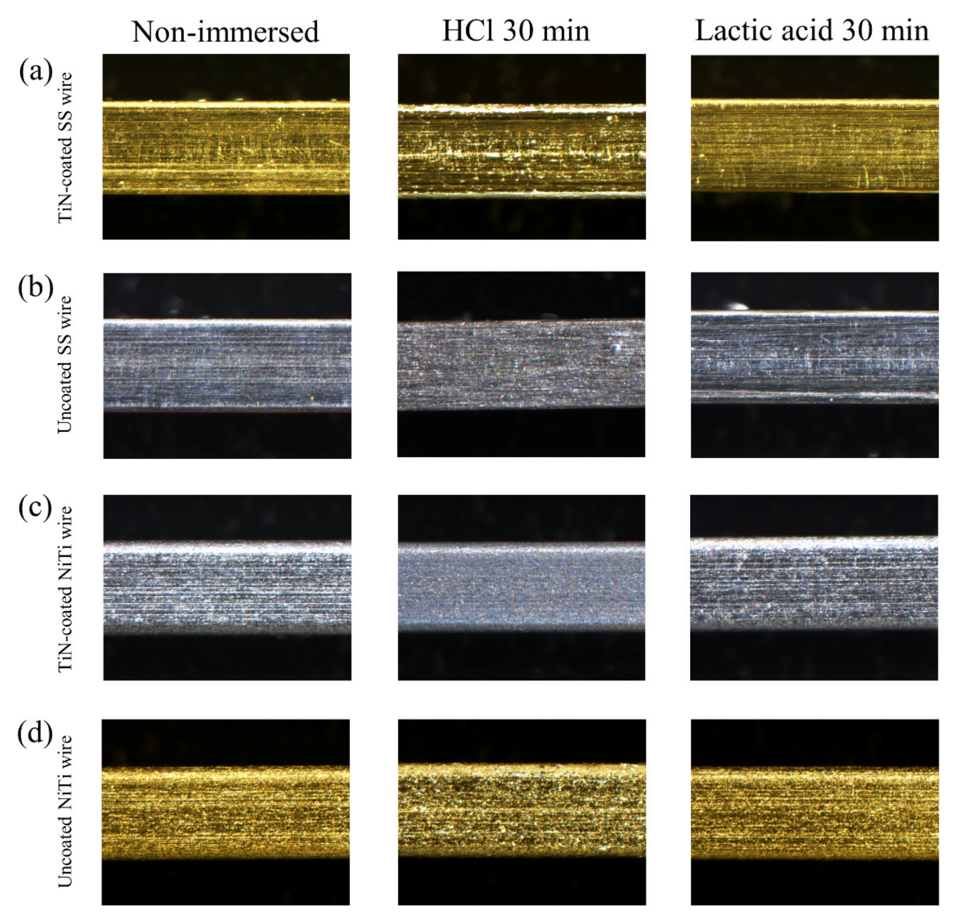
Figure 3 shows stereomicrographs of the surfaces of the wire samples. Corrosion of the surface was found on both the TiN-coated and uncoated SS and NiTi wire after immersion in HCl for 30 min. There were no differences between the non-immersed wire and wire immersed in lactic acid for 30 min.
SEM images of the wire surface are shown in Figure 4. At a magnification of 200×, corrosion was found on both the TiN-coated and uncoated SS and NiTi wires after immersion in HCl for 30 min. At a magnification of 1000×, the TiN-coated SS and NiTi wires demonstrated pitting corrosion, while the uncoated SS and NiTi wires showed uniform corrosion. There were no differences in surfaces between non-immersed wires or TiN-coated and uncoated wire immersed in lactic acid for 30 min.
3.4. Surface Roughness
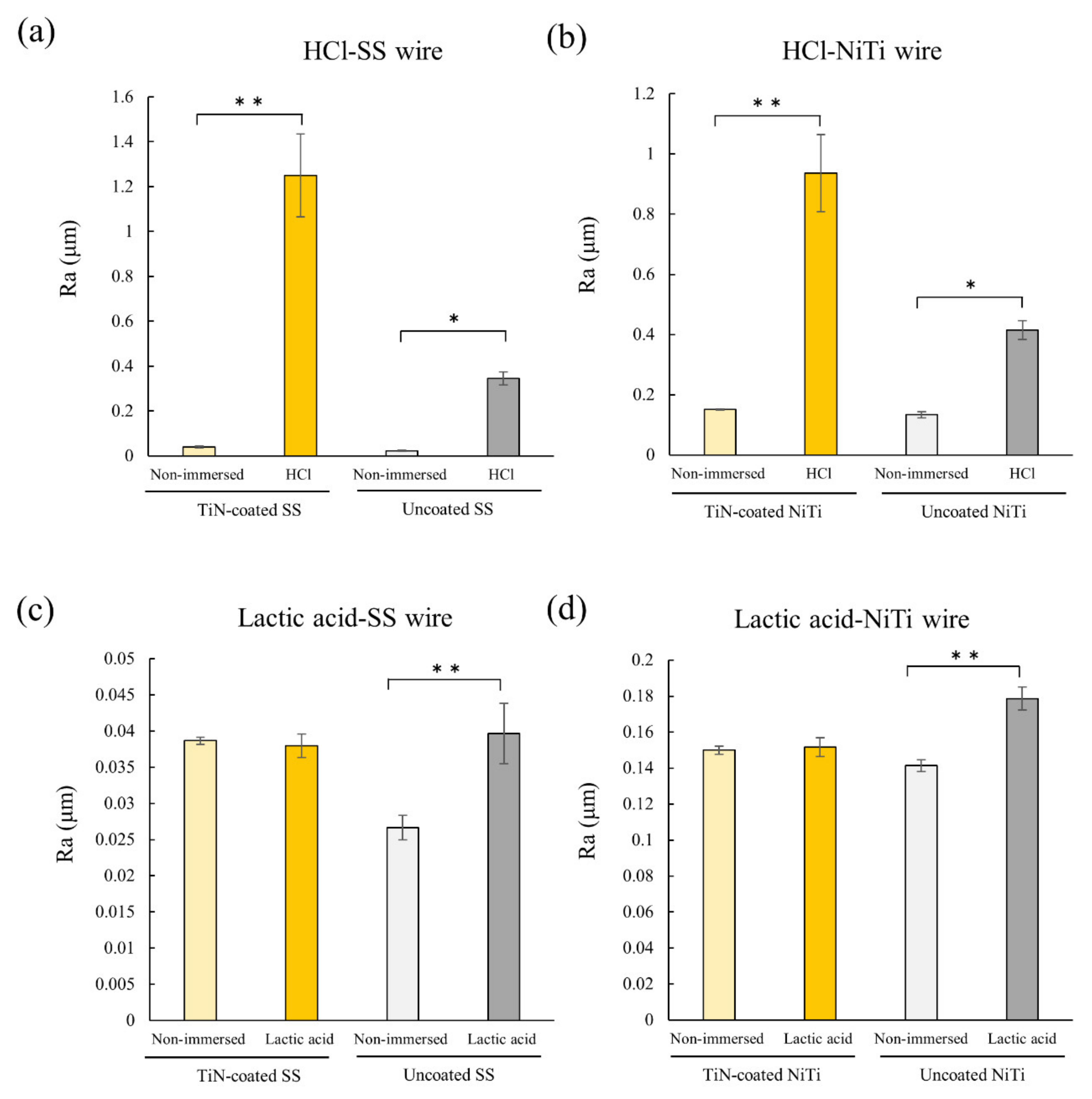
Both the uncoated and the TiN-coated SS and NiTi wires immersed in HCl for 30 min showed significantly increased surface roughness compared to the non-immersed wire (Figure 5). On the other hand, immersion in lactic acid for 30 min was associated with significantly increased surface roughness compared to non-immersed wire only for the uncoated SS and NiTi specimens.
4. Discussion
As the release of metal ions from orthodontic materials in the oral environment is closely related to their corrosion, metal surfaces are coated to improve corrosion resistance [13]. A number of plating techniques and materials are available for coating such metal components, and TiN ion plating was used in this study. The Ti of TiN components forms a passivation film, which improves the surface properties of the substrate material [25,26]. TiN ion plating results in little degradation of the mechanical properties of the substrate material because it is performed at low temperatures. Previously, we reported that TiN-coated orthodontic wire has a reduced friction coefficient, and the mechanical properties of the base material are maintained [17]. This study was performed to investigate whether the elution of metal ions can be suppressed by coating orthodontic wires with TiN film.
The ICP-MS data showed that the elution of metal ions was significantly suppressed in the TiN-coated wire compared to the uncoated wire after immersion in HCl for 30 min. Similarly, the DMG test confirmed that that the amount of Ni leached from both the SS and the NiTi wire after immersion in HCl were significantly lower for the TiN-coated than for the uncoated wire. The leaching of metal ions by HCl was suppressed by TiN coating. On the other hand, with immersion in lactic acid, Ni ions were leached from the uncoated but not the TiN-coated SS and NiTi wire, suggesting that the TiN-coated SS and NiTi wire was resistant to corrosion by lactic acid. Wire immersed in lactic acid showed no positive reaction on the DMG test, which may have been because the concentration of Ni was below the limit of detection of the test (10 μg/mL, 10 ppm) [27]. ICP-MS also showed that the amount of Ni ions eluted from the wire immersed in lactic acid was < 10 μg/mL, suggesting that it would be difficult to detect Ni elution associated with immersion in lactic acid on the DMG test. The amount of metal ions eluted from the TiN-coated SS and NiTi wire and the variations in the data were considered to be greater because of the high concentration of HCl used in this experiment. As the HCl derived from GERD has a lower concentration than that used in this study [23] and the immersion time in GERD-derived HCl is likely to be < 30 min in the oral cavity, the amount of elution is expected to be much smaller than in this experiment. However, as the threshold for Ni allergies is 1.5 μg [28], and there is a risk of onset of metal allergy when exposed to very small amounts of the metal, further experiments are required to examine the use of HCl at a concentration closer to that likely to be present in the oral cavity. Furthermore, the amount of Ni ion elution from uncoated SS wire immersed in lactic acid showed a high standard deviation. The immersion time of 30 min in lactic acid is considered to be the starting point of erosion for the SS wire used in this experiment. In some samples, the amount of Ni ion elution was small because dissolution had only just begun, and this may have resulted in the high degree of variation in the data.
The stereomicroscopy and SEM analyses showed the corrosion of all the wires following immersion in HCl, but the types of corrosion differed between the TiN-coated and uncoated wires. That is, pitting corrosion occurred in the TiN-coated SS and NiTi wire, while the uncoated SS and NiTi wire showed uniform corrosion. Such uniform corrosion is known to occur when metal is immersed in HCl [29], and this was confirmed in the present study. Pitting corrosion tends to occur in TiN with a coating thickness <6 μm [30]. In the present study, the TiN coating was 0.3 μm thick, which was intended to be used for orthodontic treatment, so the pitting corrosion may have occurred easily. Preliminary experiments showed that the peeling of TiN coating gradually became noticeable under the stereomicroscope when immersed in HCl for more than 30 min, so the immersion time was set to 30 min in this study.
The surface roughness of both the uncoated and the TiN-coated wire immersed in HCl was increased significantly compared to that of the non-immersed samples, suggesting that the wire surface was corroded by HCl, confirming the results of the microscopic analyses. Furthermore, the surface roughness of the TiN-coated wire was greater than that of the uncoated wire despite the reduced amount of metal ion elution. This may have been due to the difference in corrosion patterns between the TiN-coated and uncoated wires, as revealed by the stereomicroscopy and SEM analyses. The uniform corrosion of the uncoated wire was seen over its entire surface, but the pitting corrosion of TiN-coated wire induced by HCl in this study was randomly scattered, and the SEM images showed small fragments of coating material that seemed to have been peeled off around the pores, which may have caused an increase in the surface roughness. The surface roughness (Ra) measured in this study is a two-dimensional measurement, so a three-dimensional measurement of surface roughness (Sa) may be useful for further detailed analyses [31]. Microscopic examination indicated that the surface condition of the TiN-coated SS and NiTi wire was not altered by immersion in lactic acid and there was no change in the surface roughness, suggesting no corrosion by lactic acid. On the other hand, for the uncoated SS and NiTi wire, although microscopy revealed no changes in the surface condition, the surface roughness was increased by immersion in lactic acid, suggesting the possibility of corrosion by lactic acid.
Taken together, the results outlined above indicated that pitting corrosion occurred on the surface of TiN-coated SS and NiTi wire immersed in HCl, but that the corrosion did not spread over the entire surface of the wire, so the leaching of metal ions was significantly suppressed compared to the uncoated wire. The leaching of Ni ions from the TiN-coated SS and NiTi wire immersed in lactic acid was also shown to be significantly suppressed compared to the uncoated wire. These results indicated that TiN coating inhibits acid-induced corrosion and the elution of metal ions. GERD is associated with the regurgitation of gastric acid, mainly HCl, into the oral cavity. Thus, the results presented above suggest that the use of TiN-coated wires may be effective in preventing the leaching of metal ions from orthodontic appliances by HCl in gastric acid and reduce the rates of induction and exacerbation of metal allergy in patients with GERD. In addition, Lactobacillus acidophilus present in the oral cavity has been suggested to be related to the corrosion of metals mediated by lactic acid production [11]. The TiN-coated wire used in this study was resistant to corrosion by lactic acid, and is expected to be applicable to orthodontic treatment of patients with metal allergies.
We analyzed the acid corrosion resistance of TiN-coated wires in vitro. The following issues should be considered in future experiments. First, it is necessary to examine how long the TiN coating can be maintained in good condition in the oral cavity during orthodontic treatment. Second, the TiN coating is expensive, so cost reduction must be considered for clinical use. We plan to reduce the cost by coating as many wires as possible at once. Although TiN coating has excellent biocompatibility, as demonstrated previously in a device for atrial septal defect closure [14], it will be necessary to study its biocompatibility in the oral cavity, where it will be exposed to various chemical substances in food and drink, as well as saliva.
5. Conclusions
TiN plating of orthodontic wires can inhibit acid-mediated corrosion, and thus reduce the elution of Ni ions from the wire surface. These results suggest that TiN plating may be useful for the orthodontic treatment of patients with metal allergies. In addition, TiN plating has the potential for use in other dental applications, such as coating the metal parts of removable dentures and for wires to splint the teeth after orthodontic treatment.
Author Contributions
Conceptualization, A.I. and H.K.; methodology, A.I. and H.K.; validation, A.I. and H.K.; formal analysis, A.I.; investigation, A.I. and H.S.; resources, A.I. and H.K.; data curation, A.I.; writing—original draft preparation, A.I.; writing—review and editing, H.K.; visualization, A.I., T.N. and F.O.; supervision, H.K. and I.M.; project administration, H.K.; funding acquisition, A.I. All authors have read and agreed to the published version of the manuscript.
Funding
This work was supported in part by JSPS KAKENHI grants from the Japan Society for the Promotion of Science (No. K20K187740).
Informed Consent Statement
Not applicable.
Data Availability Statement
The data and analysis in this study are available on request from the corresponding author.
Conflicts of Interest
The authors declare there are no conflicts of interest.
Abbreviations
| TiN | titanium nitride |
| ICP-MS | inductively coupled plasma mass spectrometry |
| DMG | dimethylglyoxime |
| Ni | nickel |
| Co | cobalt |
| Cr | chromium |
| PVD | physical vapor deposition |
| SS | stainless steel |
| NiTi | nickel titanium |
| Ar | argon |
| N2 | nitrogen |
| HCl | hydrochloric acid |
| Mn | manganese |
| Fe | iron |
| SEM | scanning electron microscopy |
| GERD | gastroesophageal reflux disease |
References
- Mortz, C.G.; Bindslev-Jensen, C.; Andersen, K.E. Prevalence, incidence rates and persistence of contact allergy and allergic contact dermatitis in The Odense Adolescence Cohort Study: A 15-year follow-up. Br. J. Dermatol. 2013, 168, 318–325. [Google Scholar] [CrossRef]
- Thyssen, J.P.; Linneberg, A.; Menné, T.; Johansen, J.D. The epidemiology of contact allergy in the general population—Prevalence and main findings. Contact Dermat. 2007, 57, 287–299. [Google Scholar] [CrossRef]
- Kitaura, H.; Fujimura, Y.; Nakao, N.; Eguchi, T.; Yoshida, N. Treatment of a patient with metal hypersensitivity after orthognathic surgery. Angle Orthod. 2007, 77, 923–930. [Google Scholar] [CrossRef]
- Lindsten, R.; Kurol, J. Orthodontic appliances in relation to nickel hypersensitivity. A review. J. Orofac. Orthop. 1997, 58, 100–108. [Google Scholar] [PubMed]
- Noble, J.; Ahing, S.I.; Karaiskos, N.E.; Wiltshire, W.A. Nickel allergy and orthodontics, a review and report of two cases. Br. Dent. J. 2008, 204, 297–300. [Google Scholar] [CrossRef] [PubMed]
- Bajaj, A.K.; Saraswat, A.; Mukhija, G.; Rastogi, S.; Yadav, S. Patch testing experience with 1000 patients. Indian J. Dermatol. Venereol. Leprol. 2007, 73, 313–318. [Google Scholar] [CrossRef] [PubMed]
- Dotterud, L.K.; Smith-Sivertsen, T. Allergic contact sensitization in the general adult population: A population-based study from Northern Norway. Contact Dermat. 2007, 56, 10–15. [Google Scholar] [CrossRef]
- Adachi, N.; Kitaura, H.; Ikeda, M.; Kobayashi, K. Quantitative analysis of the surface elements of orthodontic metal materials using an X-ray fluorescence spectroscope. Orthod. Waves 2000, 59, 128–137. [Google Scholar]
- Nielsen, N.H.; Linneberg, A.; Menné, T.; Madsen, F.; Frølund, L.; Dirksen, A.; Jørgensen, T. Allergic contact sensitization in an adult Danish population: Two cross-sectional surveys eight years apart (the Copenhagen Allergy Study). Acta Derm. Venereol. 2001, 81, 31–34. [Google Scholar] [CrossRef]
- Dotterud, L.K. The prevalence of allergic contact sensitization in a general population in Tromsø, Norway. Int. J. Circumpolar Health 2007, 66, 328–334. [Google Scholar] [CrossRef] [Green Version]
- Chaturvedi, T.P.; Upadhayay, S.N. An overview of orthodontic material degradation in oral cavity. Indian J. Dent. Res. 2010, 21, 275–284. [Google Scholar] [CrossRef]
- Fors, R.; Persson, M. Nickel in dental plaque and saliva in patients with and without orthodontic appliances. Eur. J. Orthod. 2006, 28, 292–297. [Google Scholar] [CrossRef] [Green Version]
- Jabbari, Y.S.A.; Fehrman, J.; Barnes, A.C.; Zapf, A.M.; Zinelis, S.; Berzins, D.W. Titanium nitride and nitrogen ion implanted coated dental materials. Coatings 2012, 2, 160–178. [Google Scholar] [CrossRef]
- Zhang, Z.; Fu, B.; Zhang, D.Y.; Zhang, Z.; Cheng, Y.; Sheng, L.; Lai, C.; Xi, T. Safety and efficacy of nano lamellar TiN coatings on nitinol atrial septal defect occluders in vivo. Mater. Sci. Eng. C Mater. Biol. Appl. 2013, 33, 1355–1360. [Google Scholar] [CrossRef] [PubMed]
- Kim, H.; Kim, C.Y.; Kim, D.W.; Lee, I.S.; Lee, G.H.; Park, J.C.; Lee, S.J.; Lee, K.Y. Wear performance of self-mating contact pairs of TiN and TiAlN coatings on orthopedic grade Ti-6Al-4V. Biomed. Mater. 2010, 5, 044108. [Google Scholar] [CrossRef]
- Steele, J.G.; McCabe, J.F.; Barnes, I.E. Properties of a titanium nitride coating for dental instruments. J. Dent. 1991, 19, 226–229. [Google Scholar] [CrossRef]
- Sugisawa, H.; Kitaura, H.; Ueda, K.; Kimura, K.; Ishida, M.; Ochi, Y.; Kishikawa, A.; Ogawa, S.; Takano-Yamamoto, T. Corrosion resistance and mechanical properties of titanium nitride plating on orthodontic wires. Dent. Mater. J. 2018, 37, 286–292. [Google Scholar] [CrossRef] [PubMed] [Green Version]
- Smiech-Slomkowska, G.; Jablonska-Zrobek, J. The effect of oral health education on dental plaque development and the level of caries-related Streptococcus mutans and Lactobacillus spp. Eur. J. Orthod. 2007, 29, 157–160. [Google Scholar] [CrossRef] [Green Version]
- Kameda, T.; Oda, H.; Ohkuma, K.; Sano, N.; Batbayar, N.; Terashima, Y.; Sato, S.; Terada, K. Microbiologically influenced corrosion of orthodontic metallic appliances. Dent. Mater. J. 2014, 33, 187–195. [Google Scholar] [CrossRef] [Green Version]
- Dashper, S.G.; Reynolds, E.C. Lactic acid excretion by Streptococcus mutans. Microbiology 1996, 142, 33–39. [Google Scholar] [CrossRef] [PubMed] [Green Version]
- Dent, J.; El-Serag, H.; Wallander, M.A.; Johansson, S. Epidemiology of gastro-oesophageal reflux disease: A systematic review. Gut 2005, 54, 710–717. [Google Scholar] [CrossRef]
- Sulaiman, T.A.; Abdulmajeed, A.A.; Shahramian, K.; Hupa, L.; Donovan, T.E.; Vallittu, P.; Närhi, T.O. Impact of gastric acidic challenge on surface topography and optical properties of monolithic zirconia. Dent. Mater. 2015, 31, 1445–1452. [Google Scholar] [CrossRef] [PubMed]
- Elsaka, S.; Hassan, A.; Elnaghy, A. Effect of gastric acids on surface topography and bending properties of esthetic coated nickel-titanium orthodontic archwires. Clin. Oral Investig. 2021, 25, 1319–1326. [Google Scholar] [CrossRef] [PubMed]
- Nakao, N.; Kitaura, H.; Yoshida, N. Analysis of the release of nickel from orthodontic wires using the dimethylglyoxime spot test: In vitro and in vivo study. Orthod. Waves 2002, 61, 478–481. [Google Scholar]
- Iijima, M.; Endo, K.; Ohno, H.; Yonekura, Y.; Mizoguchi, I. Corrosion behavior and surface structure of orthodontic Ni-Ti alloy wires. Dent. Mater. J. 2001, 20, 103–113. [Google Scholar] [CrossRef] [PubMed]
- Lee, T.H.; Huang, T.K.; Lin, S.Y.; Chen, L.K.; Chou, M.Y.; Huang, H.H. Corrosion resistance of different nickel-titanium archwires in acidic fluoride-containing artificial saliva. Angle Orthod. 2010, 80, 547–553. [Google Scholar] [CrossRef] [PubMed] [Green Version]
- Rycroft, R.J.; Menné, T.; Frosch, P.J.; Lepoittevin, J.-P. Textbook of Contact Dermatitis; Springer: Berlin/Heidelberg, Germany, 2013. [Google Scholar]
- Emmett, E.A.; Risby, T.H.; Jiang, L.; Ng, S.K.; Feinman, S. Allergic contact dermatitis to nickel: Bioavailability from consumer products and provocation threshold. J. Am. Acad. Dermatol. 1988, 19, 314–322. [Google Scholar] [CrossRef]
- Tjahjanti, P.; Kurniawan, M.; Firdaus, R.; Iswanto, A. Experimental Study of Corrosion Rate on st60 Steel as a Result of Immersion in HCl, NaOH, and NaCl Solutions; IOP Conference Series: Materials Science and Engineering; IOP Publishing: Bristol, UK, 2020; p. 012038. [Google Scholar]
- Lang, F.; Yu, Z. The corrosion resistance and wear resistance of thick TiN coatings deposited by arc ion plating. Surf. Coat. Technol. 2001, 145, 80–87. [Google Scholar] [CrossRef]
- Ardila-Rodríguez, L.A.; Rans, C.; Poulis, J.A. Effect of surface morphology on the Ti-Ti adhesive bond performance of Ti6Al4V parts fabricated by selective laser melting. Int. J. Adhes. Adhes. 2021, 110, 102918. [Google Scholar] [CrossRef]
Figure 1.
Metal ions released from orthodontic arch wire immersed in different solutions. After immersion in HCl or lactic acid solution for 30 min, the amount of metal ions eluted from the wire was determined by ICP-MS. The concentrations of the eluted metal ions from TiN-coated SS wire and uncoated SS wire were: (a) SS immersed in HCl; (b) NiTi immersed in HCl; (c) SS immersed in lactic acid; (d) NiTi immersed in lactic acid. The statistical significance of differences was determined by the Shapiro-Wilk (* p < 0.05, ** p < 0.01).

Figure 2.
DMG test: (a) Positive reactions were scored; (b) SS wire immersed in HCl; (c) NiTi wire immersed in HCl. The statistical significance of differences was determined by the Tukey-Kramer HSD test. (* p < 0.05, ** p < 0.01).
Figure 2.
DMG test: (a) Positive reactions were scored; (b) SS wire immersed in HCl; (c) NiTi wire immersed in HCl. The statistical significance of differences was determined by the Tukey-Kramer HSD test. (* p < 0.05, ** p < 0.01).

Figure 3.
Stereomicrographs of orthodontic wire immersed in HCl or lactic acid for 30 min: (a) TiN-coated SS wire; (b) Uncoated SS wire; (c) TiN-coated NiTi wire; (d) Uncoated NiTi wire.
Figure 3.
Stereomicrographs of orthodontic wire immersed in HCl or lactic acid for 30 min: (a) TiN-coated SS wire; (b) Uncoated SS wire; (c) TiN-coated NiTi wire; (d) Uncoated NiTi wire.

Figure 4.
Scanning electron micrographs of orthodontic arch wire immersed in HCl or lactic acid for 30 min. (×200): (a) TiN-coated SS wire; (b) Uncoated SS wire; (c) TiN-coated NiTi wire; (d) Uncoated NiTi wire. (×1000): (e) TiN-coated SS wire; (f) Uncoated SS wire; (g) TiN-coated NiTi wire; (h) Uncoated NiTi wire.

Figure 5.
Comparison of surface roughness of orthodontic arch wires after immersion in HCl or lactic acid. After immersion in HCl or lactic acid for 30 min, the surface roughness (Ra) of orthodontic arch wires was assessed using a surface roughness tester: (a) SS wire immersed in HCl; (b) NiTi wire immersed in HCl; (c) SS wire immersed in lactic acid; (d) NiTi immersed in lactic acid. The statistical of differences was determined by the Tukey-Kramer HSD test. (* p < 0.05, ** p < 0.01).


Table 1.
Metal ion release from SS wire (μg/L).
| Wire | Solution | Immersion Time (min) | Cr | Mn | Fe | Ni | ||||
|---|---|---|---|---|---|---|---|---|---|---|
| Mean | SD | Mean | SD | Mean | SD | Mean | SD | |||
| TiN-coated SS wire | Sterile water | 0 | 4.78 | 6.83 | 0.76 | 0.54 | 49.26 | 52.90 | 0.43 | 0.28 |
| 30 | 3.08 | 2.85 | 0.51 | 0.31 | 22.07 | 17.00 | 0.48 | 0.26 | ||
| Physiological saline | 0 | 2.38 | 2.48 | 1.01 | 0.31 | 16.61 | 12.25 | 1.06 | 0.87 | |
| 30 | 2.21 | 0.38 | 2.54 | 1.67 | 19.33 | 15.39 | 11.82 | 12.23 | ||
| Hydrochloric acid | 0 | 5.22 | 1.32 | 1.08 | 0.35 | 70.09 | 30.67 | 8.03 | 11.02 | |
| 30 | 24,625.05 | 7557.31 | 2070.16 | 725.35 | 81,189.98 | 25,124.15 | 15,839.44 | 5752.39 | ||
| Lactic acid | 0 | 32.15 | 0.48 | 0.83 | 0.32 | 71.60 | 1.29 | 1.34 | 0.07 | |
| 30 | 31.74 | 0.51 | 0.64 | 0.10 | 77.52 | 4.71 | 1.55 | 0.10 | ||
| Uncoated SS wire | Sterile water | 0 | 4.78 | 6.83 | 0.76 | 0.54 | 49.26 | 52.90 | 0.43 | 0.28 |
| 30 | 3.29 | 2.27 | 0.86 | 0.40 | 33.03 | 14.57 | 2.25 | 1.72 | ||
| Physiological saline | 0 | 2.38 | 2.48 | 1.01 | 0.31 | 16.61 | 12.25 | 1.06 | 0.87 | |
| 30 | 3.89 | 2.72 | 1.52 | 0.24 | 22.22 | 11.94 | 17.36 | 22.61 | ||
| Hydrochloric acid | 0 | 5.22 | 1.32 | 1.08 | 0.35 | 70.09 | 30.67 | 8.03 | 11.02 | |
| 30 | 171,051.75 | 41,866.15 | 15,044.11 | 2118.26 | 653,585.22 | 116,232.28 | 82,007.05 | 15,393.43 | ||
| Lactic acid | 0 | 32.15 | 0.48 | 0.83 | 0.32 | 71.60 | 1.29 | 1.34 | 0.07 | |
| 30 | 32.33 | 0.58 | 0.77 | 0.20 | 81.63 | 2.24 | 4.14 | 3.49 | ||
Table 2.
Metal ion release from NiTi wire (μg/L).
| Wire | Solution | Immersion Time (min) | Ni | Ti | ||
|---|---|---|---|---|---|---|
| Mean | SD | Mean | SD | |||
| TiN-coated SS wire | Sterile water | 0 | 0.43 | 0.28 | 1.82 | 0.99 |
| 30 | 1.35 | 0.95 | 2.57 | 2.11 | ||
| Physiological saline | 0 | 1.06 | 0.87 | 1.76 | 0.77 | |
| 30 | 4.16 | 2.05 | 7.29 | 10.30 | ||
| Hydrochloric acid | 0 | 8.03 | 11.02 | 13.01 | 10.59 | |
| 30 | 13,536.28 | 10,012.85 | 12,005.95 | 9871.59 | ||
| Lactic acid | 0 | 1.34 | 0.07 | 24.19 | 0.27 | |
| 30 | 1.44 | 0.03 | 24.56 | 0.33 | ||
| Uncoated SS wire | Sterile water | 0 | 0.43 | 0.28 | 1.82 | 0.99 |
| 30 | 2.38 | 1.47 | 2.18 | 2.43 | ||
| Physiological saline | 0 | 1.06 | 0.87 | 1.76 | 0.77 | |
| 30 | 3.37 | 0.91 | 1.17 | 0.29 | ||
| Hydrochloric acid | 0 | 8.03 | 11.02 | 13.01 | 10.59 | |
| 30 | 215,499.84 | 64,463.60 | 173,761.82 | 73,627.09 | ||
| Lactic acid | 0 | 1.34 | 0.07 | 24.19 | 0.27 | |
| 30 | 2.29 | 0.19 | 25.37 | 0.36 | ||
Table 3.
DMG test results for SS wire.
| Wire | Solution | Immersion Time (min) | Mean | SD |
|---|---|---|---|---|
| TiN-coated SS wire | Sterile water | 0 | 0.00 | 0.00 |
| 30 | 0.00 | 0.00 | ||
| Physiological saline | 0 | 0.00 | 0.00 | |
| 30 | 0.00 | 0.00 | ||
| Hydrochloric acid | 0 | 0.00 | 0.00 | |
| 30 | 1.00 | 0.00 | ||
| Lactic acid | 0 | 0.00 | 0.00 | |
| 30 | 0.00 | 0.00 | ||
| Uncoated SS wire | Sterile water | 0 | 0.00 | 0.00 |
| 30 | 0.00 | 0.00 | ||
| Physiological saline | 0 | 0.00 | 0.00 | |
| 30 | 0.00 | 0.00 | ||
| Hydrochloric acid | 0 | 0.00 | 0.00 | |
| 30 | 2.67 | 0.47 | ||
| Lactic acid | 0 | 0.00 | 0.00 | |
| 30 | 0.00 | 0.00 |
Table 4.
DMG test results for NiTi wire.
| Wire | Solution | Immersion Time (min) | Mean | SD |
|---|---|---|---|---|
| TiN-coated NiTi wire | Sterile water | 0 | 0.00 | 0.00 |
| 30 | 0.00 | 0.00 | ||
| Physiological saline | 0 | 0.00 | 0.00 | |
| 30 | 0.00 | 0.00 | ||
| Hydrochloric acid | 0 | 0.00 | 0.00 | |
| 30 | 1.00 | 0.00 | ||
| Lactic acid | 0 | 0.00 | 0.00 | |
| 30 | 0.00 | 0.00 | ||
| Uncoated NiTi wire | Sterile water | 0 | 0.00 | 0.00 |
| 30 | 0.00 | 0.00 | ||
| Physiological saline | 0 | 0.00 | 0.00 | |
| 30 | 0.00 | 0.00 | ||
| Hydrochloric acid | 0 | 0.00 | 0.00 | |
| 30 | 2.67 | 0.47 | ||
| Lactic acid | 0 | 0.00 | 0.00 | |
| 30 | 0.00 | 0.00 |
Publisher’s Note: MDPI stays neutral with regard to jurisdictional claims in published maps and institutional affiliations. |
© 2021 by the authors. Licensee MDPI, Basel, Switzerland. This article is an open access article distributed under the terms and conditions of the Creative Commons Attribution (CC BY) license (https://creativecommons.org/licenses/by/4.0/).
Share and Cite
MDPI and ACS Style
Ito, A.; Kitaura, H.; Sugisawa, H.; Noguchi, T.; Ohori, F.; Mizoguchi, I. Titanium Nitride Plating Reduces Nickel Ion Release from Orthodontic Wire. Appl. Sci. 2021, 11, 9745. https://0-doi-org.brum.beds.ac.uk/10.3390/app11209745
AMA Style
Ito A, Kitaura H, Sugisawa H, Noguchi T, Ohori F, Mizoguchi I. Titanium Nitride Plating Reduces Nickel Ion Release from Orthodontic Wire. Applied Sciences. 2021; 11(20):9745. https://0-doi-org.brum.beds.ac.uk/10.3390/app11209745
Chicago/Turabian StyleIto, Arata, Hideki Kitaura, Haruki Sugisawa, Takahiro Noguchi, Fumitoshi Ohori, and Itaru Mizoguchi. 2021. "Titanium Nitride Plating Reduces Nickel Ion Release from Orthodontic Wire" Applied Sciences 11, no. 20: 9745. https://0-doi-org.brum.beds.ac.uk/10.3390/app11209745
Note that from the first issue of 2016, this journal uses article numbers instead of page numbers. See further details here.