Article

Retrospective Case Series Regarding the Advantages of Cortico-Puncture (CP) Therapy in Association with Micro-Implant Assisted Rapid Palatal Expander (MARPE)

1
Faculty of Dental Medicine, George Emil Palade University of Medicine, Pharmacy, Science and Technology of Târgu Mureș, 540142 Târgu Mureș, Romania
2
Dudley Group of Hospitals NHS Foundation Trust, Birmingham B18 7QH, UK
*
Author to whom correspondence should be addressed.
Submission received: 27 December 2020 / Revised: 14 January 2021 / Accepted: 27 January 2021 / Published: 1 February 2021
(This article belongs to the Special Issue New Materials and Technologies in Orthodontics)

Abstract

:
Transverse maxillary deficiency currently affects 8–23% of adults. One of the most widely used orthodontic treatments today in patients with transverse maxillary defects is the maxillary skeletal expander (MSE). This was a retrospective observational imaging study regarding structural bone changes that may occur during healing after the placement of micro-implant assisted rapid palatal expanders (MARPE) in combination with cortico-puncture (CP) therapy. Regarding the magnitude of the mid-palatal suture opening, the mean split at the anterior nasal spine (ANS) and the posterior nasal spine (PNS) was 3.76 and 3.12 mm, respectively. The amount of split at the PNS was smaller than at the ANS, approximately 85% of the distance, showing that the opening of the midpalatal suture was almost parallel in the sagittal plane. On average, one-half of the anterior nasal spine (ANS) moved more than the contralateral by 0.89 mm. In the present study, we show that MARPE associated with CP therapy had a positive outcome on the midpalatal suture opening. This occurred in safe conditions, without post-surgery bleeding, and showing healing at the corticotomy level, with no signs of swelling or sepsis, which are side effects usually associated with more complex surgical treatments. Our results suggest that non-surgical palatal expansion, assisted by MARPE and CP, is achievable and predictable in young adults.

1. Introduction

Transverse maxillary deficiency currently affects 8–23% of adults and adolescents [1]. One of the most widely used orthodontic treatments today in patients with transverse maxillary defects is the maxillary skeletal expander (MSE) [2]. In clinical practice, micro-implants were first introduced in the palatal region 25 years ago. The possibility of using these micro-implants at the level of the palatal region is favored by the gingival mucosa at this level having favorable elasticity [3]. With the use of micro-implants associated with maxillary expanders, the incidence of post-interventional complications, such as tooth-borne forces leading to limited skeletal movement and the potential for undesirable tooth movement, root resorption, and lack of firm anchorage to retain sutural long-term expansion, has been reduced [4,5]. The reason is that the micro-implants play the role of the anchoring system for the maxillary expander [6,7]. Transverse jaw deficiency is a common condition in young adults. Untreated or treated incorrectly, it can affect the health of these patients over time. In this sense, modifications of the occlusal plane, damage to periodontal structures, gingival retraction, change in tongue position, asymmetry of the facial planes, and, in severe cases, even sleep apnea syndrome may occur in these patients. Given these possible co-morbidities, orthodontic treatment is particularly important in these cases and is required in clinical practice [8,9].
To obtain skeletal changes with micro-implant assisted rapid palatal expanders (MARPEs), the applied force should be enough to overcome areas of resistance located in the midface region such as the mid-palatal suture (the first that needs to be disrupted), pterygoid junctions, piriform aperture pillars, and zygomatic buttresses [10]. For this reason, using a MARPE device, force is applied directly into the center of resistance of the maxilla using micro-implants and not the tooth as in classical tooth palatal expanders. As a result of this applied force, buccal tipping of the tooth is prevented, and a more parallel suture opening is promoted [11].
Cortico-puncture (micro-perforation) was introduced in clinical practice as a surgical procedure to shorten orthodontic treatment time. It removes the cortical bone that resists orthodontic force in the jaw and keeps the blood circulation and continuity of bone tissues to reduce the risk of necrosis and facilitate tooth movement [11].
In the present study, we evaluated the treatment outcomes of adult patients with maxillary compression who underwent orthodontic cortico-puncture CP surgery in association with MARPE.

2. Materials and Method

In this retrospective study, we assessed the structural changes that occurred during healing after the placement of a micro-implant assisted rapid palatal expander MARPE in combination with cortico-puncture therapy (CP) (Figure 1). Prior to the treatment, cone-beam computed tomography (CBCT) was used to determine bone anatomy and density, palatal suture maturation, and anatomical structures that needed to be avoided during CP therapy. For image acquisition, we used a ProMax 3D CBCT unit (Planmeca, Finland). Images were acquired and saved in JPEG format.
The inclusion criteria were as follows:
-
Patients without craniofacial abnormalities;
-
No previous orthodontic treatment;
-
Transverse maxillary deficiency;
-
Unilateral or bilateral crossbite.
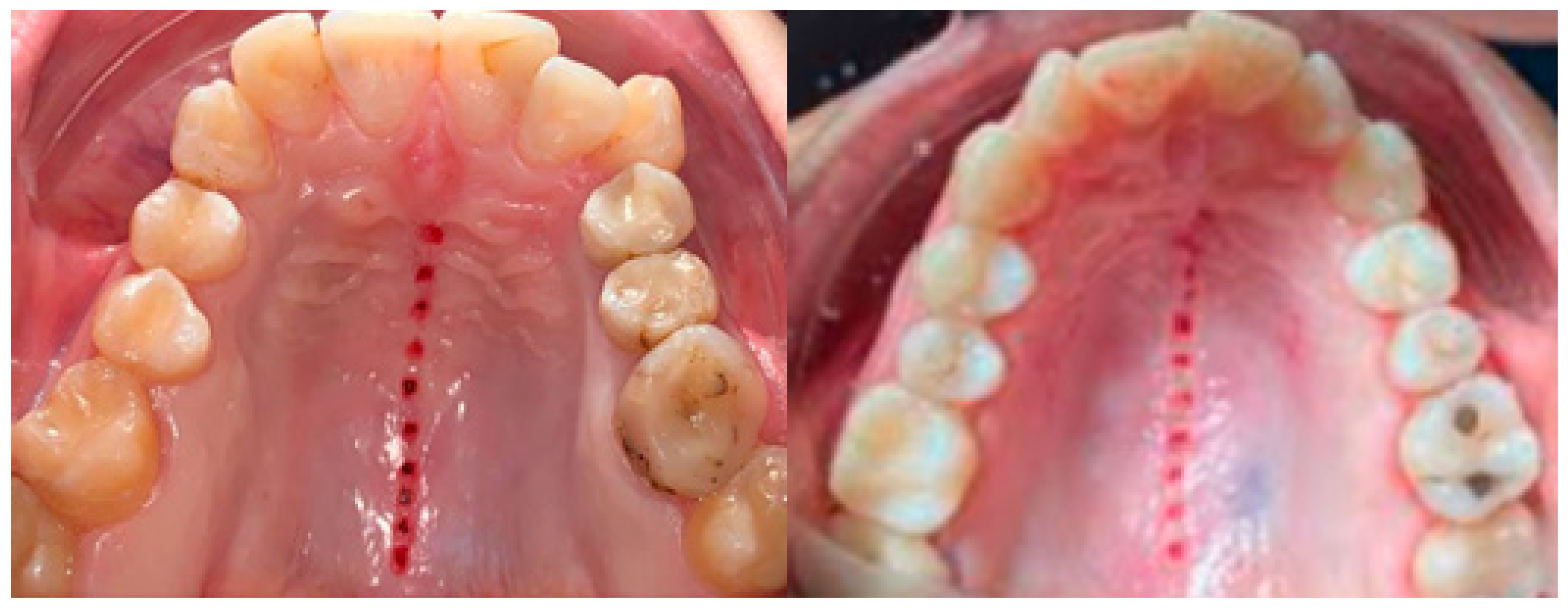
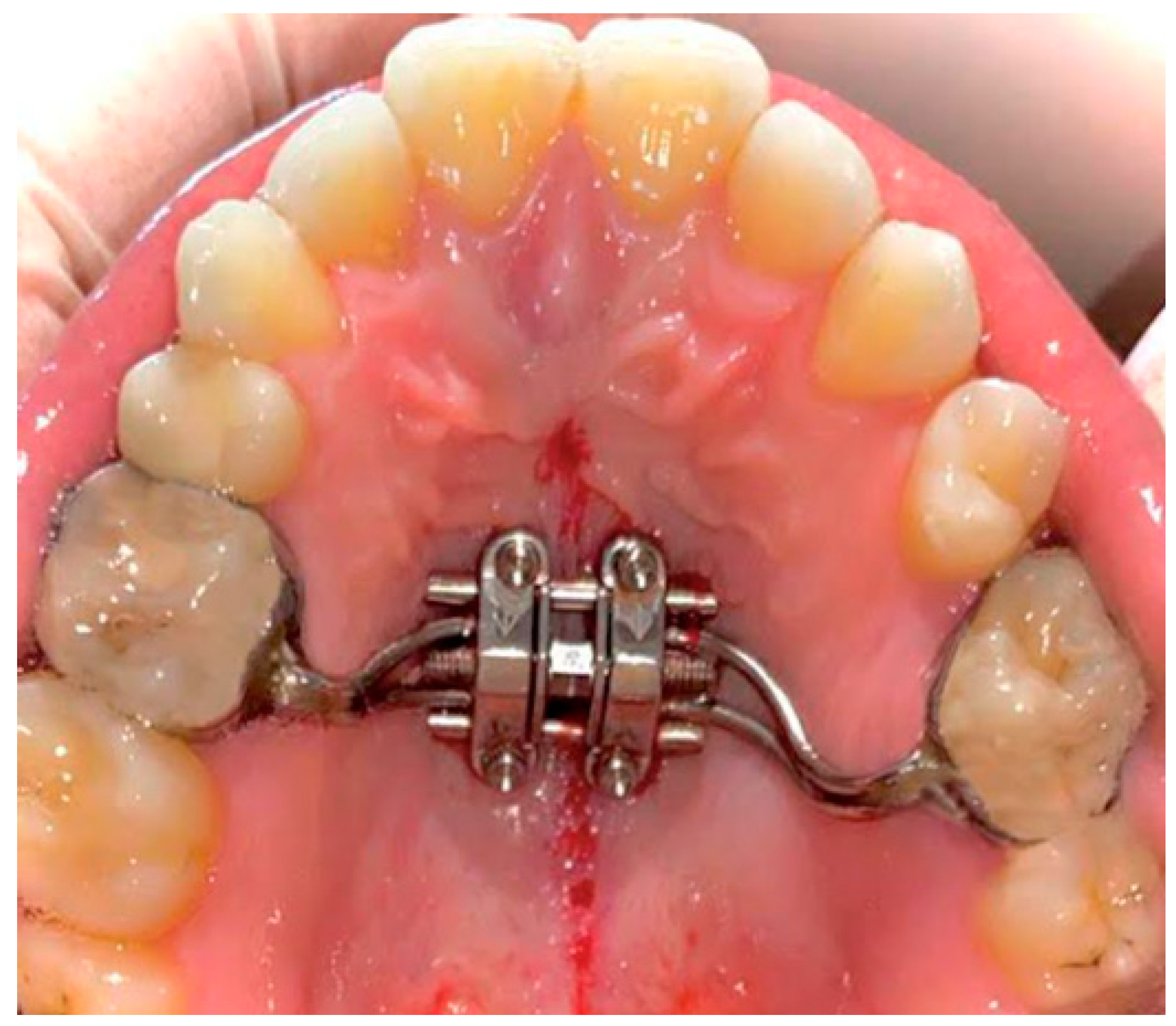
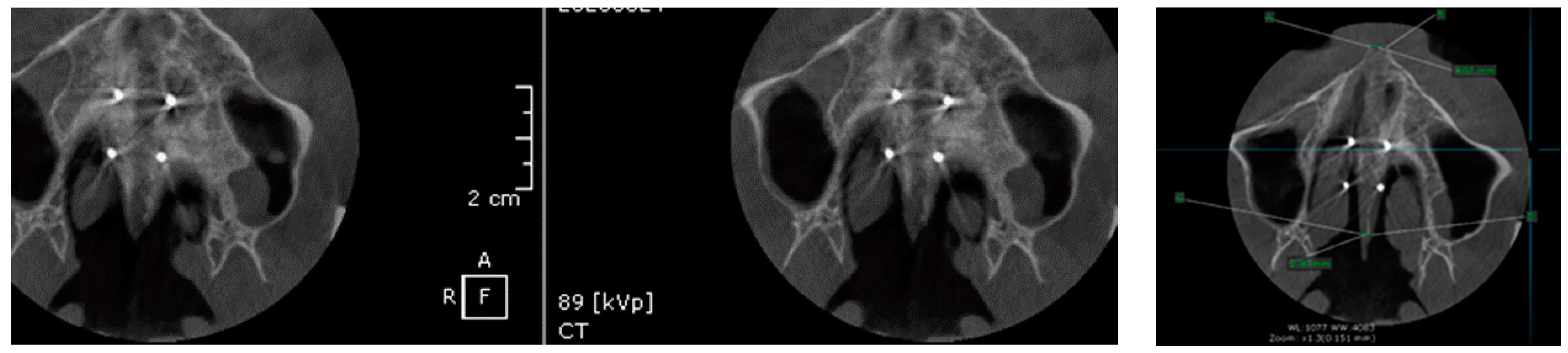
Included in the study group were twenty patients aged between 21 and 35 years (mean age 23.8 years). Thirteen were female and seven were male.
With the aid of CBCT, patients were confirmed with stage E sutural fusion as classified by Angelieri et al. [8]. The mid-palatal suture could not be identified, and the parasutural bone density was the same as in other regions of the palate (Figure 2).
All procedures performed were in accordance with the ethical standards of the Responsible Committee on Human Experimentation (institutional and national) and with the Helsinki Declaration of 1975, as revised in 2008. Informed consent was obtained from all patients for inclusion in the study. The study was approved by the ethics committee of Algocalm Private Medical Center of Târgu Mureș, Romania. (892/5 February 2020).

Surgical Protocol

All subjects were treated under the following surgical and orthodontic protocol: Local anesthesia was achieved by applying topical Lidocaine™ Septodont (Creteil, France) 2% spray applied for 1 min, followed by buccal infiltration of the hard palate of a solution of articaine hydrochloride + epinephrine 1:100,000 (ARTICAINE™ Septodont, Creteil, France), administered with the aid of a 0.30 × 38 mm gingival needle (Heraeus™, Hanau, Germany).
Cortico-puncture therapy consisted of the following steps: Ten bone perforations (cortico-punctures) were manually performed along the mid-palatal suture with the aid of a round bur 1.8 mm in diameter and a pilot drill, MIS Implant System™ (Haifa, Israel). Perforations were performed approximately 2 mm apart at a depth ranging from 2–5 mm depending on the thickness of the cortical plate, determined to be safe upon CBCT examination before surgical treatment. After the surgical procedure, analgesic medication (ibuprofen 400 mg 3 × 1/day) was prescribed, and the use of 0.12% chlorhexidine oral rinse for 10–14 days was recommended.
Mini-implant placement consisted of the following steps: After the maxillary skeletal expander II (BioMaterials™, Seoul, Korea) was cemented (Figure 3) in the patient’s oral cavity with bands around 1st molars, 4 orthodontic mini-implants (BioMaterials™, Seoul, Korea) (1.8 × 11 mm) were inserted into the palatal bone using the appliance slots as surgical guides. The MSE II was positioned slightly anterior to the soft palate between the first and second molars to direct the expansion forces against the buttress bones.
CBCT images were recorded using a tube voltage of 89 kV and a current intensity of 6 mA, using a cylindrical field of view (FOV) of 82 mm both in diameter and height (Figure 4), the maxilla being our region of interest. The voxel size was 0.2 × 0.2 × 0.2 mm, and the dental arches were positioned similarly in the FOV as presented in Figure 4 and Figure 5.
For image acquisition, we used a ProMax 3D CBCT unit (Planmeca, Finland) with the previously mentioned settings. Images were acquired and saved in JPEG format. To locate the cortico-puncture sites, bone modification, and palatal suture split, we used OnDemand 3Ddata App™ software (Seoul, Korea). At each site, our region of interest was a square volume of bone located within the median palatal area (Figure 4). All data recorded were saved using Microsoft Office Excel™, 2017 version, analysis software.
After the surgical procedure, patients were instructed on how to perform the second activation protocol for MSE II: minimum 4–6 turns/day (0.53–0.80 mm/day) until a diastema between central incisors was observed (after 10–19 days), indicating success in splitting the mid-palatal suture (Figure 6), and after diastema appeared: 2 turns/day (0.27 mm/day) until crossbite overcorrection had been achieved. After the expansion, the MSE remained inactivated for at least 2 months to stabilize the expansion.
On average, 2 months after the palatal split, new CBCT examinations were performed to assess changes that might have occurred at the skeletal level, such as the dimensions of the anterior nasal spine (ANS) split and posterior nasal spine (PNS) split, interincisal distance split at the cemental-enamel junction (CEJ), and first molar inclination after treatment (Figure 6, Figure 7, Figure 8, Figure 9, Figure 10 and Figure 11).
To determine the direction of movement that has occurred at the first molar position, a parallel line to the long axis of the tooth was drawn, and the angle between this line and the horizontal plane was measured (Figure 12).

3. Statistical Analysis

Statistical processing was performed using GraphPad Prism™ V6.01 software for Windows™. Statistical analysis involved the use of the Student’s t-test for unpaired (independent) and paired (dependent) data. The D’Agostino and Pearson omnibus normality test was used to determine the normality of the data. The chosen significance threshold was alpha = 0.05, considering p significant when p < 0.05. A non-parametric test was also chosen because the pre-expansion values of all considered parameters were equal to zero (non-normal distribution).

4. Results

Regarding the magnitude of the midpalatal suture opening, the mean split at the anterior nasal spine (ANS) and the posterior nasal spine (PNS) was 3.76 and 3.12 mm, respectively (Table 1). The magnitude of the split at the PNS was smaller than at the ANS by approximately 85% of the distance (Table 2), showing that the opening of the midpalatal suture was almost parallel in the anteroposterior direction. On average, one-half of the anterior nasal spine (ANS) moved more than the contralateral one by 0.89 mm, which was statistically significant (p < 0.05).
Regarding the magnitude of interincisal distance at the cemento-enamel junction level, the mean space obtained was 4.10 mm (Table 3) ± 0.38 standard deviation (p < 0.05).
The correlation analysis between first molar tilt, before and after treatment, is given in Table 4. The mean inclination of the first molar was 2.005° SD 0.72° (p < 0.001) after treatment, showing that tooth movement also occurred after the splitting of the palatal suture.

5. Discussion

As described in the literature, tooth-borne expanders produce a V-shaped opening of the midpalatal suture, with the greatest opening anteriorly and progressively less separation toward its posterior part [12,13,14,15]. Lione et al. [14] used a tooth-borne maxillary expander activated by 7 mm on all patients and found that the opening of the midpalatal suture was 3.01 and 1.15 mm at the ANS and PNS, respectively. Conversely, in the patients treated with MARPE in our present study, the borders of the midpalatal suture moved almost perfectly parallel to each other, as evidenced by the amount of split at the PNS, which was 85% of that at the ANS.
Although some authors recommended classic orthodontic treatment in young adult patients with transverse maxillary deficiencies, the failure rate in such cases can be high [16]. Performing an initial cone-beam computed tomography (CBCT) examination is crucial in limiting therapeutic failure in patients undergoing MARPE. The advantage of this method is that it provides important details about the local anatomy, the thickness of the palatine bone, and anatomical details about the articulation of the palatine bone with the pterygoid process to the sphenoid bone. This allows proper planning of this orthodontic treatment. Knowledge of local anatomical details plays a particularly important role in these cases [17,18].
Analyzing the transverse asymmetry of the mid-palatal split (Table 1), on average, one-half of the ANS moved more than the contralateral one by 0.89 mm. The reason for this is unclear but could be related to external forces, such as the presence of a unilateral crossbite that hampers the movement of one maxilla.
Lin et al. [19] published a comparative study between tooth-borne and bone-borne MARPE on late adolescents. The MARPE used in their study included four micro-implants embedded in two acrylic shelves supporting the jackscrew. All implants were positioned close to the dentition, inferiorly from the mid-palatal suture, but the appliance did not contact the dentition. Angular measurements were performed to assess alveolar bone bending and dental tipping using an arbitrary palatal plane. Significant alveolar bone bending and dental tipping even with this bone-borne expander treatment were found. The dentoalveolar changes may have been possible because of the force applied to the dentoalveolar region by this appliance; however, it is difficult to accept that dental movement could occur even though the expander did not have any physical contact with the dentition. This implies that angular measurements from arbitrary points cannot accurately assess the true impact of an appliance.
The activation force of a rapid maxillary expander (RME) device initially results in the compression of the periodontal ligament, bending of the alveolar bone, and tipping of the anchored teeth [20]. Therefore, a 1–24° increase in molar inclination is inevitable, probably because of alveolar bending and/or tipping of the posterior teeth [21]. Winsauer et al. [22] showed this in a study of 33 adults aged between 23 and 33 years, where 90% of the patients had successful palatal widening without a surgically assisted rapid palatal expander (SARPE) and no dental side effects [22].
Moon et al. [23] conducted a study on 48 late-adolescent patients divided into two groups according to the type of expander: MSE group (n = 24, age = 19.2 ± 5.9 years) and bone-borne expanders group (n = 24, age = 18.1 ± 4.5 years). CBCT scans were taken before treatment and 3 months after expansion. Transverse skeletal and dental expansion, alveolar inclination, tooth axis, buccal alveolar bone height, thickness, dehiscence, and fenestration were evaluated on the maxillary first molar. They concluded that the incorporation of teeth into bone-borne expanders resulted in an increase in the severity of side effects. For patients in late adolescence, tissue bone-borne expanders offer comparable skeletal effects to tooth bone-borne expanders, with fewer dentoalveolar side effects [23].
In a randomized control trial regarding the use of bone-borne expansion in the adolescent population, Celenk-Koca et al. [24] showed an increased extent of skeletal changes in the range of 1.5–2.8 times that of tooth-borne expansion and did not result in any dental side effects [24]. In another study of late-adolescent patients, Liu et al. showed that bone-borne expanders produced greater transverse skeletal expansion than tooth-borne Hyrax expanders. In the bone-borne expander group, there was less alveolar bending, less dental tipping, and less vertical alveolar bone loss at the first premolar [19].
Regarding the limitations of MARPE devices, Garib et al. reported that MARPE therapy requires a longer activation time and twice the force for the rupture of the medial palatine suture compared to SARPE [25]. Other disadvantages of MARPE: it may cause temporary inflammation of the palatal mucosa [26], difficulty in hygiene around micro-implants, and risk of infection [1]. Regarding other limitations, Choi et al. named the possibility of failure to separate the suture due to the resistance of the craniofacial structures [27] and when a patient presents a narrow and deep palate because the proper position of some MARPE devices cannot be achieved, since it should be placed close to the palatal mucosa [28]. In such cases, surgically assisted rapid palatal expanders (SARPE) is often suggested to these patients. This surgical procedure increases expansion predictability and success and reduces its side effects. One of the available SARPE techniques consists of a LeFort I osteotomy, associated with surgical rupture of the mid-palatal suture, which decreases mechanical resistance to the lateral forces that will be applied by Hyrax expanders, usually anchored to the first molars and first premolars. However, despite its benefits, SARPE requires hospitalization and increases the biological and financial costs of the treatment [16].
To minimize the surgical procedure and reduce postoperative discomfort, other techniques may be recommended. Cortico-puncture is a method that increases the expression of cytokines and chemokines responsible for stimulating the differentiation of osteoclasts in bone remodeling and thus increasing the rate of tooth movement by up to 62% [23].
Our study demonstrates the use of CP therapy as a surgical method for accelerating bone remodeling to complement the MARPE technique to facilitate suture split. In their study on maxillary expansion in rabbits, Pulver et al. suggested that greater skeletal expansions may be possible when combined with surgical methods such as cortico-puncture to promote regional acceleratory phenomenon (RAP), stimulating bone remodeling, and reducing bone volume and density [29]. In animal studies, Tsai et al. [30] compared the effects of corticotomy and bone microperforations and concluded that both techniques increased bone remodeling, and there were no significant differences between them. Suzuki et al. [31] demonstrated that when performed along the midpalatal suture, a minimally invasive surgical method such as CP accelerated bone remodeling and favored suture split when this failed to occur after the conventional protocol for MARPE activation [31].
Regarding stability after splitting in literature, Choi et al. [27], using MARPE, found an 86.96% success rate in young adult patients (mean age = 20.9 ± 2.9 years), with stable results after 30 months of follow-up.
For the treatment of transverse maxillary deficiency in adults, Hassan et al. reported that assisted expansion with corticotomy, defined as decortication on the buccal and palatal walls of the alveolar bone, is an effective technique and suggested that the technique may provide greater stability and better periodontal health than conventional expansion. However, the same study reported that there might be side effects of the corticotomy method, such as mild bone loss and loss of inserted gingiva [32]. Studies recommend the use of bone grafts to conserve the periodontium to avoid this [33]. In addition, subcutaneous hematomas and postoperative swelling and discomfort were also associated with the corticotomy procedure [32].
Our study sample comprised only 20 young adults and has a high risk of bias, which emphasized the necessity of further prospective studies involving larger numbers of patients and longer-term evaluation of stability and periodontal adaptation after MARPE.

6. Conclusions

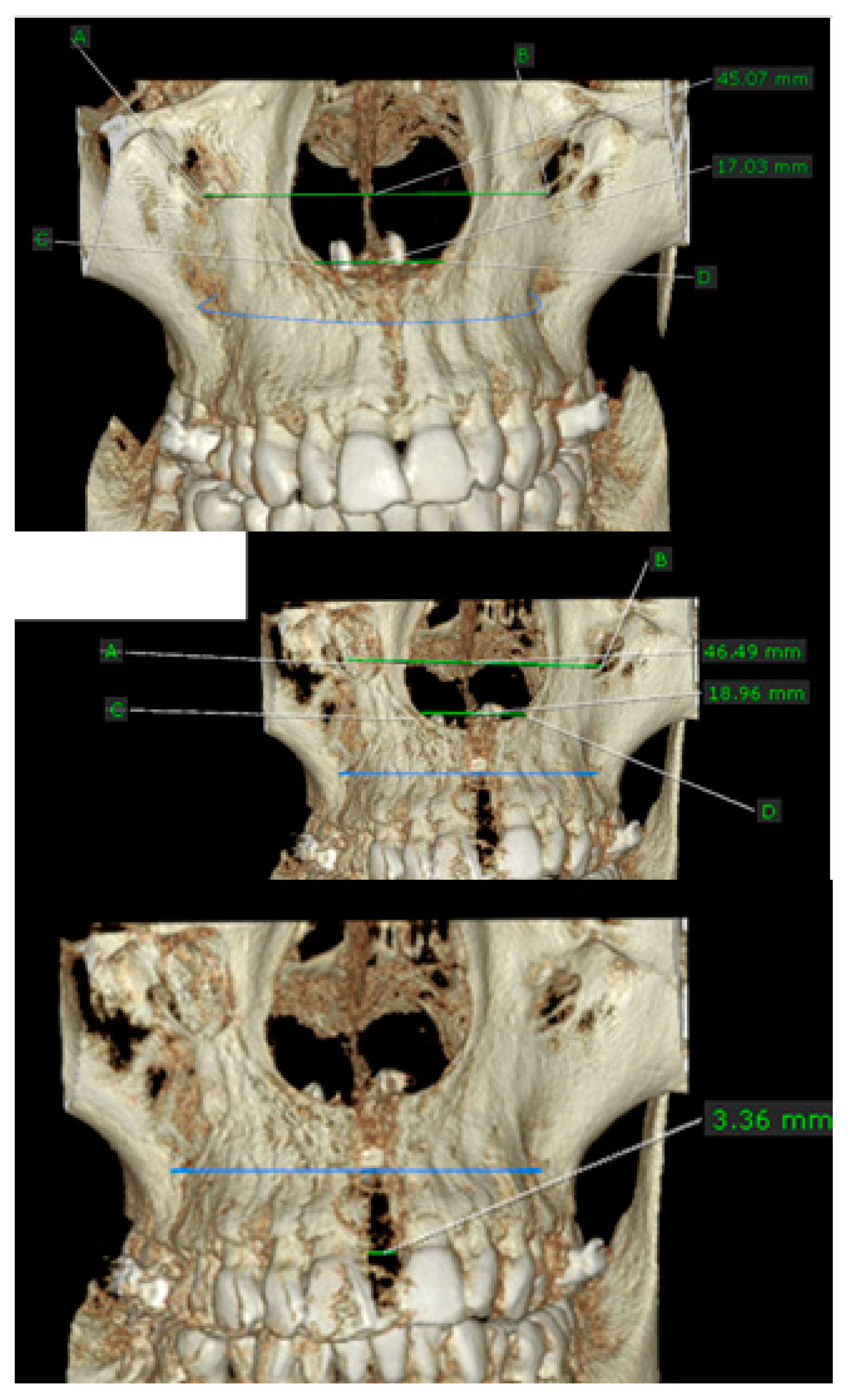
  • MARPE associated with cortico-puncture therapy efficiently split the midpalatal suture in adults. The mean split at the anterior nasal spine (ANS) and posterior nasal spine (PNS) was 3.76 and 3.12 mm, respectively. The magnitude of the split at the PNS was smaller than at the ANS (by approximately 85% of the distance), showing that the opening of the midpalatal suture was almost parallel in the anteroposterior direction.
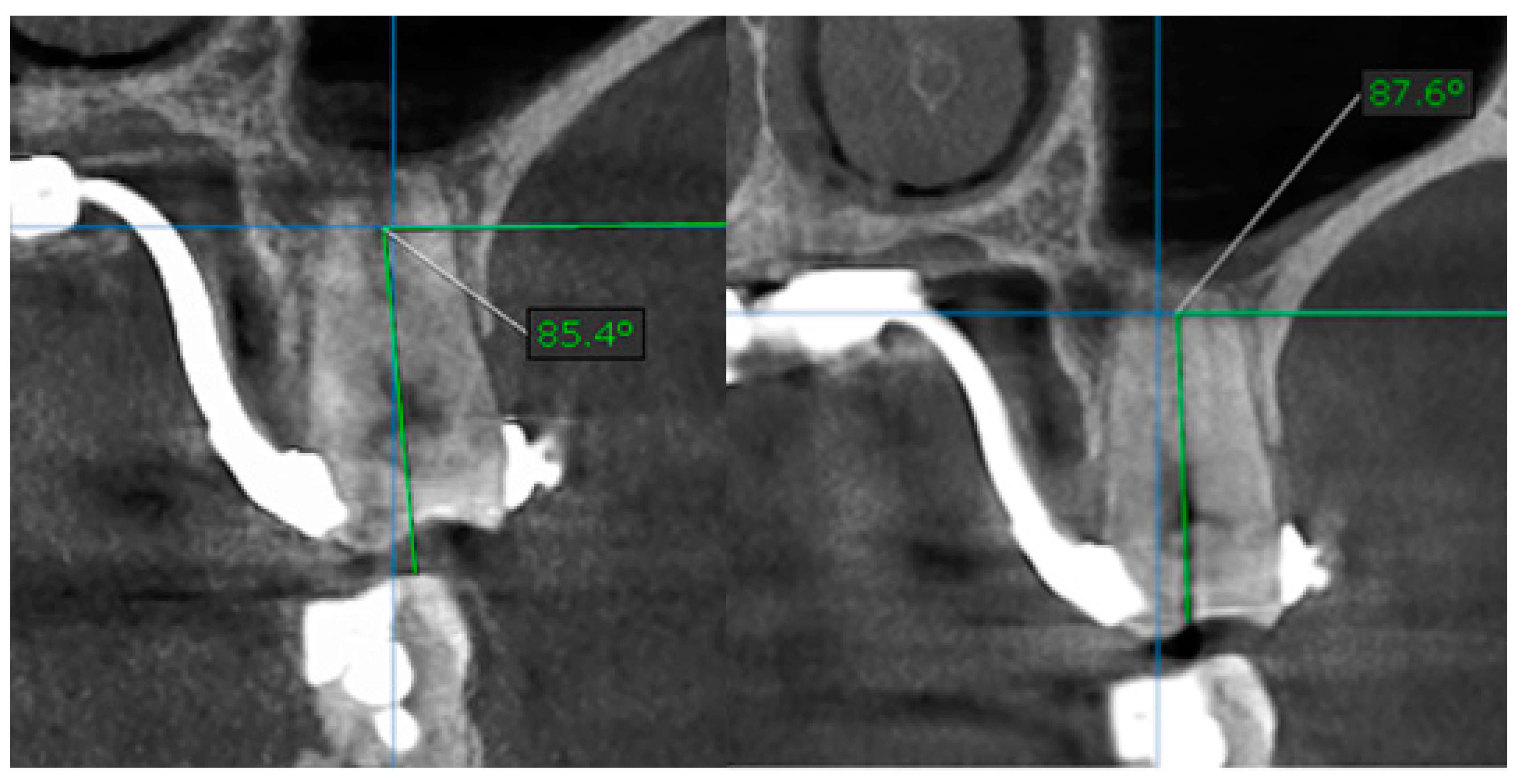
  • MARPE therapy associated with cortico-puncture therapy had a positive outcome on midpalatal suture opening and maxillary advancement, but a medium molar inclination of 2.005° was also observed, suggesting that tooth movement cannot be avoided because of the anchorage of the MARPE device at the molar level.
  • Our results suggest that non-surgical palatal expansion, assisted by micro-implants and cortico-puncture, is achievable and predictable in young adults. This occurs in safe conditions without the need for more complex surgical treatment.
  • The combination of MARPE and the cortico-puncture method proved to be a non-surgical treatment option to correct maxillary transverse deficiency in young adult patients. Cortico-puncture was able to weaken the suture interdigitation, thus facilitating the split.

Author Contributions

All authors contributed equally to this research. E.-S.B., M.P. and A.B. designed and performed the surgical phase. A.P.L., P.V. and L.L. derived the models and analyzed the data. A.V., E.-S.B. and M.P. assisted with measurements and helped carry out the statistical analysis. A.V., A.P.L. and L.L. produced the manuscript in consultation with E.-S.B., P.V. and A.B. All authors have read and agreed to the published version of the manuscript.

Funding

This research received no external funding.

Institutional Review Board Statement

The study was conducted according to the guidelines of the Declaration of Helsinki, and approved by the Ethics Committee of Algocalm Private Medical Center, Targu-Mures, Romania (892/5 February 2020).

Informed Consent Statement

Informed consent was obtained from all subjects involved in the study.

Conflicts of Interest

The authors declare no conflict of interest. Authors declare that they have no conflict of interest regarding this manuscript and we did not receive any financial support from an organization or a research grant.

References

  1. MacGinnis, M.; Chu, H.; Youssef, G.; Wu, K.W.; Machado, A.W.; Moon, W. The effects of micro-implant assisted rapid palatal expansion (MARPE) on the nasomaxillary complex—A finite element method (FEM) analysis. Prog. Orthod. 2014, 15, 52. [Google Scholar] [CrossRef] [Green Version]
  2. Hartono, N.; Soegiharto, B.M.; Widayati, R. The difference of stress distribution of maxillary expansion using rapid maxillary expander (RME) and maxillary skeletal expander (MSE)—A finite element analysis. Prog. Orthod. 2018, 19, 33. [Google Scholar] [CrossRef] [PubMed]
  3. Wehrbein, H.; Glatzmaier, J.; Mundwiller, U.; Diedrich, P. The Orthosystem—A new implant system for orthodontic anchorage in the palate. J. Orofac. Orthop. 1996, 57, 142–153. [Google Scholar] [CrossRef] [PubMed]
  4. Lagravere, M.O.; Carey, J.P.; Heo, G.; Toogood, R.W.; Major, P.W. Transverse, vertical, and anteroposterior changes from bone-anchored maxillary expansion vs traditional rapid maxillary expansion: A randomized clinical trial. Am. J. Orthod. Dentofac. Orthop. 2010, 137, 304–305. [Google Scholar] [CrossRef]
  5. Liu, S.S.; Kyung, H.; Buschang, P.H. Continuous forces are more effective than intermittent forces in expanding sutures. Eur. J. Orthod. 2010, 32, 371–380. [Google Scholar] [CrossRef] [Green Version]
  6. Tausche, E.; Hansen, L.; Schneider, M.; Harzer, W. Expansion maxillaire rapide par appui osseux avec une vis Hyrax implanto-portée: Le Dresden Distractor (DD) ou Disjoncteur de Dresde. Orthod. Fr. 2008, 79, 127–135. [Google Scholar] [CrossRef]
  7. Deeb, W.; Hansen, L.; Hotan, T.; Hietschold, V.; Harzer, W.; Tausche, E. Changes in nasal volume after surgically assisted bone-borne rapid maxillary expansion. Am. J. Orthod. Dentofac. Orthop. 2010, 137, 782–789. [Google Scholar] [CrossRef]
  8. Angelieri, F.; Cevidanes, L.H.S.; Franchi, L.; Goncalves, J.R.; Benavides, E.; McNamara, J.A., Jr. Midpalatal suture maturation: Classification method for individual assessment before rapid maxillary expansion. Am. J. Orthod. Dentofac. Orthop. 2013, 144, 759–769. [Google Scholar] [CrossRef] [Green Version]
  9. McNamara, J.A., Jr.; Lione, R.; Franchi, L.; Angelieri, F.; Cevidanes, L.H.; Darendeliler, M.A.; Cozza, P. The role of rapid maxillary expansion in the promotion of oral and general health. Prog. Orthod. 2015, 16, 33. [Google Scholar] [CrossRef] [Green Version]
  10. Möhlhenrich, S.C.; Modabber, A.; Kniha, K.; Peters, F.; Steiner, T.; Hölzle, F.; Fritz, U.; Raith, S. Simulation of three surgical techniques combined with two different bone-borne forces for surgically assisted rapid palatal expansion of the maxillofacial complex: A finite element analysis. Int. J. Oral Maxillofac. Surg. 2017, 46, 1306–1314. [Google Scholar] [CrossRef]
  11. Suri, L.; Taneja, P. Surgically assisted rapid palatal expansion: A literature review. Am. J. Orthod. Dentofac. Orthop. 2008, 133, 290–302. [Google Scholar] [CrossRef] [PubMed]
  12. Alexander, N.S.; Schroeder, J.W., Jr. Pediatric obstructive sleep apnea syndrome. Pediatr. Clin. N. Am. 2013, 60, 827–840. [Google Scholar] [CrossRef] [PubMed]
  13. Haas, A.J. The treatment of maxillary deficiency by opening of the midpalatal suture. Angle Orthod. 1965, 35, 200–217. [Google Scholar] [PubMed]
  14. Lione, R.; Ballanti, F.; Franchi, L.; Baccetti, T.; Cozza, P. Treatment and posttreatment skeletal effects of rapid maxillary expansion studied with low-dose computed tomography in growing subjects. Am. J. Orthod. Dentofac. Orthop. 2008, 134, 389–392. [Google Scholar] [CrossRef] [PubMed] [Green Version]
  15. Wertz, R.A. Skeletal and dental changes accompanying rapid midpalatal suture opening. Am. J. Orthod. 1970, 58, 41–66. [Google Scholar] [CrossRef]
  16. Stuart, D.A.; Wiltshire, W.A. Rapid palatal expansion in the young adult: Time for a paradigm shift? J. Can. Dent. Assoc. 2003, 69, 374–377. [Google Scholar]
  17. Cantarella, D.; Dominguez-Mompell, R.; Moschik, C.; Sfogliano, L.; Elkenawy, I.; Pan, H.; Mallya, S.M.; Moon, W. Zygomaticomaxillary modifications in the horizontal plane induced by micro-implant-supported skeletal expander, analyzed with CBCT images. Prog. Orthod. 2018, 19, 41. [Google Scholar] [CrossRef] [Green Version]
  18. Cunha, A.C. Expansão Maxilar Assistida por Mini-Implantes: Estudo do Complexo Sutural Craniofacial sob Perspectivas Clínicas e Microestruturais. Ph.D. Thesis, Universidade Federal do Rio de Janeiro, Rio de Janeiro, Brasil, 2017. [Google Scholar]
  19. Lin, L.; Ahn, H.W.; Kim, S.J.; Moon, S.C.; Kim, S.H.; Nelson, G. Tooth-borne vs boneborne rapid maxillary expanders in late adolescence. Angle Orthod. 2015, 85, 253–262. [Google Scholar] [CrossRef]
  20. Haas, A.J. Palatal expansion: Just the of dentofacial orthopedics. Am. J. Orthod. 1970, 57, 219–255. [Google Scholar]
  21. Lione, R.; Franchi, L.; Cozza, P. Does rapid maxillary expansion induce adverse effects in growing subjects? Angle Orthod. 2012, 83, 172–182. [Google Scholar] [CrossRef] [Green Version]
  22. Winsauer, H.; Walter, A.; Muchitsch, A.P.; Winsauer, C.; Jaeschke, D.; Katsaros, C.; Scherfler, M.; Ploder, O. Pure bone borne maxillary expansion with 4 mini-implants in adults with and without SARPE: A consecutive study of 35 patients. Presented at the 91st Congress of The European Orthodontic Society, Venice, Italy, 13 June 2015. [Google Scholar]
  23. Moon, H.-W.; Kim, M.-J.; Ahn, H.-W.; Kim, S.-J.; Kim, S.-H.; Chung, K.-R.; Nelson, G. Molar Inclination and Surrounding Alveolar Bone Change Relative to the Design of Bone-borne Maxillary Expanders: A Cbct study. Angle Orthod. 2020, 90, 13–22. [Google Scholar] [CrossRef] [PubMed] [Green Version]
  24. Celenk-Koca, T.; Erdinc, A.E.; Hazar, S.; Harris, L.; English, J.D.; Akyalcin, S. Evaluation of miniscrew-supported rapid maxillary expansion in adolescents: A prospective randomized clinical trial. Angle Orthod. 2018, 88, 702–709. [Google Scholar] [CrossRef] [PubMed] [Green Version]
  25. Garib, D.G.; de Lima Navarro, R. Francischone CE, Oltramari PVP. Expansão rápida da maxila ancorada em implantes: Uma nova proposta para expansão ortopédica na dentadura permanente. Rer. Den. Press Ortod. Ortop. Fac. 2007, 12, 75–81. [Google Scholar] [CrossRef] [Green Version]
  26. Lee, K.-J.; Park, Y.-C.; Park, J.-Y.; Hwang, W.-S. Miniscrew-assisted nonsurgical palatal expansion before orthognathic surgery for a patient with severe mandibular prognathism. Am. J. Orthod. Dentofac. Orthop. 2010, 137, 830–839. [Google Scholar] [CrossRef] [PubMed]
  27. Choi, S.-H.; Shi, K.-K.; Cha, J.-Y.; Park, Y.-C.; Lee, K.-J. Nonsurgical miniscrew-assisted rapid maxillary expansion results in acceptable stability in young adults. Angle Orthod. 2016, 86, 713–720. [Google Scholar] [CrossRef] [Green Version]
  28. Brunetto, D.P.; Sant’Anna, E.F.; Machado, A.W.; Moon, W. Non-surgical treatment of transverse deficiency in adults using microimplant-assisted rapid palatal expansion (MARPE). Den. Press J. Orthod. 2017, 22, 110–125. [Google Scholar] [CrossRef] [Green Version]
  29. Pulver, R.J.; Campbell, P.M.; Opperman, L.A.; Buschang, P.H. Miniscrew-assisted slow expansion of mature rabbit sutures. Am. J. Orthod. Dentofac. Orthop. 2016, 150, 303–312. [Google Scholar] [CrossRef]
  30. Tsai, C.; Yang, T.-K.; Hsieh, H.-Y.; Yang, L.-Y. Comparison of the effects of micro-osteoperforation and corticision on the rate of orthodontic tooth movement in rats. Angle Orthod. 2015, 86, 558–564. [Google Scholar] [CrossRef] [Green Version]
  31. Suzuki, S.S.; Braga, L.F.S.; Fujii, D.N.; Moon, W.; Suzuki, H. Corticopuncture Facilitated Microimplant-Assisted Rapid Palatal Expansion. Case Rep. Dent. 2018, 2018, 1–12. [Google Scholar] [CrossRef] [Green Version]
  32. Hassan, A.H.; Al-Fraidi, A.A.; Al-Saeed, S.H. Corticotomy-assisted orthodontic treatment: Review. Open Den. J. 2010, 4, 159–164. [Google Scholar] [CrossRef] [Green Version]
  33. Dibart, S.; Surmenian, S.; Sebaoun, J.D.; Montesani, L. Rapid treatment of class II malocclusion with piezocision: Two case reports. Int. J. Periodontics Restor. Den. 2010, 30, 487–493. [Google Scholar]
Figure 1. Cortico-puncture therapy perforations of the palatal suture.
Applsci 11 01306 g001
Figure 2. Stage E palatal suture as classified by Angelieri et al.
Applsci 11 01306 g002
Figure 3. MARPE device in position after cementation.
Applsci 11 01306 g003
Figure 4. The field of view was a cylinder of 82 mm in diameter and height.
Applsci 11 01306 g004
Figure 5. Rendering axial images showing FOV images used in the study before the palatal split. (a) Micro-implants in position, (b) Device in position.
Applsci 11 01306 g005
Figure 6. Interincisal diastema formation. (a)Axial view (b) Frontal view (c) Cross-sectional view.
Applsci 11 01306 g006
Figure 7. Rendering images showing cortico-puncture perforations and micro-implants in position.
Applsci 11 01306 g007
Figure 8. The split of the palatal suture.
Applsci 11 01306 g008
Figure 9. The fracture of the palatal plate.
Applsci 11 01306 g009
Figure 10. Cone Beam Computed Tomography CBCT images used in the study showing different measurements before and after the treatment: A: most mesial point of the infraorbital foramen right side; B: most mesial point of the infraorbital foramen left side; C: most lateral point of the nasal notch right side; D: most lateral point of the nasal notch left side.
Applsci 11 01306 g010
Figure 11. CBCT images used in the study showing different measurements before and after the treatment: Measurements on the frontal section on post-expansion CBCT: A: most mesial point of the infraorbital foramen right side; B: most mesial point of the infraorbital foramen left side; C: most lateral point of the nasal notch right side; D: most lateral point of the nasal notch left side.
Applsci 11 01306 g011
Figure 12. The first molar position before vs. after the palatal suture split.
Applsci 11 01306 g012
Table 1. The magnitude of the mid-palatal suture opening in millimeters.
Anterior Nasal Spine (ANS) Opening after TreatmentPosterior Nasal Spine (PNS) Opening after Treatment
Number of values2020
Minimum2.9702.670
25% Percentile3.2502.780
Median3.9002.995
75% Percentile4.1653.385
Maximum4.5404.110
Mean3.7663.125
SD0.49560.4070
Standard error of mean (SEM)0.11080.09102
Lower 95% CI of mean3.5342.934
Upper 95% CI of mean3.9983.316
Table 2. Comparison of the magnitude of the split at the PNS and ANS.
Table AnalyzedData 2
Column BPosterior nasal spine (PNS) opening after treatment
vs.vs.
Column AAnterior nasal spine (ANS) opening after treatment
Paired t-test
p-value0.0008
p-value summary
Significantly different? (p < 0.05)Yes
One- or two-tailed p-value?Two-tailed
t, dft = 3.964 df = 19
Number of pairs20
How big is the difference?
Mean of differences−0.6410
SD of differences0.7232
SEM of differences0.1617
95% confidence interval−0.9795 to −0.3025
R2 0.4526
How effective was the pairing?
Correlation coefficient (r)−0.2770
p-value (one tailed)0.1185
p-value summaryNs
Significant correlation? (p > 0.05)Yes
Table 3. The magnitude of the interincisal distance opening at the cemento-enamel junction (CEJ) level.
ParameterInterincisal Opening at the CEJ Level/Millimeter
Minimum3.360
25% percentile3.783
Median4.210
75% percentile4.365
Maximum4.770
Mean4.103
SD0.3881
SEM0.08679
Lower 95% CI of mean3.921
Upper 95% CI of mean4.284
Table 4. Changes at the first molar position after treatment.
D’Agnostino & Pearson Omnibus Normality Test1st Molar Tilt after Treatment1st Molar Tilt before Treatment
Number of vales4040
Minimum85.6084.00
25% Percentile87.6085.23
Median88.0085.65
75% Percentile88.6586.70
Maximum89.3087.90
Mean87.8885.87
Std. Deviation1.0011.057
Std. Error of Mean0.22380.2363
Lower 95 % CI of mean87.4185.38
Upper 95 % CI of mean88.3486.36
Table AnalyzedData 1
Column B1st Molar tilt before treatment
Vs.Vs.
Column A1st Molat tilt after treatment
Paired t test
p value< 0.0001
p value summary
Significantly different? (p < 0.05)yes
One- or two-tailed P valueTwo-tailed
t, dft = 12.33 df = 19
Number of pairs40
How big is the difference−2.005
SD of differences0.7273
SEM of differences0.1626
95% confidence interval−2.345 to −1.665
R square0.8889
Publisher’s Note: MDPI stays neutral with regard to jurisdictional claims in published maps and institutional affiliations.

