Article

Calcium Hydroxide Removal Using Four Different Irrigation Systems: A Quantitative Evaluation by Scanning Electron Microscopy

1
Department of Surgery, Medicine, Dentistry and Morphological Sciences with Transplant Surgery, Oncology and Regenerative Medicine Relevance (CHIMOMO), University of Modena and Reggio Emilia, 41124 Modena, Italy
2
Section of Human Morphology, Department of Biomedical, Metabolic and Neural Science, University of Modena and Reggio Emilia, 41124 Modena, Italy
3
Product Engineer, Beckett Thermal Solution, 41043 Formigine, Italy
4
Independent Researcher, 88900 Crotone, Italy
5
Private Practice, 40122 Bologna, Italy
6
Clinical Department of Medical, Surgical and Health Sciences, University of Trieste, 34149 Trieste, Italy
7
Department of General Surgery and Surgical-Medical Specialties, University of Catania, 95123 Catania, Italy
*
Author to whom correspondence should be addressed.
Submission received: 24 November 2021 / Revised: 21 December 2021 / Accepted: 27 December 2021 / Published: 28 December 2021
(This article belongs to the Special Issue Dental Materials: Latest Advances and Prospects)

Abstract

:
This study compares conventional endodontic needle irrigation, passive ultrasonic irrigation, apical negative pressure irrigation, and mechanical activation to remove calcium hydroxide from single straight root canals. Eighty-four mandibular premolars were prepared in a crown-down manner up to size #40. Two teeth represented a negative control, and another two served as a positive control. Calcium hydroxide paste was placed inside root canals. The remaining eighty samples were analyzed based on the activation techniques, and the cleanliness of the canals was quantified using Fiji’s software on 500× magnified SEM backscattered electron micrographs. Considering the whole canal, all instruments showed better performance than conventional endodontic needle irrigation in removing calcium hydroxide (p < 0.05). Irrisafe and XP-endo Finisher could remove a significantly higher amount of calcium hydroxide than Endovac (p < 0.05). Irrisafe and XP-endo Finisher have been able to remove more calcium hydroxide than EndoVac.

1. Introduction

The prevention and eradication of bacteria from tooth cavities still represents one of the major concerns of root canal treatment. Among the materials proposed as intracanal medicament, calcium hydroxide (Ca(OH)2) (CH) has been widely studied for its antimicrobial efficacy, and the benefits of its use are well documented [1,2,3,4]. On the other hand, the dressing material has to be removed entirely before root canal filling [5,6], since various research studies have shown that CH residues on dentin walls can compromise the canal seal by promoting micro-leakage between the sealer and gutta-percha, hindering the penetration of the sealer inside dentinal tubules and interacting with the setting of some sealers [7,8,9]. Moreover, different studies have identified an additional issue, which is the difficulty to completely remove CH from the apical third of the canals [7,10,11,12]. Despite the various protocols and techniques that have been proposed [13,14,15,16], studies have shown that instrumentation and irrigation are unable to clean completely the whole root canal system from CH [17,18]. The most commonly described root canal cleansing method is the use of a master apical file inserted until the working length (WL) and in conjunction with the abundant irrigation of sodium hypochlorite (NaOCl) and EDTA [19]. However, a recent review reported that the ultrasonic activation of irrigants seems more effective for removing CH than other techniques [9]. Kourti and Pantelidou [20] demonstrated that the use of the EndoVac (Kerr Endo, Orange County, CA, USA), an irrigation system using apical negative pressure that sucks the irrigants employing a microcannula placed at the WL, improved CH removal with respect to ultrasonic activation and conventional endodontic needle irrigation. Recently, XP-endo Finisher (FKG Dentaire SA, La Chaux de Fonds, Switzerland) has been developed to improve the efficacy of the final irrigation procedure. XP-endo Finisher exhibits a reduced core size (#25), zero taper, and the peculiar heat-treated NiTi alloy named MaxWire (Martensite-Austenite-electropolish-fileX) [21] that allows the instrument a great flexibility, leading to the removal of debris from hard-to-reach regions while limiting the damage on dentine at the same time. Moreover, the rotational movement can potentially produce agitation of the irrigant solutions, so it was suggested as a final step of disinfection procedures. The first published articles [22,23,24] confirmed the XP-endo Finisher’s superior effectiveness in cleaning the dentin walls compared to the conventional technique.
This study was aimed to evaluate quantitatively the residual amount of CH, comparing the effect of four irrigation systems: conventional endodontic needle irrigation, Irrisafe (Satelec Acteon Group, Merignac, France), EndoVac and XP-endo Finisher, by means of scanning electron microscopy equipped with an environmental-dispersive X-ray detector (SEM-EDX). The null hypothesis tested was the absence of a difference in the amount of CH removal between the four different irrigation systems employed in root canal cleaning.

2. Materials and Methods

2.1. Root Canal Instrumentation

One hundred and forty-seven mandibular single-rooted premolars were withdrawn from a pool of extracted teeth. After radiographical examination of teeth in a mesiodistal and buccolingual direction, only teeth with a single straight canal were selected. Teeth with previous root canal treatment, calcifications, apical curvature, immature apices, or resorptions were discarded. A total of eighty-four teeth were included in the study. The specimens were stored in distilled water for no longer than 15 days before the experiment. The crowns were partially removed with a size 701 high-speed fissure bur (Komet Italia, Milano, Italy) under water irrigation to standardized root lengths at 18 mm. After access cavity preparation, a #10 K-file (Dentsply Maillefer, Ballaigues, Switzerland) was introduced into the canal until its tip emerged at the apical foramen, which was observed under a microscope at 10× magnification (OPMI PICO; Carl Zeiss Meditec Inc., Jena, Germany), and the WL was determined subtracting 1 mm from the measurement. To reproduce in vivo conditions, the apex was sealed with cyanoacrylate [25]. Each tooth was instrumented in a crown-down manner by using the ProTaper Gold rotary system (Dentsply Maillefer, Ballaigues, Switzerland) to size 40 at the WL. After each instrument change, the root canal was rinsed with 1 mL 5% of NaOCl (Niclor, Ogna, Muggiò, Italy) with a side vented needle (27-gauge) inserted on a syringe (Max-i-Probe, Dentsply Rinn, Elgin, IL, USA) introduced before the binding point but at a distance greater than 2 mm from the WL. The final rinse after instrumentation was performed with 1 mL 5% NaOCl and subsequently 1 mL 17% EDTA (Ogna), which were both left in place for 1 min, and with 5 mL of sterile water (Baxter, Roma, Italy) delivered with a 27-gauge side-vented needle (Max-i-Probe). Sterile paper points (ProTaper Gold paper points F4, Dentsply Maillefer, Ballaigues, Switzerland) were employed to dry the root canals. As a negative control, two teeth were used that did not undergo any further treatment. CH paste (Calxyl®, OCO präparate, Dirmstein, Germany) was placed inside root canals with a size #30 lentulo spiral (Dentsply Maillefer, Ballaigues, Switzerland) and sterile paper points. The access cavities were filled with cotton and a temporary filling material (Cavit, 3M ESPE, Seefeld, Germany). Two radiographs of all teeth were taken in mesiodistal and bucco-lingual directions to verify the complete filling of all canals with CH paste. The lingual and buccal surfaces were marked with a longitudinal groove each, that was made with a diamond disk without penetrating the canal to facilitate the subsequent split in two halves (Figure 1A). Then, the specimens were kept at 37 °C at 100% humidity for a month in a Heracell VIOS 250i incubator (Thermo Scientific, Waltham, MA, USA).

2.2. Retreatment Procedures

After removal of the temporary fillings, two teeth did not undergo any further treatment and were used as positive controls. The remaining eighty teeth were subdivided into four experimental groups (n = 20) according to the systems used: conventional endodontic needle irrigation group (CENI), Irrisafe group (IS), EndoVac group (EV), and XP-endo Finisher group (XPF). The specimens were fixed in a tube stand and placed in a thermostatic bath heated to 37 °C to simulate the body temperature. In all groups, a master apical file ProTaper Universal size 40 at WL was used to remove the CH, and then, different techniques were applied.
The subsequent irrigation steps were performed after leaving the samples in the thermostatic bath for at least 20 min.
In the CENI group, irrigation was performed with a syringe with a 27-gauge side-vented needle placed 2 mm from the WL with 3 mL of 5% NaOCl left in place for 60 s, which was followed by 3 mL of 17% EDTA for 60 s and 5 mL of sterile water. No additional agitation of irrigants was performed.
In the IS group, the same irrigation protocol as in the CENI group was used, but ultrasonic activation of NaOCl and EDTA were obtained with the P5 Newtron (Satelec) mounting a stainless steel non-cutting 25 tip (Irrisafe, Satelec), which was inserted 2 mm from the WL for 1 min for each irrigant.
NaOCl was delivered with the master delivery tip in the EV group and simultaneously aspirated by using the macrocannula for 30 s with an up-and-down movement from a point where it started to bind to a point just below the canal orifice. NaOCl was left in place for 60 s. After this, three cycles of microirrigation were performed by inserting the microcannula at the WL for 6 s, then at 2 mm from the WL for 6 s, and eventually at WL for another 6 s. This was done until a total of 30 s was reached for each cycle. At the end of cycles, the microcannula completely aspirated the irrigant from within the canal. The first and third cycles were performed by using 5% NaOCl, whereas the second cycle used 17% EDTA.
In the XPF group, the same irrigation protocol as in the CENI group was used, but NaOCl and EDTA were activated using the XP-endo Finisher file. The file, mounted on an endodontic motor (X-Smart Plus, Dentsply Maillefer, Tulsa, OK, USA) rotated at a speed of 800 rpm and 1 N cm torque, and it was cooled down with ethyl chloride (Crio Spray, Karl Sanremo, IM, Italy). The file was operated for 60 s with vertical movements of 7–8 mm to the WL to activate NaOCl and EDTA.
The same amount of irrigants was employed in all groups. Sterile paper points were used to dry all canals, and the roots were then split along their longitudinal axis into two halves with a small chisel and a hammer. Division in thirds (apical, middle, and coronal) was determined by marking the roots at 2, 5, and 11 mm from the apex, respectively (Figure 1B).

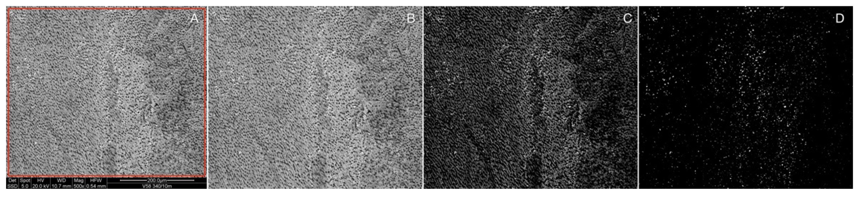
2.3. SEM-EDX Evaluation (Quantitative Analysis)

Split roots were mounted on aluminum stubs coated with 8–10 nm of gold and analyzed by SEM-EDX (Quanta 200, Fei Company, Eindhoven, Netherlands and INCA-350, Oxford instruments, Oxford, UK) in backscattered mode (BSE). Micrographs were acquired at various magnification from 60× to 15,000×. EDX microanalysis was applied to assess the chemical composition of the different phases visible in the SEM micrographs. To quantify the residual CH, image analysis was performed using Fiji’s software (National Institute of Health, Bethesda, MD, USA). The analysis was performed on 500× magnified SEM backscattered electron (BSD) micrographs, exploiting the gray scale contrast generated by different chemical compositions of dentin and CH in BSE mode. The brightness and contrast of the micrographs were adjusted to highlight the difference between the phases, and a gray scale threshold limit was imposed to obtain a binary black and white image, as shown in Figure 2. From the binary image, the area fraction occupied by the white spots was calculated. This protocol was adapted from Conserva [26] and Deari [27].

2.4. Statistical Analysis

STATA 11 (StataCorp LLC, College Station, TX, USA) software was used for statistical analysis. Values in tables are reported as the mean ± standard deviation (SD). Since most data did not follow a normal distribution, the Kruskal–Wallis test followed by the Dunn test were used to compare results among and within groups. A p -value < 0.05 was considered significant.

3. Results

Figure 3 shows a backscattered electron micrograph acquired into a CH-treated tooth canal at 15,000× magnification (Figure 3A) and the relative spot EDX spectra (Figure 3B). Based on the different gray shades of the particles visible in the micrographs and the relative results of the EDX microanalysis, two well-distinguishable phases may be identified. The darker phase, containing high amounts of Ca, P, and O, may be identified as the tooth dentin; the bright globular phase, containing mainly Ca, S, Ba, and O, may be identified as the CH paste remnants. The presence of barium sulfate (BaSO4) in the CH paste, as reported in the safety data sheet of the Calxyl® radiopaque (OCO präparate GmbH, Dirmstein, Germany), added for its X-ray opacity properties, allows clearly distinguishing the CH paste residues in the BSE images, giving to the paste a much brighter contrast compared to the dentin.
Table 1 reports the average values of residual calcium hydroxide measured inside the root canal’s selected areas according to instrument and level. The Kruskal–Wallis test shows significant differences among groups, both considering the individual levels and the whole root canal. Irrisafe and XP-endo Finisher performed significantly better at the coronal level than the conventional endodontic needle irrigation. Moreover, XP-endo Finisher performed better than Endovac at this level. All instruments removed a significantly higher amount of CH than conventional endodontic needle irrigation at the middle level; moreover, Irrisafe and XP-endo Finisher performed significantly better than Endovac. At the apical level, the amount of CH removed was significantly higher in the Irrisafe and XP-endo Finisher groups than conventional endodontic needle irrigation and Endovac irrigation.
When the whole canal is considered, all instruments showed better performance than conventional endodontic needle irrigation in removing CH. Among the three techniques, Irrisafe and XP-endo Finisher were able to remove a significantly higher amount of CH than Endovac.
When results were compared within groups to measure the performance among the three levels of the root canal, the Kruskal–Wallis test highlighted significant differences only respecting the conventional endodontic needle irrigation. Compared to the middle and coronal root canal walls, more CH was detected at the apical level. Figure 4 shows an overview of representative SEM images from the experimental groups at 2, 5, and 11 mm levels.

4. Discussion

This study was designed to assess the ability of four irrigation systems to remove Ca(OH)2 from root canals through a quantitative evaluation. CH paste is a widely used intracanal dressing during root canal therapy due to its alkalinity (pH 12.5) to reduce residual bacteria [4], but the presence of residues on dentin walls can prevent sealer penetration into dentinal tubules, thus compromising the endodontic seal and increasing apical leakage [8]. For the previously mentioned reasons, the intracanal dressings should be completely removed before permanent obturation of the root canal [24]. Many studies have been published regarding the removal of CH or other medicaments from the root canal system using other supplementary systems to the conventional irrigation [15,16,28,29,30]. The aim of this study was to compare Irrisafe, Endovac, and XP-endo Finisher efficacy with the conventional endodontic needle irrigation. One of the major strengths of the present investigation is represented by the quantitative evaluation performed through SEM images. Different methods have been used to measure residual material inside root canals, spanning from direct visualization to micro-CT, from digital microscopy to scanning electron microscopy [9,13,31]. In most of the cases, studies conducted by SEM analysis achieved a semi-quantitative evaluation. Other studies performed using the grooves model had several advantages with respect to studies investigating smooth root canals wall, because the scoring of the groove’s cleanness is presumably more reproducible than the percentage assessment of the whole canal [30]. In this study, standardized grooves were not performed in the canals because a quantitative method was used that allowed measuring the total amount of CH left without visual evaluations by different examiners. In particular, a rectangular area of images was selected. Brightness and contrast were adapted to accentuate CH remnants. The threshold function was used to select the contrasted spots to obtain a binary black and white image, permitting accurately measuring the area of the white spots inside the area selected [26]. Moreover, one of the significant criticisms of the SEM investigation to evaluate the CH removal is the difficulty of discriminating between Ca(OH)2 residues and the smear layer or dentin debris, since EDX spectroscopy analysis reveals Ca++ in both cases [9]. In the present study, this problem was overcome by using a Barium sulfate-doped paste of Ca(OH)2. Barium sulfate, probably added by the manufacturer for its X-ray opacity properties, permitted the EDX analysis to perfectly distinguish CH remnants from the dentin debris. According to Silva [31], in the present study, it was decided to preserve the tooth crown as an irrigant reservoir during the endodontic procedures, preventing the same irrigants coronal leak during ultrasonic activation.
The main finding of this investigation was that Irrisafe, Endovac, and XP-endo finisher showed significantly better performance than the conventional endodontic needle irrigation when the whole canal was considered, regardless of the coronal, middle, and apical level. Among the three techniques, XP-endo Finisher and Irrisafe were able to remove a significantly higher amount of CH than Endovac. Altogether, the results suggest that the null hypothesis has to be rejected. In a recent study, XP-endo Finisher was less efficient than ultrasonic activation in the removal of CH from apical grooves [30]. These results are in contrast with the present findings probably because, in this study, the CH removal procedures were executed at 37 °C, and at this temperature, XP-endo Finisher inside the root canal changes shape becoming curved due to the transition from the martensitic state to austenitic state [21,30].
In a comparative study between Passive Ultrasonic Irrigation (PUI) and XP-endo Finisher, the specimens were placed in a controlled-temperature water bath at 37 °C, and the whole canals were analyzed. The XP-endo Finisher was significantly more effective in removing the CH medication in the apical third than PUI [29]. Contrary to the present study, the samples were decoronated, probably causing leakage of the irrigant during ultrasonic activation.

5. Conclusions

The systems investigated in this study were unable to completely remove CH from root canals. However, Irrisafe and XP-endo Finisher were significantly more efficient than EndoVac in the elimination of calcium hydroxide. Conventional endodontic needle irrigation (CENI) was considerably less efficient than the other three systems, especially at the apical level.

Author Contributions

Conceptualization, L.G. (Luigi Generali), F.C.; Data curation, F.I., D.A. and V.C.; methodology, L.G. (Luigi Generali), F.F., F.C. and L.G. (Luciano Giardino); software, P.S.; validation, E.P., L.G. (Luciano Giardino) and L.G. (Luigi Generali); formal analysis, L.G. (Luigi Generali), P.S., F.F. and V.C.; investigation, L.G. (Luigi Generali), C.P., D.A. and C.B., writing—original draft preparation, L.G. (Luigi Generali), F.C., F.I., V.C. and E.P.; writing—review and editing, E.P., F.I., C.P., C.B. All authors have read and agreed to the published version of the manuscript.

Funding

The research was funded from the FAR project (University of Modena and Reggio Emilia Funds for Research) entitled Microstructural and Mechanical investigation of Nickel-Titanium Endodontic instruments—MMiNTEndo.

Informed Consent Statement

Written informed consent has been obtained from the patients to collect teeth for scientific purposes.

Acknowledgments

The SEM microscope is available at the imaging facility (Centro Interdipartimentale Grandi Strumenti (CIGS) of the University of Modena and Reggio Emilia. We thank Mauro Zapparoli from CIGS for technical support.

Conflicts of Interest

The authors declare no conflict of interest.

References

  1. Mohammadi, Z.; Dummer, P.M. Properties and applications of calcium hydroxide in endodontics and dental traumatology. Int. Endod. J. 2011, 44, 697–730. [Google Scholar] [CrossRef] [PubMed]
  2. Lima, R.K.; Guerreiro-Tanomaru, J.M.; Faria-Júnior, N.B.; Tanomaru-Filho, M. Effectiveness of calcium hydroxide-based intracanal medicaments against Enterococcus faecalis. Int. Endod. J. 2012, 45, 311–316. [Google Scholar] [CrossRef]
  3. Chockattu, S.J.; Deepak, B.S.; Goud, K.M. Comparison of anti-bacterial efficiency of ibuprofen, diclofenac, and calcium hydroxide against Enterococcus faecalis in an endodontic model: An in vitro study. J. Conserv. Dent. 2018, 21, 80–84. [Google Scholar] [CrossRef]
  4. Govindaraju, L.; Jenarthanan, S.; Subramanyam, D.; Ajitha, P. Antibacterial activity of various intracanal medicament against enterococcus faecalis, streptococcus mutans and staphylococcus aureus: An in vitro study. J. Pharm. Bioallied Sci. 2021, 13, S157–S161. [Google Scholar] [CrossRef] [PubMed]
  5. Türker, S.A.; Koçak, M.M.; Koçak, S.; Saglam, B.C. Comparison of calcium hydroxide removal by self—Adjusting file, EndoVac, and CanalBrush agitation techniques: An in vitro study. J. Conserv. Dent. 2013, 16, 439–443. [Google Scholar] [CrossRef] [PubMed]
  6. Margi, P.; Karkala, V.K.; Nidhi, P.S.; Maitry, P.; Krushn, S.; Vinukonda, H.B.; Das, T.D. Efficacy of removal of calcium hydroxide medicament from root canals by endoactivator and endovac irrigation techniques: A systematic review of in vitro studies. Contemp. Clin. Dent. 2019, 10, 135–142. [Google Scholar] [CrossRef]
  7. Ozyurek, E.U.; Erdogan, O.; Turker, S.A. Effect of calcium hydroxide dressing on the dentinal tubule ppenetration of 2 different root canal sealers: A confocal laser scanning microscopy study. J. Endod. 2018, 44, 1018–1023. [Google Scholar] [CrossRef] [PubMed]
  8. Kim, S.K.; Kim, Y.O. Influence of calcium hydroxide intracanal medication on apical seal. Int. Endod. J. 2002, 35, 623–628. [Google Scholar] [CrossRef] [PubMed]
  9. Yaylali, I.E.; Kececi, A.D.; Ureyen Kaya, B. Ultrasonically activated irrigation to remove calcium hydroxide from apical third of human root canal system: A systematic review of in vitro studies. J. Endod. 2015, 41, 1589–1599. [Google Scholar] [CrossRef]
  10. Yu, D.C.; Schilder, H. Cleaning and shaping the apical third of a root canal system. Gen. Dent. 2001, 49, 266–270. [Google Scholar]
  11. da Silva, J.M.; Andrade Junior, C.V.; Zaia, A.A.; Pessoa, O.F. Microscopic cleanliness evaluation of the apical root canal after using calcium hydroxide mixed with chlorhexidine, propylene glicol, or antibiotic paste. Oral Surg. Oral Med. Oral Pathol. Oral Radiol. Endodontol. 2011, 111, 260–264. [Google Scholar] [CrossRef]
  12. Faria, G.; Kuga, M.C.; Ruy, A.C.; Aranda-Garcia, A.J.; Bonetti-Filho, I.; Guerreiro-Tanomaru, J.M.; Toledo Leonardo, R. The efficacy of the self-adjusting file and protaper for removal of calcium hydroxide from root canals. J. Appl. Oral Sci. 2013, 21, 346–350. [Google Scholar] [CrossRef]
  13. Kenee, D.M.; Allemang, J.D.; Johnson, J.D.; Hellstein, J.; Nichol, B.K. A quantitative assessment of efficacy of various calcium hydroxide removal techniques. J. Endod. 2006, 32, 563–565. [Google Scholar] [CrossRef] [PubMed]
  14. Rodig, T.; Vogel, S.; Zapf, A.; Hülsmann, M. Efficacy of different irrigants in the removal of calcium hydroxide from root canals. Int. Endod. J. 2010, 43, 519–527. [Google Scholar] [CrossRef] [PubMed]
  15. Capar, I.; Ozcan, E.; Arslan, H.; Ertas, H.; Aydinbelge, H. Effect of different final irrigation methods on the removal of calcium hydroxide from an artificial standardized groove in the apical third of root canals. J. Endod. 2014, 40, 451–454. [Google Scholar] [CrossRef]
  16. Arslan, H.; Akcay, M.; Capar, I.; Saygili, G.; Gok, T.; Ertas, H. An in vitro comparison of irrigation using photon-initiated photoacoustic streaming, ultrasonic, sonic and needle techniques in removing calcium hydroxide. Int. Endod. J. 2015, 48, 246–251. [Google Scholar] [CrossRef]
  17. Haapasalo, M.; Shen, Y.; Qian, W.; Gao, Y. Irrigation in endodontics. Dent. Clin. N. Am. 2010, 54, 291–312. [Google Scholar] [CrossRef]
  18. Paqué, F.; Balmer, M.; Attin, T.; Peters, O.A. Preparation of oval-shaped root canals in mandibular molars using nickel-titanium rotary instruments: A micro-computed tomography study. J. Endod. 2010, 36, 703–707. [Google Scholar] [CrossRef] [PubMed] [Green Version]
  19. Lambrianidis, T.; Kosti, E.; Boutsioukis, C.; Mazinis, M. Removal efficacy of various calcium hydroxide/chlorhexidine medicaments from the root canal. Int. Endod. J. 2006, 39, 55–61. [Google Scholar] [CrossRef] [PubMed]
  20. Kourti, E.; Pantelidou, O. Comparison of different agitation methods for the removal of calcium hydroxide from the root canal: Scanning electron microscopy study. J. Conserv. Dent. 2017, 20, 439–444. [Google Scholar] [CrossRef]
  21. Zupanc, J.; Vahdat-Pajouh, N.; Schäfer, E. New thermomechanically treated NiTi alloy—A review. Int. Endod. J. 2018, 51, 1088–1103. [Google Scholar] [CrossRef] [PubMed] [Green Version]
  22. Wigler, R.; Dvir, R.; Weisman, A.; Matalon, S.; Kfir, A. Efficacy of XP-endo finisher files in the removal of calcium hydroxide paste from artificial standardized grooves in the apical third of oval root canals. Int. Endod. J. 2017, 50, 700–705. [Google Scholar] [CrossRef]
  23. Uygun, A.D.; Gündoğdu, E.C.; Arslan, H.; Ersoy, İ. Efficacy of XP-endo finisher and TRU Shape 3D conforming file compared to conventional and ultrasonic irrigation in removing calcium hydroxide. Aust. Endod. J. 2017, 43, 89–93. [Google Scholar] [CrossRef]
  24. Keskin, C.; Sariyilmaz, E.; Sariyilmaz, Ö. Efficacy of XP-endo finisher file in removing calcium hydroxide from simulated internal resorption cavity. J. Endod. 2017, 43, 126–130. [Google Scholar] [CrossRef] [PubMed]
  25. Giardino, L.; Cavani, F.; Generali, L. Sodium hypochlorite solution penetration into human dentin: A histochemical evaluation. Int. Endod. J. 2017, 50, 492–498. [Google Scholar] [CrossRef]
  26. Conserva, E.; Generali, L.; Bandieri, A.; Cavani, F.; Borghi, F.; Consolo, U. Plaque accumulation on titanium disks with different surface treatments: An in vivo investigation. Odontology 2018, 106, 145–153. [Google Scholar] [CrossRef]
  27. Deari, S.; Mohn, D.; Zehnder, M. Dentine decalcification and smear layer removal by different ethylenediaminetetraacetic acid and 1-hydroxyethane-1,1-diphosphonic acid species. Int. Endod. J. 2019, 52, 237–243. [Google Scholar] [CrossRef] [Green Version]
  28. van der Sluis, L.W.; Wu, M.K.; Wesselink, P.R. The evaluation of removal of calcium hy- droxide paste from an artificial standardized groove in the apical root canal using different irrigation methods. Int. Endod. J. 2007, 40, 52–57. [Google Scholar] [CrossRef]
  29. Hamdan, R.; Michetti, J.; Pinchon, D.; Diemer, F.; Georgelin-Gurgel, M. The XP-endo finisher for the removal of calcium hydroxide paste from root canals and from the apical third. J. Clin. Exp. Dent. 2017, 9, e855–e860. [Google Scholar] [CrossRef] [PubMed] [Green Version]
  30. Donnermeyer, D.; Wyrsch, H.; Bürklein, S.; Schäfer, E. Removal of calcium hydroxide from artificial grooves in straight root canals: Sonic activation using EDDY versus passive ultrasonic irrigation and XPendo finisher. J. Endod. 2019, 45, 322–326. [Google Scholar] [CrossRef]
  31. Silva, L.J.M.; Pessoa, O.F.; Teixeira, M.B.G.; Gouveia, C.H.; Braga, R.R. Micro-CT evaluation of calcium hydroxide removal through passive ultrasonic irrigation associated with or without an additional instrument. Int. Endod. J. 2015, 48, 768–773. [Google Scholar] [CrossRef] [PubMed]
Figure 1. (A) Buccal surface with the longitudinal groove made with a diamond disk. (B) A split half of a sample ready for SEM analysis.
Applsci 12 00271 g001
Figure 2. (A) Original SEM image where measures were performed; the red square shows where the image is cropped as seen in (B). (C) Enhancement of contrast highlighting white dots corresponding to CH particles, (D) image after binarization.
Applsci 12 00271 g002
Figure 3. (A) SEM image showing the globular aspect of CH residues; arrows indicate the areas analyzed with EDX, (B) EDX spectra relative to areas indicated in (A).
Applsci 12 00271 g003
Figure 4. Representative SEM images of areas used for CH residues evaluation for each kind of treatment and root level from the apex.
Applsci 12 00271 g004
Table 1. Calcium hydroxide residues % (mean ± SD) inside the root canal measured on SEM images.
Groups
CENIISEVXPFK-W
p-Value
Coronal4.83 ± 6.0 °0.88 ± 0.72.56 ± 2.7 §0.66 ± 0.70.002
Middle4.91 ± 5.6 #0.53 ± 0.91.40 ± 1.7 *0.68 ± 1.1<0.001
Apical11.52 ± 8.8 °,+0.35 ± 0.46.39 ± 11.0 *2.30 ± 5.0<0.001
Total7.09 ± 5.9 #0.59 ± 0.53.45 ± 4.6 *1.21 ± 2.0<0.001
* versus Irrisafe and XP-endo Finisher p < 0.05; § versus XP-endo Finisher p < 0.05; # versus Irrisafe, Endovac, and XP-endo Finisher p < 0.05; ° versus Irrisafe and XP-endo Finisher p < 0.05; + versus coronal and middle levels p < 0.05.
Publisher’s Note: MDPI stays neutral with regard to jurisdictional claims in published maps and institutional affiliations.

