Review

Otologic Manifestations of IgG4-Related Disease: Literature Review and Report of Two Cases

1
Department of Otolaryngology-Head and Neck Surgery, Soroka University Medical Center, Faculty of Health Sciences, Ben Gurion University, Beer-Sheva 8410501, Israel
2
Department of Pathology, Soroka University Medical Center, Faculty of Health Sciences, Ben-Gurion University, Beer-Sheva 8410501, Israel
3
Department of Diagnostic Imaging, Soroka University Medical Center, Faculty of Health Sciences, Ben-Gurion University, Beer-Sheva 8410501, Israel
4
Department of Otolaryngology-Head and Neck Surgery, Rabin Medical Center, Petach Tikva 4941492, Israel
*
Author to whom correspondence should be addressed.
Submission received: 20 July 2022 / Revised: 17 August 2022 / Accepted: 18 August 2022 / Published: 21 August 2022
(This article belongs to the Special Issue Hearing Loss: From Pathophysiology to Therapies and Habilitation)

Abstract

:
Background: IgG4-related disease (IgG4-RD) is an inflammatory process. The literature on IgG4-RD is rather limited, with mastoid involvement being uncommon. In such cases, presentation may mimic other middle ear and mastoid cavity pathologies. Objective: To summarize findings of patients with IgG4-RD involving the mastoid. Methods: Description of two new cases and summary of findings with previous reports. Results: Nineteen cases of IgG4-RD with mastoid bone involvement were reported in the literature, the earliest appearing at the beginning of the previous decade. Most frequent symptoms included hearing deterioration, tinnitus and otalgia. In 58% of the cases, the process was restricted to otologic manifestations. In 7 out of 19 cases, tissue IgG4 levels were elevated. In all histopathologic samples taken from the operative sites, a dense lymphoplasmacytic cell infiltration was observed. Following definitive pathological diagnosis, the most common treatment was corticosteroids. Generally, the time from onset to final diagnosis was usually more than six months. The treatment is corticosteroids, followed by immunosuppressive agents such as rituximab, cyclophosphamide, and methotrexate. Conclusions: IgG4-RD involving the mastoid is a challenging condition, both in diagnosis and treatment. Hence, IgG4-RD should be included in the differential diagnosis of middle ear pathologies and include a multi-disciplinary team for treatment.

1. Introduction

IgG4 related disease (IgG4-RD) is a fibro-inflammatory condition, characterized by swelling lesions in several tissues and pathological findings of organ lymphoplasmacytic infiltrate rich in IgG4 and plasma cells; the infiltrate is described as storiform fibrosis and obliterative phlebitis. Elevation of serum IgG4 concentrations often occurs, but the sensitivity and specificity of these antibodies are unknown [1,2]. Since the first description of the disease and its association with autoimmune-pancreatitis two decades ago [3], it has been described in virtually all portions of the body, including the head and neck region [4,5,6].
Head and neck involvement in IgG4-RD was reported in the temporal bone, sphenoid and maxillary sinuses, salivary glands and the internal auditory canal, among others [7,8]. Usually presenting as recurrent mastoiditis, the mastoid bone is the leading site of the inflammatory process in the case of otologic involvement [9,10,11].
The diagnosis of IgG4-RD in the otologic area remains unclear. The American College of Rheumatology/European League Against Rheumatism published guidelines and diagnostic criteria for IgG4-RD and the otologic manifestations are under-represented [12]. To date, the medical literature is based upon sporadic case reports, and the information about this specific entity of IgG-RD is sparse. Since the treatment of the disease is considered simple and safe (mainly steroid based), early diagnosis is of immense importance and could prevent unnecessary therapeutic and diagnostic steps, especially surgical intervention [13]. Therefore, understanding the symptomatology in the ear and mastoid area will help provide more appropriate treatment for those suffering from this disease.
In this study, we report two cases of IgG4-RD presented as acute mastoiditis and review the literature on the clinical, pathological, radiologic, and therapeutic aspects of IgG4-RD in the mastoid bone.

2. Methods

Data regarding two patients with IgG4-RD in the mastoid from a single tertiary medical center were collected. All laboratory, imaging, and pathology data were retrieved through computerized medical files. The study was exempt from the approval of the institutional ethics committee because the institutional policy is not to review retrospective case series up to 3 patients.
A detailed literature search was performed including the terms of IgG4-RD and mastoid bone, and mastoiditis. A search was created using a combination of keywords and controlled vocabulary in PubMed, SCOPUS, and Google Scholar. Of the initial 76 results, 9 records were duplicate and a further 53 were excluded because of irrelevant data to IgG4-RD and mastoiditis. The14 remaining sources that described 17 cases of IgG4-RD mastoiditis were analyzed and summarized as shown in Table 1.

3. Case Presentations
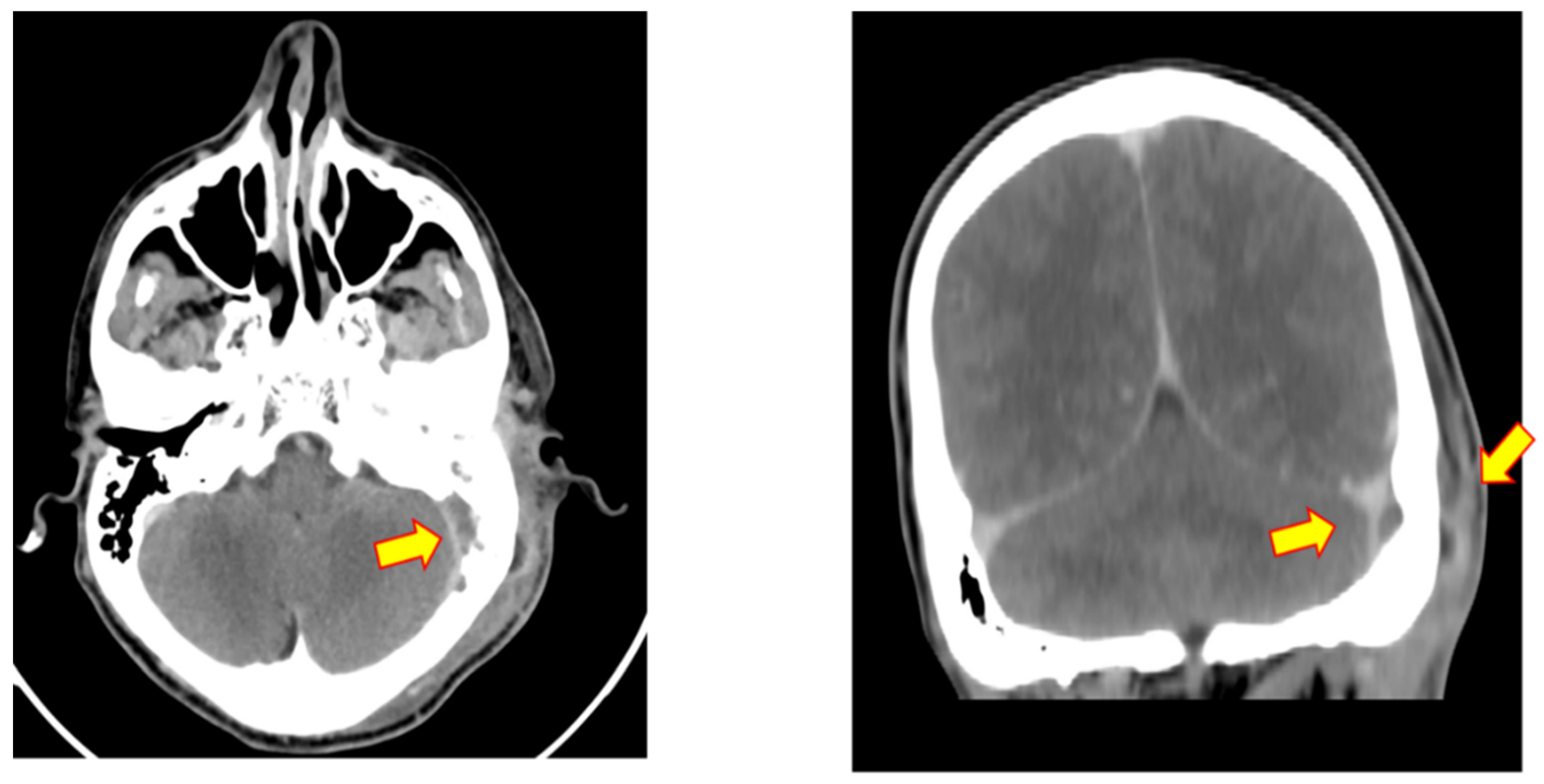
3.1. Case 1

A 44-year-old, previously healthy male presented to the emergency department due to left ear otalgia, otorrhea, and hearing impairment for a duration of 2 weeks. A clinical diagnosis of external otitis media was made, and systemic ciprofloxacin antibiotic therapy and local steroid with neomycin ear drops were initiated. The patient was discharged for follow-up; a swab of the secretion was negative, and the patient experienced a temporary relief of symptoms.
One month after the initial presentation, the patient returned to the emergency department with acute mastoiditis in the left ear with a subperiosteal abscess and was hospitalized for further evaluation and intravenous (IV) antibiotic treatment of amoxicillin with clavulanic acid. Repeated bacterial cultures were taken from the discharge, which were negative.
Laboratory evaluation was notable for normocytic, mildly hypochromic anemia (hemoglobin 11.6 g/dL), absolute leukocytosis of 14 k/uL with 56% neutrophils, and elevated C-Reactive Protein (CRP) of 7.8 mg/dL (normal <0.5 mg/dL). Blood analysis targeting autoimmune causes for chronic diffuse inflammation, including rheumatoid factor; antinucleotide antibodies; complement 3 and 4 levels within normal limits.
Audiometry revealed severe mixed hearing loss of the affected ear, with reduced speech discrimination (88%).
High resolution computed tomography (HRCT) scan (Figure 1 and Figure 2) revealed a complete opacification of both middle ear and mastoid cavity, signs of bone erosion of the mastoid and the perisinus area with a perisinus abscess.
The patient underwent a cortical mastoidectomy and decompression of the sigmoid sinus and insertion of a ventilation tube (VT) into the tympanic membrane. Intraoperative findings included granulation tissue in the middle ear, mastoid, and pus from the perisinus area. A considerable amount of granulation tissues was removed and sent for immunohistopathological examination. Post-operative management included broad-spectrum antibiotic treatment of parenteral Meropenem.
On histologic examination (Figure 3A,B), there was fibrous tissue with chronic active inflammation. Upon immunostaining (Figure 3C), up to 15 IgG cells were seen per high powered field, of them <20% were IgG4 positive. Due to the pathology results, blood serum IgG levels were taken, which were within normal limits (1188 mg/dL; normal 700–1600 mg/dL). Intravenous steroid therapy (methylprednisolone sodium succinate) was initiated, with a clinically good response.
After 16 days of hospitalization, the patient was discharged with recommendations of further investigation by an immunologist who decided on another course of steroid therapy, and additional blood and serum tests.

3.2. Case 2

A 48-year-old male was admitted to the emergency department with complaints of left ear otalgia, dizziness, tinnitus, and hearing impairment lasting for a few weeks. On physical examination there was no evidence of otorrhea. On otoscopy, retraction of the eardrum was detected, along with granulation tissue on the back wall of the ear canal. Head impulse test was positive to the left side. Blood tests were normal and bacterial cultures of ear discharge were negative.
Audiometry revealed severe to profound mixed hearing loss of the affected ear, with reduced speech discrimination and SRT of 40DB.
On CT scan of the left ear (Figure 4) complete opacification of external and middle ear was noted. In addition, extensive bone erosion, involving the mastoid ear cells, the middle ear structure, and the labyrinth was detected (lateral semicircular canal and the cochlea). The patient underwent a canal wall down mastoidectomy, with the removal of granulation tissue from the mastoid and middle ear; repair of lateral canal fistula was performed. The patient required additional revision surgery due to recurrent otorrhea and granulation tissue in the mastoid cavity.
Post-operative management included analgetic treatment and systemic antibiotic treatment (amoxicillin with clavulanic acid). The patient was discharged on the tenth day of hospitalization with significant improvement.
Four months later, the patient was readmitted to the emergency department with complaints of worsening dizziness and tinnitus, fatigue, or vomiting. On left ear micro-otoscopy, the mastoid cavity was filled with granulation tissue. According to neurological assessment, the patient was suspected as having cerebritis.
On temporal bone CT scan, post-operative changes were recorded with notable perisinus fluid and narrowing of sigmoidal sinus. Treatment with dexamethasone and pentoxifylline was initiated, resulting in rapid improvement of symptoms. The patient was discharged with a recommendation of ambulatory MRI follow-up in the timeframe of eight weeks and instructed the continuation of pentoxifylline therapy for the same period of time.
At that point, the initial pathologic sample was revisited. As shown in Figure 5A,B, histologic examination revealed fibrous tissue with foci of dense lymphoplasmacytic inflammation and storiform fibrosis. Upon immunostaining (Figure 5C), up to 25 IgG cells were seen per high powered field, of them <20% were IgG4 positive. Blood serum IgG levels were determined to be within normal limits. Intravenous steroid therapy (methylprednisolone sodium succinate) and methotrexate were initiated, resulting in good clinical response and CNS involvement improved on MRI follow-up.

4. Results

In addition to the two cases presented here, senenteen more cases of IgG4-RD with mastoid involvement were reported between 2012 and 2022. A total of nineteen cases included 5 males and 14 females, ages 45 ± 11 years (mean ± SD). The most common clinical presentations were otitis media and mastoiditis (8, 42% and 7, 37%, respectively); three patients (15.7%) had a clinical presentation of inflammatory pseudotumor.
All patients underwent surgical intervention that included mastoidectomy and surgical site biopsies. Five of the nineteen patients (26.4%) required repeated surgical intervention. Importantly, eight of the cases (42%) involved extra otologic sites: three cases of cranial fossa (middle and posterior), three cases of meningitis, and one case of cervicitis with parotiditis.
Serum IgG4 levels were available in 12 out of 19 patients. Values were elevated in seven patients (>135 mg/dL; 229 ± 50.4 mg/dL) and normal in five patients.
Symptomatology, pharmacological treatment, and chronology of the disease are described in Figure 6. The most frequent symptoms included hearing loss, otalgia, tinnitus and headache as reported in nine, six, and five patients, respectively. Otorrhea was reported in three patients (Figure 6a). The clinical presentation of IgG4-related disease in the ear was not specific nor compatible with other infectious and inflammatory processes of the middle ear or mastoid bone.
All patients underwent CT scans, while MRI was further performed in seven patients, as part of the pre-operative management. Soft tissue opacification and extensive destruction of mastoid bone were noted in 12/19 patients (63%). In two cases, imaging concluded the presence of an inflammatory pseudotumor.
The biopsies obtained from all cases were suggestive of IgG4-RD. Dense lymphoplasmacytic cells infiltrate were reported in all, while storiform fibrosis and obliterative phlebitis was noted in 14/19 cases (73.6%) and 6/19 (31.5%) cases, respectively. Information regarding IgG4/IgG ratio was published in ten cases, averaging 40% IgG4/IgG ratio. Absolute counts of IgG4 were recorded in nine cases, averaging 78 ±68.5 cells per high power field (HPF).
Treatment data (Figure 6b) were reported in all 19 case reports. In most cases, initial therapeutic management, prior to the pathological diagnosis, included antibiotics and symptomatic treatment. Except for three cases, treatment was initiated only after a final pathologic diagnosis had been rendered. The most common treatment was corticosteroids, followed by immunosuppressive agents such as rituximab, cyclophosphamide, and methotrexate. Combined regimens of corticosteroids and rituximab/methotrexate were reported in three of the cases. In these three cases, the treatment was given only after the biopsy (in order to avoid potential obscure of histologic picture). Generally, it can be said that symptom relief and recovery were recorded quickly under treatment. This fact retrospectively helped reinforce the diagnosis based on the histopathological laboratory results.
Patients frequently followed an extended clinical course, from presentation to the hospital to a final diagnosis (Figure 6c). Time elapsed to diagnosis was reported in fifteen of the patients. In six cases it lasted more than 12 months to reach a diagnosis.

5. Discussion

Since the first diagnosis of IgG-RD as autoimmune pancreatitis more than two decades ago [3], various patterns have been identified in different locations of the body. Occasionally the process is limited to a single organ, but the fibrotic and inflammatory process can be multiple organs, as well. Due to unspecified inflammatory and infiltrative pathophysiology nature, there is commonly misdiagnosis of infections, autoimmune conditions, and space-occupied lesions [27].
Regarding the head and neck region, the disease is reported practically in almost every organ, from the clivus, through meninges to the orbits. A systematic review recently published, identified 184 cases of IgG4-RD in the skull and skull base [11]. Disease involving the mastoid bone was reported in ten cases (5.3%), making it the most frequent location affected in the anatomical region of the ear.
The first case of otologic manifestations was reported in 2010 by Masterson et al. [9], and since then an ongoing number of cases have been reported. Qinzhan et al. [28] reviewed the literature and identify 22 cases of IgG-RD with 5 cases involved the mastoid. According to these case studies, otological IgG4-RD consists of progressive hearing loss, otalgia, tinnitus, and vertigo. Recurrent mastoiditis, secretory otitis media, otorrhea, and facial numbness, have been the main clinical symptoms.
IgG4-RD may mimic multiple conditions, including infections, malignancies, and autoimmune diseases [2]. Thus, differential diagnosis may be broad and include not only imaging studies but serology investigations as well, including human immunodeficiency virus (HIV) testing, anti-nuclear antibody (ANA) titers, and antineutrophil cytoplasmic antibody (ANCA) titers, in purpose to rule out other conditions [24].
The gold standard for IgG4-RD diagnosis is histopathology. The key morphologic features of IgG4-RD are threefold: a dense lymphoplasmacytic infiltrate, storiform-type fibrosis, and obliterative phlebitis [2]. In the consensus statement on the pathology of IgG4-RD, an attempt has been made to create criteria for pathological diagnosis in various organs such as the pancreas, lungs, lymph nodes, and salivary glands. Specific criteria regarding the ear and its components were not given, since cases are quite rare.
Secondary in importance, IgG4 immunostaining and calculating per HPF and measurement of IgG4/IgG ratio are recommended, with a comprehensive cutoff value when plasma cell ratio is over 40% [29]. In both cases reported here, IgG4/IgG ratio was lower than 20%, illustrating the certainty of such ratio on definitive diagnosis.
IgG4 levels in serum are not mandatory for IgG4-Rd diagnosis. There are cases where the levels are increased, but the sensitivity and specificity of the immunoglobulin levels are still in doubt [1]. Even though not mandatory, taking IgG4 levels in the serum along with the levels in HPF and IgG4/IgG ratio from the histological examination could have been valuable in the diagnosis process in our cases.
IgG4-RD lesions are well visualized on MRI and/or CT scan, particularly in cases with bone involvement, such as in the mastoid bone. In postcontrast imaging studies, IgG4-RD lesions are typically’ nonspecific- homogenously enhancing, which does not reliably distinguish between IgG4-RD and another inflammatory or tumor process in the bone and its surroundings [30].
To date, there is a lack of randomized controlled trials examining the appropriate treatment for IgG-RD, especially in cases of solely organ involvement. Glucocorticoids are usually the first line of therapy. In 2010, a consensus statement from 17 medical centers in Japan proposed initial treatment with prednisolone for 14 to 30 days [31].
In this study we reported two cases of IgG-RD involving the mastoid bone and the middle ear, showing characteristic histopathological features of the disease. In the second case we reported a long-standing disease at this site, with multiple surgical interventions due to recurrence, with disease extending to the meninges leading to cerebritis. In spite of this long-standing nature, therapy with steroids resulted in stabilization of the disease and resolution of the symptom including the neurological signs.
The diagnostic and imaging process is not a simple challenge for clinicians and may take a long time. However, whenever clinical suspicion arises, whether due to therapeutic failure, recurrence of the disease, or unusual surgical findings, the diagnostic-pathological process can be directed to provide a diagnosis within a short time.
The two cases demonstrate that IgG-RD can present with mastoid bone involvement with local complications such as cerebritis and perisinus abscess formation, without systemic involvement. We demonstrate that the middle ear and mastoid may be the main site for the diagnosis of this entity, requiring a mastoidectomy or middle ear exploration. Even though the middle ear is not one of the typical organs specified in the recent IgG4-RD categorization, its involvement has been demonstrated to be a site of interest in neuro-meningeal presentation [19].
The diagnosis of IgG-RD necessitates a high clinical index of suspicion, which is critical for early treatment to avoid disease progression and recurring revision surgery.
According to the second patient we discussed in this article, it’s not inconceivable to believe that early immunosuppressive medication combined with simple exploratory surgery and biopsy would have been a better option.
This study carries several limitations, mainly related to its nature as a review of the literature. First and foremost, some of the data was missing and did not fully match in all cases. Secondly, the cases have been published in journals from several different disciplines—pathology, radiology, rheumatology, and of course otolaryngology. This is one of the significant challenges in diagnosis and treatment. Hence, it a ‘multi-disciplinary team’ is most appropriate for handling such cases.

6. Conclusions

IgG4-RD is an uncommon disease with varying presentations, involvement of the middle ear and mastoid may mimic infectious diseases at this site. Diagnosis requires a high degree of clinical suspicion and clinic-pathological correlation with biopsies is mandatory, the middle ear and mastoid seem to be a good source for tissue sampling for a conclusive diagnosis.

Author Contributions

Conceptualization, A.S. and S.E.-S.; Writing—Original Draft Preparation, A.S. and S.E.-S.; Writing—Review and Editing, D.M.K.; Data Curation, B.S., R.N. and O.H.; Supervision, D.M.K. and S.E.-S. All authors have read and agreed to the published version of the manuscript.

Funding

This research received no external funding.

Informed Consent Statement

Informed consent was obtained from all subjects involved in the study.

Data Availability Statement

The authors confirm that the data supporting the findings of this study are available within the article and its supplementary materials.

Conflicts of Interest

The authors declare no conflict of interest.

References

  1. Deshpande, V.; Zen, Y.; Chan, J.K.; Yi, E.E.; Sato, Y.; Yoshino, T.; Klöppel, G.; Heathcote, J.G.; Khosroshahi, A.; Ferry, J.A.; et al. Consensus statement on the pathology of IgG4-related disease. Mod. Pathol. 2012, 25, 1181–1192. [Google Scholar] [CrossRef] [PubMed] [Green Version]
  2. Stone, J.H.; Zen, Y.; Deshpande, V. IgG4-related disease. N. Engl. J. Med. 2012, 366, 539–551. [Google Scholar] [CrossRef] [PubMed]
  3. Hamano, H.; Kawa, S.; Horiuchi, A.; Unno, H.; Furuya, N.; Akamatsu, T.; Fukushima, M.; Nikaido, T.; Nakayama, K.; Usuda, N.; et al. High serum IgG4 concentrations in patients with sclerosing pancreatitis. N. Engl. J. Med. 2001, 344, 732–738. [Google Scholar] [CrossRef] [PubMed]
  4. Ebbo, M.; Patient, M.; Grados, A.; Groh, M.; Desblaches, J.; Hachulla, E.; Saadoun, D.; Audia, S.; Rigolet, A.; Terrier, B.; et al. Ophthalmic manifestations in IgG4-related disease: Clinical presentation and response to treatment in a French case-series. Medicine 2017, 96, e6205. [Google Scholar] [CrossRef]
  5. Ozawa, M.; Fujinaga, Y.; Asano, J.; Nakamura, A.; Watanabe, T.; Ito, T.; Muraki, T.; Hamano, H.; Kawa, S. Clinical features of IgG4-related periaortitis/periarteritis based on the analysis of 179 patients with IgG4-related disease: A case–control study. Arthritis Res. Ther. 2017, 19, 223. [Google Scholar] [CrossRef] [Green Version]
  6. Gradoni, P.; Frausini, G.; Pandolfini, M.; Bedetta, S.; Migliori, G. Laryngeal involvement of immunoglobulin G4-related disease: Case report and. B-ENT 2019, 15, 51–54. [Google Scholar]
  7. Yim, C.D.; An, H.J.; Ahn, S.K.; Hur, D.G.; Lee, H.J. IgG4-related disease presenting as otogenic skull base osteomyelitis. Auris Nasus Larynx 2021, 48, 166–170. [Google Scholar] [CrossRef]
  8. Bittencourt, A.G.; Pereira, L.V.; Junior, F.C.; de Santes Halang, F.; de Castro Gonçalves, M.; Bento, R.F. IgG4-related sclerosing disease of the temporal bone. Otol. Neurotol. 2013, 34, e20–e21. [Google Scholar] [CrossRef]
  9. Masterson, L.; Del Pero, M.M.; Donnelly, N.; Moffat, D.A.; Rytina, E. Immunoglobulin G4 related systemic sclerosing disease involving the temporal bone. J. Laryngol. Otol. 2010, 124, 1106–1110. [Google Scholar] [CrossRef]
  10. Cho, H.K.; Lee, Y.J.; Chung, J.H.; Koo, J.W. Otologic manifestation in IgG4-related systemic disease. Clin. Exp. Otorhinolaryngol. 2011, 4, 52. [Google Scholar] [CrossRef]
  11. ler, S.J.; Sharifai, N.; Baker, B.; Dowling, J.L.; Pipkorn, P.; Yaeger, L.; Clifford, D.B.; Dahiya, S.; Chicoine, M.R. IgG4-Related Disease of the Skull and Skull Base–A Systematic Review and Report of Two Cases. World Neurosurg. 2021, 150, 179–196. [Google Scholar]
  12. Wallace, Z.S.; Naden, R.P.; Chari, S.; Choi, H.; Della-Torre, E.; Dicaire, J.F.; Hart, P.A.; Inoue, D.; Kawano, M.; Khosroshahi, A.; et al. The 2019 American College of Rheumatology/European League Against Rheumatism classification criteria for IgG4-related disease. Arthritis Rheumatol. 2020, 72, 7–19. [Google Scholar] [CrossRef] [PubMed] [Green Version]
  13. Schiffenbauer, A.I.; Wahl, C.; Pittaluga, S.; Jaffe, E.S.; Hoffman, R.; Khosroshahi, A.; Stone, J.H.; Deshpande, V.; Gahl, W.A.; Gill, F. IgG4-related disease presenting as recurrent mastoiditis. Laryngoscope 2012, 122, 681. [Google Scholar] [CrossRef] [PubMed] [Green Version]
  14. Li Li, X.; Wang, X.H.; Duan, R.S.; Wang, Z.K.; Yang, B. IgG4-Related Mastoiditis, Hypertrophic Pachymeningitis and Inflammatory Pseudotumor: A Case Report and Review of the Literature. J. Clin. Cell. Immunol. 2018, 9, 2. [Google Scholar] [CrossRef]
  15. Cheng, X.; Shu, Y.; Chen, B. A solely ear-involved IgG4-related sclerosing disease with two-years following-up. European Annals of Otorhinolaryngology. Head Neck Dis. 2019, 136, 401–403. [Google Scholar]
  16. Deshpande, V.; Zane, N.A.; Kraft, S.; Stone, J.H.; Faquin, W.C. Recurrent mastoiditis mimics IgG4 related disease: A potential diagnostic pitfall. Head Neck Pathol. 2016, 10, 314–320. [Google Scholar] [CrossRef] [Green Version]
  17. Barnado, A.L.; Cunningham, M.A. IgG4-related disease presenting as recurrent mastoiditis with central nervous system involvement. J. Investig. Med. High Impact Case Rep. 2013, 2, 2324709614553670. [Google Scholar] [CrossRef]
  18. Vuncannon, J.R.; Panella, N.J.; Magliocca, K.R.; Mattox, D.E. Diagnostic challenges in a case of IgG4-RD affecting the temporal bone. Ann. Otol. Rhinol. Laryngol. 2017, 126, 241–244. [Google Scholar] [CrossRef]
  19. Wuesthoff, C.; Allende, A.; Patel, N. IgG4 disease of the ear: Report and review. SAGE Open Med. Case Rep. 2018, 6, 2050313X18791428. [Google Scholar] [CrossRef] [Green Version]
  20. Lu, P.; Sha, Y.; Wang, F.; Wang, S. IgG4-related sclerosing disease involving middle ear. Otol. Neurotol. 2017, 38, e65–e67. [Google Scholar] [CrossRef]
  21. Shao, S.A.; Lin, C.D.; Tsai, S.T.; Lin, C.C.; Wu, P.C. Immunoglobulin G4-related disease presented as recurrent otitis media and mixed hearing loss treated with cyclophosphamide and rituximab: A case report. Arch. Rheumatol. 2019, 34, 233. [Google Scholar]
  22. Wick, C.C.; Zachariah, J.; Manjila, S.; Brown, W.C.; Malla, P.; Katirji, B.; Cohen, M.; Megerian, C.A. IgG4-related disease causing facial nerve and optic nerve palsies: Case report and literature review. Am. J. Otolaryngol. 2016, 37, 567–571. [Google Scholar] [CrossRef] [PubMed]
  23. Polianskis, M.; Ivaška, J.; Dadonienė, J.; Lengvenis, G.; Besusparis, J.; Rauba, D.; Morozas, A.; Ivaškienė, T.; Lesinskas, E. Immunoglobulin G4-Related Disease Presenting as Temporal Bone Lesion with Facial Nerve Palsy. ORL 2022, 7, 1–729. [Google Scholar] [CrossRef] [PubMed]
  24. Hofmeyr, L.M.; Herbst, G.; Pretorius, E.; Sarembock, B.; Taylor, K.; Roytowski, D. Diagnosis of petrous apex IgG4-related disease via a middle cranial fossa craniotomy and temporal bone biopsy. Front. Neurol. 2022, 9, 957. [Google Scholar]
  25. Chowsilpa, S.; Teeranoraseth, T.; Roongrotwattanasiri, K. Temporal bone involvement of IgG4-related disease: A rare condition misleading to petrous apicitis causing lateral rectus palsy. BMJ Case Rep. 2019, 12, e228550. [Google Scholar] [CrossRef]
  26. Wang, J.; Sun, Z.; Zhuo, S.; Wang, K. Sigmoid sinus occlusion infiltrated by inflammatory myofibroblastic tumor from mastoid. Head Neck 2015, 37, E4–E7. [Google Scholar] [CrossRef]
  27. Khosroshahi, A.; Wallace, Z.S.; Crowe, J.L.; Akamizu, T.; Azumi, A.; Carruthers, M.N.; Chari, S.T.; Della-Torre, E.; Frulloni, L.; Goto, H.; et al. International consensus guidance statement on the management and treatment of IgG4-related disease. Arthritis Rheumatol. 2015, 67, 1688–1699. [Google Scholar] [CrossRef]
  28. Ren, Q.; Su, J.; Zhang, D.; Ding, X. Otological IgG4-related disease with inner ear involvement: A case report and review of literature. Ear Nose Throat J. 2020, 12, 0145561320976411. [Google Scholar] [CrossRef]
  29. Sato, Y.; Kojima, M.; Takata, K.; Morito, T.; Asaoku, H.; Takeuchi, T.; Mizobuchi, K.; Fujihara, M.; Kuraoka, K.; Nakai, T.; et al. Systemic IgG4-related lymphadenopathy: A clinical and pathologic comparison to multicentric Castleman’s disease. Mod. Pathol. 2009, 22, 589–599. [Google Scholar] [CrossRef] [Green Version]
  30. Katsura, M.; Mori, H.; Kunimatsu, A.; Sasaki, H.; Abe, O.; Machida, T.; Ohtomo, K. Radiological features of IgG4-related disease in the head, neck, and brain. Neuroradiology 2012, 54, 873–882. [Google Scholar] [CrossRef]
  31. Kamisawa, T.; Okazaki, K.; Kawa, S.; Shimosegawa, T.; Tanaka, M. Japanese consensus guidelines for management of autoimmune pancreatitis: III. Treatment and prognosis of AIP. J. Gastroenterol. 2010, 45, 471–477. [Google Scholar] [CrossRef] [PubMed]
Figure 1. Pre-operative CT scan—case 1. Axial high-resolution temporal bone CT scan and coronal reconstruction show extensive bone destruction at the medial and posterior aspects of the left mastoid bone. Mastoid, middle ear, and external auditory canal are opacified—see yellow arrows).
Applsci 12 08353 g001
Figure 2. Pre-operative CT scan—case 1. Axial and coronal reconstruction post-contrast head CT shows a large filling defect and enhancement around the sigmoid sinus (delta sign, consistent with sigmoid sinus thrombosis (inner yellow arrow). In the soft tissue there are mall subperiosteal abscess formations (outer yellow arrow).
Applsci 12 08353 g002
Figure 3. Biopsy of left mastoid—case 1. Hematoxylin and eosin (H&E). Original magnification: A–D 100×, E 200×. Fibrous tissue with (A) foci of dense lymphoplasmacytic infiltration. (B,C) Storiform fibrosis is present, as well as areas suggestive of obliterative phlebitis.
Applsci 12 08353 g003
Figure 4. Pre-operative CT scan—case 2. Axial and coronal reconstruction of high-resolution temporal bone CT scan shows opacification of the mastoid air cells (right picture), and extensive bone destruction of the left mastoid bone and of the posterior wall of the external auditory canal, including the cochlea and vestibule (left picture).
Applsci 12 08353 g004
Figure 5. Biopsy of left mastoid—case 2. Hematoxylin and eosin (H&E). Original magnification: A-D 100×, E 200×. (A) Fibrous tissue with (top) foci of dense lymphoplasmacytic inflammation and (bottom) storiform fibrosis (H&E, original magnification 100×). (B) Total IgG and (C) IgG4 plasma cell population (original magnification 400×).
Applsci 12 08353 g005
Figure 6. Clinical features of IgG4 related mastoiditis.
Applsci 12 08353 g006aApplsci 12 08353 g006b
Table 1. IgG4 related disease in the mastoid.
NumberYearLocationAuthorsAgeGenderInitial
Clinical
Diagnosis
Number of Surgical
Interventions
Another
Organ
Involvement
Biopsy
Findings
Pathology IgG4 CellsIgG4 in Serum
12012USASchiffenbauer A.I. et al. [13]50fmastoiditis3-lymphoplasmacytic infiltrate and storiform fibrosisdata not available213 mg/dL
22018ChinaLi Li X. et al. [14]59fmastoiditis1-lymphoplasmacytic cells and a few eosinophils infiltrated with fibrosisIgG4:IgG. > 50%350 mg/dL
32019ChinaCheng X. et al. [15]54fotitis media1-dense lymphoplasmacytic infiltrate, storiform fibrosis, obliterative phlebitisdata not availableNormal
42016USADeshpande V. et al. [16]43fmastoiditis3-Lymphoplasmacytic infiltrate, storiform fibrosisIgG4 200 per HPF; IgG4:IgG > 50%191 mg/dL
52016USA52fmastoiditis1meningesLymphoplasmacytic infiltrate, storiform fibrosisIgG4 110 per HPF; IgG4:IgG 35%Data not available
62016USA50fserous otitis media1-Lymphoplasmacytic infiltrate, storiform fibrosisIgG4 210 per HPF; IgG4:IgG >50%213 mg/dL
72014USABarnado A.L. et al. [17]43finflammatory pseudotumor3cerebritisdense lymphoplasmacytic infiltrate and storiform fibrosisIgG4 positive cells > 200 per HPF200 mg/dL
82017USAVuncannon J.R. et al. [18]35fotitis media2parotid glanddense lymphoplasmacytic infiltrate with scattered eosinophils and storiform fibrosisdata not availableData not available
92018AustraliaWuesthhoff C. et al. [19]56mserous otitis media1-inflamed fibrous tissue, no storiform fibrosis or
obliterative phlebitis
IgG4:igG > 40%229 mg/dL
102017ChinaLu P. et al.
[20]
54fchronic mastoiditis with cholesteatoma1-lymphoplasmacytic infiltration, fibrosis, and sporadic small vasculitisdata not availableData not available
112019TaiwanShao S.A. et al. [21]43motitis media with effusion2-plasma cell granulomaIgG4:IgG > 40%Data not available
122016USAWick C. et al.
[22]
61finflammatory pseudotumor1middle and posterior cranial fossaplasmacytic infiltration, storiform fibrosis and phlebitisIgG4 positive > 50 per HPF Normal
132022LithuaniaPolianskis M. et al. [23]31fmastoiditis1middle cranial fossastoriform fibrosis, dense lymphoplasmatic infiltratesIgG4 positive >80 per HPF
IgG4:IgG 63%
Normal
142022South AfricaHofmyer L. et al. [24]29fmastoiditis1middle cranial fossalymphoplasmacytic infiltration, storifom fibrosis, IgG4 positive > 50 per HPF
IgG4:IgG > 20%
Normal
162019ThailandChowsilpa S. et al. [25]19fotitis media1petrous apexlymphoplasmacytic infiltration, fibrosisIgG4 positive 15 per HPF110 mg/dL
172015ChainaJingye W. et al. [26]38minflammatory pseudotumor1meningeslymphoplasmacytic infiltration, fibrosisIgG4 positive 56 per HPFData not available
182016IsraelPresent study48motitis media with effusion1-fibrous tissue with storiform fibrosis, lymphoplasmacytic infiltration, obliterative phlebitis IgG4 positive 15 per HPF; IgG4:IgG < 20%210 mg/dL
192021IsraelPresent study44motitis media1-fibrous tissue, dense lymphoplasmacytic inflammation, storiform fibrosis IgG4 25 per HPF; IgG4:IgG < 20%Data not available
f—female; m—male; HPF—high power field.
Publisher’s Note: MDPI stays neutral with regard to jurisdictional claims in published maps and institutional affiliations.

