Review

Encapsulated Probiotics: Potential Techniques and Coating Materials for Non-Dairy Food Applications

1
Faculty of Food Science and Nutrition, Universiti Malaysia Sabah, Jalan UMS, Kota Kinabalu 88400, Sabah, Malaysia
2
Food Technology Division, School of Industrial Technology, Universiti Sains Malaysia, Minden 11800, Penang, Malaysia
3
Renewable Biomass Transformation Cluster, School of Industrial Technology, Universiti Sains Malaysia, Minden 11800, Penang, Malaysia
4
Australasian Nanoscience and Nanotechnology Initiative, Monash University, Melbourne, VIC 3168, Australia
*
Author to whom correspondence should be addressed.
Submission received: 8 August 2022 / Revised: 28 September 2022 / Accepted: 29 September 2022 / Published: 5 October 2022
(This article belongs to the Special Issue Innovative Food Products and Processing)

Abstract

:
The growing health awareness among consumers has increased the demand for non-dairy-based products containing probiotics. However, the incorporation of probiotics in non-dairy matrices is challenging, and probiotics tend to have a low survival rate in these matrices and subsequently perform poorly in the gastrointestinal system. Encapsulation of probiotics with a physical barrier could preserve the survivability of probiotics and subsequently improve delivery efficiency to the host. This article aimed to review the effectiveness of encapsulation techniques (coacervation, extrusion, emulsion, spray-drying, freeze-drying, fluidized bed coating, spray chilling, layer-by-layer, and co-encapsulation) and biomaterials (carbohydrate-, fat-, and protein-based) on the viability of probiotics under the harsh conditions of food processing, storage, and along the gastrointestinal passage. Recent studies on probiotic encapsulations using non-dairy food matrices, such as fruits, fruit and vegetable juices, fermented rice beverages, tea, jelly-like desserts, bakery products, sauces, and gum products, were also included in this review. Overall, co-encapsulation of probiotics with prebiotics was found to be effective in preserving the viability of probiotics in non-dairy food matrices. Encapsulation techniques could add value and widen the application of probiotics in the non-dairy food market and future perspectives in this area.

1. Introduction

The growing awareness among consumers regarding healthy lifestyles has increased the demand for food that could provide additional specific health benefits beyond nutrition. Functional food is one of the leading trends in today’s food industry. The term “functional food” refers to foods containing (either present naturally or added by manufacturers) ingredients or bioactive compounds that provide extra health benefits over its adequate nutritional effects, which can beneficially affect one or more physiological mechanisms in the body, resulting in an enhancement in health and reduction in risk for disease, in the amount consumed in a diet [1]. For example, probiotics are one of the dominant groups of functional foods [2].
Probiotics, from the Greek word, “for life”, are defined as “live microorganisms that, when administered in adequate amounts, confer a health benefit to the host” by a joint United Nations Food and Agricultural Organization/World Health Organization working group in 2001 and The International Scientific Association for Probiotics and Prebiotics (ISAPP). Probiotics have also been considered functional foods due to their health-promoting abilities [3]. Among probiotic strains in use today, strains from genera of Lactobacillus and Bifidobacterium are the most frequently used. In addition, other non-pathogenic microorganisms that occur within the host gut or tissues have also been developed as probiotics. These include strains from genera Propionibacterium, Pediococcus, Bacteroides, Bacillus, Streptococcus, Escherichia, Enterococcus, and Saccharomyces. Lately, Faecalibacterium prausnitzii, Akkermansia muciniphila, and Eubacterium hallii have also been identified as potential next-generation probiotics with promising health-promoting functionalities [1,4].
By regulating the natural balance of gut bacteria in the human gastrointestinal tract, probiotics have been shown to promote a wide range of health benefits such as improving intestinal health, improving lactose digestion, enhancing the host’s immune response, reducing serum cholesterol, diarrhea diseases, and inflammatory bowel disease, counteracting allergies, and lowering the risk of certain cancers [5]. For a potential probiotic strain to exert therapeutic effects on the host, the viability of probiotics in food should be at least 6 to 7 log CFU/mL (or CFU/g) when reaching the small intestine and colon. In this regard, the viability of at least 8 to 9 log CFU/mL (or CFU/g) of probiotics in food before ingestion is necessary [3,6].
Probiotics must be stable throughout the digestive tract and able to adhere to human epithelial cells when they reach the intestine. However, the survival of probiotics is greatly affected by the harsh conditions of the gastrointestinal tract, including the acidic pH of the gastric environment and bile acids (a loss of around 2 log CFU/mL or CFU/g during digestion) [7]. Several intrinsic (e.g., pH, water activity, molecular oxygen, the composition of the food, food additives added, and oxidation-reduction potential) and extrinsic factors (e.g., temperature, relative humidity, and gas composition) have also been observed to negatively affect the viability and stability of probiotics during food preparation and food processing, as well as over a prolonged storage period [5,7,8].
Traditionally, dairy products have been recognized as the best carriers of probiotics. Current probiotics have been formulated into numerous dairy products, such as fermented milk, yogurt, cheese, and ice cream. However, consumers’ preferences today lie more with non-dairy-based probiotic products because of the ongoing trend of vegetarianism and awareness of drawbacks associated with the intake of dairy products, such as lactose intolerance, high cholesterol content, and milk protein allergy [2,9]. In recent years, non-dairy matrices, such as fruits [10,11,12], fruit and vegetable juices [7,11,12,13,14,15,16,17,18,19,20,21,22,23,24,25,26], fermented rice beverages [27], tea [28,29], jelly-like desserts [30], bakery products [31,32,33], cereal bars [34], sauces [35], gum products [36], and powdered functional drink [37] have been explored as vehicles to deliver probiotics. Although non-dairy food matrices are more versatile (absent of lactose, dairy allergens, and cholesterol) than dairy food matrices, the delivery of probiotics using non-dairy food matrices is more challenging. As an example of a dairy food matrix, milk, which is rich in proteins and fats, could effectively act as a protective matrix to protect the probiotics throughout the digestive tract [38]. In contrast, non-dairy food matrices, such as fruit and vegetable juices, have considerable amounts of organic acids, dissolved oxygen, and inherently low pH values that could negatively affect the viability of inoculated probiotics [9]. Dairy food matrices are usually stored at refrigerated temperature (4 °C), and therefore, the viability of probiotics can be well-maintained throughout the product’s shelf life. In contrast to dairy food matrices, non-dairy food matrices are often stored at ambient temperature, which could adversely affect the viability of probiotics [2]. The sensory qualities of non-dairy food matrices could also be enhanced or deteriorated by the metabolic compounds produced through the interaction between the probiotics and food matrices [2,9].
To address these challenges, encapsulation techniques have been implemented to preserve the viability of probiotics. Encapsulation can be defined as “a process in which small solid particles, liquid droplets, or gases are entrapped by a coating layer, or incorporated into a homogeneous or heterogeneous matrix, yielding small capsules with useful properties in immobilization, protection, controlled release, structuration, and functionalization” [39]. In other words, encapsulation is a technique of retaining a substance (core material, such as probiotics) within another (wall material). When applied successfully, the encapsulation technique may improve the resistance of probiotics to the harsh gastric environment and hence, facilitate the controlled release and successful delivery of probiotics to the site of action. By restricting the probiotics from being directly in contact with food components, encapsulation could maintain the viability of probiotics during the food manufacturing process and long-term storage. Through encapsulation techniques, probiotic cultures can be transformed into concentrated dry powder form, which is more stable and easier to incorporate into many food matrices [1,40,41,42]. This article aims to review and analyze the effectiveness of encapsulation techniques and supplementation of coating materials on the viability of probiotics in non-dairy food and beverage products during storage, as well as while transiting through our gastrointestinal tract.

2. Encapsulation

To date, encapsulation is one of the most promising techniques in protecting active compounds against adverse environments. Encapsulation technology has been widely used in the pharmaceutical, medicine, nutritional, food science, biological, agriculture, toiletries, and cosmetics industries for over 50 years. The goal of encapsulation is to protect the encapsulated active compound (core material) against unfavorable or adverse environments (such as light, moisture, temperature, and oxygen). In food industries, a broad range of products (including probiotics, antioxidants, antimicrobials, flavors, enzymes, and nucleic acids) are encapsulated to (a) prevent the core material from degradation, (b) slow down the evaporation rate of volatile core material, (c) separate the components that would otherwise react with each other, (d) modify the nature of the core material for easier handling, (e) increase the stability, (f) to mask undesired tastes, colors, and odors, (g) enable sustained and controlled release (release slowly over time at a constant rate), (h) control oxidative reactions, (i) use with bacteriophages to control foodborne pathogens, and (j) extend the shelf life. Indeed, encapsulation is one of the new and effective methods to protect probiotics from the harsh conditions they encounter throughout food processing, shelf storage, and gastrointestinal transit [1,40,41,42].

3. Probiotic Encapsulation Techniques
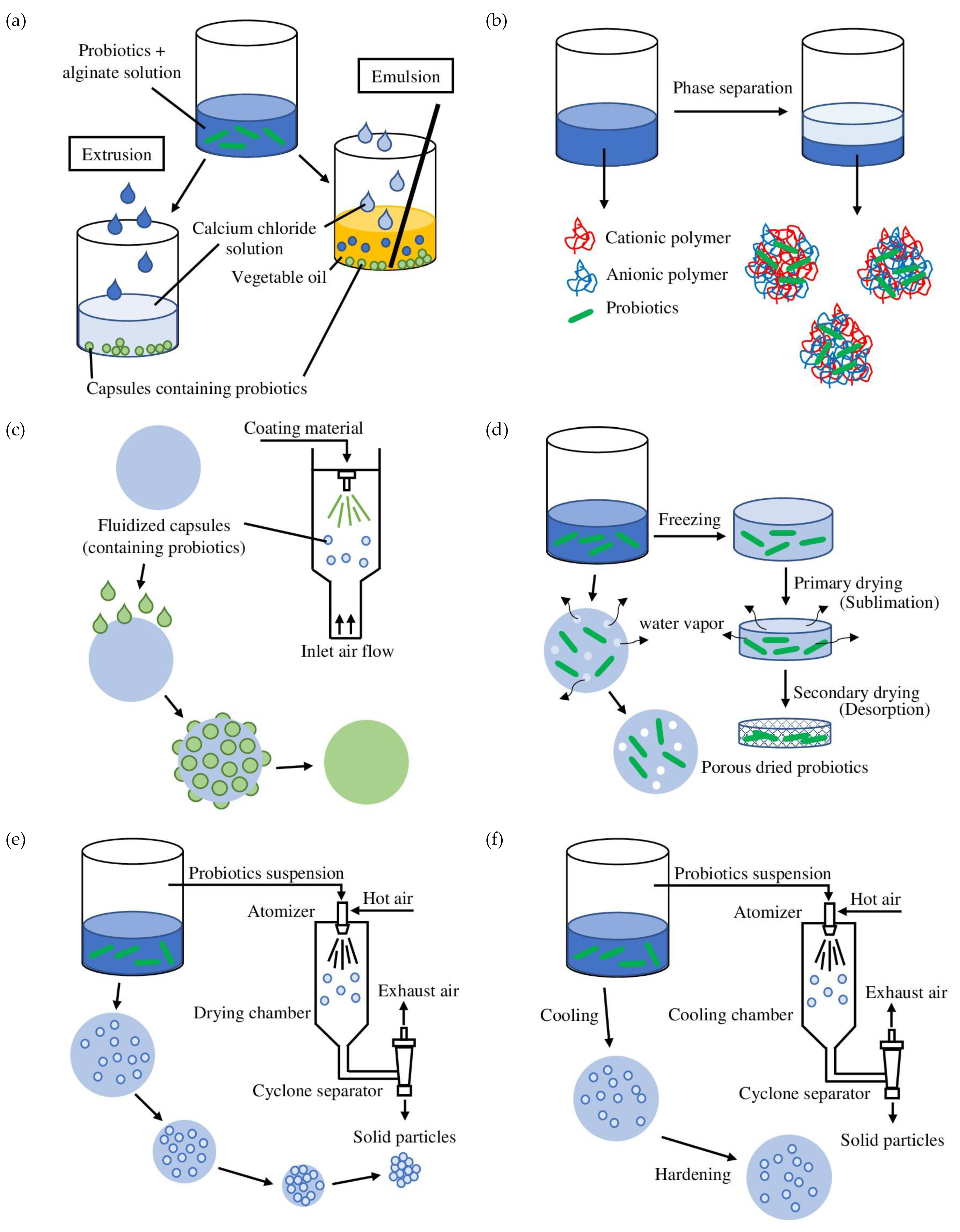
Numerous encapsulation technologies have been developed and adopted to protect probiotics. All the techniques aim to protect the viability and stability of probiotics. However, their concepts, operation methods, and properties of produced capsules are different. Each technique also has its own strengths and drawbacks. Figure 1 illustrates different types of probiotics encapsulation techniques and the morphologies of corresponding microcapsules obtained. Various aspects must be taken into consideration before the selection of encapsulation techniques. Selecting a suitable encapsulation technique depends on several parameters, such as the nature of the probiotics, the operational conditions of the encapsulation technique, the properties of the biomaterials used, the particle size needed to deliver the adequate probiotics load without affecting the sensory properties, the release mechanism and release rate, the composition of the target food application, the storage conditions of the food products before consumption, and lastly, the cost limitation of production [43,44].

3.1. Extrusion

Extrusion (also known as external ionic gelation, which produces capsules with sizes of 100 μm to 5 mm) is the oldest and the most common physical technique for encapsulating the probiotic cell. In the extrusion technique, probiotics are first suspended in a biopolymer solution. The suspension is then fed into an extruder (pilot scale) or a syringe needle (laboratory scale) and drips off into a hardening solution (most commonly, calcium chloride) with gentle stirring [40,45].
The extrusion technique is relatively simple, direct, straightforward, and gentle (does not involve extreme temperature, pH condition, and organic solvents), thus resulting in relatively high viability (low cell harm) of probiotic microorganisms and requiring a lower operational cost. This technique can be conducted under both aerobic and anaerobic conditions. It is biocompatible and flexible as it does not require any harmful solvents. Using alginate/shellac blend and sunflower oil as wall and core materials, respectively [40,41,43,45], Silva et al. [46] demonstrated that the co-extrusion encapsulation technique increased the probiotic survival of L. acidophilus LA3 by about 80% in simulated gastrointestinal conditions and 83% of the probiotics loaded into dried particles were viable after 60 days storage at room temperature (25 °C). Kim et al. [47] also demonstrated that encapsulation of probiotic L. acidophilus by ionic gelation between phytic acid and chitosan followed by the addition of calcium carbonate and starch with electrostatic extrusion provided buffering effects and protection against acid injury during simulated gastric conditions and refrigerated storage. The extrusion technique has also been utilized in non-dairy probiotic foods such as E. faecium in cherry juice [13], L. lactis ABRIINW-N19 in orange juice [17], L. casei DSM 20011 in pineapple, raspberry, and orange juices [18], L. acidophilus TISTR 2365 in sweet fermented rice sap beverage [27], L. acidophilus NCFM in mulberry tea [29], L. casei Lc-01 and L. acidophilus La5 in mayonnaise [35], and L. reuteri in chewing gum [36].
However, the size of beads produced through the extrusion technique is relatively big (up to 5 mm), and the process of bead solidification is also relatively slow. Hence, this technique is not suitable to be used in large-scale production [40,43,45]. Over the last decades, an evolving extrusion technique (vibrating nozzle method) has been focused on and studied. This new extrusion technique uses vibrating technology (mechanical principle), in which, when a defined amplitude is enforced, the vibrational frequency will break the extruded fluid into pre-defined-sized droplets. The size of the droplets generated using this technique can be controlled through the diameter of the jet, the velocity of the extrusion process, the viscosity and the surface tension of the fluid, and the frequency of disturbance [41].

3.2. Emulsion

In the emulsion (also known as internal ionic gelation, which produces capsules with sizes of 200 nm to 1 mm) encapsulation processes, the suspension containing probiotics cell and polymer (disperse phase) is first dispersed into vegetable oil (continuous phase) and homogenized to form a water-in-oil (W/O) emulsion in the presence of a surfactant (emulsifier). After emulsification, calcium chloride (cross-linking agent) is added to insolubilize and harden (fast gelling process) the water-soluble biopolymer. The gel beads can then be harvested by filtration or centrifugation [40,43].
Compared to the extrusion technique, the emulsion technique is easier to scale up for mass production. Hence, it is more suitable for application at the industrial level [45]. Additionally, high survival of probiotics was also reported after encapsulation using the emulsion technique [43]. Singh et al. [48] found that probiotic L. rhamnosus GG encapsulated in a homogeneous system of carboxymethyl cellulose/gelatin blend survived better under simulated intestinal tract conditions compared to free probiotics. Picone et al. [49] revealed that encapsulated L. rhamnosus in gelled water-in-oil emulsions had a survival rate of more than 77% against in vitro digestion. Probiotics (B. bifidum [7], L. acidophilus PTCC1643, B. bifidum PTCC 1644 [15], L. plantarum, L. fermentum, L. casei, L. sphaericus, S. boulardii [16], and Lactobacillus salivarius spp. salivarius CECT 4063 [10]) encapsulated using the emulsion technique have also been reported as suitable to be used in non-dairy food matrices such as grape [7,15], tomato, and carrot [16] juices, and apple matrix [10]. Furthermore, the emulsion encapsulation technique is flexible since it can adjust and control the beads’ size. According to Oberoi et al. [40], the diameter of the beads produced through the emulsion technique can be reduced to 25 µm, which cannot be achieved using extrusion methods. However, the emulsion technique has a high operational cost due to the high price of vegetable oil (such as soy, sunflower, corn–millet, and light paraffin oil) [40,45]. In addition, the microcapsule produced is not suitable for use in low-fat food products due to the oil residual in the capsule [39].
Considering that conventional emulsions are thermodynamically unstable, advanced emulsion technologies such as nano-emulsion, Pickering emulsion, and Pickering high internal phase emulsion are implemented for efficient probiotics encapsulation. Nano-emulsion is a relatively stable emulsion system with smaller droplet sizes ranging from 50 to 200 nm. In the study by Vaishanavi and Preetha [50], nano-emulsions containing soy protein isolate, Tween 80, and gum Arabic were prepared for encapsulating L. delbrueckii subsp. bulgaricus. The stability and survivability of the probiotics loaded in nano-emulsions were well-maintained throughout the storage period of 40 days. Contrary to nano-emulsions, Pickering emulsion is an emulsion system that does not require emulsifiers in the stabilization process. Pickering emulsion is stabilized rather by solid particles (more effective with hydrophobic particles). Pickering-type double emulsion (water-in-oil-in-water, stabilized with polyglycerol polyricinoleate) has also been used to encapsulate probiotics (L. acidophilus) [51]. The viability (gastric digestion = 93.59%, intestinal digestion = 84.24%) and colon-adhesion efficiency (43.27%) of probiotics entrapped in the double emulsion were higher than the free probiotics (viability after 1 h gastric digestion = 0%, colon-adhesion efficiency = 14.20%) during storage (14 days) and after exposure to simulated gastrointestinal conditions. Pickering high internal phase emulsion is a Pickering-type emulsion with a high internal oil phase fraction. By limiting the probiotics from contact with water and oxygen, Pickering high internal phase emulsion is known to possess high encapsulation efficiency and serve as a promising delivery system for probiotics. In a study conducted by Qin et al. [52], Pickering high internal phase emulsion stabilized with the covalent conjugates of whey protein isolate and (-)-epigallocatechin-3-gallate was used to encapsulate and protect probiotics (L. Plantarum). The probiotics encapsulated in the emulsion showed higher viability after storage (14 days) and were more resistant to acidic medium, bile salts, and digestive enzyme digestion when compared to the free probiotics.

3.3. Coacervation

Coacervation (phase separation, which produces capsules with sizes of 1 μm to 1 mm) is a process whereby an initial solution is separated into a polymer-rich phase (coacervate) and a polymer-poor phase (coacervation medium). Coacervation techniques can be further categorized into simple and complex coacervations. Simple coacervation involves only a single polymer. In simple coacervation, phase separation can be induced when a strongly hydrophilic substance, water-miscible non-solvent, or inorganic salt (desolvation of the polymer) is added into a colloid solution. On the other hand, complex coacervation refers to the ionic interactions between two or more polymers (usually a protein and a polysaccharide) of opposite charges. During complex coacervation, when the charge is neutralized, the polymers separate, deposit on the droplet, and form coacervates [41,53]. Therefore, complex coacervation is preferable in probiotics encapsulation and the food industry [43].
Complex coacervation is known to produce capsules with high loading capacity that can incorporate a high number of probiotics. This technique provides high encapsulation efficiency, even at a very high (99%) payload. Complex coacervation is a simple process that does not involve high temperatures and hence, is safe for probiotics. Sharifi et al. [54] showed that probiotic L. plantarum and phytosterols, co-entrapped by heteroprotein complex coacervation utilizing whey protein isolate and gum Arabic, resulted in increased probiotic viability in Iranian white cheese. Silva et al. [46] demonstrated improved resistance to simulated gastrointestinal tract conditions of the microcapsules of probiotic L. acidophilus encapsulated by complex coacervation followed by transglutaminase crosslinking (up to 9.07 log CFU/g survival) and maintained probiotic viability (up to 9.59 log CFU/g) for 60 days at freezing (−18 °C) temperature.
The complex coacervation technique also produces microcapsules with water immiscibility which leads to optimal controlled-release properties. Complex coacervation can produce microcapsules with sizes ranging from 1 to 100 µm. However, complex coacervation is hard to scale up, as the solute used to form coacervates must be in liquid form. The range of polymers employed in this technique is also limited as coacervates are only stable within a range of pH, ionic strength, and temperature. Gelatin is the most common polymer employed in complex coacervation. However, the use of animal-based protein is limited in certain situations [55]. Zhao et al. [56] demonstrated that in comparison with the protein/polysaccharide complex coacervation, the encapsulated probiotic in water/water emulsion via type-A gelatin/sodium caseinate coacervation had a better survival rate after heating, ambient storage, and simulated digestion. The authors indicated that the increased protection of the type-A gelatin/sodium caseinate matrix was associated with lower hygroscopicity, solubilization, and wettability and could also be caused by the significantly higher hydrophobicity. Complex coacervation is also regarded as a costly technique because an additional hardening process is required.
Complex coacervation is suitable for non-dairy probiotic foods. In the study by Silva et al. [22], probiotic orange and apple juices were produced with the aid of complex coacervation associated with enzymatic crosslinking. As indicated by the results, encapsulated L. acidophilus LA-02 incorporated in fruit juices can survive throughout a storage period of 63 days (4 °C). In addition, complex coacervation was also used by Holkem et al. [14] to encapsulate B. animalis subsp. lactis in the development of probiotic sugar cane juice. The viability of B. animalis during storage and delivery was enhanced through complex coacervation.

3.4. Drying Method

3.4.1. Spray-Drying

In food industries, spray-drying is the most used method to dry the encapsulated mixture into powdered probiotics (capsule sizes: 5–150 μm). The principle in spray-drying is the simultaneous mass and heat transfer processes between hot air and droplets. There are three main processes involved in the spray-drying process (i) atomization of a solution comprising probiotics and core material into fine droplets, (ii) droplets evaporation in a heated gas stream, and finally, (iii) separation and collection of spray-dried powder [43,44].
The advantages of this drying technique include (i) the process is rapid and continuous, (ii) this technique does not require a high operational cost (10 times cheaper compared to freeze-drying), (iii) highly reproducible, easy for scaling up and suitable for industrial application, and (iv) the spray-dried products are typically dry, low in water activity, highly stable, and have low bulk density. Studies about the encapsulation of probiotics by spray-drying have been extensively reported. For example, Arslan et al. [57] showed that probiotic Saccharomyces cerevisiae var. boulardii microencapsulated with gum Arabic, pea protein, and gelatin by spray-drying were more resistant to simulated stomach solution. Jantzen et al. [58] also demonstrated that probiotic L. reuteri cultivated in whey slurry microencapsulated by direct spray-drying showed a 32% greater survival rate upon exposure to simulated digestive juice than those without encapsulation. Numerous studies have used spray-drying encapsulation in non-dairy probiotic food. Vivek et al. [20] demonstrated that spray-dried Sohiong juice fermented with L. plantarum remained viable (6.12 log CFU/g) for 36 days of storage at 25 °C. A study by Hernández-Barrueta et al. [28] showed that the viability of spray-dried L. rhamnosus GG in a matrix of whey protein isolate and hydrolyzed extruded huauzontle starch was stable in a ready-to-drink green tea beverage during the 5 weeks refrigerated storage.
The drawback of this technique is the harsh processing conditions, which can cause adverse effects on the stability, viability, and survivability of the probiotics [1,40,43]. For instance, the high temperature and osmotic stress applied during spray-drying can kill the probiotics. Furthermore, high air velocities during spray-drying can result in microcapsules formed with poor uniformity in terms of particle size and morphology.

3.4.2. Freeze-Drying

Freeze-drying, also known as lyophilization or cryodesiccation, is a process whereby the water vapor in the frozen sample is removed through the sublimation of ice. This technique produces capsules with sizes of 1–1.5 mm. It is commonly used to preserve thermosensitive components such as probiotics. The process of freeze-drying can be divided into three phases, (i) the initial freezing process of the probiotics (together with the carrier material), (ii) the primary drying (sublimation) phase, and (iii) secondary drying to eliminate the remaining traces of water due to absorption [43].
Rishabh et al. [23] used freeze-drying and spray-drying to encapsulate E. faecalis incorporated in carrot juice using gum Arabic and maltodextrin as coating materials. Compared to spray-drying, heat injuries to the probiotics are lower in the freeze-drying technique. Raddatz et al. [59] reported that L. acidophilus microencapsulated in the form of emulsification/internal gelation followed by freeze-drying using a blend of pectin micro-particles with prebiotic rice bran maintained probiotic viability for 120 days at 25 °C. In another study, Massounga Bora et al. [25] used freeze-drying to encapsulate L. acidophilus and L. casei using whey protein isolate and fructooligosaccharides as wall material in the development of probiotics-enriched freeze-dried banana powder. During the 30 days of refrigerated (at 4 °C) storage, the encapsulated probiotics had higher survivability compared to the free cells. The encapsulated probiotics were also more resistant to simulated gastric intestinal fluid.
However, in another investigation by Shoji et al. [60], the authors did not obtain the same positive findings using microencapsulation of L. acidophilus Lac-04 through complex coacervation followed by freeze-drying. The authors observed a significant decrease in viability (p < 0.05) after 30 days at 37 °C. The microencapsulated probiotics failed to withstand the pH condition of the human stomach. Although the freeze-drying technique has been reported to provide shelf stability to probiotics, sometimes the crystal formation during the freezing process can result in cell injury and eventually lead to cell death. Therefore, cryoprotectants that exert protection for the probiotics are necessary [61]. Cryoprotectants protect probiotics from freezing damage by inhibiting rapid cellular dehydration and ice formation during freeze-drying. Furthermore, freeze-drying is an expensive procedure with high operational and maintenance costs and is not easy to scale up [40].

3.4.3. Spray Chilling

Spray chilling (cooling or congealing, which produces capsules with sizes of 20–200 µm) is like spray-drying, but it injects cold air to atomize and solidify the particles instead of hot air. In the spray chilling process, the encapsulated agent is first dispersed in a molten lipid matrix before atomization in a chamber with cold air injection [62]. In this technique, lipids are utilized as the encapsulation material. During the passage through the gastrointestinal tract, when the temperature reaches the melting point of the carrier material (lipid), the lipases in the intestines digest the lipid wall materials and release the probiotics. Therefore, spray chilling was found to be very promising in the controlled release of probiotics. Spray chilling is reported to be the cheapest encapsulation technique as it provides a high process yield regardless of continuous or batch production. Spray chilling has also been recognized as more environmentally friendly as it requires only mild processing conditions, low operation energy, and time. Since spray chilling does not require heat, the viability of probiotics can be retained. Furthermore, this technique can be operated continuously with the elimination of hold times between manufacturing steps, making it suitable to be scaled up for industrial-scale production [43,48].
S. boulardii, L. acidophilus, and B. bifidum have been encapsulated using the spray chilling technique [63]. The results showed that the survivability of spray-chilled (S. boulardii, 97.89%; L. acidophilus, 83.57%; B. bifidum, 88.50%) and spray-dried (S. boulardii, 97.51%; L. acidophilus, 84.05%; B. bifidum, 90.10%) probiotics under simulated gastric conditions were similar. The spray chilling technique has also been used to encapsulate probiotics in probiotics-enriched cream-filled cakes [31] and savory cereal bars [34]. Spray chilling improved the survivability of S. boulardii, L. acidophilus, and B. bifidum incorporated in cream-filled and marmalade-filled cake samples during refrigerated storage [31]. Similarly, the viabilities of spray-chilled L. acidophilus and B. animalis subsp. lactis were higher than freeze-dried and free probiotics in the savory cereal bars after being stored for 90 days at 4 °C [34]. However, low encapsulation capacities on the beads produced through this technique have been reported. A lower load (10–20%) was obtained when compared to spray-drying (5–50%) [64]. The beads produced through the spray chilling technique have low melting points (32–42 °C). Probiotics encapsulated using the spray chilling technique were also found to protrude from the beads during storage. Hence, proper handling and storage conditions are required to preserve spray-chilled probiotics [43,44].

3.4.4. Fluidized Bed Drying

Fluidized bed drying, or fluidized bed coating, is a modified spray-drying method that involves intensive, simultaneous heat and mass transfers between solid particles in a suspension (produces capsules with sizes of 5–5000 μm). In the fluidized bed drying process, dried pre-encapsulated probiotics are first suspended in a hot air flow. Subsequently, the surfaces of the particles are fluidized with the biopolymer solution. The biopolymer coating is then solidified into a homogeneous layer surrounded by the pre-encapsulated probiotics [44,50]. In the fluidized bed drying process, the aqueous medium is dried in a uniform airflow, and the dried particles are suspended in the heated air. Hence, the particles are evenly dried with much less agglomeration and are uniformly coated. Compared with spray and freeze-drying processes, fluidized bed drying requires less energy consumption, and therefore, it is comparatively economical. Compared to other techniques, a lower drying temperature (ranges from ambient temperature to 120 °C) can be set and used in fluidized bed drying. Hence, it can preserve heat-sensitive probiotics. For example, Sánchez-Portilla et al. [65] proved that the viability of Bifidobacterium sp. was retained for more than 2 years, with a concentration exceeding 5 log CFU/g, as well as resistance to acid and complete enteric-targeted release, through the fluidized bed drying technique. The fluidized bed drying technique is ideal for food industries as it is easy to scale up and can be prepared in large batch volumes and high throughputs. Fluidized bed drying can provide multi-coating layers. Thus, it can contribute to a variety of functional properties. Nevertheless, this technique is time-consuming (~2 h), likely to kill the probiotics, and it is not easy to master. Therefore, probiotics should be encapsulated before drying in a fluidized bed dryer [43,44,62,66].
Fluidized bed drying techniques have been successfully used by Galvão et al. [11], Mirzamani et al. [32], and Nilubol and Wanchaitanawong [26] to preserve the viability of probiotics in non-dairy food matrices. Galvão et al. [11] dried and coated apple cubes with a mixture containing hydroxyethyl cellulose and polyethylene glycol containing B. coagulans using the fluidized bed drying technique. The viability of probiotics in the dried apple snacks was well preserved during the storage period. The fluidized bed drying technique has also been used by Mirzamani et al. [32] to develop probiotic bread. The double-layered (first layer: microcrystalline cellulose powder and alginate or xanthan gum; second layer: gellan or chitosan) microcapsules produced through the fluidized bed drying technique had higher heat resistance and could protect the encapsulated probiotics (L. Sporogenes) under baking conditions. In the study of Nilubol and Wanchaitanawong [26], carrot tablets containing L. plantarum TISTR 2075 were produced using a fluidized bed drying technique employing gelatin. The finding indicated that the L. plantarum TISTR 2075 encapsulated in carrot tablets (survivability: 77.68–87.30%) had higher tolerance against heat digestion treatments than free cells (39.52%).

3.5. Layer-by-Layer Method (Multilayer Technique)

For better performance, encapsulated probiotics are coated with more than one layer, using different polymers for each layer. The layer-by-layer method (multilayer technique) was proven to increase the survivability of probiotics against the conditions of processing, storage, and along the gastrointestinal tract [50,67]. For instance, Beldarrain-Iznaga et al. [68] revealed that microencapsulation of L. casei using a combination of layer (canola oil)-by-layer (sodium caseinate) double emulsion and ionic gelation technique could enhance the thermal stability and cell viability of L. casei during storage and digestion. The functional characteristics of L. casei C24 were also retained through microencapsulation using the layer (alginate)-by-layer (chitosan) double emulsion technique [68]. In another study, layer (carboxymethyl cellulose)-by-layer (zein protein) encapsulating L. plantarum 299v was applied to apple slices [12]. The two-layer coating was able to protect the probiotics both under storage and during simulated gastrointestinal conditions.
The layer-by-layer technique involves the alternative adsorption of positively and negatively charged materials on surfaces through the chemical electrostatic deposition technique. This technique produced a protective outer layer on a microencapsulated probiotic by immersing the capsule in a biopolymer solution. This layer coating process can be repeated several times until the desired number of layers or thickness is obtained. The strength of the multilayer-coated capsule can be enhanced by increasing the interaction intensity between the charged materials. This is made possible by modification of the pH, concentration, and ionic strength of the polymer solution [31,45,60].
This technique does not involve high operational costs, as only mild conditions, aqueous solutions, and naturally charged materials are used in the coating process. The thickness, permeability, strength, and morphology of the layers can be tailored depending on the desired application. However, the adhesion times of each layer are between 1 and 60 min, which is not instantaneous. This leads to a certain degree of aggregation of the capsules during the adhesion of the subsequent layer, reducing the available surface area for consecutive layer adhesions [53,69].

3.6. Co-Encapsulation

Co-encapsulation is an encapsulation method that utilizes the synergistic effect of two or more bioactive substances that can positively influence each other to enhance the function/viability of the encapsulated substances. This technique has been used in drugs and bioactive components in pharmaceutical industries [62]. However, in recent years, considerable attention has been given to co-encapsulation processes in food industries. Co-encapsulation has been proven to be able to sustain and enhance the viability of probiotics [17,18,27,29,35,36,70].
Co-encapsulation of probiotics together with prebiotics has received attention from food researchers. The effect of co-encapsulation of probiotics with arrowroot starch for yogurt was investigated in the study of Samedi and Charles [71]. After being stored in ambient and refrigerated conditions for 90 days, the co-encapsulated probiotics had higher viability when compared to the free probiotics. The probiotics co-encapsulated with arrowroot starch with low digestibility and prebiotic potential were more resistant to the harsh conditions in the gastrointestinal tract and acidic conditions in yogurt. Furthermore, Zaeim et al. [72] investigated the protective role of polysaccharide matrix (inulin or resistant starch in calcium-alginate/chitosan microcapsules) on the co-encapsulated probiotics (L. plantarum ATCC 8014 and B. animalis subsp. lactis) under gastrointestinal conditions and storage at -18, 4, and 25 °C. The presence of inulin and resistant starch in the microcapsules improved the survivability of these probiotics. Shinde et al. [73] also demonstrated that co-extrusion encapsulation of probiotic L. acidophilus with apple skin polyphenol extract using an aqueous delivery system possessed >96% microencapsulation efficiency and improved viability under low pH conditions (pH 2, 37 °C, 120 min) and after 50 days refrigeration storage (4 °C) in milk. Overall, encapsulated probiotics with resistant starch had stronger resistance against gastrointestinal conditions compared to the ones with inulin. Resistant starch could prevent gastrointestinal acid from diffusing into the microcapsules by entrapping within the porous alginate matrix. As the carbon source, resistant starch could improve the survival of probiotics during storage and also enhance the colonization and proliferation of probiotics in the intestines [72].
Table 1 shows the main properties, advantages, and disadvantages of encapsulation techniques that can be applied in multilayer and co-encapsulation techniques of probiotics.

4. Biomaterials Utilized for Probiotics Encapsulation

To be an effective encapsulation material (core or wall material), the biomaterial used must be able to protect the encapsulated probiotics along the gastrointestinal tract until reaching the target site (small intestine/large intestine), where they can exert their health-promoting effects. The encapsulation material should only release the encapsulated probiotics when it is exposed and triggered by certain environmental conditions (such as temperature, pH, and enzyme activity). In other words, the capsules containing probiotics should remain protected inside the encapsulation material during the passage through the stomach and only decompose after reaching the target site to release the probiotics. The commonly used biomaterials in probiotic encapsulation include carbohydrates, proteins, and lipids, which will be discussed in detail in the coming subsections. Their specific advantages and limitations in probiotic encapsulation are also summarized in Table 2.

4.1. Carbohydrate Polymers

4.1.1. Alginate

Among the carbohydrate polymers used, the most common biomaterial is alginate. Alginate can be produced by various brown seaweeds (Laminaria digitata, Laminaria hyperborea, Laminaria japonica, Macrocystis pyrifera, and Ascophyllum nodosum) and two genera of bacteria (Pseudomonas and Azotobacter), making it abundant and comparatively low in cost [45]. Alginate is the preferred biomaterial in probiotic encapsulation owing to its non-harmful nature, ease in producing strong beads, and being promptly accessible. Alginate has good gelling, balancing out, and thickening properties, and is easy to manipulate, biocompatible, and biodegradable [1,40,45,79]. Alginate is a pH-responsive polymer that is stable at lower pH and unstable in higher pH conditions which is beneficial in customizing release profiles. During the delivery, alginate beads tend to shrink in low acidic gastric environments. Hence, it prevents the release of the encapsulated probiotics from the beads. Once the beads reach the small intestine with alkaline conditions, the alginate transforms into a soluble alginic acid layer. Subsequently, they swell and release the encapsulated probiotics [75]. Unfortunately, alginate is sensitive to heat treatment, porous, unstable, and has poor barrier properties because of its high molecular mobility and weak interaction between the molecular chains [1,45]. The weakness of alginate can be overcome through a crosslinking reaction with divalent cations or co-encapsulation with starch or by coating the alginate capsules with an extra layer (multilayer technique) made of a different type of biomaterial [92]. The ionic crosslinking of alginate chains with calcium cations could result in a strong gel structure. The presence of calcium cations could also disrupt the water coordination of the alginate network [79]. The synergistic effects of alginate and starch of the alginate capsules could protect the entrapped probiotics [45]. With the interaction of the negatively charged carboxylic groups of alginate with positively charged amine groups chitosan, stronger, ordered three-dimensional gel networks can be produced. The resulting capsules also have smoother surfaces with decreased water permeability [67].

4.1.2. Chitosan

Chitosan originates from chitin which is naturally synthesized by algae and the shell waste of crab, shrimp, and crawfish [45]. Chitosan-based hydrogels have been extensively employed to deliver probiotics owing to their unique cationic character, non-toxicity, high biocompatibility, biodegradability, bio-adhesiveness, inexpensive nature, antimicrobial, and antifungal properties [67,75,93]. Chitosan also has high tolerance against the deteriorative effects of calcium chelating and anti-gelling agents [67]. However, chitosan is a pH-sensitive material that tends to degrade in low pH conditions and is water-insoluble at pH > 5.4. Therefore, it is less effective in the delivery of probiotics to the gut [75]. Moreover, using chitosan as a polymer for entrapping live lactic acid bacteria (LAB) could exhibit inhibitory effects on the LAB [1]. Therefore, it is commonly applied as a coating or shell rather than a capsule matrix. Chitosan has been extensively used in combination with other biomaterials, including alginate, starch, whey protein isolate, and xanthan gum [1,45]. Chitosan coating could enhance the porosity of alginate beads, thus, reducing leakage of encapsulated bacteria and improving the pH stability of beads [67]. Chitosan coating increased the release rate of probiotics from alginate/starch beads and enhanced the survivability of probiotics in low pH conditions [94]. The chitosan coating on alginate/whey protein isolates beads increased the resistance to thermal, storage, and simulated gastrointestinal environment [95]. In addition, the heat resistance of microcrystalline cellulose/Xanthan gum beads has also been enhanced by chitosan coating [32]. Encapsulation of probiotics using microcrystalline cellulose powder and alginate, or Xanthan gum followed by coating with chitosan (0.5%) as the outermost layer is effective in protecting probiotics (L. Sporogenes) against the baking process (90 °C for 15 min) in bread making.

4.1.3. Gums

Xanthan gum has been proven as an excellent embodiment in conferring protection against harsh gastrointestinal conditions and elevated temperatures (up to 90 °C for 5 s) to probiotics. It is an exopolysaccharide obtained through fermentation by Xanthomonas campestris from agro-industrial wastes [79]. Xanthan gum possesses an anionic character, is non-toxic, biodegradable, biocompatible, highly soluble in cold and hot water, and has excellent gelling properties. Furthermore, it has excellent heat and acid stability and is highly resistant to gastrointestinal digestion and enzymatic decomposition [41,82]. The study of Thang et al. [33] indicated the protective effect of Xanthan gum on the viability of L. acidophilus incorporated in bread under simulated gastric and intestinal conditions compared to using alginate alone. Xanthan gum has a negative charge structure that could bind to H+ ions and minimize the effect of an acidic condition on the probiotics. Unfortunately, it has some limitations. It is susceptible to microbial contamination, unstable viscosity, and uncontrollable hydration rate, as well as producing gels with poor shear resistance, mechanical strength, and thermal properties when used solely [41,83]. Therefore, to enhance the coating properties of Xanthan gum in probiotic encapsulation, it is combined and used with other biomaterials, including alginate, chitosan, gellan, and β-cyclodextrin [41]. In contrast to alginate beads, the combination of xanthan and gellan gums produces capsules with higher resistance toward acid conditions [1,41].
Gellan gum is a microbial polysaccharide, industrially produced through fermentation by Sphingomonas elodea and Pseudomonas elodea. It is available in two forms, low acyl gellan gum (deacylated; Kelcogel) and high acyl gellan gum (acylated; Gelrite). Upon cooling, gellan gum with lower acyl contents (gel setting temperature: 40 °C) forms a more rigid and brittle gel, whereas gellan gum with higher acyl contents (gel setting temperature: 65 °C) tends to produce gels with a softer and more flexible texture. In probiotic encapsulation, low acyl gellan gum is commonly used [41]. In general, gellan gum is negatively charged, non-toxic, biocompatible, biodegradable, relatively cheap, and water-soluble. Gellan gum has high resistance against heat, enzymatic degradation, acidic environments, and swells in alkaline conditions, allowing it to be suitable as a controlled release polymer. However, the gel formed by gellan gum is considerably poor in mechanical strength and unstable in physiological conditions. The high gel-setting temperature (80–90 °C) of gellan gum also causes heat injuries to probiotics. Usually, it is used in combination with other biomaterials such as gelatin, sodium caseinate, and alginate in probiotic encapsulation [44,84]. Gellan gum has been used to increase the thermal stability of probiotics in a study on probiotic bread [32]. Results demonstrated that the gellan gum (1.5%) coating layer increased the survivability of L. Sporogenes encapsulated in alginate beads 24 h after baking.
Gum Arabic (or gum acacia) is another common biomaterial used in probiotic encapsulation. It is an arabinogalactan polysaccharide-protein anionic complex that provides surface activity, foaming abilities, and emulsifying characteristics. Gum Arabic possesses acid stability, high water solubility, and low viscosity even at a high concentration [41]. Gum Arabic has high water solubility, relatively low viscosity, and good film-forming and emulsifying properties, which reduces the hygroscopicity and degree of caking of the obtained powder. At the same time, it can prevent complete dehydration of probiotics during the drying process and storage. Hence, gum Arabic has been extensively used in spray-drying [78]. For instance, gum Arabic provided good protection to L. acidophilus from spray-drying damage. The viability of L. acidophilus encapsulated with gum Arabic was reduced by only 1 log CFU/g after being treated with spray-drying [70]. Gum Arabic tends to produce microcapsules with irregular shapes and rough surfaces, which can reduce the ability to retain the probiotics. Gum Arabic is also a comparatively expensive ingredient because of frequent supply shortages. It shows partial protection against oxygen. Hence, it is used together with other biomaterials such as maltodextrin, gelatin, and whey protein isolates [41,84]. The use of gum Arabic in combination with maltodextrin (survivability of probiotics: 71.0%) was proven to provide better protection to probiotics during storage (10 weeks) than gum Arabic (35.3%) or maltodextrin (30.2%) alone [69]. Gum Arabic has also been used with β-cyclodextrin to produce the spray-dried probiotics (S. boulardii, L. acidophilus, and B. bifidum) in the production of probiotics-enriched cream-filled cake, marmalade-filled cake, and chocolate coated cake [31].

4.1.4. Starch

Starches have received great attention in the probiotic encapsulation process because they are generally recognized as safe, abundant, inexpensive, non-allergenic, able to produce a gel with a strong and flexible structure, transparent, colorless, flavorless, and odorless gel that is semi-permeable to water, carbon dioxide, and oxygen [76]. Probiotics can survive in gastrointestinal and colon environments when embodied in the starch granules [40]. Moreover, the utilization of starch with combinations of biomaterials, such as alginate and chitosan, was reported to protect the probiotics [41]. Chemically modified starches (e.g., succinated, cross-linked, substituted, oxidized, and acid-treated) possess higher solubility and better mechanical properties and have also been used in probiotic encapsulation [41,76]. Starch, i.e., resistant starch, can also serve as a potential prebiotic since this type of starch cannot be digested in our small intestines. The prebiotic effects of resistant starch allow a higher release of the probiotics in the large intestine. The adherence of the probiotics is also higher with resistant starch due to its robustness and resilience to environmental stresses [1]. Starch adhesion increases the initial cell load of probiotics and improves the targeted delivery of probiotics. However, starch often exhibits high viscosity in solution. Thus, it negatively affects the efficiency of encapsulation [67]. The starch viscosity can be adjusted through starch modifications.

4.1.5. Cellulose and Cellulose Derivatives

Cellulose is the most abundant biopolymer found in nature. Cellulose and its derivatives have been extensively used in probiotic encapsulation owing to their non-toxic character, biocompatibility, tunable surface properties, and pH-controlled release ability. Cellulose is insoluble at pH ≤ 5 but soluble at pH ≥ 6. Hence, it is effective in delivering probiotics to the colon. Common celluloses used in probiotic encapsulation are carboxymethyl cellulose, methyl cellulose, hydroxyethyl cellulose, hydroxypropyl cellulose, hydroxypropyl methyl cellulose, and microcrystalline cellulose. An optimized delivery with a sustained and slow release of probiotics to the targeted region (intestine tracts) has been developed on cellulose-based gel beads [77]. Youssef et al. [94] reported that the viability of L. salivarius subsp. salivarius CGMCC No. 1.1881 encapsulated in alginate and coated with cellulose (carboxymethyl cellulose) was higher than the probiotics encapsulated in alginate without coating under thermal treatment, storage, and simulated gastrointestinal conditions. The main limitation of celluloses is that they cannot form gel beads using the extrusion technique [77].

4.1.6. Maltodextrin

Maltodextrin is one of the most common wall materials used in spray-drying to encapsulate probiotics. It is a starch hydrolysate produced from any starch via partial acidic or enzymatic hydrolysis. Maltodextrin is abundant, inexpensive, non-toxic, bland in taste, possesses low hygroscopicity, shows excellent thermal stability, has high water solubility, and low viscosity, even when at a high solid content [41,78]. These properties prevent particle agglomeration and contribute to the easy spray-drying of maltodextrin. It also possesses moderate prebiotic properties and is beneficial in probiotic encapsulation. However, maltodextrin has a weak emulsifying capacity. Hence, it shows a low encapsulation efficiency. In this regard, maltodextrin is often used together with other biomaterials such as gum Arabic and sodium caseinate [41]. Thang et al. [33] reported that the survivability of L. acidophilus during the bread-baking process was increased through the addition of maltodextrin in the encapsulation matrix. A higher protective effect was observed when maltodextrin was used with Xanthan gum.

4.1.7. Carrageenans

Carrageenans are natural hydrophilic polymers extracted from red seaweeds (Rhodophyta). Among the three commercially available carrageenans (κ-, ι-, and λ-carrageenan), κ-carrageenan is the most widely used in the encapsulation of probiotics [1]. This is due to the thermosensitive and thermoreversible characteristics of κ-carrageenan, making it a suitable material to deliver probiotics as the release can be controlled with temperature [79]. In general, encapsulation using κ-carrageenan involves the addition of probiotics to melted (at 80–90 °C) κ-carrageenan during the cooling period (at 40–50 °C). The encapsulation is completed when gelation occurs, i.e., when the reaction mixture is cooled to ambient temperature [1]. Nevertheless, the gels produced using κ-carrageenan are brittle and weak [41]. The properties of the formed gel can be enhanced by combining it with ι-carrageenan, locust bean gum, alginate, and carboxymethyl cellulose [41,79]. κ-carrageenan hydrogels have been used to deliver probiotics. It is observed to increase the viability of probiotics under gastrointestinal conditions and storage at refrigeration conditions (4 °C) and room temperature at 22 °C [79]. However, the rate of probiotic release from carrageenan-based hydrogels is much slower than from alginate-based hydrogels [41]. Carrageenan (κ-carrageenan) has also been used to enhance the viability of B. bifidum incorporated in grape juice [7]. The viability of B. bifidum encapsulated with carrageenan (7.09 log CFU/mL) was higher than the free probiotics (6.58 log CFU/mL) after being stored for 35 days.

4.1.8. Pectin

Pectin is a heteropolysaccharide that can be extracted from various kinds of fruits but commonly from the peels of citrus fruits. Pectin has been extensively used as a substitute for expensive biomaterials in the encapsulation of probiotics owing to its abundance, affordable price, anionic and non-toxic character, biocompatibility, biodegradability, bioadhesives, antimicrobial, and antiviral properties. Pectin possesses excellent gelling, thickening, and water-binding properties. In addition, it can form emulsions at low concentrations, making it suitable to be incorporated into spray-drying techniques. Pectin has been used with maltodextrins as probiotics (L. casei Shirota, L. casei Immunitas, and L. acidophilus Johnsonii) carrier in the production of probiotic enriched orange powder [24]. The combination of pectin and maltodextrins effectively enhanced the stability of probiotics during the spray-drying process. Pectin is resistant to gastric and intestinal enzymes but can be rapidly fermented by gut microbiota, thus facilitating the controlled release of probiotics in the gut. It is also an effective prebiotic that can enhance growth, increase acid tolerance, and improve the survival of encapsulated probiotics [75,80,81]. However, due to the high solubility of pectin in the aqueous medium, the bead produced by pectin shows limitations in its rate of diffusion and release of probiotics. Pectin beads have high porosity, low thermal stability, and mechanical strength. During the gelation of pectin, sucrose content was observed to increase. Therefore, pectin-based beads were not recommended for patients with diabetes [81].

4.2. Protein

In the past, proteins have been widely used as biomaterials in the encapsulation of probiotics. Potential plant-based proteins include maize (zein) and soy proteins, whereas animal-based proteins include gelatin, whey proteins, and milk proteins. In general, proteins possess an amphiphilic character, high emulsifying capacity, gel-forming ability, film formation capability, water solubility, biocompatibility, and biodegradability, allowing them to be excellent encapsulating materials. However, protein conformation and encapsulation efficiency depend on the pH, ionic strength, and temperature. For instance, proteins are commonly used in combination with carbohydrate-based biomaterials. The main concern of using proteins as encapsulants is their allergenicity. Usually, plant-based proteins are less allergenic than animal-based proteins. The applications of animal-based proteins have also been limited by vegetarian and kosher trends, lactose intolerance, and other dietary restrictions [41].

4.2.1. Plant-Based Proteins

Zein Proteins

Zein is the major protein of maize. Owing to its amino acid residues with polar and non-polar side chains, zein exhibits an amphiphilic character. It is suitable for use as a biomaterial for encapsulations and delivery of water-insoluble probiotics. In addition, zein has high resistance against gastric juice. Hence, it can extend the release of probiotics in the small intestines. Despite its high surface hydrophobicity, zein is highly unstable and tends to aggregate in aqueous solutions. For instance, zein beads are commonly coated with a layer of emulsifiers such as sodium caseinate and Tween 20 or ionic polysaccharides such as alginate and pectin [89].
Riaz et al. [96] used zein protein-coated alginate microbeads to encapsulate B. bifidum. The probiotics encapsulated in zein protein (1, 3, 5, 7, and 9% (w/v))-coated alginate microbeads were higher in viability compared to those encapsulated in alginate microbeads (105 log CFU/g) and free cells (103 log CFU/g) after being stored for 32 days at 4 °C. Zein protein coating also enhanced the resistance of encapsulated B. bifidum against the harsh conditions in gastrointestinal transit. Zein protein (5% (w/v)) coating was also observed to increase the viability of carboxymethyl cellulose-coated L. plantarum 299v in apple slices under simulated gastrointestinal conditions [12].

Soy Proteins

To date, the utilization of soy protein in the encapsulation of probiotics is rare. Protein isolated from soybean is a potential probiotic encapsulation biomaterial owing to its high nutritional value, less allergenic nature, and good emulsifying, absorbing, and film-forming properties [41,89]. Soy proteins also possess high resistance against gastric juice. Therefore, they are efficient in delivering and controlling the release of probiotics to the gut. Nevertheless, heat-induced gel formation of soy proteins can affect the viability of heat-sensitive probiotics. Heat treatment could also cause protein denaturation, resulting in loss of functionality [90].
Soy protein isolates have been used with gum Arabic to prepare nano-emulsion to encapsulate L. delbreuckii subsp. Bulgaricus [50]. The presence of soy protein isolates in the emulsion can increase the stability and enhance the survival rate of probiotics during storage (at 27 °C for 40 days). In another study, soy protein isolates (20% (w/v)) were employed with sodium alginate (4% (w/v)) to encapsulate L. plantarum using the extrusion technique. The inclusion of soy protein isolates increased the thermal resistance of L. plantarum. The viability of encapsulated L. plantarum (a slight decrease from 9.10 log to 8.11 log CFU/mL) in mango juice remained high after the pasteurization process [21].

4.2.2. Animal-Based Proteins

Gelatin

Gelatin is a heterogeneous mixture of water-soluble proteins that can be obtained through the partial hydrolysis of collagen derived from various sources, e.g., bones, skin, scales, and connective tissues of animals [85]. When dissolved in hot water, gelatin forms a thermoreversible gel, which has been used (both on its own and with other biomaterials) to encapsulate probiotics [1,85].
Gelatin can combine with many different polysaccharides, making it one of the most studied proteins in probiotic encapsulation [1,41]. Amphoteric gelatin can be used with anionic polysaccharides (synergistic effects) to form capsules that are tolerant against cracking and breaking. The linear structure of gelatin also provides a better oxygen barrier when compared to globular proteins [1].

Whey Proteins

Whey proteins are a complex mixture of globular proteins isolated from whey, which refers to the liquid part of milk (by-product) that separates during the cheese-making process. Whey is mainly constituted of β-lactoglobulin (85%), α-lactalbumin (10%), and bovine serum albumin (5%). β-lactoglobulin, the major protein in whey, is rich in rigid β-sheet structure and two disulfide bonds. These two unique features of β-lactoglobulin provide whey a high resistance and stability against pepsin digestion, making whey protein a suitable encapsulation material for the controlled release of probiotics [86]. Previously, whey proteins have been found to increase the resistance of probiotics against gastrointestinal conditions for up to 3 h [45,88]. Whey proteins, including whey protein concentrates (35−85% protein) and whey protein isolates (>95% protein), have been used in probiotic food products as encapsulating materials [41]. Whey proteins are a suitable medium to preserve and deliver probiotics owing to their high nutritional composition (containing soluble milk proteins and lactose). Whey proteins also possess amphoteric character, good gelation properties, thermal stability, hydration, and emulsification properties (pre-treated by heat-induced denaturation). Hence, they can interact, entrap, and protect probiotics components [87]. In probiotic encapsulation, whey proteins have been used as wall materials together with gum Arabic, maltodextrin, and pectin. The synergistic effects between whey proteins and polysaccharides have been reported to enhance the encapsulation efficiency of whey proteins [86].

Caseins

Caseins are a promising encapsulating material for probiotics owing to their structural and physicochemical properties. Caseins have excellent gelation properties and can form gels under mild conditions through different techniques, including extrusion, emulsification, spray-drying, and acid- and enzyme-induced gelation. As one of the protein components in milk, casein accounts for almost 80% of milk’s total protein content. Moderate viscosities of caseins have contributed to easy dispersion of the probiotics, producing gel beads with high density and better protection for the encapsulated probiotics. The strong amphiphilic character allows caseins to encapsulate both hydrophilic and hydrophobic probiotics. Caseins can also produce gel beads of varying sizes (ranging from 1 to 1000 µm). In probiotic encapsulation, sodium caseinate is most used, owing to its excellent emulsifying properties and high resistance to thermal denaturation [41,88,89].

4.3. Lipids

Lipid matrices, such as fatty acids, diglycerides, monoglycerides, vegetable-based oils, waxes, and resins, are commonly used to encapsulate hydrophilic probiotics [81,82]. Lipid-based biomaterials are naturally low in polarity, exhibit excellent water barrier properties, and are thermally stable [76,92]. However, lipid-based biomaterials have weak mechanical properties and are chemically unstable. Therefore, lipids are often combined with other biomaterials, such as polysaccharides or proteins, to increase their performances in probiotic encapsulation [76]. In addition, when used with other biomaterials, capsules with low gas migration can be produced [41,76]. Compared to free L. casei and B. pseudolongum, lipid encapsulated probiotics were observed to have higher viability under simulated intestinal conditions [41]. However, this improvement was not observed during storage. In addition, lipid-based biomaterials were reported to have adverse effects on the overall sensory characteristics of the food product carrying the probiotics owing to lipid oxidation [41,76].

5. Application of Probiotics Encapsulation in Non-Dairy-Based Food and Beverage Products

The growing demand for non-dairy probiotic food products has encouraged scientists and researchers to explore more new non-dairy food matrices (Table 3). Recent studies have proved that non-dairy food matrices (known to be free of lactose, dairy allergens, and cholesterol and rich in nutrients) are promising vehicles for probiotic delivery. Furthermore, the probiotics were also observed to adapt well to encapsulation using non-dairy food matrices owing to their richness in nutrients. However, researchers still face some challenges, such as the maintenance of probiotic viability and sensory properties of probiotic food products [2,9]. For instance, the composition, pH value, and storage condition of the non-dairy food substrate could negatively affect the viability of inoculated probiotics. Under certain conditions, the metabolic compounds produced through the interaction between the probiotics and food matrices could negatively affect the sensory qualities of non-dairy food products. While probiotics do not usually replicate in non-dairy matrices, it is necessary to keep the viability of probiotics at an adequate level. In addition, components such as carbohydrates, proteins, and flavoring agents in the food matrix could also negatively affect the viability of probiotics. Encapsulated probiotics with bigger particle sizes were also reported to be adverse to the mouthfeel sensation.

5.1. Fruit and Vegetable-Based

In contrast to dairy products, fruit and vegetable juices do not contain allergens, lactose, and cholesterol. In addition, the main macronutrients in fruit and vegetable juices are carbohydrates and dietary fibers, and they are rich in vitamins, minerals, polyphenols, phytochemicals, and antioxidants. In the sensory aspect, fruit and vegetable juices are refreshing and usually do not have undesirable tastes and flavors. Therefore, fruit and vegetable juices have been recognized as promising carriers for probiotics for all age and economic groups [2,9].
Several factors could limit the survivability of probiotics in fruit and vegetable juices, including the type of probiotic strain used, the conditions of medium (e.g., pH, water activity, oxygen stress, presence of antimicrobial compounds, dyes, flavors, and preservatives), as well as the process of juice production (e.g., pasteurization process, storage temperature, type of packaging material used, and food handling practices) [9]. Among the factors, the pH condition of the medium used has the most effect on the viability of probiotics. Fruit juices naturally have a low pH value, while vegetable juices are generally less acidic. It has been reported that Lactobacilli can resist and survive in pH conditions ranging from 3.7 to 4.3; however, Bifidobacteria are less acid tolerant. Recently, encapsulated probiotics (B. animalis, B. bifidum, E. faecium, L. acidophilus, L. casei, L. fermentum, L. lactis, L. plantarum, L. sphaericus, and S. boulardii) were incorporated into fruit and vegetable juices, such as carrot, cherry, grape, mandarin fruit, mango, orange, passion fruit, pineapple, raspberry, Sohiong, sugar cane, and tomato juices [7,13,14,15,16,17,18,20,21,22].
Sour cherry juice has an approximate pH value of 3.5, rendering it an unsuitable medium for delivering probiotics. Encapsulation (technique: extrusion, material: sodium alginate) increased the viability of E. faecium in sour cherry juice during storage (from 2.18 to 5.39 log CFU/mL, 4 °C for 60 days; from 4.30 to 6.25 log CFU/mL, 25 °C for 21 days) and its tolerance against heat, acid, and digestion treatments [13]. Although alginate is the most used biomaterial in protecting probiotics, it is susceptible to low acid conditions. Low-acidic conditions change the particle shape of alginate beads, resulting in adverse effects on the release rate. In a recent study [17], Persian Gum was used with alginate and prebiotics (FOS and inulin) to encapsulate L. lactis ABRIINW-N19 before being added to orange juice. Among the formulations tested in the study, alginate–Persian Gum + 2% inulin was the best as it contributed the highest encapsulation efficiency and best protection for the probiotics against harsh gastrointestinal conditions. Alginate–Persian Gum + 2% inulin-encapsulated L. lactis also showed the highest viability during the storage period. In addition, it exhibited the best cell release activity and buffering ability in orange juice. The application of evolved extrusion technique (vibrating nozzle method) to encapsulate L. casei DSM 20011 was demonstrated by Olivares et al. [18]. However, the vibrating nozzle method and biomaterial used (alginate) were reported to be insufficient in protecting the probiotics as the acidic conditions could still negatively affect the viability of L. casei even when encapsulated. According to Olivares et al. [18], the addition of antimicrobial compounds, such as anthocyanins, can affect the viability of probiotics. Praepanitchai et al. [21] also utilized the extrusion technique to encapsulate L. plantarum in the developing probiotics-enriched mango juice. Soy protein isolate (20% (w/v)) used in encapsulation increased the thermal resistance of L. plantarum in mango juice, i.e., a slight decrease in the viability of encapsulated L. plantarum was observed after the pasteurization.
Generally, the pH value of grape juice ranges between 3.0 and 4.0. Using the emulsion technique, Mokhtari et al. [15] and Afzaal et al. [7] showed that the survivability of probiotics in grape juice can be improved. Both researchers encapsulated their probiotics in alginate beads, while Afzaal et al. [7] also encapsulated B. bifidum with κ-carrageenan. Similar findings were observed in both studies, whereby the viability of encapsulated probiotics in grape juice is higher than those of non-encapsulated. The survivability of L. acidophilus and B. bifidum with encapsulation (8.67 and 8.27 log CFU/mL, respectively) was higher than free probiotics (7.57 and 7.53 log CFU/mL, respectively) after being kept refrigerated (4 °C) for up to 2 months [15]. The survivability of B. Bifidum was enhanced from 6.58 to 8.51 log CFU/mL (encapsulated with sodium alginate) and 7.09 log CFU/mL (encapsulated with κ-carrageenan) after 35 days of storage [7]. The encapsulated probiotics were also observed to have stronger resistance against simulated GI conditions when compared to free probiotics [7].
Similarly, Naga Sivudu et al. [16] also utilized the emulsion technique to encapsulate probiotics (L. plantarum, L. fermentum, L. casei, L. sphaericus, and S. boulardii) in juices (tomato and carrot juices), but with an additional of chitosan coating at the outer layer of alginate capsule. Although encapsulated probiotics had higher viability than free probiotics during refrigerated storage (4 °C for 5–6 weeks), the beads negatively influenced the sensory quality of the juice. The vegetable juices with encapsulated probiotics were reported as hard to swallow and highly turbid.
Sugarcane juice is a relatively new matrix used to deliver probiotics. In the study carried out by Holkem et al. [14], B. animalis was co-encapsulated with concentrated whey protein, gum Arabic, and proanthocyanidin-rich cinnamon extract through a complex coacervation technique. The encapsulation showed an increment in the probiotics’ survivability and retention of the phenolic and proanthocyanidin compounds in the sugarcane juice. However, encapsulated probiotics and proanthocyanidin-rich cinnamon extract altered the viscosity of sugarcane juice. This is adverse to the sensory properties of the juice. The complex coacervation technique has also been used by Silva et al. [22] in probiotic orange and apple juices. The encapsulated L. acidophilus LA-02 incorporated in fruit juices survived throughout the refrigerated storage (4 °C for 63 days).
By using the spray-drying technique, Vivek et al. [20], Gervasi et al. [24], and Santos Monteiro et al. [19] successfully obtained fruit powder rich in probiotics. Encapsulation with magnesium carbonate and maltodextrin, the viability of L. plantarum (6.12 log CFU/g) in Sohiong juice powder was maintained for 36 days without refrigeration [19]. In the study of Gervasi et al. [24], L. casei Shirota, L. casei Immunitas, and L. acidophilus Johnsonii were encapsulated by using pectin and maltodextrin before spray-drying together with orange juice. The combination of pectin and maltodextrin was reported to enhance the stability of probiotics during the spray-drying process. On the other hand, Santos Monteiro et al. [19] claimed that a blend of gelatin and maltodextrin retained the viability of L. reuteri and phenolic compounds in passion fruit pulp against harsh conditions of the spray-drying process. In another study [23], freeze-drying and spray-drying were used to encapsulate E. faecalis incorporated in carrot juice using gum Arabic and maltodextrin as coating materials. The results showed that freeze-drying exerted fewer heat injuries on the probiotics than those spray-dried. Massounga Bora et al. [25] also used freeze-drying to encapsulate probiotics (L. acidophilus and L. casei), using whey protein isolate and fructooligosaccharides as wall material, in the production of banana powder. Freeze-dried probiotics were observed to possess higher survivability under storage (4 °C for 30 days) and simulated gastrointestinal conditions than free probiotics. Probiotics (L. plantarum TISTR 2075) enriched carrot tablets were developed using the fluidized bed drying technique [26]. The results showed that the encapsulated probiotics in the tablets were more resistant to heat and digestion treatments when compared to the free probiotics.
Fruit pieces are also potential vehicles to deliver probiotics. In a recent study by Ester et al. [10], L. salivarius was encapsulated in alginate beads through the emulsion technique before adding to mandarin juice. The probiotic-supplemented mandarin juice was then used to incorporate L. salivarius into apple discs. The probiotics-impregnated apple discs were then dried and stored for 30 days. From the study, the encapsulated L. salivarius was found to have higher viability than free cells, indicating that encapsulation had improved the heat resistance properties of the probiotics. The encapsulation also proved to exert stronger resistance onto the probiotics against simulated gastrointestinal conditions. Wong et al. [12] also encapsulated probiotics (L. plantarum 299v) in apple slices. The L. plantarum 299v was coated with carboxymethyl cellulose followed by zein protein, and the coatings were reported to increase the resistance of probiotics under simulated gastrointestinal conditions. In another study, Galvão et al. [11] utilized a fluidized bed drying technique to dry and coat apple cubes with a mixture of hydroxyethyl cellulose and polyethylene glycol containing B. coagulans. The encapsulation was able to preserve the viability of probiotics in the dried apple snacks throughout the storage period.
Nowadays, non-edible parts of fruits have received much attention from researchers due to their abundance of bioactive compounds and promising functional properties. Recently, a powdered premix was developed using grape pomace, pomegranate, beetroot peel extract powders, and L. casei 431 co-encapsulated in quince seed gum-alginate hydrogel beads. Encapsulation increased the survival rate of L. casei throughout the freeze-drying process, from 42.16 (free cells) to 86.40% (normal encapsulation without the inclusion of prebiotic) and 87.56% (co-encapsulation with prebiotic). Quince seed gum–alginate hydrogel beads showed high encapsulation efficiency of 95.20% and maintained the viability of L. casei for up to 2 months [37].

5.2. Other Non-Dairy Based Products

In addition to fruit and vegetable juices, tea and sap beverages have also been used as vehicles to deliver probiotics. Green tea is rich in polyphenols and was found to have various health-promoting effects. The presence of polyphenols has been reported to be able to improve the survival of oxygen-sensitive probiotics in aqueous solutions [73,97]. During storage, fermentation by the probiotics can occur, affecting the sensory acceptability of green tea. Furthermore, the polyphenols in green tea can also be adversely impacted, leading to the loss of its antioxidant activity. To address these adverse effects, Hernández-Barrueta et al. [28] encapsulated L. rhamnosus in a matrix of whey protein isolate in combination with modified huauzontle starch by spray-drying before incorporating it into green tea. After refrigerated storage (4 °C for 23 days), the green tea displayed high viability of probiotics (7 log CFU/mL). There was also no evidence of the occurrence of fermentation and insignificant variation in the antioxidant and polyphenolic contents of green tea.
In another work by Yee et al. [29], L. acidophilus NCFM (L-NCFM) was encapsulated in beads prepared using locust bean gum with and without mannitol (prebiotic) to develop a mulberry tea fortified with probiotics. Findings from the study revealed that L-NCFM encapsulated with the presence of mannitol showed the highest survivability (78.89%) and viable count (6.80 log CFU/mL) in the tea after a month of storage at 4 and 25 °C, respectively. Higher survivability was also observed in co-encapsulated L-NCFM under simulated gastrointestinal conditions compared to free and regular encapsulated (extrusion without prebiotic) probiotics. Similarly, using a co-encapsulation technique, Srisuk et al. [27] successfully introduced L. acidophilus TISTR 2365 into a sweet fermented rice sap beverage. During the encapsulation of probiotics into alginate beads, egg and fruiting bodies of bamboo mushrooms were added as prebiotics. The incorporation of an egg of bamboo mushroom at 3% was observed to increase the survival of L. acidophilus in the beverage most efficiently. The total phenolic contents and DPPH radical scavenging activities were also increased with the addition of the prebiotic.
Bakery products are recognized as staple foods worldwide, commonly consumed as breakfast, afternoon tea, and even evening snacks. However, bakery products are usually perceived as unhealthy as they contain high amounts of simple sugars and fats while being low in dietary fiber [98]. Hence, attempts have been made to improve the negative perception of bakery products, including incorporating probiotics into bakery products. Under typical probiotic incorporation into bakery products, whereby probiotics were added to the dough, a significant loss of viable probiotics in the bakery products is inevitable as these probiotics were killed by the high temperature used during baking. Although the loss of viability can be minimized by incorporating the probiotics directly into the cream filling or spreading them on the surface of the baked bakery product, not all bakery products are cream-filled. Arslan-Tontul et al. [31] investigated using single- and double-layered coated capsules to protect S. boulardii, L. acidophilus, and B. bifidum in cake. Double-layered encapsulation was found able to preserve the probiotics during the baking process. In a recent study, Mirzamani et al. [32] used an encapsulation method (fluidized bed drying) to protect the L. Sporogenes in bread production. The encapsulated L. sporogenes in alginate (1%) capsules were observed to tolerate the simulated gastric acid condition. Incorporating chitosan (0.5%) into the outer layer increased the ability of probiotics to withstand heat. The highest survivability 24 h after baking was observed in encapsulated L. sporogenes with an outer layer coated with 1.5% gellan. In another study by Thang et al. [33], probiotics were incorporated into bread. It was reported that the survivability of L. acidophilus during the bread baking process was enhanced through the addition of maltodextrin and Xanthan gum in the encapsulation matrix.
Mayonnaise is used as an adjunct on salads, vegetables, and sandwiches. The high fat and high water activity of mayonnaise make mayonnaise a suitable carrier for probiotics in the human gut. In the study by Bigdelian and Razavi [35], L. casei Lc-01 and L. acidophilus La5 were added into mayonnaise in free and encapsulated forms (with and without prebiotic). Both L. casei and L. acidophilus encapsulated with high amylose maize starch (7.204 and 8.45 log CFU/mL) had higher viability than those without prebiotic added (7.1 and 7.94 log CFU/mL) and free cell (6.23 and 6.039 log CFU/mL) after refrigerated storage (4 °C for 91 days). Co-encapsulated probiotic cells had higher viability in mayonnaise throughout the storage than normal encapsulated (extrusion without prebiotic) probiotics. In addition, fewer chemical changes were observed in the mayonnaise sample supplemented with co-encapsulated probiotics.
Confectionery products are food products with minimal nutritional value and high sugar content. Over the years, the popularity of confectionery products has been on the rise among children. In this case, attempts have been carried out to incorporate probiotics into confectionery products, hoping to bring health benefits to consumers, especially children. Among the confectionery products, jelly and chewing gum are extensively consumed by all age groups. While high thermal treatments and low acidic conditions are unavoidable in producing jelly, Wulandari et al. [30] managed to maintain the viability of L. plantarum Mar8 (9 log CFU/mL) in black grass jelly for 14 days during refrigerated storage (4 °C) through microencapsulation using carrageenan. Alternately, a combination of inulin and lecithin was used as prebiotic sources with wall material alginate to co-encapsulate probiotics in the preparation of synbiotic chewing gum [36]. The prebiotics in encapsulation retained the viability of the L. reuteri during storage (for 21 days) without affecting the sensory properties of the chewing gum. The viability of L. reuteri was also reported to increase with the concentration of inulin and lecithin.

6. Conclusions

With the ongoing popular trend of vegetarianism and an increasing number of lactose-intolerant and dairy-allergic consumers, the development of non-dairy delivery systems without lactose, dairy allergens, and cholesterol for probiotics has shown tremendous growth in recent years. Nevertheless, the development of non-dairy delivery systems is quite challenging because the composition, pH value, and storage condition of the non-dairy food matrices could negatively affect the viability of inoculated probiotics. Although encapsulation has been widely reported to be effective in preserving the viability of probiotics during storage, manufacturing, and gastrointestinal digestion, the techniques and biomaterials used are greatly dependent on the probiotic strain, the food matrix, and the food preparation method. Therefore, it is crucial to select appropriate techniques and biomaterials for the encapsulation and delivery of probiotics. Based on cited studies, co-encapsulation of probiotics with prebiotics was found to be most effective in preserving the viability of probiotics in non-dairy food matrices.

Author Contributions

Project administration, funding acquisition, investigation, supervision, W.Y.K.; writing-original draft, visualization, writing—review and editing, W.Y.K. and X.X.L.; writing—review and editing, T.-C.T., R.K. and B.R. All authors have read and agreed to the published version of the manuscript.

Funding

This research was supported by a grant provided by Universiti Malaysia Sabah, SPLB scheme (Grant No.: SLB2228).

Institutional Review Board Statement

Not applicable.

Informed Consent Statement

Not applicable.

Data Availability Statement

Not applicable.

Conflicts of Interest

The authors declare no conflict of interest.

References

  1. Sarao, L.K.; Arora, M. Probiotics, prebiotics, and microencapsulation: A review. Crit. Rev. Food Sci. Nutr. 2017, 57, 344–371. [Google Scholar] [CrossRef]
  2. Min, M.; Bunt, C.R.; Mason, S.L.; Hussain, M.A. Non-dairy probiotic food products: An emerging group of functional foods. Crit. Rev. Food Sci. Nutr. 2019, 59, 2626–2641. [Google Scholar] [CrossRef]
  3. FAO/WHO. FAO/WHO Joint Working Group Report on Drafting Guidelines for the Evaluation of Probiotics in Food (30 April 2002 and 1 May 2002); Scientific Research Publishing: London, ON, Canada, 2002. [Google Scholar]
  4. Sanders, M.E.; Goh, Y.J.; Klaenhammer, T.R. Probiotics and Prebiotics. In Food Microbiology: Fundamentals and Frontiers; ASM Press: Washington, DC, USA, 2019; pp. 831–854. [Google Scholar]
  5. Murua-Pagola, B.; Castro-Becerra, A.L.; Abadia-Garcia, L.; Castano-Tostado, E.; Amaya-Llano, S.L. Protective effect of a cross-linked starch by extrusion on the survival of Bifidobacterium breve ATCC 15700 in yogurt. J. Food Process. Preserv. 2021, 45, e15097. [Google Scholar] [CrossRef]
  6. da Silva, M.N.; Tagliapietra, B.L.; dos Santos Richards, N.S.P. Encapsulation, storage viability, and consumer acceptance of probiotic butter. LWT 2021, 139, 110536. [Google Scholar] [CrossRef]
  7. Afzaal, M.; Saeed, F.; Saeed, M.; Ahmed, A.; Ateeq, H.; Nadeem, M.T.; Tufail, T. Survival and stability of free and encapsulated probiotic bacteria under simulated gastrointestinal conditions and in pasteurized grape juice. J. Food Process. Preserv. 2020, 44, e14346. [Google Scholar] [CrossRef]
  8. Thamacharoensuk, T.; Boonsom, T.; Tanasupawat, S.; Dumkliang, E. Optimization of microencapsulated Lactobacillus rhamnosus GG from whey protein and glutinous rice starch by spray drying. Key Eng. Mater. 2020, 859, 265–270. [Google Scholar] [CrossRef]
  9. Aspri, M.; Papademas, P.; Tsaltas, D. Review on non-dairy probiotics and their use in non-dairy based products. Fermentation 2020, 6, 30. [Google Scholar] [CrossRef] [Green Version]
  10. Ester, B.; Noelia, B.; Laura, C.-J.; Francesca, P.; Cristina, B.; Rosalba, L. Probiotic survival and in vitro digestion of L. salivarius spp. salivarius encapsulated by high homogenization pressures and incorporated into a fruit matrix. LWT 2019, 111, 883–888. [Google Scholar]
  11. Galvão, A.M.; Rodrigues, S.; Fernandes, F.A. Probiotic dried apple snacks: Development of probiotic coating and shelf-life studies. J. Food Process. Preserv. 2020, 44, e14974. [Google Scholar] [CrossRef]
  12. Wong, C.H.; Mak, I.E.K.; Li, D. Bilayer edible coating with stabilized Lactobacillus plantarum 299v improved the shelf life and safety quality of fresh-cut apple slices. Food Packag. Shelf Life 2021, 30, 100746. [Google Scholar] [CrossRef]
  13. Azarkhavarani, P.R.; Ziaee, E.; Hosseini, S.M.H. Effect of encapsulation on the stability and survivability of Enterococcus faecium in a non-dairy probiotic beverage. Food Sci. Technol. 2019, 25, 233–242. [Google Scholar] [CrossRef]
  14. Holkem, A.T.; Neto, E.J.S.; Nakayama, M.; Souza, C.J.; Thomazini, M.; Gallo, F.A.; Favaro-Trindade, C.S. Sugarcane juice with co-encapsulated Bifidobacterium animalis subsp. lactis BLC1 and proanthocyanidin-rich cinnamon extract. Probiotics Antimicrob. Proteins 2020, 12, 1179–1192. [Google Scholar] [CrossRef]
  15. Mokhtari, S.; Jafari, S.M.; Khomeiri, M. Survival of encapsulated probiotics in pasteurized grape juice and evaluation of their properties during storage. Food Sci. Technol. 2019, 25, 120–129. [Google Scholar] [CrossRef]
  16. Naga Sivudu, S.; Ramesh, B.; Umamahesh, K.; Vijaya Sarathi Reddy, O. Probiotication of tomato and carrot juices for shelf-life enhancement using micro-encapsulation. J. Food Sci. Technol. 2016, 6, 13–22. [Google Scholar]
  17. Nami, Y.; Lornezhad, G.; Kiani, A.; Abdullah, N.; Haghshenas, B. Alginate-persian gum-prebiotics microencapsulation impacts on the survival rate of Lactococcus lactis ABRIINW-N19 in orange juice. LWT 2020, 124, 109190. [Google Scholar] [CrossRef]
  18. Olivares, A.; Soto, C.; Caballero, E.; Altamirano, C. Survival of microencapsulated Lactobacillus casei (prepared by vibration technology) in fruit juice during cold storage. Electron. J. Biotechnol. 2019, 42, 42–48. [Google Scholar] [CrossRef]
  19. Santos Monteiro, S.; Albertina Silva Beserra, Y.; Miguel Lisboa Oliveira, H.; Pasquali, M.A.d.B. Production of probiotic passion fruit (Passiflora edulis Sims f. Flavicarpa Deg.) drink using Lactobacillus reuteri and microencapsulation via spray drying. Foods 2020, 9, 335. [Google Scholar] [CrossRef] [PubMed] [Green Version]
  20. Vivek, K.; Mishra, S.; Pradhan, R.C. Characterization of spray dried probiotic Sohiong fruit powder with Lactobacillus plantarum. LWT 2020, 117, 108699. [Google Scholar] [CrossRef]
  21. Praepanitchai, O.-A.; Noomhorm, A.; Anal, A.K. Survival and behavior of encapsulated probiotics (Lactobacillus plantarum) in calcium-alginate-soy protein isolate-based hydrogel beads in different processing conditions (pH and temperature) and in pasteurized mango juice. Biomed Res. Int. 2019, 2019, 9768152. [Google Scholar] [CrossRef] [PubMed] [Green Version]
  22. da Silva, T.M.; Pinto, V.S.; Soares, V.R.F.; Marotz, D.; Cichoski, A.J.; Zepka, L.Q.; Lopes, E.J.; da Silva, C.B.; de Menezes, C.R. Viability of microencapsulated Lactobacillus acidophilus by complex coacervation associated with enzymatic crosslinking under application in different fruit juices. Food Res. Int. 2021, 141, 110190. [Google Scholar] [CrossRef] [PubMed]
  23. Rishabh, D.; Athira, A.; Preetha, R.; Nagamaniammai, G. Freeze dried probiotic carrot juice powder for better storage stability of probiotic. J. Food Sci. Technol. 2021, 58, 1–9. [Google Scholar] [CrossRef]
  24. Gervasi, C.; Pellizzeri, V.; Vecchio, G.L.; Vadalà, R.; Foti, F.; Tardugno, R.; Cicero, N.; Gervasi, T. From by-product to functional food: The survival of L. casei shirota, L. casei immunitas and L. acidophilus johnsonii, during spray drying in orange juice using a maltodextrin/pectin mixture as carrier. Nat. Prod. Res. 2022, 36, 1–8. [Google Scholar] [CrossRef] [PubMed]
  25. Massounga Bora, A.F.; Li, X.; Zhu, Y.; Du, L. Improved viability of microencapsulated probiotics in a freeze-dried banana powder during storage and under simulated gastrointestinal tract. Probiotics Antimicrob. Proteins 2019, 11, 1330–1339. [Google Scholar] [CrossRef] [PubMed]
  26. Nilubol, S.; Wanchaitanawong, P. Viability of Lactobacillus plantarum TISTR 2075 in carrot tablet after fluidized bed drying. In Proceedings of the The National and International Graduate Research Conference, Khon Kaen, Thailand, 10 March 2017; pp. 155–165. [Google Scholar]
  27. Srisuk, N.; Nopharatana, M.; Jirasatid, S. Co-encapsulation of Dictyophora indusiata to improve Lactobacillus acidophilus survival and its effect on quality of sweet fermented rice (Khoa-Mak) sap beverage. J. Food Sci. Technol. 2021, 58, 3598–3610. [Google Scholar] [CrossRef]
  28. Hernández-Barrueta, T.; Martínez-Bustos, F.; Castaño-Tostado, E.; Lee, Y.; Miller, M.J.; Amaya-Llano, S.L. Encapsulation of probiotics in whey protein isolate and modified huauzontle’s starch: An approach to avoid fermentation and stabilize polyphenol compounds in a ready-to-drink probiotic green tea. LWT 2020, 124, 109131. [Google Scholar] [CrossRef]
  29. Yee, W.L.; Yee, C.L.; Lin, N.K.; Phing, P.L. Microencapsulation of Lactobacillus acidophilus NCFM incorporated with mannitol and its storage stability in mulberry tea. Cienc. Agrotecnologia 2019, 43, 1–11. [Google Scholar] [CrossRef] [Green Version]
  30. Wulandari, N.; Suharna, N.; Yulinery, T.; Nurhidayat, N. Probiotication of black grass jelly [Mesona chinensis (Benth.)] by encapsulated Lactobacillus plantarum Mar8 for a ready to drink (RTD) beverages. J. Agric. Sci. Technol. 2019, 15, 375–386. [Google Scholar]
  31. Arslan-Tontul, S.; Erbas, M.; Gorgulu, A. The use of probiotic-loaded single-and double-layered microcapsules in cake production. Probiotics Antimicrob. Proteins 2019, 11, 840–849. [Google Scholar] [CrossRef]
  32. Mirzamani, S.; Bassiri, A.; Tavakolipour, H.; Azizi, M.; Kargozari, M. Fluidized bed microencapsulation of Lactobacillus sporogenes with some selected hydrocolloids for probiotic bread production. J. Food Sci. Technol. 2021, 11, 23–34. [Google Scholar]
  33. Thang, T.D.; Hang, H.T.T.; Luan, N.T.; KimThuy, D.T.; Lieu, D.M. Survival survey of Lactobacillus acidophilus in additional probiotic bread. TURJAF 2019, 7, 588–592. [Google Scholar]
  34. Bampi, G.B.; Backes, G.T.; Cansian, R.L.; de Matos, F.E.; Ansolin, I.M.A.; Poleto, B.C.; Corezzolla, L.R.; Favaro-Trindade, C.S. Spray chilling microencapsulation of Lactobacillus acidophilus and Bifidobacterium animalis subsp. lactis and its use in the preparation of savory probiotic cereal bars. Food Bioproc. Technol. 2016, 9, 1422–1428. [Google Scholar]
  35. Bigdelian, E.; Razavi, S. Evaluation of survival rate and physicochemical properties of encapsulated bacteria in alginate and resistant starch in mayonnaise sauce. Biotechnol. Bioprocess Eng. 2014, 4, 1. [Google Scholar]
  36. Qaziyani, S.D.; Pourfarzad, A.; Gheibi, S.; Nasiraie, L.R. Effect of encapsulation and wall material on the probiotic survival and physicochemical properties of synbiotic chewing gum: Study with univariate and multivariate analyses. Heliyon 2019, 5, e02144. [Google Scholar] [CrossRef] [PubMed] [Green Version]
  37. Jouki, M.; Khazaei, N.; Rashidi-Alavijeh, S.; Ahmadi, S. Encapsulation of Lactobacillus casei in quince seed gum-alginate beads to produce a functional synbiotic drink powder by agro-industrial by-products and freeze-drying. Food Hydrocoll. 2021, 120, 106895. [Google Scholar] [CrossRef]
  38. Kumar, B.V.; Vijayendra, S.V.N.; Reddy, O.V.S. Trends in dairy and non-dairy probiotic products-a review. J. Food Sci. Technol. 2015, 52, 6112–6124. [Google Scholar] [CrossRef] [Green Version]
  39. Maciel, M.I.S.; de Souza, M.M.B. Prebiotics and Probiotics-Potential Benefits in Human Nutrition and Health. In Prebiotics and Probiotics-Potential Benefits in Nutrition and Health; IntechOpen: London, UK, 2020. [Google Scholar]
  40. Oberoi, K.; Tolun, A.; Sharma, K.; Sharma, S. Microencapsulation: An overview for the survival of probiotic bacteria. J. Microbiol. Biotechnol. Food Sci. 2021, 2021, 280–287. [Google Scholar] [CrossRef]
  41. Pech-Canul, A.d.l.C.; Ortega, D.; García-Triana, A.; González-Silva, N.; Solis-Oviedo, R.L. A brief review of edible coating materials for the microencapsulation of probiotics. Coatings 2020, 10, 197. [Google Scholar] [CrossRef]
  42. Yao, M.; Xie, J.; Du, H.; McClements, D.J.; Xiao, H.; Li, L. Progress in microencapsulation of probiotics: A review. Compr. Rev. Food Sci. Food Saf. 2020, 19, 857–874. [Google Scholar] [CrossRef] [Green Version]
  43. Frakolaki, G.; Giannou, V.; Kekos, D.; Tzia, C. A review of the microencapsulation techniques for the incorporation of probiotic bacteria in functional foods. Crit. Rev. Food Sci. Nutr. 2021, 61, 1515–1536. [Google Scholar] [CrossRef]
  44. Rodrigues, F.; Cedran, M.; Bicas, J.; Sato, H. Encapsulated probiotic cells: Relevant techniques, natural sources as encapsulating materials and food applications-a narrative review. Food Res. Int. 2020, 137, 1016. [Google Scholar] [CrossRef]
  45. Khalil, K.A. A review on microencapsulation in improving probiotic stability for beverages application. Sci. Lett. 2020, 14, 49–61. [Google Scholar] [CrossRef]
  46. Silva, M.P.; Tulini, F.L.; Martins, E.; Penning, M.; Favaro-Trindade, C.S.; Poncelet, D. Comparison of extrusion and co-extrusion encapsulation techniques to protect Lactobacillus acidophilus LA3 in simulated gastrointestinal fluids. LWT 2018, 89, 392–399. [Google Scholar] [CrossRef]
  47. Kim, J.U.; Kim, B.; Shahbaz, H.M.; Lee, S.H.; Park, D.; Park, J. Encapsulation of probiotic Lactobacillus acidophilus by ionic gelation with electrostatic extrusion for enhancement of survival under simulated gastric conditions and during refrigerated storage. Int. J. Food Sci. Technol. 2017, 52, 519–530. [Google Scholar] [CrossRef]
  48. Singh, P.; Medronho, B.; Miguel, M.G.; Esquena, J. On the encapsulation and viability of probiotic bacteria in edible carboxymethyl cellulose-gelatin water-in-water emulsions. Food Hydrocoll. 2018, 75, 41–50. [Google Scholar] [CrossRef]
  49. Picone, C.S.F.; Bueno, A.C.; Michelon, M.; Cunha, R.L. Development of a probiotic delivery system based on gelation of water-in-oil emulsions. LWT 2017, 86, 62–68. [Google Scholar] [CrossRef]
  50. Vaishanavi, S.; Preetha, R. Soy protein incorporated nanoemulsion for enhanced stability of probiotic (Lactobacillus delbrueckii subsp. bulgaricus) and its characterization. Mater. Today 2021, 40, S148–S153. [Google Scholar]
  51. Wang, L.; Song, M.; Zhao, Z.; Chen, X.; Cai, J.; Cao, Y.; Xiao, J. Lactobacillus acidophilus loaded pickering double emulsion with enhanced viability and colon-adhesion efficiency. LWT 2020, 121, 108928. [Google Scholar] [CrossRef]
  52. Qin, X.-S.; Gao, Q.-Y.; Luo, Z.-G. Enhancing the storage and gastrointestinal passage viability of probiotic powder (Lactobacillus Plantarum) through encapsulation with pickering high internal phase emulsions stabilized with WPI-EGCG covalent conjugate nanoparticles. Food Hydrocoll. 2021, 116, 106658. [Google Scholar] [CrossRef]
  53. Tylkowski, B.; Trojanowska, A.; Giamberini, M.; Tsibranska, I.; Nowak, M.; Marciniak, Ł.; Jastrzab, R. Microencapsulation in food chemistry. J. Membr. Sci. Res. 2017, 3, 265–271. [Google Scholar]
  54. Sharifi, S.; Rezazad-Bari, M.; Alizadeh, M.; Almasi, H.; Amiri, S. Use of whey protein isolate and gum Arabic for the co-encapsulation of probiotic Lactobacillus plantarum and phytosterols by complex coacervation: Enhanced viability of probiotic in Iranian white cheese. Food Hydrocoll. 2021, 113, 106496. [Google Scholar] [CrossRef]
  55. Timilsena, Y.P.; Akanbi, T.O.; Khalid, N.; Adhikari, B.; Barrow, C.J. Complex coacervation: Principles, mechanisms and applications in microencapsulation. Int. J. Biol. Macromol. 2019, 121, 1276–1286. [Google Scholar] [CrossRef]
  56. Zhao, M.; Huang, X.; Zhang, H.; Zhang, Y.; Gänzle, M.; Yang, N.; Nishinari, K.; Fang, Y. Probiotic encapsulation in water-in-water emulsion via heteroprotein complex coacervation of type-A gelatin/sodium caseinate. Food Hydrocoll. 2020, 105, 105790. [Google Scholar] [CrossRef]
  57. Arslan, S.; Erbas, M.; Tontul, I.; Topuz, A. Microencapsulation of probiotic Saccharomyces cerevisiae var. boulardii with different wall materials by spray drying. LWT 2015, 63, 685–690. [Google Scholar]
  58. Jantzen, M.; Göpel, A.; Beermann, C. Direct spray drying and microencapsulation of probiotic Lactobacillus reuteri from slurry fermentation with whey. J. Appl. Microbiol. 2013, 115, 1029–1036. [Google Scholar] [CrossRef]
  59. Raddatz, G.C.; Poletto, G.; de Deus, C.; Codevilla, C.F.; Cichoski, A.J.; Jacob-Lopes, E.; de Menezes, C.R. Use of prebiotic sources to increase probiotic viability in pectin microparticles obtained by emulsification/internal gelation followed by freeze-drying. Food Res. Int. 2020, 130, 108902. [Google Scholar] [CrossRef] [PubMed]
  60. Shoji, A.S.; Oliveira, A.C.; Balieiro, J.D.C.; Freitas, O.D.; Thomazini, M.; Heinemann, R.J.B.; Favaro-Trindade, C.S. Viability of L. acidophilus microcapsules and their application to buffalo milk yoghurt. Food Bioprod. Process. 2013, 91, 83–88. [Google Scholar] [CrossRef]
  61. Halim, M.; Mustafa, N.A.M.; Othman, M.; Wasoh, H.; Kapri, M.R.; Ariff, A.B. Effect of encapsulant and cryoprotectant on the viability of probiotic Pediococcus acidilactici ATCC 8042 during freeze-drying and exposure to high acidity, bile salts and heat. LWT 2017, 81, 210–216. [Google Scholar] [CrossRef]
  62. Liu, H.; Cui, S.W.; Chen, M.; Li, Y.; Liang, R.; Xu, F.; Zhong, F. Protective approaches and mechanisms of microencapsulation to the survival of probiotic bacteria during processing, storage and gastrointestinal digestion: A review. Crit. Rev. Food Sci. Nutr. 2019, 59, 2863–2878. [Google Scholar] [CrossRef] [PubMed]
  63. Arslan-Tontul, S.; Erbas, M. Single and double layered microencapsulation of probiotics by spray drying and spray chilling. LWT 2017, 81, 160–169. [Google Scholar] [CrossRef]
  64. Haffner, F.B.; Diab, R.; Pasc, A. Encapsulation of probiotics: Insights into academic and industrial approaches. AIMS Mater. Sci. 2016, 3, 114–136. [Google Scholar] [CrossRef]
  65. Sánchez-Portilla, Z.; Melgoza-Contreras, L.M.; Reynoso-Camacho, R.; Pérez-Carreón, J.I.; Gutiérrez-Nava, A. Incorporation of Bifidobacterium sp. into powder products through a fluidized bed process for enteric targeted release. J. Dairy Sci. 2020, 103, 11129–11137. [Google Scholar] [CrossRef] [PubMed]
  66. Broeckx, G.; Vandenheuvel, D.; Claes, I.J.; Lebeer, S.; Kiekens, F. Drying techniques of probiotic bacteria as an important step towards the development of novel pharmabiotics. Int. J. Pharm. 2016, 505, 303–318. [Google Scholar] [CrossRef] [PubMed]
  67. Călinoiu, L.-F.; Ştefănescu, B.E.; Pop, I.D.; Muntean, L.; Vodnar, D.C. Chitosan coating applications in probiotic microencapsulation. Coatings 2019, 9, 194. [Google Scholar] [CrossRef] [Green Version]
  68. Beldarrain-Iznaga, T.; Villalobos-Carvajal, R.; Leiva-Vega, J.; Armesto, E.S. Influence of multilayer microencapsulation on the viability of Lactobacillus casei using a combined double emulsion and ionic gelation approach. Food Bioprod. Process. 2020, 124, 57–71. [Google Scholar] [CrossRef]
  69. Ramos, P.E.; Cerqueira, M.A.; Teixeira, J.A.; Vicente, A.A. Physiological protection of probiotic microcapsules by coatings. Crit. Rev. Food Sci. Nutr. 2018, 58, 1864–1877. [Google Scholar] [CrossRef]
  70. Colín-Cruz, M.; Pimentel-González, D.; Carrillo-Navas, H.; Alvarez-Ramírez, J.; Guadarrama-Lezama, A. Co-encapsulation of bioactive compounds from blackberry juice and probiotic bacteria in biopolymeric matrices. LWT 2019, 110, 94–101. [Google Scholar] [CrossRef]
  71. Samedi, L.; Charles, A.L. Viability of 4 probiotic bacteria microencapsulated with arrowroot starch in the simulated gastrointestinal tract (GIT) and yoghurt. Foods 2019, 8, 175. [Google Scholar] [CrossRef] [Green Version]
  72. Zaeim, D.; Sarabi-Jamab, M.; Ghorani, B.; Kadkhodaee, R. Double layer co-encapsulation of probiotics and prebiotics by electro-hydrodynamic atomization. LWT 2019, 110, 102–109. [Google Scholar] [CrossRef]
  73. Shinde, T.; Sun-Waterhouse, D.; Brooks, J. Co-extrusion encapsulation of probiotic Lactobacillus acidophilus alone or together with apple skin polyphenols: An aqueous and value-added delivery system using alginate. Food Bioproc. Tech. 2014, 7, 1581–1596. [Google Scholar] [CrossRef]
  74. Gheorghita Puscaselu, R.; Lobiuc, A.; Dimian, M.; Covasa, M. Alginate: From food industry to biomedical applications and management of metabolic disorders. Polymers 2020, 12, 2417. [Google Scholar] [CrossRef]
  75. Martău, G.A.; Mihai, M.; Vodnar, D.C. The use of chitosan, alginate, and pectin in the biomedical and food sector-biocompatibility, bioadhesiveness, and biodegradability. Polymers 2019, 11, 1837. [Google Scholar] [CrossRef] [Green Version]
  76. Pavli, F.; Tassou, C.; Nychas, G.-J.E.; Chorianopoulos, N. Probiotic incorporation in edible films and coatings: Bioactive solution for functional foods. Int. J. Mol. Sci. 2018, 19, 150. [Google Scholar] [CrossRef] [Green Version]
  77. Mettu, S.; Hathi, Z.; Athukoralalage, S.; Priya, A.; Lam, T.N.; Ong, K.L.; Lin, C.S.K. Perspective on constructing cellulose-hydrogel-based gut-like bioreactors for growth and delivery of multiple-strain probiotic bacteria. J. Agric. Food Chem. 2021, 69, 4946–4959. [Google Scholar] [CrossRef]
  78. Arepally, D.; Goswami, T.K. Effect of inlet air temperature and gum Arabic concentration on encapsulation of probiotics by spray drying. LWT 2019, 99, 583–593. [Google Scholar] [CrossRef]
  79. Kwiecień, I.; Kwiecień, M. Application of polysaccharide-based hydrogels as probiotic delivery systems. Gels 2018, 4, 47. [Google Scholar] [CrossRef] [Green Version]
  80. Lara-Espinoza, C.; Carvajal-Millán, E.; Balandrán-Quintana, R.; López-Franco, Y.; Rascón-Chu, A. Pectin and pectin-based composite materials: Beyond food texture. Molecules 2018, 23, 942. [Google Scholar] [CrossRef] [PubMed] [Green Version]
  81. Noreen, A.; Akram, J.; Rasul, I.; Mansha, A.; Yaqoob, N.; Iqbal, R.; Zia, K.M. Pectins functionalized biomaterials; a new viable approach for biomedical applications: A review. Int. J. Biol. Macromol. 2017, 101, 254–272. [Google Scholar] [CrossRef]
  82. Zhu, Y.; Wang, Z.; Bai, L.; Deng, J.; Zhou, Q. Biomaterial-based encapsulated probiotics for biomedical applications: Current status and future perspectives. Mater. Des. 2021, 210, 110018. [Google Scholar] [CrossRef]
  83. Patel, J.; Maji, B.; Moorthy, N.H.N.; Maiti, S. Xanthan gum derivatives: Review of synthesis, properties and diverse applications. RSC Adv. 2020, 10, 27103–27136. [Google Scholar] [CrossRef] [PubMed]
  84. Razavi, S.; Janfaza, S.; Tasnim, N.; Gibson, D.L.; Hoorfar, M. Microencapsulating polymers for probiotics delivery systems: Preparation, characterization, and applications. Food Hydrocoll. 2021, 120, 106882. [Google Scholar] [CrossRef]
  85. Al-Hassan, A. Gelatin from camel skins: Extraction and characterizations. Food Hydrocoll. 2020, 101, 105457. [Google Scholar] [CrossRef]
  86. Li, X.; Chen, W.; Jiang, J.; Feng, Y.; Yin, Y.; Liu, Y. Functionality of dairy proteins and vegetable proteins in nutritional supplement powders: A review. Int. Food Res. J. 2019, 26, 1651–1664. [Google Scholar]
  87. Minj, S.; Anand, S. Whey proteins and its derivatives: Bioactivity, functionality, and current applications. Dairy 2020, 1, 233–258. [Google Scholar] [CrossRef]
  88. Abd El-Salam, M.H.; El-Shibiny, S. Preparation and properties of milk proteins-based encapsulated probiotics: A review. Dairy Sci. Technol. 2015, 95, 393–412. [Google Scholar] [CrossRef] [Green Version]
  89. Fathi, M.; Donsi, F.; McClements, D.J. Protein-based delivery systems for the nanoencapsulation of food ingredients. Compr. Rev. Food Sci. Food Saf. 2018, 17, 920–936. [Google Scholar] [CrossRef] [Green Version]
  90. Dong, Q.Y.; Chen, M.Y.; Xin, Y.; Qin, X.Y.; Cheng, Z.; Shi, L.E.; Tang, Z.X. Alginate-based and protein-based materials for probiotics encapsulation: A review. Int. J. Food Sci. Technol. 2013, 48, 1339–1351. [Google Scholar] [CrossRef]
  91. Nahum, V.; Domb, A.J. Recent developments in solid lipid microparticles for food ingredients delivery. Foods 2021, 10, 400. [Google Scholar] [CrossRef] [PubMed]
  92. Serna-Cock, L.; Vallejo-Castillo, V. Probiotic encapsulation. Afr. J. Microbiol. Res. 2013, 7, 4743–4753. [Google Scholar]
  93. Dordevic, S.; Dordevic, D.; Sedlacek, P.; Kalina, M.; Tesikova, K.; Antonic, B.; Tremlova, B.; Treml, J.; Nejezchlebova, M.; Vapenka, L. Incorporation of Natural Blueberry, Red Grapes and Parsley Extract By-Products into the Production of Chitosan Edible Films. Polymers 2021, 13, 3388. [Google Scholar] [CrossRef] [PubMed]
  94. Khumpouk, P.; Saichanaphan, N.; Khimmakthong, U. The efficiency of polysaccharide microencapsulation in improving survival of probiotic bacteria. Songklanakarin J. Sci. Technol. 2022, 44, 184–190. [Google Scholar]
  95. Youssef, M.; Korin, A.; Zhan, F.; Hady, E.; Ahmed, H.Y.; Geng, F.; Chen, Y.; Li, B. Encapsulation of Lactobacillus salivarius in single and dual biopolymer. J. Food Eng. 2021, 294, 110398. [Google Scholar] [CrossRef]
  96. Riaz, T.; Iqbal, M.W.; Saeed, M.; Yasmin, I.; Hassanin, H.A.; Mahmood, S.; Rehman, A. In vitro survival of Bifidobacterium bifidum microencapsulated in zein-coated alginate hydrogel microbeads. J. Microencapsul. 2019, 36, 192–203. [Google Scholar] [CrossRef] [PubMed]
  97. Gaudreau, H.; Champagne, C.P.; Remondetto, G.E.; Bazinet, L.; Subirade, M. Effect of catechins on the growth of oxygen-sensitive probiotic bacteria. Food Res. Int. 2013, 53, 751–757. [Google Scholar] [CrossRef]
  98. Peris, M.; Rubio-Arraez, S.; Castelló, M.L.; Ortolá, M.D. From the laboratory to the kitchen: New alternatives to healthier bakery products. Foods 2019, 8, 660. [Google Scholar] [CrossRef] [PubMed]
Figure 1. Different types of probiotics encapsulation techniques and the morphologies of corresponding microcapsules obtained: (a) ionic gelation (emulsion, extrusion); (b) coacervation; (c) fluidized bed coating; (d) freeze-drying; (e) spray-drying; (f) spray chilling; (g) layer-by-layer method; (h) co-encapsulation.
Applsci 12 10005 g001aApplsci 12 10005 g001b
Table 1. Overview of common probiotic encapsulation techniques.
MethodsProperties of EncapsulationAdvantagesDisadvantagesReferences
Extrusion (external ionic gelation)Produces capsules with sizes of 100 μm to 3 mm.
Can encapsulate hydrophilic and hydrophobic/lipophilic compounds.
Monodispersity.
Simple and mild process.Can be conducted under both aerobic and anaerobic conditions.
Low operation cost.
High survival rate of probiotics.
Produces relatively large beads.Slow solidification process.
Not suitable for mass production.
Additional drying process is required.
[40,41,43,45]
Emulsion (internal ionic gelation)Produces capsules with sizes of 200 nm to 1 mm.
Can encapsulate hydrophilic and hydrophobic compounds.
Simple process.
Produces relatively small beads.
Suitable for mass production.
High survival rate of bacteria.
Polydispersity.
High operation cost.
Conventional emulsions are thermodynamically unstable.
Not suitable for low-fat food matrices.
Additional drying process is required.
[39,40,43,45]
Coacervation (complex coacervation)Produces capsules with sizes of 1 μm to 1 mm.
Encapsulates hydrophobic compounds.
Simple and mild process.
Suitable for the food industry.
High encapsulation efficiency.
Controlled release potential.
High operational cost.
Not suitable for mass production.Animal-based protein is commonly used.
Only stable at a narrow pH, ionic strength, and temperature range.
[43,55]
Spray-dryingProduces capsules with sizes of 5–150 μm.
Encapsulateshydrophilic and hydrophobic compounds.
Monodispersity.
Fast, continuous process.L
ow operation cost.
Suitable for mass production.
Produces dry beads with low bulk density, water activity, and high stability.
Low cell viability.
Produces beads with low uniformity.Biomaterials used have to be water-soluble.
[1,40,43,44]
Freeze-dryingProduces capsules with sizes of 1–1.5 mm.
Encapsulates hydrophilic and hydrophobic/lipophilic compounds.
Suitable for temperature-sensitive probiotics.
Dried end product is suitable for most food applications.
High operation cost.
Not suitable for mass production.
Cryoprotectants are needed.
[40,61]
Spray chillingProduces capsules with sizes of 20–200 µm.
Encapsulates hydrophobic compounds.
Monodispersity.
Fast, continuous, mild process.
Low operation cost.
Suitable for mass production.Promising in controlled release of probiotics.
Low encapsulation efficiency.
Rapid release of the encapsulated probiotics.
Special storage conditions can be required.
[43,44,48,64]
Fluidized bed coatingProduces capsules with sizes of 5–5000 μm. Encapsulateshydrophilic and hydrophobic compounds.Mild process.Low operation cost.
Suitable for mass production.
Can provide multi-coating layers.
Suitable for temperature-sensitive probiotics.
Slow process.
Probiotics have to be pre-encapsulated and dried.
[43,44,62,66]
Table 2. Common biomaterials for encapsulating probiotics.
CategoryBiomaterialCharacteristics and AdvantagesLimitationsRemarksReferences
CarbohydrateAlginatesAnionic character, non-toxic, biocompatibility, biocompostability, cell affinity, strong bioadhesion, absorption characteristics, antioxidative, anti-inflammatory, and low in cost.
Stable (shrink) in the low acidic stomach gastric solution and gradually dissolve (swell and release encapsulated probiotics) under alkaline conditions in the small intestine.
Sensitive to heat treatment, highly porous, poor stability and barrier properties.Technique: extrusion, emulsion.Could form a strong gel network by interacting with cationic material (e.g., chitosan).
Combination: pectin, starch, chitosan.
[74,75]
ChitosanCationic character, non-toxic, biodegradability, bioadhesiveness, antimicrobial, antifungal, low in cost, high film-forming properties, great probiotics biocompatibility, resistance to the damaging effects of calcium chelating and anti-gelling agent, generate strong beads.Degrade easily in low pH conditions, water-insoluble at pH > 5.4. Pose inhibitory effect against lactic acid bacteria.Technique: extrusion, layer-by-layer (LbL), emulsion.Normally used as a coating rather than as a capsule.
Combination: alginate, pectin.
[67,75]
Starch and starch derivativesGRAS is abundant, low in cost, non-allergenic, and biodegradable. Could produce gels with strong but flexible structure, transparent, colorless, flavorless, and odorless gel that is semi-permeable to water, carbon dioxide, and oxygen. Resistant to pancreatic enzymes. Pose prebiotic properties.Exhibit high viscosity in solution.Technique: extrusion, emulsion.
Combination: alginate.
[67,76]
Cellulose and cellulose derivativesAbundant, low in cost, biodegradability, biocompatibility, tunable surface properties. Insoluble at pH ≤ 5 but soluble at pH ≥ 6, effective in delivering probiotics to the colon.Cannot form gel beads by extrusion technique.Technique: emulsion, spray-drying.
Combination: alginate, protein.
[77]
MaltodextrinNon-toxic, bland in taste, abundant, low in cost, good solubility, low viscosity even at high solid content. Excellent thermal stability. Pose (moderate) prebiotic properties.Low emulsifying capacity.Technique: spray-drying.
Combination: gum Arabic, sodium caseinate.
[41,78]
Carrageenan (κ-carrageenan)Pose thermosensitive and thermoreversible characteristics, the probiotic release can be controlled with temperature.The gel beads produced are irregular in shape, brittle and weak, and their probiotic release rate is much slower than alginate beads.Technique: extrusion, emulsion.
Dissolves at 80–90 °C. Addition of probiotics at 40–50 °C. Gelation at room temperature.
Combination: milk protein, alginate, locust bean gum (LBG), carboxymethyl cellulose.
[41,79]
PectinAnionic character, abundant, non-toxic, water-soluble, biocompatibility, biodegradability, bioadhesiveness, antimicrobial, antiviral, good gelling, emulsifying, thickening and water binding properties, prebiotic effect.Low in thermal stability, poor mechanical properties. High water solubility. High concentration of sucrose contents.Technique: spray-drying.
Combination: a variety of carbohydrate-based biomaterials.
[75,80,81]
GumsXanthan gumAnionic character, non-toxic, biodegradable, biocompatible, excellent gelling properties, highly soluble in both cold and hot water. Excellent heat and acid stability. Resistant to gastrointestinal digestion and enzymatic decomposition. Could also act as a source prebiotic.High susceptibility to microbial contamination, unstable viscosity, and uncontrollable hydration rate. Gels produced solely using xanthan gum are relatively weak.Technique: spray and freeze-drying.
Combination: alginate, chitosan, gellan, and β-cyclodextrin.
[41,82,83]
Gellan gumAnionic character, non-toxic, biocompatible, biodegradable, water-soluble, and low in cost. High resistance against heat, acidic environments, and enzymatic degradation. Swell at high pH.High gel-setting temperatures (80–90 °C) cause heat injuries to probiotics.Technique: spray-drying.
Combination: gelatin, sodium caseinate, and alginate.
[41,44,84]
Gum ArabicAnionic character, acid stability, highly water soluble, low in viscosity. Exhibit surface activity, foaming, and emulsifying abilities. Could prevent complete dehydration of probiotics during the drying process and storage.Restricted availability and high cost. Show only partial protection against oxygen.Technique: spray-drying.
Combination: maltodextrin, gelatin, whey protein isolates.
[41,78,84]
Animal-based proteinsGelatinAmphoteric character, could form complexes with anionic polymers. Could produce beads with strong structure and impermeable to oxygen.High solubility.Technique: extrusion, complex coacervation, spray chilling, spray-drying, lyophilization.
Combination: alginate, pectin.
[1,41,85]
Whey proteinAmphoteric character, highly nutritious, high resistance and stability against pepsin digestion, great gelation properties, thermal stability, hydration, and emulsification properties.The gel beads or matrices produced are weak.Technique: extrusion.
Combination: gum Arabic, pectin, maltodextrin.
[41,86,87]
Milk protein (casein)Amphiphilic character, abundant, low in cost, possess excellent gelling and emulsifying properties, self-assembling properties, biocompatibility, biodegradability, produce gel beads with varying sizes (range from 1 to 1000 µm), higher density and better protection, high resistance to thermal denaturation (sodium caseinate).Immunogenicity and allergenicity.Technique: extrusion, emulsification, spray-drying, enzyme-induced gelation.
Combination: a variety of carbohydrate-based biomaterials.
[41,88,89]
Plant-based proteinsZein proteinAmphiphilic character, biocompatible, biodegradable, water-insoluble, high resistance against gastric juice.Highly unstable, aggregate in aqueous solutions.Technique: electro-spinning, electro-spraying, spray-drying.
Combination: sodium caseinate, alginate, pectin.
[89]
Soy proteinHigh nutritional value, less allergenic, surface active, good emulsifying, absorbing, film forming properties, high resistance against gastric juice.Heat-induced gel formation.Technique: extrusion, spray-drying, coacervation.
Combination: carrageenan, pectin.
[41,89,90]
LipidsNatural waxes, vegetable oils, diglycerides, monoglycerides, fatty acids, resinsLow in polarity, excellent water barrier properties, thermally stable, and could encapsulate hydrophilic substances.Weak mechanical properties, chemically unstable, might negatively affect the sensory characteristics of food products due to lipid oxidation.Technique: spray chilling, spray coating.Have melting points ranging from 50–85 °C.
Combination: polysaccharides or proteins.
[91]
Table 3. Examples of recent application of probiotics encapsulation in non-dairy-based products.
CategoryTechnologyProbiotic/LAB StrainEncapsulating AgentFood ProductResultsReference
Fruit and vegetable-basedEmulsionBifidobacterium bifidum60 mL sodium alginate, κ-carrageenan, 5 g Tween 80Grape juiceThe viability of B. bifidum was enhanced from 6.58 log CFU/mL (free) to 8.51 log CFU/mL (sodium alginate-encapsulated) and 7.09 log CFU/mL (κ-carrageenan-encapsulated) after 35 days of storage.[7]
ExtrusionEnterococcus faecium2% (w/w) sodium alginateCherry juiceEncapsulated probiotics had higher viability during storage (4 and 25 °C) and stronger tolerance against heat, acid, and digestion treatments than free probiotics.[13]
EmulsionLactobacillus salivarius spp. salivarius CECT 4063100 mL of sodium alginate (3%), 1 mL Tween 80Apple matrixEncapsulated L. salivarius spp. Salivarius had higher survivability (3%) than those non-encapsulated (19%) after 30 days of storage.[10]
Complex coacervationBifidobacterium animalis subsp. lactis6% whey protein concentrate, 1% gum Arabic, 5% (w/w) proanthocyanidin-rich cinnamon extract (bioactive compound)Sugar cane juiceCo-encapsulation of compounds was effective in protecting the viability of B. animalis and the stability of proanthocyanidins during storage and allowing simultaneous delivery.[14]
EmulsionLactobacillus acidophilus PTCC1643, Bifidobacterium bifidum PTCC 16442% (v/w) sodium alginate, 5 g/L Span 80 emulsifierGrape juiceThe survivability of L. acidophilus and B. Bifidum in the encapsulated samples (8.67 and 8.27 log CFU/mL) was higher than free probiotics (7.57 and 7.53 log CFU/mL) after 60 days of storage at 4 °C.[15]
Emulsion followed by coatingLactobacillus plantarum, Lactobacillus fermentum, Lactobacillus casei, Lysinibacillus sphaericus, Saccharomyces boulardiiEmulsion: 20 mL of sodium alginate (2%), 0.1% Tween 80
Coating: 0.4% chitosan in acidified distilled water
Tomato and carrot juicesEncapsulated probiotics had higher viability than free probiotics during storage of 5–6 weeks at 4 °C. Lys. sphaericus was observed to have higher viability and stability than other probiotics.[16]
Co-encapsulation (extrusion)Lactococcus lactis ABRIINW-N191.5, 2% alginate-0.5% Persian gum (hydrogels), 1, 1.5, 2% fructooligosaccharides (FOS; prebiotic), and 1, 1.5, 2% inulin (prebiotic)Orange juiceAll formulations used were able to retain the viability of L. lactis during 6 weeks of storage at 4 °C. Encapsulated L. lactis were only released after 2 h and remained stable for up to 12 h in colonic conditions.[17]
Vibrating nozzle method (evolved extrusion)Lactobacillus casei DSM 200112% sodium alginatePineapple, raspberry, and orange juicesAfter 28 days of storage at 4 °C, some microcapsules were observed as broken in pineapple juice, but the viability was 100% (2.3 × 10⁷ CFU/g spheres). 91% viability (5.5 × 10⁶ CFU/g spheres) was observed in orange juice. Raspberry juice was not a suitable medium for L. casei.[18]
Co-encapsulation (spray-drying)Lactobacillus reuteri60 g maltodextrin, 0−2% gelatinPassion fruit juice powderThe use of gelatin in combination with maltodextrin was more efficient in maintaining the cellular viability and retention of phenolic compounds than maltodextrin alone.[19]
Spray-dryingLactobacillus plantarum0.5% (w/w) magnesium carbonate, 12% (w/w) maltodextrinSohiong (Prunus nepalensis L.) juice powderThe quality of probiotic Sohiong juice powder and viability of L. plantarum (6.12 log CFU/g) could be maintained for 36 days without refrigeration (25 °C and 50% relative humidity).[20]
Fluidized bed dryingBacillus coagulansMixture of 0.0125 g/mL hydroxyethyl cellulose and 1.17 µL/mL polyethylene glycolDried apple snackEncapsulated Bacillus coag-ulans in dried apple snacks had high viability (>8 log CFU/portion) after 90 days of storage at 25 °C.[11]
ExtrusionLactobacillus plantarumMixtures (1:2, 1:4, 1:8, 1:12) of 4% (w/v) sodium alginate and 20% (w/v) soy protein isolateMango juiceHomogenous aqueous solutions of alginate and soy protein isolate (1:8) increased the thermal resistance of L. plantarum against pasteurization process. The viability of L. plantarum remained high after the pasteurization process (8.11 log CFU/mL; reduced 0.99 log CFU/mL).[21]
Layer-by-layer (Coating)Lactobacillus plantarum 299vFirst layer: 1% (w/v) carboxymethyl cellulose (CMC) and 50% w/w (based on CMC weight) glycerol; Second layer: 5% (w/v) zein proteinApple slicesThe viability of CMC-zein protein-coated L. plantarum 299v was higher than CMC-coated L. plantarum 299v in apple slices under simulated gastrointestinal conditions (120 min digestion; CMC-zein protein-coated: 1.00 log CFU/g reduction, CMC-coated: 2.18 log CFU/g reduction).[12]
Complex coacervation (associated with enzymatic crosslinking)Lactobacillus acidophilus LA-02Complex co-acervation: 2.5% gelatin, 2.5% gum Arabic; Crosslinking: 2.5, 5.0 U/g transglutaminaseApple and orange juicesEncapsulated L. acidophilus LA-02 incorporated in fruit juices was able to survive throughout the storage period of 63 days (4 °C).[22]
Freeze-drying, spray-dryingEnterococcus faecalis (K13)Gum Arabic and maltodextrinCarrot juice powderHeat injuries to the probiotics are lower in the freeze-drying technique compared to spray-drying. After being stored for 1 month, the viability of freeze-dried E. faecalis remained high (6–7 log CFU/g).[23]
Spray-dryingLactobacillus casei Shirota, Lactobacillus casei Immunitas, and Lactobacillus acidophilus JohnsoniiMaltodextrin and pectin at weight ratio of 10:1Orange juice powderThe combination of pectin and maltodextrins effectively protected the probiotics during the spray-drying process and storage (4 °C)[24]
Freeze-dryingLactobacillus acidophilus, Lactobacillus caseiWhey protein isolate, fructooligosaccharides, and combination of whey protein isolate, fructooligosaccharides (1:1)Banana powderL. acidophilus and L. casei encapsulated with the combination of whey protein isolate and fructooligosaccharides had higher survivability after being stored for 30 days at 4 °C and more resistant to the simulated gastric fluid intestinal fluid than free probiotics.[25]
Fluidized bed dryingLactobacillus plantarum TISTR 20753% (w/w) gelatin and 5% (w/w) of monosodium glutamate, maltodextrin, inulin, and fructooligosaccharideCarrot tabletEncapsulated L. plantarum TISTR 2075 in carrot tablet (survivability: 77.68–87.30%) had higher tolerance against heat digestion treatments than free cells (39.52%).[26]
Other beveragesSpray-dryingLactobacillus rhamnosus GG (LGG)Mixtures (1:1.6 (w/w)) of 7.5% (w/v) whey protein isolate and 20% (w/v) modified huauzontle’s starch (acid hydrolysis-extrusion), supplemented with ascorbic acidGreen tea beverageThe viability of LGG remained above the recommended 7 log CFU/mL after 5 weeks of storage at 4 °C.[28]
Co-encapsulation (extrusion)Lactobacillus acidophilus TISTR 2365Alginate, egg (0, 0.8, 1, and 3%, w/v), and fruiting body of bamboo mushroom (prebiotic)Sweet fermented rice (Khoa-Mak) sap beverageAll formulations used were able to provide high encapsulation yields (95.72−98.86%) and high viability of L. acidophilus (>8 log CFU/g) in Khoa-Mak sap beverages for 35 days of storage at 4 °C. Encapsulation with involvement of 3% egg of bamboo mushroom increased the survival of L. acidophilus the most.[27]
Co-encapsulation (extrusion)Lactobacillus acidophilus NCFM (L-NCFM)Co-extrusion: 0–2% (w/v) LBG, 0–5% (w/v) mannitol (prebiotic)
Coating: sodium alginate
Mulberry teaL-NCFM encapsulated with LBG and mannitol (0.5% (w/v) and 3% (w/v), respectively) showed microencapsulation efficiency and viability of 96.81% and 8.92 log CFU/mL, respectively.
Among other samples, L-NCFM microencapsulated with mannitol showed the highest survivability (78.89%) and viable count (6.80 log CFU/mL) after 4 weeks of storage at 4 and 25 °C.
[29]
Bakery productsDouble-layered microencapsulation, combination of spray chilling and spray-dryingSaccharomyces boulardii, Lactobacillus acidophilus, Bifidobacterium bifidumSpray chilling: 5% (v/w) blend of gum Arabic and β-cyclodextrin solution (9:1 (w/w), 20 g in total), 1% lecithin
Spray-drying: 5% (v/w) blend of gum Arabic and β-cyclodextrin solution, 20 g hydrogenated palm oil, 2% Tween 80 emulsifier
CakeThe survivability of probiotics
during the cake baking process was improved by double-layered microencapsulation.
[31]
Fluidized bed dryingLactobacillus sporogenesFirst layer: 10 g microcrystalline cellulose powder and alginate or xanthan
gum
Second layer: gellan or chitosan
BreadEncapsulated L. sporogenes in alginate (1%) capsule tolerated the simulated gastric
acid condition the best. The incorporation of chitosan (0.5%) as an outer layer improved the heat tolerance of L. sporogenes. Encapsulated L. sporogenes with an outer layer coated with 1.5% gellan showed the highest survivability 24 h after baking.
[32]
EmulsionLactobacillus acidophilus ATCC 43561. Alginate 2%; 2. Alginate 2% + maltodextrin 1%; 3. Alginate 2% + xanthan gum 0.1%; 4. Alginate 2% + maltodextrin 1% + 0.1% xanthan gumBreadAmong the encapsulation agents, probiotics encapsulated using the combination of maltodextrin, xanthan gum, and alginate (4) had the highest survivability under storage (7.7 log CFU/bread) and simulated gastrointestinal conditions.[33]
SauceCo-encapsulation (extrusion)Lactobacillus casei Lc-01, Lactobacillus acidophilus La54% (w/v) sodium alginate and 2% alginate mixture in distilled watercontaining 2% high amylose maize starch (prebiotic), 0.2% Tween 80MayonnaiseThe viability of L. casei and L. acidophilus encapsulated with high amylose maize starch (7.204 and 8.45 log CFU/mL, respectively) was higher than free probiotics (6.23 and 6.039 log CFU/mL, respectively) and those without high amylose maize starch (7.1 and 7.94 log CFU/mL, respectively) after 91 days of storage at 4°C.[35]
OthersExtrusion followed by freeze-dryingLactobacillus casei (L. casei 431)3% (w/v) quince seed gum, sodium alginate, quince seed gum-sodium alginatePowdered functional drinkQuince seed gum-alginate microcapsules provided encapsulation efficiency of 95.20% and increased the survival rate of L. casei to 87.56%. The powdered functional drink was shelf stable for 2 months.[37]
Spray chillingLactobacillus acidophilus and Bifidobacterium animalis subsp. lactisVegetable fat (Tri-HS-48)Savory cereal barsThe viabilities of spray-chilled probiotics were higher than freeze-dried and free probiotics in the savory cereal bars after being stored for 90 days at 4 °C.[34]
Co-encapsulation (extrusion)Lactobacillus reuteri2% (w/v) sodium alginate, 5 mL of inulin and lecithin solution (0, 0.5, and 1%)Chewing gumAfter storing for 21 days with encapsulation, L. reuteri remained viable. The viability of the probiotic increased with the concentration of inulin and lecithin.[36]
Publisher’s Note: MDPI stays neutral with regard to jurisdictional claims in published maps and institutional affiliations.

