Article

Effects of Oxidized Pyrenes on the Biological Responses in the Human Bronchial Epithelial Cells

1
Graduate School of Global Environmental Studies, Kyoto University, Kyoto 615-8530, Japan
2
Graduate School of Engineering, Kyoto University, Kyoto 615-8510, Japan
3
School of Nursing, University of Shizuoka, Shizuoka 422-8526, Japan
4
Graduate School of Energy Science, Kyoto University, Kyoto 606-8501, Japan
5
Department of Hygiene, Graduate School of Medicine, Hokkaido University, Sapporo 060-8638, Japan
*
Author to whom correspondence should be addressed.
Submission received: 27 August 2022 / Revised: 21 September 2022 / Accepted: 22 September 2022 / Published: 26 September 2022

Abstract

:
Although polycyclic aromatic hydrocarbons (PAHs) are toxic, the effects of oxidized PAHs on health and biological responses remain unclear. In this study, we examined the in vitro effects of varying concentrations of pyrene, a type of PAH, and its quinone forms, namely 4,5-pyrenequinone (PyQ) and 1,8-PyQ + 1,6-PyQ, on human lung epithelial (BEAS-2B) cells. We evaluated cell viability, apoptosis, and the production of interleukin (IL)-6, IL-8, soluble intercellular adhesion molecule-1 (sICAM-1), and reactive oxygen species (ROS). Exposure to 1 μM 4,5-PyQ or 1,8-PyQ + 1,6-PyQ increased the cellular activity. At 3 µM, 4,5-PyQ increased the number of late apoptotic and/or necrotic cells compared with those in the control, whereas 1,8-PyQ + 1,6-PyQ increased the number of dead cells. Exposure to 4,5-PyQ at 10 µM decreased IL-6 production and exposure to both 4,5-PyQ and 1,8-PyQ + 1,6-PyQ at 3 or 10 µM decreased IL-8 production. sICAM-1 production was increased after 1,8-PyQ + 1,6-PyQ exposure at 10 µM. In the presence of cells, 4,5-PyQ and 1,8-PyQ + 1,6-PyQ increased ROS production significantly in a concentration-dependent manner; similar results were observed with 1,8-PyQ + 1,6-PyQ without cells. Overall, our results suggest that oxidized PAHs induce stronger respiratory toxicity/inflammatory responses than PAHs.

1. Introduction

Short- and long-term exposure to particulate matter (PM) in polluted air can cause serious respiratory problems. In particular, fine particulate matter (aerodynamic diameter ≤ 2.5 μm, PM2.5) can penetrate the bronchial and alveolar layers of the airway and respiratory tract, resulting in lung and cardiovascular diseases [1,2]. Previous experimental and epidemiological studies have shown that PM2.5 causes several respiratory illnesses, including pneumonia, asthma, acute bronchitis, and lung cancer [3,4]. However, despite the available information on the respiratory toxicity of PM, the contribution of each PM component remains unclear. Atmospheric PM is a mixture of inorganic compounds (including oxides of transition metals), dust, smoke, elemental metals, various liquid and solid substances, and biological agents including bacteria, fungi, and viruses [5]. Among these, organic components, such as polycyclic aromatic hydrocarbons (PAHs), created by the combustion of fossil fuels, have been studied [6,7]. In our previous study [8], we used a simulated experimental system to demonstrate that PAHs are nitrated on the surface of yellow sand, a form of PM, by the catalytic action of clay minerals. In this study, PAHs, especially pyrenes, in PMs are assumed to be readily oxidized in the same manner by ozone, a major oxidant produced from high concentrations of precursors generated in East Asia and transported beyond its borders. Furthermore, oxidized metabolites of pyrenes, such as 4,5-PyQ and 1,8-PyQ + 1,6-PyQ, also known as pyrene quinones, might have potential cytotoxic effects, as pyrene is considered an important toxic PAH [9].
Previously, we examined the respiratory effects of several quinone compounds and found that trans-airway exposure to some of these compounds induced [10] and exacerbated allergic airway inflammation [11,12,13]. However, comprehensive respiratory toxicity, including cell viability, cell death pattern, inflammatory traits, and oxidative stress of other environmental quinone compounds remain to be investigated; in addition, the hazardous effects of PAHs and those of their quinone forms remain to be compared.
Therefore, in this study, we evaluated the respiratory toxicity of pyrene, a PAH, and its quinone forms using in vitro experiments.

2. Materials and Methods

2.1. Chemicals and Reagents

Pyrene and 4,5-pyrenequinone (PyQ) were obtained from Sigma-Aldrich (St. Louis, MO, USA), and 1,8-PyQ + 1,6-PyQ was synthesized by Kanto Kagaku (Tokyo, Japan) (Supplementary Figure S1).

2.2. Cell Culture

We used BEAS-2B human bronchial epithelial cells transformed with the adenovirus 12-SV40 hybrid virus, purchased from the European Collection of Cell Cultures (Salisbury, Wiltshire, UK). These cells were seeded on 12- or 96-well plates coated with collagen I. The plates were then incubated for 72 h to semi-confluency in serum-free LHC-9 medium (Life Technologies, Carlsbad, CA, USA) at 37 °C in a humidified atmosphere with 5% CO2.

2.3. Treatment with Pyrene and Its Quinone Forms

Pyrene and its quinone forms were dissolved in dimethyl sulfoxide (DMSO). After BEAS-2B cells reached semi-confluency in LHC-9 medium, they were exposed to pyrene, 4,5-PyQ, and 1,8-PyQ + 1,6-PyQ (as separation of 1,8-PyQ and 1,6-PyQ was difficult, we examined the mixture of these PyQs: Supplementary Figure S1) at 0, 1, 3, or 10 μM for 24 h. Cell viability, apoptosis, release of interleukin (IL)-6, IL-8, soluble intercellular adhesion molecule-1 (sICAM-1), and reactive oxygen species (ROS) production were evaluated after treatment.

2.4. Cell Viability Assay

A water-soluble tetrazolium salt (WST)-1 assay was performed to measure cell viability using the Premix WST-1 Cell Proliferation Assay System (TaKaRa Bio, Kusatsu, Shiga, Japan). Briefly, WST-1 reagent was added to each well of a 96-well plate and mixed by gentle shaking. After incubating BEAS-2B cells with WST-1 reagent at 37 °C for 3 h, absorbance was measured using an iMarkMicroplate Absorbance Reader (Bio-Rad Laboratories, Hercules, CA, USA) at a wavelength of 450 nm and a reference wavelength of 630 nm. Cell viability results were expressed as the percentage of viable cells compared to untreated cells (control, 0.1% DMSO).

2.5. Detection of Cell Death via Apoptosis and Necrosis

Following treatment with pyrene and its quinone forms, the cells were washed, and cell death via apoptosis and necrosis was measured using an ANNEXIN V-FITC/7AAD KIT (Beckman Coulter, Brea, CA, USA) on a fluorescence-activated cell sorter (FACS Calibur, Becton Dickinson, San Jose, CA, USA).

2.6. Quantitation of Inflammatory Proteins in the Culture Supernatant

After the cells were treated with pyrene and their quinone forms, cell culture medium was collected and centrifuged at 300× g for 5 min to remove floating cells. The resulting supernatant was stored at −80 °C until further use. The IL-6, IL-8, and sICAM-1 levels in the culture medium were quantified using an enzyme-linked immunosorbent assay (ELISA) kit, according to the manufacturer’s instructions (Thermo Scientific, Waltham, MA, USA; Abcam, Cambridge, MA, USA). The absorbance was measured on the iMark microplate absorbance reader at 450 nm with a reference wavelength of 550 nm. The detection limits of IL-6, IL-8, and sICAM-1 assays were <2.0 pg/mL, 2.6 pg/mL, and 0.16 ng/mL, respectively.

2.7. Quantification of ROS Generation

We used a fluorescent probe, 5-(and-6)-chloromethyl-2′,7′-dichlorodihydrofluorescein diacetate acetyl ester (CM-H2DCFDA), to measure intracellular (with cell condition) and extracellular (without cell condition) ROS generation. To measure intracellular ROS, the cells were incubated with 5 μM CM-H2DCFDA for 30 min, allowing the dye to enter the cells. The cells were then washed to remove extracellular dye and exposed to pyrene and its quinones. Fluorescence intensity during 0–3 h (excitation at 485 nm and emission at 530 nm) was measured.
To measure extracellular ROS, 1 mM CM-H2DCFDA was hydrolyzed with 0.01 M NaOH at 37 °C under protection from light for 30 min to react with ROS under cell-free conditions. Pyrene and its quinone forms were mixed with 5 μM CM-H2DCFDA. Fluorescence intensity during 0–3 h (excitation at 485 nm and emission at 530 nm) was measured.

2.8. Statistical Analysis

Data are presented as the mean ± standard error for each experimental group (n = 4). We analyzed the differences among groups using Tukey’s multiple comparison test (Excel Statistics 2012; Social Survey Research Information Co., Ltd., Tokyo, Japan). The null hypothesis was that the cellular responses induced by quinone forms do not differ from those of untreated cells (control, 0.1% DMSO), and that their influences do not differ depending on the type of quinone (position of oxygen). Differences were considered statistically significant at p < 0.05.

3. Results

3.1. Effect of Pyrene and Its Quinone Forms on the Cellular Viability of Bronchial Epithelial Cells

BEAS-2B cells were treated with varying concentrations of pyrene, 4,5-PyQ, and 1,8-PyQ + 1,6-PyQ (1, 3, and 10 µM), and their viability was evaluated. No significant difference in cellular viability was observed after treatment with pyrene at all concentrations compared with that in the control cells. Increased viability was observed after treatment with 1 µM 4,5-PyQ and 1,8-PyQ + 1,6-PyQ (p < 0.01 vs. control). Notably, at a concentration of 3 µM, cellular viability decreased after 4,5-PyQ exposure but increased after 1,8-PyQ + 1,6-PyQ exposure compared with that in the control (p < 0.01 vs. control). After exposure to 10 µM 4,5-PyQ and 1,8-PyQ + 1,6-PyQ, cellular viability decreased (p < 0.01 vs. control).
A relative comparison between compounds at 1 µM revealed higher cellular viability after 4,5-PyQ and 1,8-PyQ + 1,6-PyQ exposure than that after pyrene exposure (p < 0.01). When the effects of 4,5-PyQ and 1,8-PyQ + 1,6-PyQ were compared, cellular viability was found to be relatively lower after 4,5-PyQ exposure (p < 0.01). At 3 µM, cellular viability decreased after exposure to 4,5-PyQ and increased after exposure to 1,8-PyQ + 1,6-PyQ compared with that after pyrene exposure (p < 0.01). A comparison between 4,5-PyQ and 1,8-PyQ + 1,6-PyQ exposure revealed decreased cellular viability after 4,5-PyQ exposure (p < 0.01). At 10 µM, the relative cellular viability was lower after exposure to 4,5-PyQ and 1,8-PyQ + 1,6-PyQ than that after pyrene exposure (p < 0.01). In contrast, when exposures at 4,5-PyQ and 1,8-PyQ + 1,6-PyQ were compared, cellular viability decreased after exposure to 4,5-PyQ (p < 0.01) (Figure 1).
In summary, fluctuations in cellular viability were observed after exposure to 4,5-PyQ and 1,8-PyQ + 1,6-PyQ, with relatively higher viability observed after treatment with 1 µM 4,5-PyQ and 1,8-PyQ + 1,6-PyQ. However, cellular viability did not change significantly after exposure to pyrene.

3.2. Effects of Pyrene and Its Quinone Forms on Airway Epithelial Cell Apoptosis and Necrosis

No significant differences in cell death were observed after treatment with 3 µM pyrene, 4,5-PyQ, and 1,8-PyQ + 1,6-PyQ compared with that in the control. However, exposure to 4,5-PyQ increased the number of late apoptotic or necrotic cells, whereas exposure to 1,8-PyQ + 1,6-PyQ increased cell death compared to that in the control, even though the number of dead cells was greater after 4,5-PyQ exposure (Figure 2).

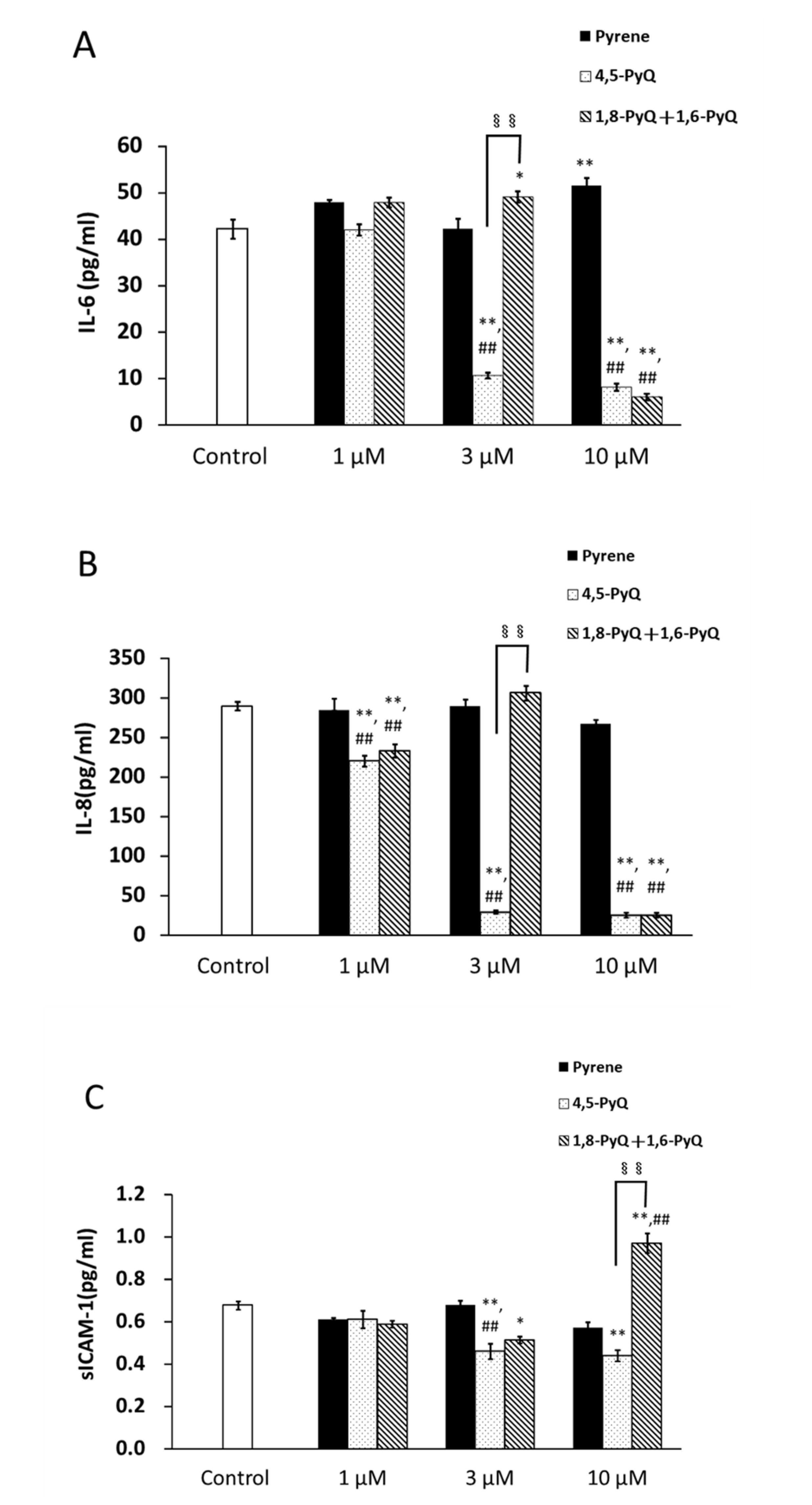
3.3. Effect of Pyrene and Its Quinone Forms on IL-6, IL-8, and sICAM-1 Production by Bronchial Epithelial Cells

We evaluated IL-6 production from BEAS-2B cells after exposure to 1, 3, and 10 µM pyrene, 4,5-PyQ, and 1,8-PyQ + 1,6-PyQ, and found no significant difference after exposure to 1 µM of all compounds compared with that in the control. At 3 µM, compared with that in the control, IL-6 production was unaltered after pyrene exposure; however, IL-6 production decreased after 4,5-PyQ exposure (p < 0.01 vs. control) and increased after 1,8-PyQ + 1,6-PyQ exposure (p < 0.05). At 10 µM, IL-6 production increased after pyrene exposure, whereas it decreased after 4,5-PyQ and 1,8-PyQ + 1,6-PyQ exposure (p < 0.01) compared to that in the control. The comparison of relative effects between the compounds revealed no significant difference in IL-6 production between any treatment condition at 1 µM. At 3 µM, IL-6 production after 4,5-PyQ exposure was lower than that after pyrene exposure (p < 0.01), without any significant difference after 1,8-PyQ + 1,6-PyQ exposure (slight elevation). Furthermore, increased IL-6 production was observed after 1,8-PyQ + 1,6-PyQ exposure compared to that after 4,5-PyQ exposure (p < 0.01). At 10 µM, IL-6 production after treatment with 4,5-PyQ and 1,8-PyQ + 1,6-PyQ was lower than that after exposure to pyrene (p < 0.01). No significant difference in IL-6 production was observed between the 4,5-PyQ and 1,8-PyQ + 1,6-PyQ exposure groups (Figure 3A).
IL-8 production was assessed after treatment with 1, 3, and 10 µM pyrene, 4,5-PyQ, and 1,8-PyQ + 1,6-PyQ, and no significant difference was observed after 1 µM pyrene exposure compared with that in the control. However, cells treated with 4,5-PyQ and 1,8-PyQ + 1,6-PyQ showed lower IL-8 levels than the control (p < 0.01). Similarly, there was no significant difference in IL-8 levels among cells treated with 3 µM pyrene and 1,8-PyQ + 1,6-PyQ compared with those in the control; lower IL-8 levels were observed in 4,5-PyQ-treated cells (p < 0.01). At 10 µM, the IL-8 levels after pyrene exposure were similar to those in the control, whereas its levels decreased in the 4,5-PyQ- and 1,8-PyQ + 1,6-PyQ-treated cells (p < 0.01). A relative comparison between compounds revealed that at 1 µM, the 4,5-PyQ and 1,8-PyQ + 1,6-PyQ groups showed lower IL-8 levels than the pyrene group (p < 0.01); in contrast, no significant differences were observed between the 4,5-PyQ and 1,8-PyQ + 1,6-PyQ groups. At 3 µM, compared to that in pyrene-treated cells, IL-8 production decreased in the 4,5-PyQ group (p < 0.01), whereas that in the 1,8-PyQ + 1,6-PyQ group showed no significant difference. IL-8 levels were higher in the 1,8-PyQ + 1,6-PyQ group than in the 4,5-PyQ-treated group (p < 0.01). At 10 µM, IL-8 levels in the 4,5-PyQ and 1,8-PyQ + 1,6-PyQ groups were lower than those in the pyrene group (p < 0.01). No significant differences were observed between the 4,5-PyQ and 1,8-PyQ + 1,6-PyQ groups (Figure 3B).
No changes in the sICAM-1 levels were observed after treatment with 1 µM pyrene, 4,5-PyQ, and 1,8-PyQ + 1,6-PyQ compared with those in the control. At 3 µM, there was no significant difference in sICAM-1 levels between the pyrene-treated cells and the control, whereas lower sICAM-1 levels were observed in the 4,5-PyQ and 1,8-PyQ + 1,6-PyQ groups (p < 0.01 and p < 0.05, respectively). At 10 µM, there was no significant difference in sICAM-1 production upon pyrene exposure compared with that in the control; however, sICAM-1 production in 4,5-PyQ-exposed cells decreased (p < 0.01 vs. control) and that in 1,8-PyQ + 1,6-PyQ-exposed cells increased (p < 0.01 vs. control). When sICAM-1 levels between groups were compared, no significant difference was found across all groups at 1 µM. At 3 µM, sICAM-1 levels were lower in the 4,5-PyQ group than in the pyrene exposure group (p < 0.01), but no significant difference was observed for the 1,8-PyQ + 1,6-PyQ group. Furthermore, there was no significant difference in sICAM-1 production between the 4,5-PyQ and 1,8-PyQ + 1,6-PyQ groups. At 10 µM, sICAM-1 levels were not significantly different between the pyrene- and 4,5-PyQ-treated cells (although the value in the 4,5-PyQ group was lower); nonetheless, sICAM-1 levels increased in 1,8-PyQ + 1,6-PyQ-treated cells (p < 0.01). When sICAM-1 levels were compared between cells treated with 4,5-PyQ or 1,8-PyQ + 1,6-PyQ, higher levels were found in the 1,8-PyQ + 1,6-PyQ group (p < 0.01; Figure 3C).
Overall, although the levels of inflammatory mediators such as IL-6, IL-8, and sICAM-1 changed after treatment with certain concentrations of 4,5-PyQ and 1,8-PyQ + 1,6-PyQ, they did not follow a specific, dose-dependent pattern.

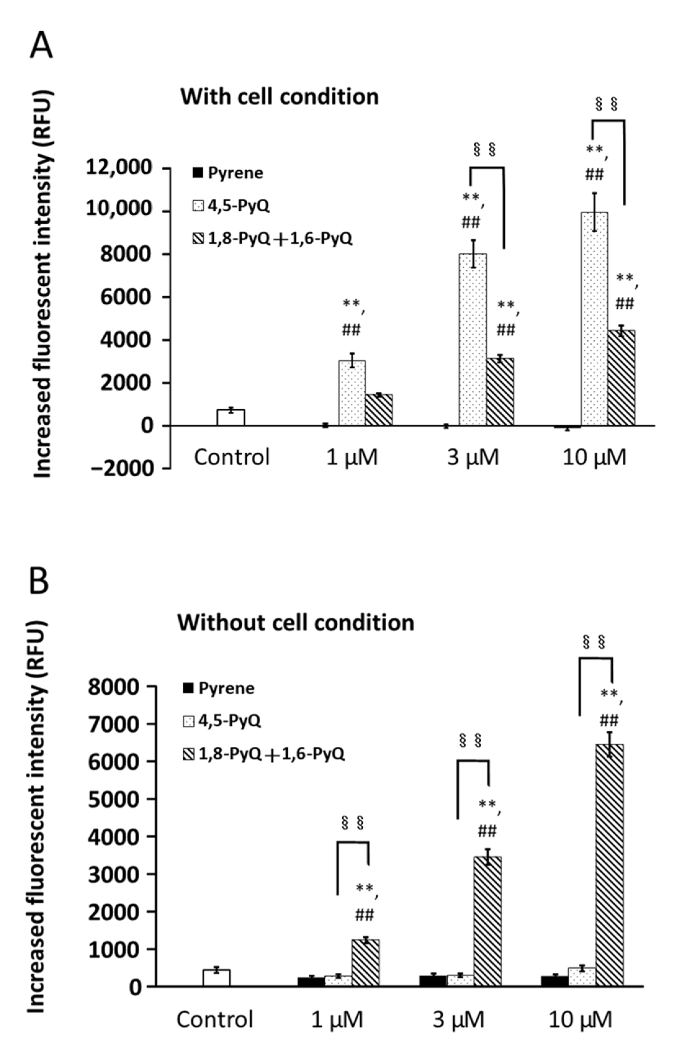
3.4. Effects of Pyrene and Its Quinone Forms on ROS Production by Bronchial Epithelial Cells

BEAS-2B cells were treated with 1, 3, and 10 µM pyrene, 4,5-PyQ, and 1,8-PyQ + 1,6-PyQ, and ROS production was examined. No significant difference in ROS production was observed when the cells were treated with 1 µM pyrene or 1,8-PyQ + 1,6-PyQ compared with that in the control. However, ROS production increased after 4,5-PyQ exposure (p < 0.01 vs. control). At 3 and 10 µM, no significant difference in ROS production was observed upon pyrene exposure compared with that in the control; nonetheless, ROS production increased after exposure to 4,5-PyQ or 1,8-PyQ + 1,6-PyQ (p < 0.01).
Exposure to 1 µM 4,5-PyQ increased ROS production compared to that after pyrene exposure (p < 0.01), whereas no significant difference was observed after 1,8-PyQ + 1,6-PyQ exposure. There were no significant differences in ROS production between 4,5-PyQ- and 1,8-PyQ + 1,6-PyQ-treated groups. At 3 and 10 µM, increased ROS production was observed after 4,5-PyQ and 1,8-PyQ + 1,6-PyQ exposure compared to that after pyrene exposure (p < 0.01). When the effects of 4,5-PyQ and 1,8-PyQ + 1,6-PyQ exposure were compared, higher ROS levels were observed in the 4,5-PyQ group (p < 0.01) (Figure 4A).
In the absence of cells, treatment with 1, 3, and 10 µM of pyrene, 4,5-PyQ, and 1,8-PyQ + 1,6-PyQ showed no significant differences in ROS production after exposure to 1 µM pyrene and 4,5-PyQ compared with that in the control, whereas increased ROS production was observed after 1,8-PyQ + 1,6-PyQ exposure at 1 µM (p < 0.01 vs. control). Similar results were observed in groups treated with 3 and 10 µM of pyrene quinones.
In the mutual comparison at 1 µM, ROS production was not significantly different after 4,5-PyQ exposure compared with that after pyrene exposure, but it increased after 1,8-PyQ + 1,6-PyQ treatment (p < 0.01). ROS levels increased after exposure to 1,8-PyQ + 1,6-PyQ compared to those after 4,5-PyQ exposure (p < 0.01) (Figure 4B). Similar results were observed in cells treated with 3 and 10 µM of each quinone derivative (Figure 4B).
Overall, significantly higher ROS production was induced in bronchial epithelial cells after treatment with oxidized pyrenes, following a dose-dependent pattern.

4. Discussion

Several in vivo studies have shown that intratracheal exposure to PM induces airway inflammation [14,15,16]. Previous in vivo studies, including those by our group, have demonstrated that diesel exhaust particles (DEPs) can affect the respiratory system by inducing edematous changes [17], carcinogenesis [18], airway inflammation with hyperresponsiveness [19], enhancement of allergic lung inflammation [20,21], and neutrophilic lung inflammation [22]. However, the exact components of PM/DEP that cause these issues remain unknown. Environmental quinones have toxic effects on living organisms. The toxic effects of DEPs are attributed to PAH quinones [23,24]. These have been found in ambient PM [25,26], automotive exhaust emissions [27], and wood smoke particles [28]. Quinones can cause nephrotoxicity, neurotoxicity, and carcinogenesis [29], along with mitochondrial dysfunction [30]. Moreover, certain quinones can generate ROS, including superoxides, hydrogen peroxide, and hydroxyl radicals, which result in cellular damage [31]. Phenanthraquinone is a quinone found at significant concentrations in DEPs [26,32]. We have previously shown that a single intratracheal instillation of phenanthraquinone in mice can induce the recruitment of inflammatory cells, such as neutrophils and eosinophils, to the airway [10]. Additionally, naphthoquinone, another quinone, can exacerbate allergic lung inflammation [12,13]. In this study, 1 μM of 4,5-PyQ or 1 and 3 μM of a mixture of 1,8-PyQ + 1,6-PyQ were found to significantly induce epithelial cell activation. Combined with previous findings, the results of this study indicate that quinones might contribute to PM-mediated respiratory toxicity, thereby resulting in airway pathophysiology. Future in vivo studies are required to further understand their role, including studies determining the harmful effects of these quinones on airway diseases such as asthma, pneumonia, and chronic obstructive pulmonary disease (COPD).
ICAM-1 (CD54) is an adhesion molecule belonging to the immunoglobulin superfamily, and it is essential for the accumulation of inflammatory cells including neutrophils, eosinophils, and T lymphocytes. Tosi et al. [14] demonstrated that neutrophil adhesion to the epithelial cell layer is regulated by ICAM-1 because this adhesion is abrogated after treatment with an anti-ICAM-1 antibody. Moreover, anti-ICAM-1 antibodies significantly blocked inflammatory responses in animal models of asthma [33]. Increased levels of serum and/or bronchoalveolar lavage fluid sICAM-1 have been reported in several inflammatory lung disorders such as sarcoidosis and bronchial asthma [34,35]. Hence, ICAM-1 expression might be responsible for regulating local inflammatory responses in the lungs. Consistent with this, the DEP-induced upregulation of ICAM-1 in the epithelium may increase neutrophil attachment in vitro [36]. We have previously shown that DEP facilitates the local ICAM-1 expression in a murine model of acute respiratory distress syndrome, which is concomitant with neutrophilic lung inflammation and enhanced expression of keratinocyte-derived chemokines in the lung in vivo [22]. Therefore, the DEP-induced upregulation of ICAM-1 may be related to its adjuvant, as previously shown in animals [20]. Direct exposure to DEP is reported to induce the upregulation of cell adhesion molecules in endothelial cells [37]. In this study, we observed that at 10 μM, 1,8-PyQ + 1,6-PyQ induced significantly higher levels of sICAM-1 than those in the control, implying that these quinones might cause PM-induced pulmonary toxicity concomitant with ICAM-1 activation. Nevertheless, the conflicting relationship between inflammatory cytokines and sICAM-1 following 1,8-PyQ + 1,6-PyQ exposure at this concentration remains unclear and warrants further experiments.
We observed that exposure to more than 3 μM of 4,5-PyQ or 10 μM of 1,8-PyQ + 1,6-PyQ induced cell death, but its pattern was different in cells treated with both quinones because 4,5-PyQ possibly induced cellular apoptosis. Airway epithelial barrier dysfunction is a major adverse event caused by air pollution [38], and may occur due to abnormalities in tight junctions and loss of epithelial cells [39,40]. Aberrant apoptosis may induce the loss of airway epithelial cells, thereby causing epithelial barrier dysfunction. Apoptosis, also known as programmed cell death, is responsible for the elimination of aging or damaged cells under physiological conditions. DEPs can alter gene regulation resulting in uncontrolled apoptosis. Moreover, because ROS act as secondary messengers in several intracellular signaling cascades, excessive ROS production might exacerbate the cellular apoptosis induced by these pollutants [41]. In contrast, PM2.5-related cell death patterns reportedly involve autophagy, necrosis, pyroptosis, and ferroptosis [42]. Therefore, future studies are required to examine the effects of quinones on these mechanisms of death. Overall, these results imply that several types of quinones may affect respiratory tissues, including induction of epithelial cell damage and death, thereby resulting in complex adverse effects on these systems.
To the best of our knowledge, this is the first study to show that oxidized pyrene activates airway cells to a greater extent compared to pyrene itself. Quinones can be reduced by reducing substances in the body and converted to unstable semiquinone radicals, which generate ROS when oxidized by dissolved oxygen. These semiquinone radicals are reoxidized to the original quinone in the redox cycle. This redox cycle is associated with ROS generation, which may be responsible for quinone toxicity. In this study, 4,5-PyQ produced ROS in a dose-dependent manner under cellular conditions, whereas it did not produce ROS in the absence of cells. However, 1,8-PyQ + 1,6-PyQ produced ROS in a dose-dependent manner with or without cell conditions. Pyrene did not produce any ROS under in vitro conditions. Taken together, these results suggest that ROS generation in cells is responsible for the adverse biological effects of quinones. However, further detailed investigations are required to understand the mechanisms underlying the toxicological effects of 4, 5-PyQ and 1, 8-PyQ + 1, 6-PyQ. Mechanisms other than oxidative stress need to be further elucidated both in vitro and in vivo. Further detailed studies using endothelial cells and/or in vivo studies are needed to elucidate the effects of these quinones.

5. Conclusions

In this study, we investigated the respiratory toxicity of pyrene, a type of PAH, and its quinone forms in vitro by assessing the effects of varying concentrations of pyrene, 4,5- PyQ, and 1,8-PyQ + 1,6-PyQ, on human lung epithelial (BEAS-2B) cells. We evaluated cell viability, apoptosis, and the production of IL-6, IL-8, sICAM-1, and ROS. At 1 μM, increased cellular activity was observed after exposure to 4,5-PyQ or 1,8-PyQ + 1,6-PyQ. At 3 µM, 4,5-PyQ increased the number of late apoptotic and/or necrotic cells compared with the control, whereas 1,8-PyQ + 1,6-PyQ exposure increased the number of dead cells. IL-6 production was decreased after exposure to 10 µM 4,5-PyQ and IL-8 production was decreased after exposure to 3 and 10 µM of both 4,5-PyQ and 1,8-PyQ + 1,6-PyQ. sICAM-1 production was higher after 1,8-PyQ + 1,6-PyQ exposure at 10 µM. In the presence of cells, 4,5-PyQ and 1,8-PyQ + 1,6-PyQ increased ROS production significantly in a concentration-dependent manner, whereas similar results were observed with 1,8-PyQ + 1,6-PyQ under cell-free conditions.
This study has the following limitations: (1) it only examined in vitro effects; thus, the effects of these pyrene quinones on respiratory pathophysiology such as asthma, pneumonia, and COPD and (2) the contribution of ROS and/or other molecular mechanisms in these epithelial cell toxicities, including cell death patterns induced by pyrene quinones, remain unclear. Therefore, additional in vivo studies and molecular assessments are needed to address these limitations.
Overall, these results show that the oxidized quinone forms of pyrene exert more harmful cytotoxic effects on respiratory organs than those of pyrene, resulting in exacerbation of respiratory diseases. Thus, future toxicological studies on the effects of PM should focus more on the oxidized forms of PAHs.

Supplementary Materials

The following supporting information can be downloaded at: https://0-www-mdpi-com.brum.beds.ac.uk/article/10.3390/app12199664/s1, Figure S1: Chemical structural formula of pyrene and its quinone forms.

Author Contributions

Conceptualization, T.K. and H.T.; Investigation, A.H. and S.T.; Resources, T.K.; Data curation, S.T.; Writing—original draft preparation, A.H. and K.-i.I.; Writing—review and editing, A.H., K.-i.I., S.T., T.K., K.U. and H.T.; Supervision, K.-i.I., T.K., K.U. and H.T.; Project administration, A.H.; Funding acquisition, T.K. and H.T. All authors have read and agreed to the published version of the manuscript.

Funding

This work was supported by CREST, JST (JPMJCR19H3: to H. Takano), and the Japan Society for the Promotion of Science Grants in Aid for Scientific Research (B: 17H01906: to T. Kameda).

Institutional Review Board Statement

Not applicable.

Informed Consent Statement

Not applicable.

Data Availability Statement

The data presented in this study are available upon request from the corresponding author. The data are not publicly available due to privacy.

Acknowledgments

We thank Megumi Nagao for their technical assistance.

Conflicts of Interest

The authors declare no conflict of interest.

References

  1. Zanobetti, A.; Franklin, M.; Koutrakis, P.; Schwartz, J. Fine particulate air pollution and its components in association with cause-specific emergency admissions. Environ. Health 2009, 8, 58. [Google Scholar] [CrossRef] [PubMed]
  2. Shi, J.; Chen, R.; Yang, C.; Lin, Z.; Cai, J.; Xia, Y.; Wang, C.; Li, H.; Johnson, N.; Xu, X.; et al. Association between fine particulate matter chemical constituents and airway inflammation: A panel study among healthy adults in China. Environ. Res. 2016, 150, 264–268. [Google Scholar] [CrossRef] [PubMed]
  3. Schwarze, P.E.; Ovrevik, J.; Låg, M.; Refsnes, M.; Nafstad, P.; Hetland, R.B.; Dybing, E. Particulate matter properties and health effects: Consistency of epidemiological and toxicological studies. Hum. Exp. Toxicol. 2006, 25, 559–579. [Google Scholar] [CrossRef] [PubMed]
  4. Dockery, D.W.; Pope, C.A., 3rd; Xu, X.; Spengler, J.D.; Ware, J.H.; Fay, M.E.; Ferris, B.G., Jr.; Speizer, F.E. An association between air pollution and mortality in six U. S. cities. N. Engl. J. Med. 1993, 329, 1753–1759. [Google Scholar] [CrossRef] [PubMed]
  5. Piao, C.H.; Fan, Y.; Nguyen, T.V.; Shin, H.S.; Kim, H.T.; Song, C.H.; Chai, O.H. PM(2.5) Exacerbates Oxidative Stress and Inflammatory Response through the Nrf2/NF-κB Signaling Pathway in OVA-Induced Allergic Rhinitis Mouse Model. Int. J. Mol. Sci. 2021, 22, 8173. [Google Scholar] [CrossRef] [PubMed]
  6. Dishaw, L.; Yost, E.; Arzuaga, X.; Luke, A.; Kraft, A.; Walker, T.; Thayer, K. A novel study evaluation strategy in the systematic review of animal toxicology studies for human health assessments of environmental chemicals. Environ. Int. 2020, 141, 105736. [Google Scholar] [CrossRef]
  7. Pan, S.; Qiu, Y.; Li, M.; Yang, Z.; Liang, D. Recent Developments in the Determination of PM(2.5) Chemical Composition. Bull. Environ. Contam. Toxicol. 2022, 108, 819–823. [Google Scholar] [CrossRef]
  8. Kameda, T.; Azumi, E.; Fukushima, A.; Tang, N.; Matsuki, A.; Kamiya, Y.; Toriba, A.; Hayakawa, K. Mineral dust aerosols promote the formation of toxic nitropolycyclic aromatic compounds. Sci. Rep. 2016, 6, 24427. [Google Scholar] [CrossRef]
  9. Liao, K.; Yu, J.Z. Abundance and sources of benzo[a]pyrene and other PAHs in ambient air in Hong Kong: A review of 20-year measurements (1997–2016). Chemosphere 2020, 259, 127518. [Google Scholar] [CrossRef]
  10. Hiyoshi, K.; Takano, H.; Inoue, K.; Ichinose, T.; Yanagisawa, R.; Tomura, S.; Cho, A.K.; Froines, J.R.; Kumagai, Y. Effects of a single intratracheal administration of phenanthraquinone on murine lung. J. Appl. Toxicol. 2005, 25, 47–51. [Google Scholar] [CrossRef]
  11. Hiyoshi, K.; Takano, H.; Inoue, K.I.; Ichinose, T.; Yanagisawa, R.; Tomura, S.; Kumagai, Y. Effects of phenanthraquinone on allergic airway inflammation in mice. Clin. Exp. Allergy 2005, 35, 1243–1248. [Google Scholar] [CrossRef] [PubMed]
  12. Inoue, K.; Takano, H.; Ichinose, T.; Tomura, S.; Yanagisawa, R.; Sakurai, M.; Sumi, D.; Cho, A.K.; Hiyoshi, K.; Kumagai, Y. Effects of naphthoquinone on airway responsiveness in the presence or absence of antigen in mice. Arch. Toxicol. 2007, 81, 575–581. [Google Scholar] [CrossRef]
  13. Inoue, K.; Takano, H.; Hiyoshi, K.; Ichinose, T.; Sadakane, K.; Yanagisawa, R.; Tomura, S.; Kumagai, Y. Naphthoquinone enhances antigen-related airway inflammation in mice. Eur. Respir. J. 2007, 29, 259–267. [Google Scholar] [CrossRef]
  14. He, M.; Ichinose, T.; Yoshida, S.; Ito, T.; He, C.; Yoshida, Y.; Arashidani, K.; Takano, H.; Sun, G.; Shibamoto, T. PM2.5-induced lung inflammation in mice: Differences of inflammatory response in macrophages and type II alveolar cells. J. Appl. Toxicol. 2017, 37, 1203–1218. [Google Scholar] [CrossRef] [PubMed]
  15. Laskin, D.L.; Mainelis, G.; Turpin, B.J.; Patel, K.J.; Sunil, V.R. Pulmonary effects of inhaled diesel exhaust in young and old mice: A pilot project. Res. Rep. Health Eff. Inst. 2010, 151, 3–31. [Google Scholar]
  16. Sun, B.; Shi, Y.; Li, Y.; Jiang, J.; Liang, S.; Duan, J.; Sun, Z. Short-term PM(2.5) exposure induces sustained pulmonary fibrosis development during post-exposure period in rats. J. Hazard. Mater. 2020, 385, 121566. [Google Scholar] [CrossRef]
  17. Ichinose, T.; Furuyama, A.; Sagai, M. Biological effects of diesel exhaust particles (DEP). II. Acute toxicity of DEP introduced into lung by intratracheal instillation. Toxicology 1995, 99, 153–167. [Google Scholar] [CrossRef]
  18. Ichinose, T.; Yamanushi, T.; Seto, H.; Sagai, M. Oxygen radicals in lung carcinogenesis accompanying phagocytosis of diesel exhaust particles. Int. J. Oncol. 1997, 11, 571–575. [Google Scholar] [CrossRef]
  19. Sagai, M.; Furuyama, A.; Ichinose, T. Biological effects of diesel exhaust particles (DEP). III. Pathogenesis of asthma like symptoms in mice. Free Radic. Biol. Med. 1996, 21, 199–209. [Google Scholar] [CrossRef]
  20. Takano, H.; Yoshikawa, T.; Ichinose, T.; Miyabara, Y.; Imaoka, K.; Sagai, M. Diesel exhaust particles enhance antigen-induced airway inflammation and local cytokine expression in mice. Am. J. Respir. Crit. Care Med. 1997, 156, 36–42. [Google Scholar] [CrossRef]
  21. Takano, H.; Ichinose, T.; Miyabara, Y.; Shibuya, T.; Lim, H.B.; Yoshikawa, T.; Sagai, M. Inhalation of diesel exhaust enhances allergen-related eosinophil recruitment and airway hyperresponsiveness in mice. Toxicol. Appl. Pharmacol. 1998, 150, 328–337. [Google Scholar] [CrossRef] [PubMed]
  22. Takano, H.; Yanagisawa, R.; Ichinose, T.; Sadakane, K.; Yoshino, S.; Yoshikawa, T.; Morita, M. Diesel exhaust particles enhance lung injury related to bacterial endotoxin through expression of proinflammatory cytokines, chemokines, and intercellular adhesion molecule-1. Am. J. Respir. Crit. Care Med. 2002, 165, 1329–1335. [Google Scholar] [CrossRef] [PubMed]
  23. Oh, S.M.; Kim, H.R.; Park, Y.J.; Lee, S.Y.; Chung, K.H. Organic extracts of urban air pollution particulate matter (PM2.5)-induced genotoxicity and oxidative stress in human lung bronchial epithelial cells (BEAS-2B cells). Mutat. Res. 2011, 723, 142–151. [Google Scholar] [CrossRef] [PubMed]
  24. Dergham, M.; Lepers, C.; Verdin, A.; Billet, S.; Cazier, F.; Courcot, D.; Shirali, P.; Garçon, G. Prooxidant and proinflammatory potency of air pollution particulate matter (PM2.5–0.3) produced in rural, urban, or industrial surroundings in human bronchial epithelial cells (BEAS-2B). Chem. Res. Toxicol. 2012, 25, 904–919. [Google Scholar] [CrossRef] [PubMed]
  25. Fraser, M.P.; Buzcu, B.; Yue, Z.W.; McGaughey, G.R.; Desai, N.R.; Allen, D.T.; Seila, R.L.; Lonneman, W.A.; Harley, R.A. Separation of fine particulate matter emitted from gasoline and diesel vehicles using chemical mass balancing techniques. Environ. Sci. Technol. 2003, 37, 3904–3909. [Google Scholar] [CrossRef] [PubMed]
  26. Cho, A.K.; di Stefano, E.; You, Y.; Rodriguez, C.E.; Schmitz, D.A.; Kumagai, Y.; Miguel, A.H.; Eiguren-Fernandez, A.; Kobayashi, T.; Avol, E.; et al. Determination of four quinones in diesel exhaust particles, SRM 1649a, and atmospheric PM2.5 special issue of aerosol science and technology on findings from the fine particulate matter supersites program. Aerosol Sci. Technol. 2004, 38, 68–81. [Google Scholar] [CrossRef]
  27. Schuetzle, D.; Lee, F.S.; Prater, T.J. The identification of polynuclear aromatic hydrocarbon (PAH) derivatives in mutagenic fractions of diesel particulate extracts. Int. J. Environ. Anal. Chem. 1981, 9, 93–144. [Google Scholar] [CrossRef]
  28. Fine, P.M.; Cass, G.R.; Simoneit, B.R. Chemical characterization of fine particle emissions from fireplace combustion of woods grown in the northeastern United States. Environ. Sci.Technol. 2001, 35, 2665–2675. [Google Scholar] [CrossRef]
  29. Monks, T.J.; Lau, S.S. Toxicology of quinone-thioethers. Crit. Rev. Toxicol. 1992, 22, 243–270. [Google Scholar] [CrossRef]
  30. Henry, T.R.; Wallace, K.B. Differential mechanisms of cell killing by redox cycling and arylating quinones. Arch. Toxicol. 1996, 70, 482–489. [Google Scholar] [CrossRef]
  31. Bolton, J.L.; Trush, M.A.; Penning, T.M.; Dryhurst, G.; Monks, T.J. Role of quinones in toxicology. Chem. Res. Toxicol. 2000, 13, 135–160. [Google Scholar] [CrossRef] [PubMed]
  32. Schuetzle, D. Sampling of vehicle emissions for chemical analysis and biological testing. Environ. Health Perspect. 1983, 47, 65–80. [Google Scholar] [CrossRef] [PubMed]
  33. Traub, S.; Nikonova, A.; Carruthers, A.; Dunmore, R.; Vousden, K.A.; Gogsadze, L.; Hao, W.; Zhu, Q.; Bernard, K.; Zhu, J.; et al. An anti-human ICAM-1 antibody inhibits rhinovirus-induced exacerbations of lung inflammation. PLoS Pathog. 2013, 9, e1003520. [Google Scholar] [CrossRef] [PubMed]
  34. Kim, D.S.; Paik, S.H.; Lim, C.M.; Lee, S.D.; Koh, Y.; Kim, W.S.; Kim, W.D. Value of ICAM-1 expression and soluble ICAM-1 level as a marker of activity in sarcoidosis. Chest 1999, 115, 1059–1065. [Google Scholar] [CrossRef]
  35. Marguet, C.; Dean, T.P.; Warner, J.O. Soluble intercellular adhesion molecule-1 (sICAM-1) and interferon-gamma in bronchoalveolar lavage fluid from children with airway diseases. Am. J. Respir. Crit. Care Med. 2000, 162, 1016–1022. [Google Scholar] [CrossRef]
  36. Takizawa, H.; Abe, S.; Ohtoshi, T.; Kawasaki, S.; Takami, K.; Desaki, M.; Sugawara, I.; Hashimoto, S.; Azuma, A.; Nakahara, K.; et al. Diesel exhaust particles up-regulate expression of intercellular adhesion molecule-1 (ICAM-1) in human bronchial epithelial cells. Clin. Exp. Immunol. 2000, 120, 356–362. [Google Scholar] [CrossRef]
  37. Rui, W.; Guan, L.; Zhang, F.; Zhang, W.; Ding, W. PM2.5-induced oxidative stress increases adhesion molecules expression in human endothelial cells through the ERK/AKT/NF-κB-dependent pathway. J. Appl. Toxicol. 2016, 36, 48–59. [Google Scholar] [CrossRef]
  38. Nasreen, N.; Khodayari, N.; Sriram, P.S.; Patel, J.; Mohammed, K.A. Tobacco smoke induces epithelial barrier dysfunction via receptor EphA2 signaling. Am. J. Physiol. Cell Physiol. 2014, 306, C1154–C1166. [Google Scholar] [CrossRef]
  39. Chen, W.Y.; Wang, M.; Zhang, J.; Barve, S.S.; McClain, C.J.; Joshi-Barve, S. Acrolein Disrupts Tight Junction Proteins and Causes Endoplasmic Reticulum Stress-Mediated Epithelial Cell Death Leading to Intestinal Barrier Dysfunction and Permeability. Am. J. Pathol. 2017, 187, 2686–2697. [Google Scholar] [CrossRef]
  40. Fischer, A.; Gluth, M.; Pape, U.F.; Wiedenmann, B.; Theuring, F.; Baumgart, D.C. Adalimumab prevents barrier dysfunction and antagonizes distinct effects of TNF-α on tight junction proteins and signaling pathways in intestinal epithelial cells. Am. J. Physiol. Gastrointest. Liver Physiol. 2013, 304, G970–G979. [Google Scholar] [CrossRef]
  41. Long, J.; Manchandia, T.; Ban, K.; Gao, S.; Miller, C.; Chandra, J. Adaphostin cytoxicity in glioblastoma cells is ROS-dependent and is accompanied by upregulation of heme oxygenase-1. Cancer Chemother. Pharmacol. 2007, 59, 527–535. [Google Scholar] [CrossRef] [PubMed]
  42. Wang, Y.; Zhong, Y.; Liao, J.; Wang, G. PM2.5-related cell death patterns. Int. J. Med. Sci. 2021, 18, 1024–1029. [Google Scholar] [CrossRef] [PubMed]
Figure 1. Effects of pyrene and its quinone forms on the viability of human bronchial epithelial cells. Cells were treated with the specified concentrations of pyrene and its quinone forms for 24 h, and their viability was assessed using the WST-1 assay. The data are represented as the percentage of viability of the control treatment (0 μmol/L) and are shown as the mean ± standard error of the mean (SEM) from four individual cultures. ** p < 0.01 vs. control, ## p < 0.01 vs. pyrene at the same concentration, §§ p < 0.01 vs. each other.
Applsci 12 09664 g001
Figure 2. Effects of pyrene and its quinone forms on the apoptosis and necrosis of human bronchial epithelial cells. Dot-plot of flow cytometry, Q1, necrotic cells; Q2, late apoptotic or necrotic cells; Q3, early apoptotic cells; Q4, healthy cells.
Applsci 12 09664 g002
Figure 3. Effect of pyrene and its quinone forms on interleukin (IL)-6, IL-8, and soluble intercellular adhesion molecule (sICAM)-1 production from human bronchial epithelial cells. The protein levels of IL-6 (A), IL-8 (B), and sICAM-1 (C) in the culture supernatants, after exposure to pyrene and its quinone forms for 24 h, were measured using enzyme-linked immunosorbent assay (ELISA). The data are expressed as the mean ± standard error of the mean (SEM) of four individual cultures. * p < 0.05, ** p < 0.01 vs. control, ## p < 0.01 vs. pyrene at the same concentration, §§ p < 0.01 vs. each other.
Applsci 12 09664 g003
Figure 4. Effects of pyrene and its quinone forms on reactive oxygen species (ROS) production by airway epithelial cells. The intracellular (with cell condition) (A) and extracellular (without cell conditions) (B) ROS production during 3 h exposure. The data are expressed as the mean ± standard error of the mean (SEM) from four individual cultures. ** p < 0.01 vs. control, ## p < 0.01 vs. pyrene at the same concentration, §§ p < 0.01 vs. each other.
Applsci 12 09664 g004
Publisher’s Note: MDPI stays neutral with regard to jurisdictional claims in published maps and institutional affiliations.

