Article

The Association between Inflammatory Biomarkers and Cardiovascular Autonomic Dysfunction after Bacterial Infection

1
Department of Preventive Medicine and Public Health, Universidad de Navarra, 31008 Pamplona, Spain
2
IdiSNA, Navarra Institute for Health Research, 31008 Pamplona, Spain
3
Biomedical Research Network Center for Pathophysiology of Obesity and Nutrition (CIBEROBN), Carlos III Health Institute, 28029 Madrid, Spain
4
Faculty of Health and Social Sciences, Bournemouth University, Bournemouth BH8 8GP, UK
5
Department of Medicine for Older People, University Hospitals Dorset, Bournemouth BH7 7DW, UK
6
Department of Brain Sciences, Imperial College, London W12 0NN, UK
*
Authors to whom correspondence should be addressed.
Submission received: 9 March 2022 / Revised: 25 March 2022 / Accepted: 26 March 2022 / Published: 29 March 2022

Abstract

:
Heart rate variability (HRV) is a known measure of cardiac autonomic function. A cardiovascular autonomic dysfunction (CAD), measured as changes in HRV, is usually presented after an infectious process. The aim of the present study is to assess the association between serum inflammatory markers and CAD. For this purpose, 50 volunteers (13 of them recovering from an infection) were recruited and followed-up for 6 weeks. Their serum inflammatory biomarkers (CRP, IL1, IL4, IL6, IL10, and TNFalpha) were quantified throughout those weeks, along with their HRV resting, in response to the Valsalva maneuver, metronome breathing, standing and sustained handgrip. The correlation of within-subject changes in both HRV and inflammatory biomarkers was assessed to evaluate the concurrent changes. An inverse within-subject correlation was found between CRP and HRV in response to the Valsalva maneuver (rho (95% CI): −0.517 (−0.877 to −0.001); p = 0.032) and HRV standing (rho (95% CI): −0.490 (−0.943 to −0.036); p = 0.034). At the beginning, increased values of CRP are found along with reduced levels of HRV. Then, the CRP was reduced, accompanied by an improvement (increase) in HRV. These results suggest that CRP is a potential marker of CAD. Whether it is the cause, the consequence or a risk indicator non-causally associated is still to be determined.

1. Introduction

The role of the parasympathetic nervous system in modulating the inflammation process has been widely discussed, with a range of evidence supporting the involvement of vagal stimulation on decreasing the amount of pro-inflammatory markers in response to inflammation [1,2]. The “cholinergic anti-inflammatory pathway” is the process by which, in the event of an infection and consequent secretion of pro-inflammatory cytokines in the periphery, the central nervous system (CNS) responds by secreting acetylcholine via different routes, including the vagal route [3,4,5,6]. This acetylcholine interacts with the nicotinic acetylcholine receptor subunit α7 (α7nAChR) expressed on the membrane of the innate immune cells to stop the secretion of pro-inflammatory cytokines [7,8,9,10]. As a result, this avoids an over exposure to pro-inflammatory cytokines, as that may damage tissue, impair organ function and could even be lethal [11,12].
The cholinergic anti-inflammatory pathway has been widely studied. In a study by Van Westerloo et al. [13], a group of mice with induced peritoneal sepsis underwent unilateral vagotomy. These authors showed that there was an enhanced host response to infection as, by suppressing the effect of the vagus nerve on the cells of the innate system, there was no release of acetylcholine, which, in turn, did not inhibit the secretion of pro-inflammatory biomarkers. Schulte et al. [14] performed a unilateral cervical vagotomy in lipopolysaccharide (LPS)-induced septic rats, and they found that their levels of pro-inflammatory markers were increased because of the vagotomy. Similarly, Li-Sha et al. [15] concluded in their study that the vagotomy was associated with an inhibition of the cholinergic anti-inflammatory pathway, and therefore an over-secretion of pro-inflammatory cytokines. In order to reach these conclusions, they performed a unilateral right cervical vagotomy in a group of mice (n = 40), which had viral myocarditis, and they measured the levels of inflammatory biomarkers IL1, IL6 and TNFα. The results of this group were compared to a group of mice that were subjected to a sham surgery (n = 40) and showed that levels of measured cytokines were significantly higher in the vagotomy group compared to the sham group (p < 0.05). All these studies demonstrate that vagus nerve inhibition enhances the inflammatory response to body injury by increasing the release of inflammatory biomarkers, which can potentially cause effects that are worse than the infection itself [16,17].
All these studies corroborate the involvement of the autonomic nervous system (ANS) on modulating the initial inflammatory response to infection by means of the cholinergic anti-inflammatory pathway. However, it is worth noting that most of the research found on this topic comes from experimental studies conducted on animal populations under controlled laboratory conditions (such as induced sepsis). This point was also noticed by Marsland et al. [18] who agreed that, to their knowledge, there was no clinical study to explore the effect of vagal activity on the immune response to injury and/or pathogens. They therefore used a sample of 183 healthy adults, and they studied the relationship between HRV and the production of lipopolysaccharide-induced inflammatory cytokines and they found out that, as suggested by animal studies, autonomic vagal dysfunction in humans is correlated with an enhanced secretion of pro-inflammatory cytokines (IL1, IL6 and TNFα) without affecting the secretion of anti-inflammatory cytokines. The study conducted by Marsland et al. [18] is pioneering in the human study of vagal activity and its association with the innate immune response. Furthermore, Williams et al. [19] carried out a meta-analysis on HRV and inflammatory markers in humans from 1996 onwards (159 studies), and they found that most research suggested a negative correlation between HRV and inflammatory markers. The authors concluded there was a need to further understand the complex mechanisms related to why this association existed, and therefore recommended the need for further research. Moreover, a point that needs to be noted is that Williams et al. [19] included in their meta-analysis any human study that assessed HRV and inflammatory biomarkers. However, a great number of the studies that were included in this meta-analysis focused on the relationship between HRV and inflammatory biomarkers, either in healthy subjects or in cardiovascular disease and depression. Only three studies [20,21,22] explored the association between HRV and a suite of inflammatory makers in sepsis (C-reactive protein, IL6 and IL10).
Papaioannou et al. [20] carried out a study to explore whether CAD was associated with increased levels of CRP and IL6. They assessed, during 6 days, the HRV of 45 critically ill septic patients, while being sedated and intubated in an ICU, and they measured their IL6 and CRP levels. Papaioannou et al. [20] agreed with what has previously been discussed, as they found that autonomic dysfunction as assessed by HRV was associated with a higher inflammatory response in septic patients. In addition to this, Taheishi et al. [21,22] conducted similar studies to Papaionnou et al. [20], as they evaluated the association between the HRV of patients admitted to an ICU with sepsis and their levels of IL6. These authors concurred with the previous authors, as they concluded that an inverse correlation appeared to exist between the levels of IL6 and HRV. However, it needs to be highlighted that all the latter studies [20,21,22] used a sample of critically ill septic patients who had been admitted to an ICU and, as Papaioannou et al. [20] state, factors, such as sedation time, medications used and breathing patterns when on mechanical support, need to be considered as they can all affect the HRV values. Furthermore, as the patients in those studies were critically ill, the HRV was assessed while resting and no other stimuli could be applied to further evaluate the integrity of the ANS, as suggested by Ewing [23]. Lastly, it needs to be considered that, in these studies, patients were not followed-up for a long period of time, and therefore it can be argued that the recovery pattern of both the HRV and inflammatory markers was not able to be fully evaluated. It is for these reasons that further research is needed to explore the relationship between HRV and inflammatory markers in the context of an infection. The current study is therefore designed to provide further knowledge of the association between CAD and inflammatory biomarkers in a sample of individuals with community-acquired bacterial infections, which did not required ICU admission and that were hemodynamically stable to undertake the HRV assessment as developed by Ewing [23].
Cardiovascular autonomic dysfunction is the alteration of the normal ANS function that has an adverse effect on an individual’s health [24]. The heart rate variability (HRV), which is the measurement of the temporal variability in between heart beats in relation to the mean HR [25,26,27], appears to be one of the best markers to assess cardiac autonomic function [28,29,30]. Cardiovascular autonomic dysfunction is associated with infectious processes [31,32]. Furthermore, de Castilho et al. [33], Pandey et al. [34] and Tang et al. [35] concluded that abnormalities in autonomic cardiovascular reflexes as measured by the heart rate variability (HRV) are associated with a higher risk of death.
In this observational longitudinal study of patients with an acute bacterial infection and healthy-control participants, the serum inflammatory biomarker concentrations are measured (CRP, IL1, IL4, IL6, IL10 and TNFα), and the cardiovascular autonomic function assessed. It is hypothesized that, during the recovery period from an acute bacterial infection, some inflammatory markers may be a useful biomarker to promptly identify cardiovascular autonomic dysfunction.

2. Materials and Methods

An exploratory longitudinal observational study was conducted to explore the relationship between cardiovascular autonomic dysfunction and systemic inflammation induced by bacterial infection. The study was conducted at the Royal Bournemouth Hospital (University Hospitals Dorset, Bournemouth) in the UK and it was approved by the Local Research Ethics Committee (Ref 08/H0201/23). Each patient received verbal information about the study, a patient information sheet and a consent form. Patients were assured that their participation was anonymous and would not interfere with their medical treatment, and that they could withdraw from the study at any time. The study was conducted according to the principles stated in the Declaration of Helsinki.

2.1. Inclusion and Exclusion Criteria

The general inclusion criteria were: (a) any person who was over 18 years old and was able to freely agree to participate; (b) participants needed to be clinically stable by the time the first cardiovascular autonomic assessment was conducted (week 1 from hospital admission); and (c) participants needed to be able to satisfactorily perform the autonomic function tests.
The exclusion criteria were: (a) patients with significant communication difficulties (e.g., severe aphasia); (b) patients with severe cognitive impairment (mini-mental state examination (MMSE) 24 or less); (c) patients who were unable to stand or hold their breath to perform the Valsalva maneuver; (d) patients who had a past history of, or active, vascular or cardiovascular problems, such as myocardial infarction, coronary artery disease, heart failure and stroke, as the HRV of these patients could already be compromised; (e) patients with Parkinson’s disease; (f) diabetic individuals; (g) patients with a known autonomic dysfunction or orthostatic hypotension; (h) patients with depression; and (i) patients who were on drugs known to affect the autonomic function, such as beta blockers and antidepressants.

2.2. Subjects and Groups

The participants who met the inclusion criteria and agreed to participate were included in one of the two different groups:
Infection group: 13 patients who were admitted with a diagnosis of an active and symptomatic bacterial infection were recruited. Patients in this group were included regardless of when the infection had started, as long as the patient was symptomatic and required inpatient treatment. Those patients whose infections were hospital-acquired were excluded because this group of patients was more likely to be particularly ill or immunosuppressed, which could therefore introduce bias into the autonomic assessment results. Patients who were seriously ill or were deemed unlikely to recover sufficiently to take part in the study within the subsequent 6 weeks were not approached. At the time of their first autonomic function tests (1 week after agreeing to participate), participants were clinically stable: adequately hydrated with an improving biochemical profile, apyrexial (Ta < 37.8 °C), systolic blood pressure > 90 mmHg, heart rate ≤ 100 beats/minute, respiratory rate ≤ 20 respirations/minute and pulse oximetry ≥ 90%, treatment started and well enough to participate in the assessment protocol [36,37,38,39].
Healthy control group: 37 subjects were included in this group. Individuals in this group had not had a bacterial infection requiring treatment for the previous 6 months. Participants in this group were selected with age and sex as similar as possible to the infection group.
Sample size was calculated a priori for a different objective than the one presented in this manuscript, to ensure 90% power for the comparison of CRP between groups for an expected difference of at least 60 mg/dL.

2.3. Assessment of the Heart Rate Variability (HRV)

The baseline autonomic function assessment was performed 1 week from hospital admission (week 1), and thereafter at the follow-up visits (week 2 and week 6) (in the healthy group, the baseline autonomic assessment was conducted in week 1 and then 6 weeks later). Autonomic function tests were based on the widely used Ewing method [23]. This method was validated by the American Diabetes Association and American Academy of Neurology in 1988. The latter Association launched a consensus statement in 1988 that declared that the Ewing method “had been validated and had been shown to be reliable and reproducible to correlate with each other and with tests of peripheral somatic nerve function”. They therefore recommended its use both for clinical and research purposes [40]. The acquisition system used a multi-channel biosensor manufactured by Procomp Infinity, with an ECG, photoplethysmography (PPG) and chest wall movement sensor. Recording was at a very fast sampling rate of 2048 Hz.
Participants were asked to have a light breakfast and refrain from smoking or drinking coffee and alcohol for at least 2 h before performing the test. Furthermore, all participants were asked to adopt a 30-to-45 degrees tilt position. All participants’ appointments were made during the morning or early hours of the afternoon. In addition to this, participants were all assessed in a quiet room with a comfortable temperature (approximately 23 °C). Participants were asked to rest for 10 min before commencing the tests in an attempt to obtain the baseline conditions for HR and BP.
The following sequence of tests was used to assess the cardiac autonomic reflexes:
  • HRV in response to the Valsalva maneuver (40 mmHg maintained for 10 s).
  • HRV in response to metronome breathing (at 6 breaths per minute for 2 min).
  • HRV in response to standing up.
  • HRV in response to sustained handgrip (30% of maximum strength for 3 min).

2.4. Measurement of Serum Inflammatory Markers

Serum concentrations of C-reactive protein, IL1, IL4, IL6, IL10 and TNFα were measured in participants in the infection group in weeks 1, 2 and 6. Participants in the healthy group had the levels of the inflammatory biomarkers measured in weeks 1 and 6. Cytokines were measured by using commercialized and standardized kits, which used the traditional enzyme-linked immunosorbent assay (ELISA).

2.5. Data Analysis

Baseline characteristics of participants were described using the mean and standard deviation for quantitative variables and absolute frequency and percentages for qualitative variables. Associations between the continuous variables of cardiovascular autonomic function and inflammatory biomarkers at baseline (in both groups and only in the infection group) were estimated using the Spearman’s Rho Correlation Coefficient. Furthermore, to study the correlation between HRV and blood cytokines, repeated measures correlation were performed. The within-subject correlation coefficients are presented to account for the correlation between the changes in both assessed variables. Analyses were performed using STATA/SE version 16.1 (StataCorp, College Station, TX, USA), two-sided p-values were used and the statistical significance threshold was set a priori at 0.05.

3. Results

3.1. Study Population

A total of 50 participants who met the inclusion criteria was enrolled in this study. There were 37 and 13 participants in the healthy group and infection group, respectively (see Table 1).
Most participants in the infection group were admitted to the hospital with pneumonia (38.5%). One individual suffered from a urinary tract infection and another one with cellulitis. In addition to this, 46.15% presented with different types of infection, including lower respiratory tract infection, infective exacerbation of COPD, infective exacerbation of asthma and pyelonephritis. All of them were discharged from the hospital before week 6 of the follow-up.

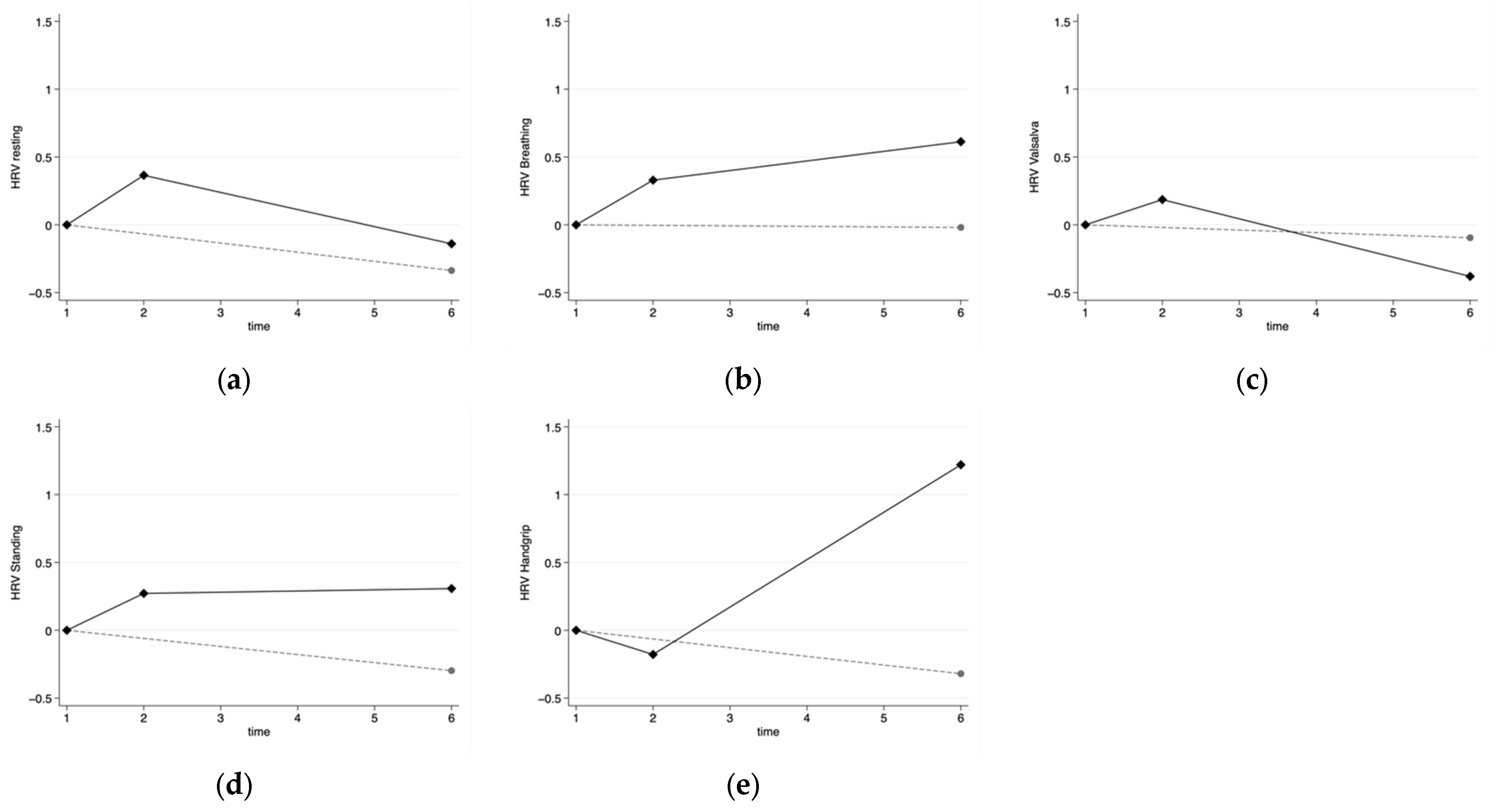
3.2. Cardiovascular Autonomic Function

The great majority of individuals in the healthy group presented with a normal cardiovascular autonomic function both in weeks 1 and 6. On the other hand, the results of the cardiovascular autonomic function assessment in the infection group showed that 40% of individuals had an abnormal cardiovascular autonomic function in the first visit. Among the participants with available information in week 6, 33.3% had an abnormal function, although they were already at home. In Figure 1 (and Figure S1), the evolution of HRV throughout the study time is presented for both groups. The data have been standardized in relation to baseline measurements to enhance comparability. As it can be seen in the figure, improvements in HRV can be observed in the infection group, but not for all indicators. Crude summaries of HRV over time for each group can be found in supplemental Table S1. Disbalances between groups may, to some extent, explain the differences between the infection and control groups.

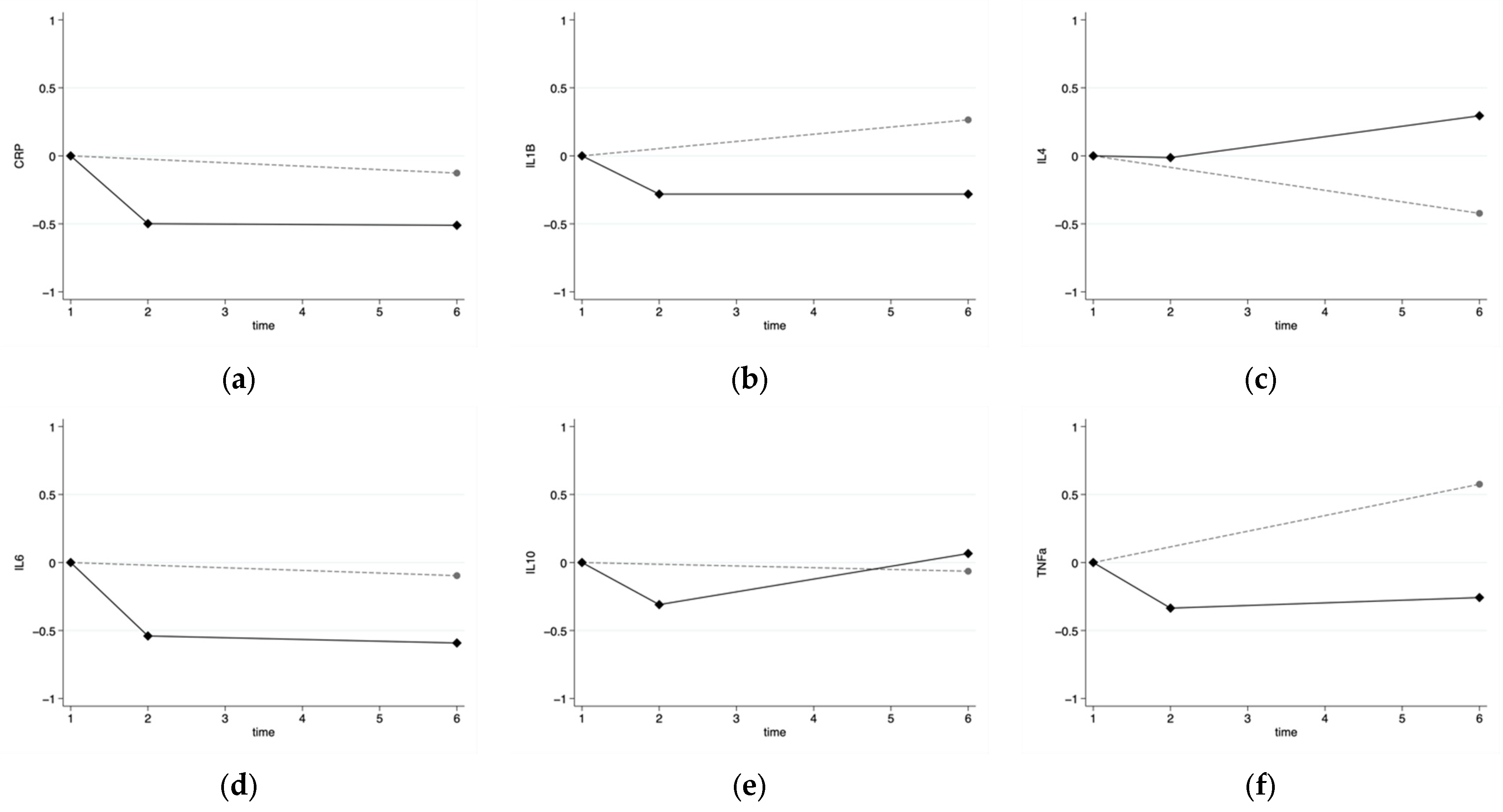
3.3. Inflammatory Markers

The inflammatory markers profile was vastly abnormal in the infection group in week 0, and those values seemed to improve throughout the weeks. Expectedly so, the inflammatory markers profile was in the majority normal in the healthy control group. In Figure 2, the progression of the inflammatory markers’ levels throughout the weeks is presented both for the infection and control groups. The data have been standardized in relation to the baseline measurements to enhance comparability. As it can be seen in the figure, the inflammatory markers improve from week 1 to week 6 in the infection group, except for the IL4 and IL10.

3.4. Cardiovascular Autonomic Dysfunction and Inflammatory Markers

The correlation between the inflammatory markers and HRV was analyzed at weeks 1 and 6, and the results show that the inflammatory biomarkers generally show an inverse association with the HRV parameters in both weeks 1 and 6 in the infection group. There was a very strong inverse association between the CRP and HRV in response to the Valsalva maneuver (rho = −0.826), which was statistically significant (p = 0.011). Furthermore, the IL6 and the resting HRV showed a moderate inverse association (rho= −0.661), which was also statistically significant (p = 0.038). These inverse associations were mirrored in week 6 in the infection group, but, as it can be seen in Table S2, those negative associations were generally weaker than it was appreciated in week 6. However, it is worth noting that IL6 still presented strong inverse associations with the resting HRV and the HRV in response to standing up (rho = −0.757 and rho= −0.766, respectively), which were statistically significant (p = 0.018 and p = 0.016) (see Table 2).
The correlation of the within-subject changes was assessed to evaluate the concurrent changes. When both groups were analyzed, an inverse within-subject correlation was found between CRP and HRV in response to the Valsalva maneuver (rho (95% CI): −0.258 (−0.508 to −0.008); p = 0.040) and HRV standing (rho (95% CI): −0.325 (−0.549 to −0.089); p = 0.007) (Table 3). In addition to this, a positive within-subject correlation was found between IL4 and HRV standing (rho (95% CI): 0.304 (0.027 to 0.580); p = 0.032). When the results from the infection group were explored, the results showed an inverse within-subject correlation between the CRP and the HRV in response to the Valsalva maneuver (rho (95% CI): −0.517 (−0.877 to −0.001); p = 0.032) and HRV standing (rho (95% CI): −0.490 (−0.943 to −0.036); p = 0.034). Moreover, IL6 appeared to show a tendency towards an inverse within-subject correlation with the HRV standing (rho (95% CI): −0.448 (−0.812 to −0.141); p = 0.071) and the HRV in response to metronome breathing (rho (95% CI): −0.406 (−0.978 to −0.266); p = 0.226) (Table 3).

4. Discussion

The data of this study revealed that the levels of a number of inflammatory markers (CRP, IL6) are inversely associated with the HRV for most of the parameters in weeks 1 and 6 in the infection group. This would mean that the lower the HRV at those points (and therefore more abnormal), the higher the level of those inflammatory markers. In addition to this, the results show that this inverse association existed throughout the weeks, so that, as individuals were recovering from the infection, the cardiovascular autonomic function was also recovering (HRV was increasing), and therefore the inflammatory markers decreased. This is particularly relevant for the CRP and the IL6, which showed the strongest negative associations both in weeks 1 and 6 and throughout the weeks.
CRP is an acute phase protein that is mainly produced in the liver in response to the elevation of pro-inflammatory cytokines, such as IL6 [41,42]. CRP rises 4 to 6 h from infection onset and is known to increase exponentially in the acute stage of infection in response to the higher levels of IL6 [43,44,45], reaching a maximum peak level 36–50 h after the inflammatory stimulus onset [46]. Furthermore, the levels of CRP rapidly decrease once the stimuli ends [46]. On the other hand, IL6 levels commence to rise just a few minutes from the infection onset [47]. Zhang et al. [48] and Endeman et al. [49] further state that IL6 in the acute stage of a bacterial infection is both needed and expected, as IL6 is one of the essential pro-inflammatory cytokines that initiates the early innate immune response to tissue damage as it attracts the macrophages and the monocytes, which work on clearing the invading pathogen. However, IL6 levels should decreased shortly after the infection starts (within the following 24 h) [46,50]. Moreover, Sun et al. [51] and McNicholas et al. [52] state that IL6 levels should considerably decrease 2 days from the beginning of antibiotic therapy. In addition to this, a number of studies have shown that persistently higher levels of IL6 lead to worse clinical outcomes [53,54].
The kinetics of the IL6 profile post-bacterial infection cannot be clearly defined as other factors related to the inflammatory response and the host response to the antibiotics need to be considered. However, what seems to be clear is that the levels of IL6 should decrease by week 1, post-antibiotic therapy and most definitely 6 weeks later. In addition to this, and considering that IL6 is a potent CRP secretion inducer, the CRP is also known to decrease exponentially post-acute infection and once the stimuli for its secretion ends [46].
It is interesting to note that, although the TNFα and IL-1 did not show statistically significant correlations with the HRV, the trend and direction of those correlations were generally negative when the parameters were evaluated for week 1 in the infection group. This would make sense as those biomarkers have also been observed to be elevated in the presence of autonomic dysfunction [4,14]. On the other hand, this negative trend was not fully appreciated when the within-subject associations were evaluated for IL1 and HRV in the infection group.
Some limitations of the present work should be acknowledged. First, the sample size of the study is small and that may limit the power to detect some correlations as statically significant. However, some comparisons were sufficiently powered as some statistically significant results were obtained. Second, the infection group and the healthy control group are different in some baseline characteristics. However, this limitation would have only affected the results of the comparison between groups. The main result is based on within-person changes in inflammatory markers and HRV, and therefore not confounded by between-group disbalances. On the other hand, some strengths should also be noted. The repeated measurement of both HRV and inflammatory biomarkers allowed us to study the process of recovery from an acute bacterial infection.
The inverse association that was found between those inflammatory markers and HRV in the acute stage of a bacterial infection indicate that the cardiac autonomic dysfunction could potentially lead, as discussed in this piece of work, to an enhanced inflammatory response, as the cholinergic anti-inflammatory pathway would be dysfunctional. Therefore, these inflammatory markers could be utilized as biomarkers for the presence of cardiac autonomic dysfunction. This would help to promptly identify those patients that may require additional assessments and care in relation to the cardiac autonomic dysfunction.

5. Conclusions

HRV appears to be reduced in the recovery period of an acute bacterial infection, and reductions in HRV appeared to be inversely associated with on-going subclinical inflammation. The existence of cardiovascular autonomic dysfunction during the recovery period of an acute bacterial infection could increase the risk of suffering from cardiovascular problems, and the over exposure to pro-inflammatory cytokines could be more damaging than the infection process itself. This study suggests that CRP and IL6 need to be considered as potential biomarkers of cardiovascular autonomic dysfunction. Whether it is the cause, the consequence or a risk indicator non-casually associated is still to be determined.

Supplementary Materials

The following supporting information can be downloaded at: https://0-www-mdpi-com.brum.beds.ac.uk/article/10.3390/app12073484/s1, Table S1: Crude summaries of heart-rate variability (HRV) for both the healthy control group and infection group throughout the weeks. Table S2: Spearman’s correlation coefficients and p-values for the week 6 association between the heart-rate variability (HRV) and inflammatory markers. Figure S1: Progression of heart rate variability parameters throughout the weeks in both the control (dotted grey line) and the infection (solid black line) group: (a) HRV resting; (b) HRV breathing; (c) HRV Valsalva Manoeuvre; (d) HRV Standing, (e) HRV sustained handgrip. Figure S2: Progression of inflammatory markers ‘levels throughout the weeks in both the control (dotted grey line) and the infection (solid black line) group: (a) C-reactive protein; (b) IL1; (c) IL4; (d) IL6, (e) IL10; (f) TNFα.

Author Contributions

Conceptualization, M.A.-C., A.K., M.V., S.C.A. and J.K.; methodology, M.A.-C., A.K., M.V., S.C.A. and J.K.; formal analysis, M.A.-C., A.G., A.K., M.V., S.C.A. and J.K.; investigation, M.A.-C., A.K., M.V., S.C.A. and J.K.; resources, M.A.-C., A.K., M.V., S.C.A. and J.K.; data curation, M.A.-C., A.G., A.K., M.V., S.C.A. and J.K.; writing—original draft preparation, M.A.-C. and A.G.; writing—review and editing, M.A.-C., A.G., A.K., M.V., S.C.A. and J.K.; visualization, M.A.-C. and A.G.; supervision, A.G., A.K., M.V., S.C.A. and J.K.; project administration, A.K., M.V., S.C.A. and J.K.; funding acquisition, A.G., A.K., M.V., S.C.A. and J.K. All authors have read and agreed to the published version of the manuscript.

Funding

This research was funded by the Medicine for Older People Department, Royal Bournemouth Hospital (University Hospitals Dorset, Bournemouth). The APC was funded by the CIBEROBN, Instituto de Salud Carlos III.

Institutional Review Board Statement

The study was conducted in accordance with the Declaration of Helsinki, and approved by the Institutional Review Board (or Ethics Committee) of the Local Research Ethics Committee Royal Bournemouth Hospital (University Hospitals Dorset, Bournemouth) (Ref 08/H0201/23).

Informed Consent Statement

Informed consent was obtained from all the subjects involved in the study.

Data Availability Statement

The data presented in this study are available on request from the corresponding author. The data are not publicly available due to privacy.

Conflicts of Interest

The authors declare no conflict of interest.

References

  1. Haensel, A.; Mills, P.J.; Nelesen, R.A.; Ziegler, M.G.; Dimsdale, J.E. The relationship between heart rate variability and inflammatory markers in cardiovascular diseases. Psychoneuroendocrinology 2008, 33, 1305–1312. [Google Scholar] [CrossRef] [PubMed] [Green Version]
  2. Lanza, G.A.; Barone, L.; Scalone, G.; Pitocco, D.; Sgueglia, G.A.; Mollo, R.; Nerla, R.; Zaccardi, F.; Ghirlanda, G.; Crea, F. Inflammation-related effects of adjuvant influenza A vaccination on platelet activation and cardiac autonomic function. J. Intern. Med. 2010, 269, 118–125. [Google Scholar] [CrossRef] [PubMed]
  3. Tracey, K.J. Reflex control of immunity. Nat. Rev. Immunol. 2009, 9, 418–428. [Google Scholar] [CrossRef] [PubMed] [Green Version]
  4. Pavlov, V.A.; Tracey, K.J. Neural circuitry and immunity. Immunol. Res. 2015, 63, 38–57. [Google Scholar] [CrossRef] [PubMed]
  5. Wells, R.; Tonkin, A. Clinical approach to autonomic dysfunction. Intern. Med. J. 2016, 46, 1134–1139. [Google Scholar] [CrossRef]
  6. Fairchild, K.D.; Srinivasan, V.; Moorman, J.R.; Gaykema, R.P.A.; Goehler, L.E. Pathogen-induced heart rate changes associated with cholinergic nervous system activation. Am. J. Physiol. 2011, 300, R330–R339. [Google Scholar] [CrossRef] [Green Version]
  7. Bellinger, D.L.; Lorton, D. Autonomic regulation of cellular immune function. Auton. Neurosci. 2014, 182, 15–41. [Google Scholar] [CrossRef]
  8. Sternberg, E.M. Neural regulation of innate immunity: A coordinated nonspecific host response to pathogens. Nat. Rev. 2006, 6, 318–328. [Google Scholar] [CrossRef] [Green Version]
  9. Báez-Pagán, C.A.; Delgado-Velez, M.; Lasalde-Dominicci, J.A. Activation of the Macrophage α7 Nicotinic Acetylcholine Receptor and Control of Inflammation. J. Neuroimmune Pharmacol. 2015, 10, 468–476. [Google Scholar] [CrossRef] [Green Version]
  10. Pavlov, V.A.; Tracey, K.J. The vagus nerve and the inflammatory reflex—Linking immunity and metabolism. Nat. Rev. 2012, 8, 743–754. [Google Scholar] [CrossRef]
  11. Huston, J.M.; Tracey, K.J. The pulse of inflammation: Heart rate variability, the cholinergic anti-inflammatory pathway and implications for therapy. J. Intern. Med. 2010, 269, 45–53. [Google Scholar] [CrossRef] [PubMed]
  12. Cicchese, J.M.; Evans, S.; Hult, C.; Joslyn, L.R.; Wessler, T.; Millar, J.A.; Marino, S.; Cilfone, N.A.; Mattila, J.T.; Linderman, J.J.; et al. Dynamic balance of pro- and anti-inflammatory signals controls disease and limits pathology. Immunol. Rev. 2018, 285, 147–167. [Google Scholar] [CrossRef] [PubMed]
  13. Van Westerloo, D.J.; Giebelen, I.A.J.; Florquin, S.; Daalhuisen, J.; Bruno, M.J.; De Vos, A.F.; Tracey, K.J.; Van Der Poll, T. The Cholinergic Anti-Inflammatory Pathway Regulates the Host Response during Septic Peritonitis. J. Infect. Dis. 2005, 191, 2138–2148. [Google Scholar] [CrossRef] [PubMed] [Green Version]
  14. Schulte, A.; Lichtenstern, C.; Henrich, M.; Weigand, M.A.; Uhle, F. Loss of vagal tone aggravates systemic inflammation and cardiac impairment in endotoxemic rats. J. Surg. Res. 2014, 188, 480–488. [Google Scholar] [CrossRef]
  15. Ge, L.-S.; Chen, X.-X.; Wu, L.-P.; Zhou, D.-P.; Li, X.-W.; Lin, J.-F.; Li, Y.-C. Right Cervical Vagotomy Aggravates Viral Myocarditis in Mice Via the Cholinergic Anti-inflammatory Pathway. Front. Pharmacol. 2017, 8, 25. [Google Scholar] [CrossRef] [Green Version]
  16. Borovikova, L.V.; Ivanova, S.; Zhang, M.; Yang, H.; Botchkina, G.I.; Watkins, L.R.; Wang, H.; Abumrad, N.; Eaton, J.W.; Tracey, K.J. Vagus nerve stimulation attenuates the systemic inflammatory response to endotoxin. Nature 2000, 405, 458–462. [Google Scholar] [CrossRef]
  17. Kessler, W.; Diedrich, S.; Menges, P.; Ebker, T.; Nielson, M.; Partecke, L.I.; Traeger, T.; Cziupka, K.; Van Der Linde, J.; Puls, R.; et al. The Role of the Vagus Nerve: Modulation of the Inflammatory Reaction in Murine Polymicrobial Sepsis. Mediat. Inflamm. 2012, 2012, 467620. [Google Scholar] [CrossRef]
  18. Marsland, A.L.; Gianaros, P.J.; Prather, A.A.; Jennings, J.R.; Neumann, S.A.; Manuck, S.B. Stimulated Production of Proinflammatory Cytokines Covaries Inversely with Heart Rate Variability. Psychosom. Med. 2007, 69, 709–716. [Google Scholar] [CrossRef]
  19. Williams, D.P.; Koenig, J.; Carnevali, L.; Sgoifo, A.; Jarczok, M.N.; Sternberg, E.M.; Thayer, J.F. Heart rate variability and inflammation: A meta-analysis of human studies. Brain Behav. Immun. 2019, 80, 219–226. [Google Scholar] [CrossRef]
  20. Papaioannou, V.E.; Dragoumanis, C.; Theodorou, V.; Gargaretas, C.; Pneumatikos, I. Relation of heart rate variability to serum levels of C-reactive protein, interleukin 6, and 10 in patients with sepsis and septic shock. J. Crit. Care 2009, 24, 625.e1–625.e7. [Google Scholar] [CrossRef]
  21. Taheishi, Y.; Hirasawa, H.; Oda, S.; Moriguchi, T.; Kuwaki, T. Blood interleukin—6 levels are inversely correlated with heart rate variability in septic patients. Crit. Care Med. 2005, 33, A163. [Google Scholar] [CrossRef]
  22. Tateishi, Y.; Oda, S.; Nakamura, M.; Watanabe, K.; Kuwaki, T.; Moriguchi, T.; Hirasawa, H. Depressed heart rate variability is associated with high IL-6 blood level and decline in the blood pressure in septic patients. Shock 2007, 28, 549–553. [Google Scholar] [CrossRef] [PubMed]
  23. Ewing, D.J.; Clarke, B.F. Diagnosis and management of diabetic autonomic neuropathy. BMJ 1982, 285, 916–918. [Google Scholar] [CrossRef] [PubMed] [Green Version]
  24. Frith, J.; Newton, J.L. Autonomic dysfunction in chronic liver disease. Liver Int. 2009, 29, 483–489. [Google Scholar] [CrossRef] [Green Version]
  25. Mccraty, R.; Shaffer, F. Heart Rate Variability: New Perspectives on Physiological Mechanisms, Assessment of Self-regulatory Capacity, and Health Risk. Glob. Adv. Health Med. 2015, 4, 46–61. [Google Scholar] [CrossRef] [PubMed] [Green Version]
  26. Samsudin, M.I.; Liu, N.; Prabhakar, S.M.; Chong, S.-L.; Lye, W.K.; Koh, Z.X.; Guo, D.; Rajesh, R.; Ho, A.F.W.; Ong, M.E.H. A novel heart rate variability based risk prediction model for septic patients presenting to the emergency department. Medicine 2018, 97, e10866. [Google Scholar] [CrossRef]
  27. Thanou, A.; Stavrakis, S.; Dyer, J.W.; Munroe, M.E.; James, J.A.; Merrill, J.T. Impact of heart rate variability, a marker for cardiac health, on lupus disease activity. Arthritis Res. Ther. 2016, 18, 197. [Google Scholar] [CrossRef] [Green Version]
  28. Chapleau, M.W.; Sabharwal, R. Methods of assessing vagus nerve activity and reflexes. Heart Fail. Rev. 2011, 16, 109–127. [Google Scholar] [CrossRef] [Green Version]
  29. Malik, M. Task Force of the European Society of Cardiology and The North American Society of Pacing and Electrophysiology. Heart Rate Variability Standards of measurement, physiological interpretation and clinical use. Eur. Heart J. 1996, 17, 354–381. [Google Scholar] [CrossRef] [Green Version]
  30. Shaffer, F.; Ginsberg, J.P. An Overview of Heart Rate Variability Metrics and Norms. Front. Public Health 2017, 5, 258. [Google Scholar] [CrossRef] [Green Version]
  31. Junior, J.C.B.; Caruso, F.R.; Mendes, R.G.; Da Silva, T.R.; Biazon, T.M.P.D.C.; Rangel, F.; Phillips, S.A.; Arena, R.; Borghi-Silva, A. Noninvasive measurements of hemodynamic, autonomic and endothelial function as predictors of mortality in sepsis: A prospective cohort study. PLoS ONE 2019, 14, e0213239. [Google Scholar] [CrossRef]
  32. Carod-Artal, F.J. The enteric nervous system: Another forgotten autonomic target in viral infections? Clin. Auton. Res. 2017, 27, 137–138. [Google Scholar] [CrossRef] [PubMed] [Green Version]
  33. De Castilho, F.M.; Ribeiro, A.L.P.; Da Silva, J.L.P.; Nobre, V.; De Sousa, M.R. Heart rate variability as predictor of mortality in sepsis: A prospective cohort study. PLoS ONE 2017, 12, e0180060. [Google Scholar] [CrossRef] [Green Version]
  34. Pandey, N.R.; Bian, Y.-Y.; Shou, S.-T. Significance of blood pressure variability in patients with sepsis. World J. Emerg. Med. 2014, 5, 42–47. [Google Scholar] [CrossRef] [PubMed] [Green Version]
  35. Tang, Y.; Sorenson, J.; Lanspa, M.; Grissom, C.K.; Mathews, V.; Brown, S.M. Systolic blood pressure variability in patients with early severe sepsis or septic shock: A prospective cohort study. BMC Anesthesiol. 2017, 17, 82. [Google Scholar] [CrossRef] [PubMed] [Green Version]
  36. Aliberti, S.; Zanaboni, A.M.; Wiemken, T.; Nahas, A.; Uppatla, S.; Morlacchi, L.C.; Peyrani, P.; Blasi, F.; Ramirez, J. Criteria for clinical stability in hospitalised patients with community-acquired pneumonia. Eur. Respir. J. 2013, 42, 742–749. [Google Scholar] [CrossRef]
  37. Halm, E.A.; Fine, M.J.; Marrie, T.J.; Coley, C.M.; Kapoor, W.N.; Obrosky, D.S.; Singer, D.E. Time to Clinical Stability in Patients Hospitalized with Community-Acquired Pneumonia: Implications for practice guidelines. JAMA 1998, 279, 1452–1457. [Google Scholar] [CrossRef] [Green Version]
  38. Mandell, L.A.; Wunderink, R.G.; Anzueto, A.; Bartlett, J.G.; Campbell, G.D.; Dean, N.C.; Dowell, S.F.; File, T.M.; Musher, D.M.; Niederman, M.S.; et al. Infectious Diseases Society of America/American Thoracic Society Consensus Guidelines on the Management of Community-Acquired Pneumonia in Adults. Clin. Infect. Dis. 2007, 44 (Suppl. 2), S27–S72. [Google Scholar] [CrossRef]
  39. Niederman, M.S.; Mandell, L.A.; Anzueto, A.; Bass, J.B.; Broughton, W.A.; Campbell, G.D.; Dean, N.; File, T.; Fine, M.J.; Gross, P.A.; et al. Guidelines for the Management of Adults with Community-acquired Pneumonia. Diagnosis, Assessment of Severity, Antimicrobial Therapy, and Prevention. Am. J. Respir. Crit. Care Med. 2001, 163, 1730–1754. [Google Scholar] [CrossRef]
  40. American Diabetes Association; American Academy of Neurology. Report and Recommendations of the San Antonio Conference on Diabetic Neuropathy. Consensus Statement. Diabetes 1988, 37, 1000–1004. [Google Scholar] [CrossRef]
  41. Dinarello, C.A. Targeting the Pathogenic Role of Interleukin 1β in the Progression of Smoldering/Indolent Myeloma to Active Disease. Mayo Clin. Proc. 2009, 84, 105–107. [Google Scholar] [CrossRef] [PubMed] [Green Version]
  42. Richter, K.; Sagawe, S.; Hecker, A.; Küllmar, M.; Askevold, I.; Damm, J.; Heldmann, S.; Pöhlmann, M.; Ruhrmann, S.; Sander, M.; et al. C-Reactive Protein Stimulates Nicotinic Acetylcholine Receptors to Control ATP-Mediated Monocytic Inflammasome Activation. Front. Immunol. 2018, 30, 1604. [Google Scholar] [CrossRef] [PubMed]
  43. Farrokhpour, M.; Kiani, A.; Mortaz, E.; Taghavi, K.; Farahbod, A.M.; Fakharian, A.; Kazempour-Dizaji, M.; Abedini, A. Procalcitonin and Proinflammatory Cytokines in Early Diagnosis of Bacterial Infections after Bronchoscopy. Open Access Maced. J. Med. Sci. 2019, 7, 913–919. [Google Scholar] [CrossRef] [PubMed] [Green Version]
  44. Holub, M.; Lawrence, D.A.; Andersen, N.; Davidová, A.; Beran, O.; Marešová, V.; Chalupa, P. Cytokines and Chemokines as Biomarkers of Community-Acquired Bacterial Infection. Mediat. Inflamm. 2013, 2013, 190145. [Google Scholar] [CrossRef] [PubMed]
  45. Sproston, N.R.; Ashworth, J.J. Role of C-Reactive Protein at Sites of Inflammation and Infection. Front. Immunol. 2018, 9, 754. [Google Scholar] [CrossRef] [PubMed]
  46. Weidhase, L.; Wellhöfer, D.; Schulze, G.; Kaiser, T.; Drogies, T.; Wurst, U.; Petros, S. Is Interleukin-6 a better predictor of successful antibiotic therapy than procalcitonin and C-reactive protein? A single center study in critically ill adults. BMC Infect. Dis. 2019, 19, 150. [Google Scholar] [CrossRef] [Green Version]
  47. Jawa, R.S.; Anillo, S.; Huntoon, K.; Baumann, H.; Kulaylat, M. Interleukin-6 in Surgery, Trauma, and Critical Care Part II: Clinical Implications. J. Intensive Care Med. 2011, 26, 73–87. [Google Scholar] [CrossRef]
  48. Zhang, Q.; Raoof, M.; Chen, Y.; Sumi, Y.; Sursal, T.; Junger, W.; Brohi, K.; Itagaki, K.; Hauser, C.J. Circulating mitochondrial DAMPs cause inflammatory responses to injury. Nature 2010, 464, 104–107. [Google Scholar] [CrossRef] [Green Version]
  49. Endeman, H.; Meijvis, S.C.A.; Rijkers, G.T.; van Velzen-Blad, H.; van Moorsel, C.H.M.; Grutters, J.C.; Biesma, D.H. Systemic cytokine response in patients with community-acquired pneumonia. Eur. Respir. J. 2011, 37, 1431–1438. [Google Scholar] [CrossRef] [Green Version]
  50. Lin, S.; Huang, Z.; Wang, M.; Weng, Z.; Zeng, D.; Zhang, Y.; Zhu, Y.; Jiang, J. Interleukin-6 as an early diagnostic marker for bacterial sepsis in patients with liver cirrhosis. J. Crit. Care 2015, 30, 732–738. [Google Scholar] [CrossRef]
  51. Sun, D.; Wang, Q.; Zhang, X.; Zhao, X.; Zhang, H.; Liu, A. Clinical Application of Serum Inflammatory Factors Combined with Dynamic Detection in the Diagnosis and Treatment of Neonatal Sepsis. Iran. J. Public Health 2021, 50, 325–332. [Google Scholar] [CrossRef]
  52. McNicholas, S.; Talento, A.F.; O’Gorman, J.; Hannan, M.M.; Lynch, M.; Greene, C.M.; Humphreys, H.; Fitzgerald-Hughes, D. Cytokine responses to Staphylococcus aureusbloodstream infection differ between patient cohorts that have different clinical courses of infection. Infect. Dis. 2014, 14, 580. [Google Scholar] [CrossRef] [PubMed] [Green Version]
  53. Jekarl, D.W.; Kim, J.Y.; Lee, S.; Kim, M.; Kim, Y.; Han, K.; Woo, S.H.; Lee, W.J. Diagnosis and evaluation of severity of sepsis via the use of biomarkers and profiles of 13 cytokines: A multiplex analysis. Clin. Chem. Lab. Med. 2015, 53, 575–581. [Google Scholar] [CrossRef] [PubMed]
  54. Beneyto, L.A.P.; Luis, O.R.; Sánchez, C.S.; Simón, O.C.; Rentero, D.B.; Bayarri, V.M. Prognostic value of interleukin 6 for death of patients with sepsis. Med. Clin. 2016, 147, 281–286. [Google Scholar] [CrossRef]
Figure 1. Progression of the heart-rate variability parameters throughout the weeks in both the control (dotted gray line) and infection (solid black line) groups: (a) HRV resting; (b) HRV breathing; (c) HRV Valsalva maneuver; (d) HRV standing; and (e) HRV sustained handgrip.
Applsci 12 03484 g001
Figure 2. Progression of the inflammatory markers’ levels throughout the weeks in both the control (dotted gray line) and infection (solid black line) groups: (a) C-reactive protein; (b) IL1; (c) IL4; (d) IL6; (e) IL10; and (f) TNFα.
Applsci 12 03484 g002
Table 1. Baseline characteristics according to the study group.
Characteristic 1Healthy Control GroupInfection Group
N3713
Age (years; mean (range))52.4 (33–76)47.9 (24–69)
Sex (% females)67.6%46.2%
Smokers (%)16.2%38.5%
Alcohol intake >14 units/week (%)5.4%7.7%
Independent with ADLs (%)100%100%
White blood cells × 109/L- 215.4 (5.9)
CRP (mg/L)- 2145 (107)
Oxygen sat (%)- 296 (3)
Pyrexia (%)- 261.5%
1 Data are the means and standard deviations, unless otherwise stated. 2 These data were not collected in the healthy control group.
Table 2. Spearman’s correlation coefficients and p-values for the week 1 association between heart-rate variability (HRV) and inflammatory markers. 1
Group Resting HRVHRV in BreathingHRV ValsalvaHRV HandgripHRV Standing
Total sampleCRP−0.153; p = 0.309−0.168; p = 0.269−0.303; p = 0.046−0.096; p = 0.539−0.252; p = 0.099
IL1β−0.050; p = 0.7360.065; p = 0.6670.124; p = 0.418−0.063; p = 0.684−0.032; p = 0.835
IL40.100; p = 0.5040.170; p = 0.2600.083; p = 0.5860.087; p = 0.5740.444; p = 0.002
IL6−0.226; p = 0.127−0.132; p = 0.383−0.003; p = 0.985−0.299; p = 0.0490.129; p = 0.400
IL10−0.025; p = 0.8690.195; p = 0.1940.102; p = 0.5050.182; p = 0.2360.192; p = 0.207
TNFα0.028; p = 0.853−0.256; p = 0.0860.308; p = 0.040−0.047; p = 0.763−0.225; p = 0.137
Infection groupCRP−0.317; p = 0.406−0.224; p = 0.533−0.826; p = 0.0110.000; p = 1.000−0.417; p = 0.265
IL1β−0.058; p = 0.8730.378; p = 0.252−0.138; p = 0.724−0.407; p = 0.2430.174; p = 0.631
IL40.394; p = 0.259−0.019; p = 0.956−0.257; p = 0.5050.039; p = 0.9150.575; p = 0.082
IL6−0.661; p = 0.038−0.464; p = 0.151−0.510; p = 0.160−0.322; p = 0.364−0.115; p = 0.751
IL100.355; p = 0.3140.089; p = 0.7940.312; p = 0.4140.236; p = 0.511−0.082; p = 0.822
TNFα−0.333; p = 0.347−0.309; p = 0.3550.318; p = 0.404−0.103; p = 0.776−0.406; p = 0.244
1 Data presented are the Spearman’s rho and p-value.
Table 3. Spearman’s correlation coefficients and p-values for the week 1 within-subject association between the heart-rate variability (HRV) and inflammatory markers. 1
Group Resting HRVHRV in BreathingHRV ValsalvaHRV HandgripHRV Standing
Total sampleCRP−0.160; p = 0.196−0.027; p = 0.853−0.258; p = 0.040−0.006; p = 0.967−0.325; p = 0.007
IL1β−0.110; p = 0.4720.020; p = 0.8770.258; p = 0.051−0.092; p = 0.5330.079; p = 0.546
IL40.109; p = 0.4900.258; p = 0.1260.211; p = 0.112−0.099; p = 0.4370.304; p = 0.032
IL6−0.261; p = 0.043−0.106; p = 0.426−0.060; p = 0.650−0.193; p = 0.134−0.148; p = 0.302
IL10−0.193; p = 0.227−0.025; p = 0.886−0.006; p = 0.9630.003; p = 0.9830.143; p = 0.320
TNFα0.039; p = 0.766−0.158; p = 0.296−0.110; p = 0.4320.124; p = 0.423−0.245; p = 0.103
Only infection groupCRP−0.427; p = 0.123−0.357; p = 0.235−0.517; p = 0.032−0.105; p = 0.704−0.490; p = 0.034
IL1β0.218; p = 0.1750.218; p = 0.1590.306; p = 0.0440.044; p = 0.8080.306; p = 0.052
IL40.261; p = 0.4310.220; p = 0.5090.306; p = 0.368−0.410; p = 0.1510.328; p = 0.259
IL6−0.315; p = 0.330−0.406; p = 0.226−0.273; p = 0.380−0.042; p = 0.909−0.448; p = 0.071
IL10−0.268; p = 0.4150.094; p = 0.7790.022; p = 0.949−0.076; p = 0.8300.087; p = 0.786
TNFα−0.336; p = 0.334−0.189; p = 0.558−0.308; p = 0.3330.242; p = 0.446−0.287; p = 0.340
1 Data presented are the within-subject Spearman’s rho and p-value.
Publisher’s Note: MDPI stays neutral with regard to jurisdictional claims in published maps and institutional affiliations.

