Article

Expression of C Reactive Protein in Gingival Crevicular Fluid of Patients with Periodontitis Wearing Metal-Ceramic Dental Crowns

by
Alina-Ramona Dimofte
1,
Dorin Nicolae Gheorghe
2,
Dora Maria Popescu
2,
Ioana Mitruț
3,*,
Petre Costin Mărășescu
3,
Horia Octavian Manolea
4,
Mihail Virgil Boldeanu
5 and
Petra Şurlin
2,*
1
Doctoral School, University of Medicine and Pharmacy of Craiova, 200349 Craiova, Romania
2
Research Center of Periodontal-Systemic Implications, Department of Periodontology, Faculty of Dental Medicine, University of Medicine and Pharmacy of Craiova, 200349 Craiova, Romania
3
Department of Dental Prosthesis Technology, Faculty of Dental Medicine, University of Medicine and Pharmacy of Craiova, 200349 Craiova, Romania
4
Department of Dental Materials, Faculty of Dental Medicine, University of Medicine and Pharmacy of Craiova, 200349 Craiova, Romania
5
Department of Immunology, Faculty of Medicine, University of Medicine and Pharmacy of Craiova, 200349 Craiova, Romania
*
Authors to whom correspondence should be addressed.
Submission received: 24 May 2023 / Revised: 21 September 2023 / Accepted: 21 September 2023 / Published: 5 October 2023

Abstract

:
Metal-ceramic crowns (MC) have long been considered as gold standard for dental prosthetic restorations. Our study aims to evaluate the influence of MC, with conventional technology used for the framework, on the periodontal parameters and levels of C reactive protein, an inflammatory marker, in gingival crevicular fluid in patients diagnosed with periodontitis. Two groups were established: the test group T-28 teeth from the lateral area wearing a MC crown and the control group C-28 teeth without any wearing. These parameters were evaluated: probing depth, Gingival Index, and longevity of crowns. The quantitative assessment through enzyme-linked immunosorbent assay of the targeted pro-inflammatory mediator was performed. There is a difference statistically significant between the groups T and C regarding the marker’s levels and gingival index, a very strong correlation between these levels and probing depth and a strong correlation between longevity and Gingival Index. Periodontal clinical parameters are more pronounced the longer the time elapsed since the application of the crowns. Expression of C reactive protein is higher in the presence of MC crowns.

1. Introduction

Periodontal disease is an inflammatory disorder that affects the supporting tissues of the teeth, gingiva, periodontal ligaments, cementum and alveolar bone. It is caused by the accumulation of oral bacteria, which organizes into a complex biofilm at the level of the gingival sulcus [1]. The clinical manifestations and evolution of the disease differ between individuals, being determined by the microbial composition of the subgingival biofilm and the host immune response. Moreover, the development of periodontal disease is influenced by a series of local and systemic predisposing factors [2]. The clinical manifestations of periodontal disease can vary from the inflammation of the gingiva to periodontal pockets, clinical attachment loss, bleeding on probing, deep resorption of the alveolar bone, progressive destruction of the periodontal ligament and finally, loss of teeth [2].
Local predisposing factors could act as biofilm retentors [3,4] including dental calculus, caries, self-inflicted injuries, malocclusions, improperly designed or adapted prosthetic and orthodontic appliances. Most local risk factors are modifiable and correctable so as to offer a favorable status for good plaque control and resolution of inflammation, thus enhancing the risk profiles for periodontal and other common chronic illnesses [5]. Design, fabrication, delivery, and materials of tooth-supported fixed dental prostheses (FDP) are often associated with plaque retention and loss of periodontal attachment. Gingival inflammation and, potentially, gingival recession might be linked to the placement of restoration margins within the junctional epithelium. Additionally, the techniques used for creating fixed prostheses and dental restorations may severely damage the supporting tissues in the periodontium. Nevertheless, the same analysis concluded that if patients adhere to self-performed plaque management and routine maintenance, restoration margins located inside the gingival sulcus do not result in gingivitis [6].
Metal-ceramic (MC) crowns have long been used for dental prosthetic restorations [7], considered superior to composite crowns in terms of prognosis and complications [8] and with no significant differences in these parameters compared to all-ceramic ones [9]. There are studies that compare the implications of MC on the periodontal hygiene and inflammatory status, in regard to the technology used for frameworks, with other types of crowns [10,11]. After prosthetic rehabilitation, one study showed that in comparison with patients wearing fixed partial dentures, single metal-ceramic crowns had no significant impact on plaque index and gingival inflammation after 14 days, respectively six months [11]. The adaptability of dental restorations is one of the most crucial variables that influence how long they survive. Significant marginal discrepancies expose the luting agent to saliva, which accelerates the process of dissolution. This enables bacterial infiltration to take place, which causes either tooth decay with endodontic problems, or periodontal complications with inflammation and bleeding gums. Eventually, all of these factors result in the failure of dental crowns, in terms of function or aesthetics [7,8,10].
In periodontal disease, local host inflammatory mediators are triggered in response to bacterial aggression. These mediators subsequently initiate a localized inflammatory response, which eventually leads to a serum antibody response to the bacteria. The synthesis of acute-phase proteins such as C reactive protein (CRP), macroglobulin, and serum amyloid may be enhanced as a result of bacterial infections, as these infections often serve as a potent trigger for a systemic acute phase response [4]. CRP, an important component of the body’s normal defense against infection or inflammation, is an acute-phase protein that belongs to the pentraxins family and is considered to be an inflammatory marker. In periodontitis, high serum and gingival crevicular fluid (GCF) levels of CRP were detected [4,12] and it was shown that periodontal therapy reduced blood levels of IL-6 and hs-CRP [4,12]. Its assessment might help to identify patients with an increased risk of periodontal disease and even cardiovascular disease [12,13]. Regardless of the presence of coronary disorders, periodontitis is linked with an increase in C-reactive protein and fibrinogen levels. The values of systemic inflammatory indicators including CRP and interleukin-6 (IL-6) have been shown to rise, and this association between elevated levels of these markers and cardiovascular illnesses has been demonstrated [4].
Our study aims to evaluate the levels of CRP in GCF in patients diagnosed with periodontitis wearing MC crowns with conventional technology used for framework and the influence of this FDP on the periodontal parameters.

2. Materials and Methods

2.1. Patient Selection

The study was approved by the Ethical Research Committee of the University of Medicine and Pharmacy of Craiova, Romania fulfilling the requirements of the European Union’s General Data Protection Regulation (GDPR) on patient data protection and discretion and the 1975–2013 Declaration of Helsinki. Written informed consent was obtained from all selected patients.
Inclusion criteria were: patients with (i) age ≥ 18, (ii) diagnosis of periodontitis, (iii) no previous periodontal treatment, (iv) at least one individual MC wearing a crown in the lateral area regardless the arch, (v) the presence of a correspondent tooth in another quadrant of the arches without any wearing.
Exclusion criteria were: (i) systemic diseases that could affect the periodontal status, (ii) smoking, (iii) anti-inflammatory or antibiotic medication in the last 30 days prior to the sampling of GCF, (iv) treatment with drugs that could cause gingival overgrowth, (v) pregnancy, (vi) provisional crowns.

2.2. Study Setting

After the inclusion/exclusion criteria had been applied, 28 participants were included, 17 women, and 11 men, aged 38–56 years and two groups were established: the test group T-28 teeth from the lateral area wearing a MC crown and the control group C-28 teeth from the lateral area without any wearing.
The patients underwent an oral clinical examination by the same well-trained dentist, and they were diagnosed with periodontitis according to the 2018 classification of periodontal diseases and conditions [13]. The periodontal chart, was utilized for the statistical analysis of probing depth (PPD), Plaque Index (PI) and Gingival Index (GI) for the control or test tooth examined.
The teeth were examined with a UNC15 periodontal probe (Hu-Friedy, Chicago, IL, USA) (except the 3rd molars and any remaining root tips) at 6 sites for each tooth (mesio-, centro-, disto-buccal, mesio-, centro- and disto-lingual/palatal), for the assessment of PPD, recording the immediate full millimeter and in four sites for GI. For the PI assessment, the tooth was examined in the same 6 sites as for the PPD, and the PI was recorded as a score (0–3). We summed the number of sites where PI was present, then divided it by the number of total sites examined and GI also as a score from 0 to 3 (0 = normal gingiva; 1 = mild inflammation: slight change in color, slight edema, no bleeding on probing; 2 = moderate inflammation: redness, edema, and glazing, or bleeding on probing; 3 = severe inflammation: marked redness and edema, tendency toward spontaneous bleeding, ulceration). The PPD was expressed in millimeters.
It was also noted the longevity of crowns (L) expressed in years.

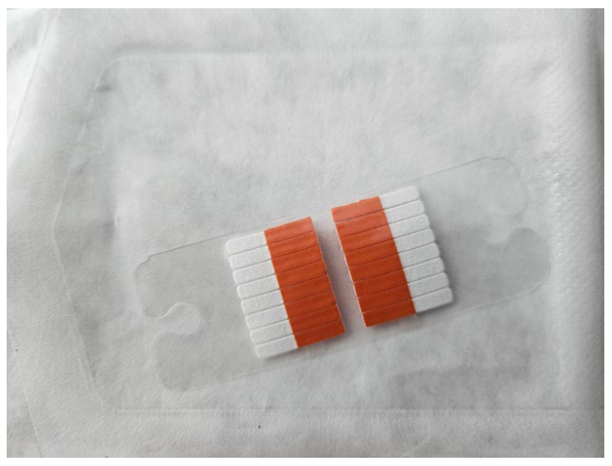
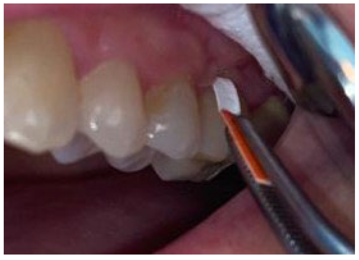
2.3. Gingival Crevicular Fluid Sampling

Gingival crevicular fluid samples were collected from each participant, using absorbent paper strips (PerioPaper, Oraflow Inc., Smithtown, NY, USA) (Figure 1). For the sampling procedure, the tooth wearing a dental crown with the deepest periodontal pocket was chosen as the test and the deepest pocket of the tooth without a dental crown in the lateral area as the control. Preventive measures for sample contamination were taken, such as the use of cotton rolls and air suction (for no contact with saliva). The strips were inserted and kept in place for 30 s (Figure 2). Consequent to their removal from the periodontal pocket they were visually inspected for lack of blood stains and were subjected to GCF quantity standardization using the Periotron 8000 device (Oraflow Inc., Smithtown, NY, USA). The resulting two paper strips were afterward placed separately into polyethylene microtubes with 50 µL saline buffer solution (PBS). The microtubes were preserved at −20 degrees Celsius, until sampling completion.

2.4. Immunological Assessment

The quantitative assessment of the targeted pro-inflammatory mediator CRP in the GCF samples was performed by the Immunology Laboratory of the University of Medicine and Pharmacy of Craiova, through enzyme-linked immunosorbent assay (ELISA). Commercial kits were used—Quantikine CRP Human ELISA Kit, RandDSystems (Minneapolis, MN, USA, range 0.8–50 ng/mL) according to the manufacturer’s indications. A standard optical analyzer at 450 nm wavelength was used with a correction at 540 nm.

2.5. Statistical Analysis

The data were statistically analyzed using the GraphPad 9.5.0 data analysis tool (GraphPad Prism, La Jolla, CA, USA), applying the Mann–Whitney U-test for comparisons between groups and the Spearman test (−1 < rho < 1) for correlations between the parameters assessed for the T group, p < 0.05 was set for statistical significance, after performing the sample size power computation using G*Power 3.1.9.7 that yielded a power level (1-β) of 52% for our analysis, at a 95% confidence level for each of the groups, given the effect size and desired alpha level, using 2-sided testing, assuming an alpha level of 0.05.

3. Results

The 28 patients were aged range 38–56 years (46.96 ± 5.72 years), 17 women, and 11 men. L for the MC was ranged 3–11 years (6.5 ± 1.79 years) (Table 1).

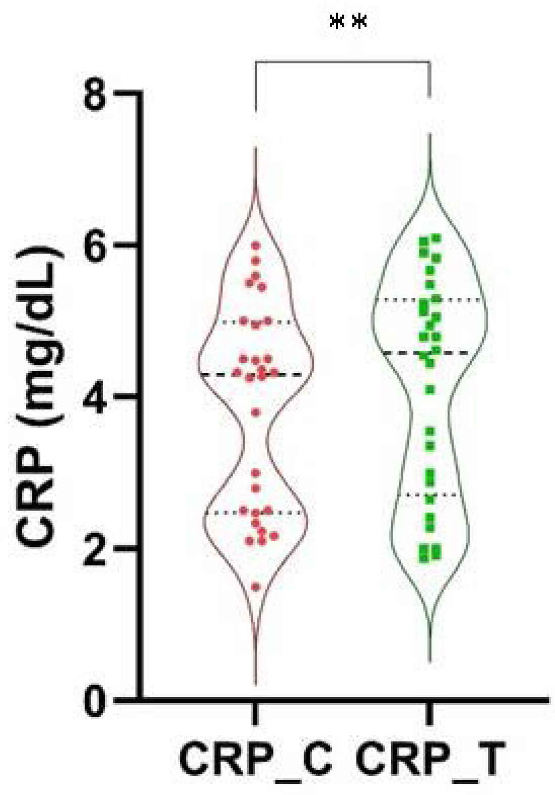
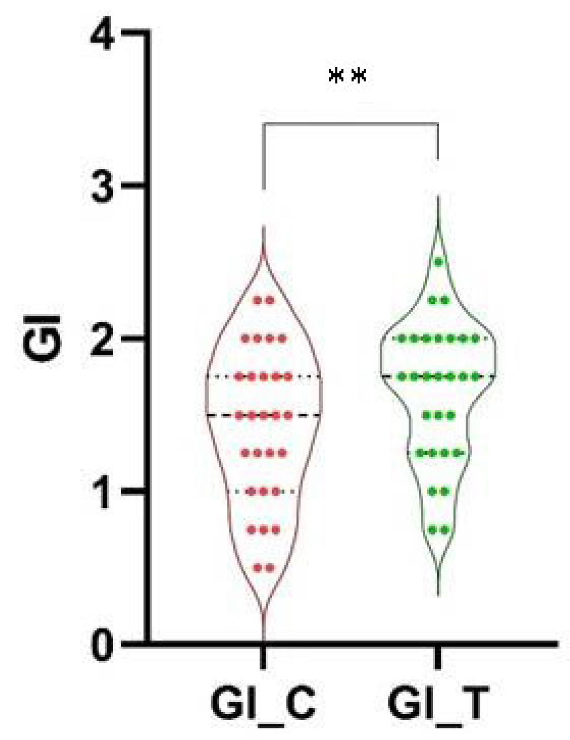
3.1. Comparisons between Groups

There are slightly higher values in T than in the C group with a statistical difference between the group T (4.14 ± 1.44) and C (3.85 ± 1.36) regarding the CRP, p = 0.036 (Figure 3) and also regarding the GI in group T (1.65 ± 0.45) compared to C (1.43 ± 0.51), at the limit of significance, p = 0.05 (Figure 4). PI was slightly higher and not significant, p = 0.1, in the T group (1.71 ± 0.67) than in C (1.5 ± 0.7) and no statistical significance was found regarding the PPD, even if the PPD in the T group (5.0 ± 0.97) was slightly higher than in C (4.83 ± 0.98), p = 0.711.

3.2. Correlations Statistically Significant between Periodontal Parameters, Longevity of Crown and Levels of CRP in T Group

There is a very strong correlation between levels of CRP and GI (rho = 0.882, p < 0.0001) and between levels of CRP and PPD (rho = 0.917, p < 0.0001). A strong correlation was found between L and levels of CRP (rho = 0.796, p < 0.0001) and between L and GI (rho = 0.715, p < 0.0001). A very strong correlation was found between L and PPD (rho = 0.847, p < 0.0001).

4. Discussion

During a routine dental examination, knowing and understanding the influence of restorative therapy on periodontal health can contribute to the establishment of an early diagnosis of periodontal disease, and implicitly facilitate its treatment. Any partial FDP in direct contact with the periodontal tissues can influence their health status, possibly through different characteristics, such as material biocompatibility, FDP marginal adaptation and topography [14].
In maintaining the health of the periodontal status in patients with fixed prosthetic restorations, oral hygiene has an important role. The study of Zlatanovska et al. emphasized the fact that adequate training in oral hygiene of patients presenting uni-dental or pluri-dental metal or metal-ceramic FDP increases the quality of oral hygiene and contributes to the reduction in plaque index and GI values [15].
The abutment teeth that present a crown as part of a pluri-dental FDP show higher GI values compared to the levels obtained from sites of intact teeth [16]. The presence of FDP, possibly by favoring plaque retention, causes inflammation in the gingival margin, increasing the GI score. This is also highlighted in the results of the current study, achieving a statistically significant difference between the T and C groups regarding the GI values, even the PI is not significantly higher, possibly due to the performance of recent domestic oral and dental hygiene before the visit in the dental office for examination, while the gingival inflammation expressed by GI is due to the biofilm accumulation.
There are studies that evaluated the influence of different biomaterials used in dental prosthetics on biofilm formation, as the placement in the oral cavity of different materials for dental treatment could generally impact the periodontal status [17,18]. Bremer et al. revealed a lower accumulation of biofilm in the case of zirconia, compared to different types of dental ceramics [19] but the study by Hahnel et al. demonstrated that between different types of ceramics, the streptococcal adhesion values show only small and random, statistically insignificant differences [20].
Another periodontal clinical parameter that seems to be influenced by the presence of a FDP is PPD. Medium values of PPD show higher values in the case of abutment teeth, compared to teeth without FDP [16]. The results of the current study also demonstrate a difference but without the statistical significance of PPD between teeth with metal-ceramic crowns and teeth in the control group.
The study by KC Basnyat S. et al. evaluates oral hygiene and gingival health in patients with uni-dental and multi-dental FDP over a period of 6 months. In the dental crown’s case, 14 days and 6 months after cementation that was accompanied by appropriate oral hygiene training, no statistically significant differences in plaque index and GI were obtained. Nor did the correlation of IP and GI values between metal and metal-ceramic materials reveal statistically significant differences [11]. Another study analyzes the gingival status and oral hygiene in patients with FDP, over a period of 3 months after their oral hygiene training. Statistically significant GI values compared between the first visit and 14 days later, were observed in the case of semi-physiognomic metal-acrylic pluri-dental FDP [15]. The research of Al-Sinaidi A. et al. shows that the highest medium values of the periodontal clinical parameters (IP, GI, PPD) are found at the level of abutment teeth, whose FDP was applied more than 5 years ago [16]. Similarly, in the actual study, within the T group, a highly significant correlation was obtained between metal-ceramic crown longevity and GI values, and between L and PPD, respectively. Thus, the longer the time elapsed since the application of FDP is, the higher the average values of periodontal clinical parameters are, as a result of the evolution of the periodontal inflammatory process.
Performing a comparison of IP values, probing depth, and sulcus bleeding index, between ceramic crowns on zirconia support obtained by CAD/CAM and metal-ceramic restorations with Ni-Cr-based metal substructure 12 months after application, they showed higher values in the case of the second category. Thus, ceramic crowns on a zirconium support promote the health of the marginal periodontium, due to the biocompatibility of the material [21]. Another study confirming the reduction in the inflammation process and the maintenance of the health of oral tissues by fixed prosthetic restorations on zirconium support obtained by the CAD/CAM technique, compared to those whose substructures are made of metal alloys, is that of SrimaneepongV. [22]. FDP with zirconium support manufactured by CAD/CAM technique shows better results on the health of periodontal tissues compared to a metal-ceramic prosthetic with Co-Cr-based metal substructure obtained either by conventional technique or by CAD/CAM technique [10] so, our patients are more exposed to a worsening of their periodontal status.
In the present study, the CRP value from the GCF content, a pro-inflammatory marker, was evaluated. There is a statistically significant difference between the average CRP values obtained in teeth with metal-ceramic crowns compared to those from intact teeth. Moreover, a strong and statistically significant correlation resulted between CRP and GI, and CRP and PPD. These results confirm/emphasize the idea that the presence of FDP causes inflammation in the periodontal tissues (translated by higher average GI values in the T group). As a result of the initiation and evolution of this inflammation, there is also an increase in CRP levels within the GCF, the more significant, the more advanced being the state of inflammation (translated by higher average values of GI and PPD). Furthermore, the older the MC, the more pronounced the degree of periodontal tissue inflammation is (highly significant correlation between L and CRP within the T group).
Periodontal diseases are correlated with elevated CRP values, both in serum and in GCF, the patients with high values of IL-1β (locally) or CRP (systemically) have a high probability of being affected by periodontal disease [23,24].
CRP values in GCF and serum increased proportionally with the severity of periodontal diseases. It also showed positive correlations with periodontal clinical parameters [25]. In the case of chronic periodontal diseases, as well as in the case of aggressive periodontal diseases, at the level of untreated sites, the IL-6, TNF-α, CRP and ALP values from the analysis of the collected GCF showed higher values compared to the sites at the level of marginal periodontium with intact status [26]. In severe chronic gingivitis, CRP and TNF-alpha levels may be reduced in GCF following treatment [27].
Referring to the longevity of FDP, in our group of periodontally-affected patients, the mean age was 6.5 years with a maximum longevity of 11 years. There are noteworthy studies that evaluate the clinical performance over time in the presence of MC crowns in comparison with crowns made of other types of materials, related or not to topography at the level of the dental arch by analyzing the survival rate of the prosthetic construction according to the occurrence of clinical complications represented by diseases of the teeth, or damage to the integrity of the FDP, requiring its removal.
The survival rate after 8 years since the application of the MC crown is 94.3% ± 1.8% [28], and after >8 years it varies between 92.3% and 95.9% [9]. The study by Boeckler et al. finds a survival rate after 3 years of 94.9% of ceramic crowns with titanium metal substructure obtained by CAD/CAM technology, due to mechanical complications but without the presence of biological ones [29]. Very close values of the survival rate were also obtained in the case of the MC crown with a metal substructure made of noble alloys 7 years after their application in the oral cavity, this being 95.5%; the reasons for removal were both mechanical and biological complications [30]. The long-term survival of the same types of MC crown with noble metal substructure is also proven by Behr M. et al. The frequency of periodontal diseases 10 years after the application of the MC crown is higher by approximately 13% compared to their prevalence obtained 5 years after the moment of cementation. Thus, it seems that the frequency of occurrence of periodontal disease increases with the longevity of the MC crown [31], a conclusion that coincides with the results of the current study, which obtained a strongly significant correlation between L and GI and CRP, as well as a highly statistically significant correlation between L and PPD, these correlations demonstrating a directly proportional relationship between the severity of the inflammatory process at the level of the periodontium and the longevity of the MC crown.
Another study on the survival rate of dental crowns related to their longevity (at 5, 10, 15 and 20 years after application) shows a value of this rate of 61.8% after 20 years, which is approximately 30% lower compared to the value obtained 5 years after application; among the biological complications tooth mobility and bone loss were mentioned [32]. There are studies that point out different survival rates of the MC crown compared to other materials, but from a biological point of view, the study by Pjetursson et al. found no differences at 5 years between all-ceramic crowns and MC crowns [33].
In the lateral area of the arch, at a period of 5 years after application, the survival rate is also increased in favor of the MC crown compared to the composite crowns, with the latter showing much more complications over time [8]. Twenty years after the application, the survival rate of MC crowns in the posterior area of the arch is 78.8%, approximately 10% higher than pluri-dental FDP in the same region, the authors taking into account both types of complications, technological and biological. Thus, in the posterior area, MC crowns seem to be a more viable long-term option [34]. Placed on molars, MC crowns have higher survival and success rates from a biological point of view, taking into account probing depth, attachment loss and bleeding on probing, compared to zirconia-supported or all-over ceramic crowns [35,36], Olley R.’s study obtaining an average survival value of these restorations of 47.53 years, under the conditions of maintaining good oral hygiene [37]. Due to the reduced number of participants, we did not divide them into subgroups in regard to the detailed diagnosis of periodontitis in stages and grades, this being a limitation of our study. the reduced number of patients was caused by the application of multiple exclusion criteria, for the research’s accuracy and avoiding bias. Thus, within the limitations of this study, in view of the present results, keeping MC in the oral cavity for a longer period of time, can lead, in the presence of periodontal inflammation, to an increase in immune and clinical inflammatory expression.

5. Conclusions

In the presence of MC crowns, high GI values and up-regulated expression of CRP were detected, which clinically translated into exacerbation of periodontal inflammation in patients with periodontitis.

Author Contributions

Conceptualization, A.-R.D. and P.Ş.; methodology, A.-R.D., M.V.B. and P.Ş.; validation, D.M.P. and I.M.; formal analysis, D.M.P.; investigation, D.N.G. and P.C.M.; data curation, D.N.G., I.M. and H.O.M.; writing—original draft preparation, A.-R.D., M.V.B. and P.Ş.; writing—review and editing, D.N.G., D.M.P., I.M., P.C.M. and H.O.M.; supervision, H.O.M. All authors have read and agreed to the published version of the manuscript.

Funding

This research received no external funding.

Institutional Review Board Statement

The study was conducted in accordance with the Declaration of Helsinki and approved by the Ethics Committee of University (Nr. 104/2018).

Informed Consent Statement

Informed consent was obtained from all subjects involved in the study.

Data Availability Statement

The data used to support the findings of this study are available from the corresponding author upon reasonable request.

Acknowledgments

The authors expressed their gratitude to Adina Turcu-Stiolica for her support in the performance of the statistical analysis.

Conflicts of Interest

The authors declare no conflict of interest.

References

  1. Cortelli, J.R.; Aquino, D.R.; Cortelli, S.C.; Fernandes, C.B.; de Carvalho-Filho, J.; Franco, G.C.N.; Kawai, T. Etiological analysis of initial colonization of periodontal pathogens in oral cavity. J. Clin. Microbiol. 2008, 46, 1322–1329. [Google Scholar] [CrossRef] [PubMed]
  2. Surlin, P.; Foia, L.; Solomon, S.; Popescu, D.M.; Gheorghe, D.N.; Camen, A.; Martu, M.A.; Rauten, A.M.; Olteanu, M.; Pitru, A.; et al. Cytokines’ Involvement in Periodontal Changes. In Cytokines; Behzadi, P., Ed.; IntechOpen: London, UK, 2020. [Google Scholar]
  3. Kamath, D.G.; Nayak, S.U. Detection, removal and prevention of calculus: Literature Review. Saudi Dent. J. 2014, 26, 7–13. [Google Scholar] [CrossRef]
  4. Hinrichs, J.E.; Thumbigere-Math, V. Chapter 13: The Role of Dental Calculus and Other Local Predisposing Factors. In Newman and Carranza’s Clinical Periodontology, 13th ed.; Elsevier: Amsterdam, The Netherlands, 2019. [Google Scholar]
  5. Reynolds, M.A. Modifiable risk factors in periodontitis: At the intersection of aging and disease. Periodontology 2000 2014, 64, 7–19. [Google Scholar] [CrossRef] [PubMed]
  6. Ercoli, C.; Caton, J.G. Dental prostheses and tooth-related factors. J. Periodontol. 2018, 89 (Suppl. 1), S223–S236. [Google Scholar] [CrossRef] [PubMed]
  7. Savencu, C.E.; Şerban, C.; Porojan, L. Adaptability Evaluation of Metal-Ceramic Crowns Obtained by Additive and Subtractive Technologies. Appl. Sci. 2020, 10, 5563. [Google Scholar] [CrossRef]
  8. Overmeer, J.; Narby, B.; Hjalmarsson, L.; Arnrup, K.; Eliasson, A. A retrospective multicenter study comparing metal-ceramic and composite single crowns performed in public general dentistry: 5-year results. Acta Biomater. Odontol. Scand. 2016, 2, 43–48. [Google Scholar] [CrossRef] [PubMed]
  9. Porcelain-Fused-to-Metal Crowns versus All-Ceramic Crowns: A Review of the Clinical and Cost-Effectiveness [Internet]; Canadian Agency for Drugs and Technologies in Health: Ottawa, ON, Canada, 29 May 2015. Available online: https://0-www-ncbi-nlm-nih-gov.brum.beds.ac.uk/books/NBK304693 (accessed on 14 August 2022).
  10. Avetisyan, A.; Markaryan, M.; Rokaya, D.; Tovani-Palone, M.R.; Zafar, M.S.; Khurshid, Z.; Vardanyan, A.; Heboyan, A. Characteristics of Periodontal Tissues in Prosthetic Treatment with Fixed Dental Prostheses. Molecules 2021, 26, 1331. [Google Scholar] [CrossRef]
  11. Kc Basnyat, S.; Sapkota, B.; Shrestha, S. Oral Hygiene and Gingival Health in Patients with Fixed Prosthodontic Appliances—A Six Month Follow-up. Kathmandu Univ. Med. J. (KUMJ) 2015, 13, 328–332. [Google Scholar]
  12. Paraskevas, S.; Huizinga, J.D.; Loos, B.G. A systematic review and meta-analyses on C-reactive protein in relation to periodontitis. J. Clin. Periodontol. 2008, 35, 277–290. [Google Scholar] [CrossRef]
  13. Tonetti, M.S.; Greenwell, H.; Kornman, K.S. Staging and grading of periodontitis: Framework and proposal of a new classification and case definition. J. Periodontol. 2018, 89 (Suppl. 1), S159–S172. [Google Scholar] [CrossRef]
  14. Panagakos, F.S.; Davies, R.M. Gingival Diseases—Their Aetiology, Prevention and Treatment; IntechOpen: London, UK, 2011; 248p. [Google Scholar]
  15. Zlatanovska, K.; Dimova, C.; Zarkova-Atanasova, J.; Korunoska-Stevkovska, V.; Gigovski, N.; Kocovski, D. Oral hygiene in patients with fixed prosthodontic restorations. J. Hyg. Eng. Des. 2017, 21, 83–89. [Google Scholar]
  16. Al-Sinaidi, A.; Preethanath, R. The effect of fixed partial dentures on periodontal status of abutment teeth. Saudi J. Dent. Res. 2014, 5, 104–108. [Google Scholar] [CrossRef]
  17. Aminov, L.; Vataman, M.; Maxim, D.C.; Salceanu, M.; Surlin, P.; Checherita, L.E. Comparative biochemical evaluation of Ca, P and Mg, after subcutaneous implantation of some biomaterials used in endodontic treatment. Rev. Mater. Plast. 2014, 51, 246–251. [Google Scholar]
  18. Kanjevac, T.; Taso, E.; Stefanovic, V.; Petkovic-Curcin, A.; Supic, G.; Markovic, D.; Djukic, M.; Djuran, B.; Vojvodic, D.; Sculean, A.; et al. Estimating the Effects of Dental Caries and Its Restorative Treatment on Periodontal Inflammatory and Oxidative Status: A Short Controlled Longitudinal Study. Front. Immunol. 2021, 12, 716359. [Google Scholar] [CrossRef] [PubMed]
  19. Bremer, F.; Grade, S.; Kohorst, P.; Stiesch, M. In vivo biofilm formation on different dental ceramics. Quintessence Int. 2011, 42, 565–574. [Google Scholar] [PubMed]
  20. Hahnel, S.; Rosentritt, M.; Handel, G.; Bürgers, R. Surface characterization of dental ceramics and initial streptococcal adhesion in vitro. Dent. Mater. 2009, 25, 969–975. [Google Scholar] [CrossRef]
  21. Shang, L.-J.; Wu, Y.; Xu, Y.-J. Effect of the CAD/CAM zirconia all-ceramic crown restoration on periodontal tissue. Chin. J. of Tissue Eng. Res. 2014, 18, 4804–4809. [Google Scholar]
  22. Srimaneepong, V.; Heboyan, A.; Zafar, M.S.; Khurshid, Z.; Marya, A.; Fernandes, G.V.O.; Rokaya, D. Fixed Prosthetic Restorations and Periodontal Health: A Narrative Review. J. Funct. Biomater. 2022, 13, 15. [Google Scholar] [CrossRef]
  23. Megson, E.; Fitzsimmons, T.; Dharmapatni, K.; Bartold, P.M. C-reactive protein in gingival crevicular fluid may be indicative of systemic inflammation. J. Clin. Periodontol. 2010, 37, 797–804. [Google Scholar] [CrossRef]
  24. Fitzsimmons, T.R.; Sanders, A.E.; Bartold, P.M.; Slade, G.D. Local and systemic biomarkers in gingival crevicular fluid increase odds of periodontitis. J. Clin. Periodontol. 2010, 37, 30–36. [Google Scholar] [CrossRef]
  25. Pradeep, A.R.; Manjunath, R.G.; Kathariya, R. Progressive periodontal disease has a simultaneous incremental elevation of gingival crevicular fluid and serum CRP levels. J. Investig. Clin. Dent. 2010, 1, 133–138. [Google Scholar] [CrossRef] [PubMed]
  26. Zhang, Q.; Chen, B.; Zhu, D.; Yan, F. Biomarker levels in gingival crevicular fluid of subjects with different periodontal conditions: A cross-sectional study. Arch. Oral. Biol. 2016, 72, 92–98. [Google Scholar] [CrossRef]
  27. Alzahrani, A.S.; Bissada, N.F.; Jurevic, R.J.; Narendran, S.; Nouneh, I.E.; Al-Zahrani, M.S. Reduced systemic inflammatory mediators after treatment of chronic gingivitis. Saudi Med. J. 2013, 34, 415–419. [Google Scholar] [PubMed]
  28. Reitemeier, B.; Hänsel, K.; Kastner, C.; Weber, A.; Walter, M.H. A prospective 10-year study of metal ceramic single crowns and fixed dental prosthesis retainers in private practice settings. J. Prosthet. Dent. 2013, 109, 149–155. [Google Scholar] [CrossRef] [PubMed]
  29. Boeckler, A.F.; Lee, H.; Stadler, A.; Setz, J.M. Prospective observation of CAD/CAM titanium ceramic single crowns: A three-year follow up. J. Prosthet. Dent. 2009, 102, 290–297. [Google Scholar] [CrossRef] [PubMed]
  30. Reitemeier, B.; Hänsel, K.; Kastner, C.; Walter, M.H. Metal-ceramic failure in noble metal crowns: 7-year results of a prospective clinical trial in private practices. Int. J. Prosthodont. 2006, 19, 397–399. [Google Scholar] [PubMed]
  31. Behr, M.; Zeman, F.; Baitinger, T.; Galler, J.; Koller, M.; Handel, G.; Rosentritt, M. The clinical performance of porcelain-fused-to-metal precious alloy single crowns: Chipping, recurrent caries, periodontitis, and loss of retention. Int. J. Prosthodont. 2014, 27, 153–160. [Google Scholar] [CrossRef] [PubMed]
  32. Hawthan, M.; Chrcanovic, B.R.; Larsson, C. Retrospective clinical study of tooth-supported single crowns: A multifactor analysis. Eur. J. Oral. Sci. 2022, 130, e12871. [Google Scholar] [CrossRef]
  33. Pjetursson, B.E.; Sailer, I.; Zwahlen, M.; Hammerle, C.H.F. A systematic review of the survival and complication rates of all-ceramic and metal–ceramic reconstructions after an observation period of at least 3 years. Part I: Single crowns. Clin. Oral. Implants Res. 2007, 18 (Suppl. 3), 73–85. [Google Scholar] [CrossRef]
  34. Reitemeier, B.; Hänsel, K.; Range, U.; Walter, M.H. Prospective study on metal ceramic crowns in private practice settings: 20-year results. Clin. Oral. Investig. 2019, 23, 1823–1828. [Google Scholar] [CrossRef]
  35. Anusavice, K.J. Standardizing failure, success, and survival decisions in clinical studies of ceramic and metal-ceramic fixed dental prostheses. Dent. Mater. 2012, 28, 102–111. [Google Scholar] [CrossRef] [PubMed]
  36. Sailer, I.; Gottnerb, J.; Kanelb, S.; Hammerle, C.H. Randomized controlled clinical trial of zirconia-ceramic and metal-ceramic posterior fixed dental prostheses: A 3-year follow-up. Int. J. Prosthodont. 2009, 22, 553–560. [Google Scholar] [PubMed]
  37. Olley, R.C.; Andiappan, M.; Frost, P.M. An up to 50-year follow-up of crown and veneer survival in a dental practice. J. Prosthet. Dent. 2018, 119, 935–941. [Google Scholar] [CrossRef] [PubMed]
Figure 1. PerioPaper strips.
Applsci 13 10993 g001
Figure 2. Gingival crevicular fluid sampling.
Applsci 13 10993 g002
Figure 3. Comparison between T and C groups of CRP levels, C-Control group, t-test group, CRP-C reactive protein, ** p < 0.05.
Applsci 13 10993 g003
Figure 4. Comparison between T and C groups of GI, C-Control group, t-test group, CRP-C reactive protein, ** p = 0.05.
Applsci 13 10993 g004
Table 1. Patient data.
n%Mean ± Standard Deviation, Range
GenderM1139.28-
F1760.71-
EnvironmentUrban28100-
Rural00-
Age--46.96 ± 5.72, 38–56
Longevity of crowns281006.5 ± 1.79, 3–11
n, number of patients; %, percentage of patients.
Disclaimer/Publisher’s Note: The statements, opinions and data contained in all publications are solely those of the individual author(s) and contributor(s) and not of MDPI and/or the editor(s). MDPI and/or the editor(s) disclaim responsibility for any injury to people or property resulting from any ideas, methods, instructions or products referred to in the content.

