Article

Prognostic Value of Lymph Node Density in Lingual Squamous Cell Carcinoma

by
Carlos Navarro Cuéllar
1,
Ángela Sada Urmeneta
1,
Raquel Lorenzo Marcos
1,
Raúl Antúnez-Conde
1,*,
Ana López López
1,
José Luis del Castillo Pardo de Vera
2,
Javier González Martín-Moro
2,
José Luis Cebrián Carretero
2,
Francisco Alijo Serrano
3,
Giovanni Dell’Aversana Orabona
4,
Jose J. Zamorano-León
5 and
Ignacio Navarro Cuéllar
1
1
Maxillofacial Surgery Department, Hospital General Universitario Gregorio Marañón, C/Doctor Esquerdo 46, 28007 Madrid, Spain
2
Maxillofacial Surgery Department, Hospital Universitario La Paz Madrid, P.º de la Castellana, 261, 28046 Madrid, Spain
3
Pathological Anatomy Department, Hospital General Universitario Gregorio Marañón, C/Doctor Esquerdo 46, 28007 Madrid, Spain
4
Maxillofacial Surgery Department, Università “Federico II”, 80131 Naples, Italy
5
Public Health and Maternal & Child Health Department, School of Medicine, Universidad Complutense, 28040 Madrid, Spain
*
Author to whom correspondence should be addressed.
Submission received: 20 February 2023 / Revised: 10 April 2023 / Accepted: 23 April 2023 / Published: 1 May 2023
(This article belongs to the Special Issue Current Advances in Dentistry)

Abstract

:
Lymph node density (LND)—the proportion of positive nodes among the total number of resected nodes—has emerged as a reliable prognostic factor in solid tumors. This study aims to assess the importance of LND in lingual squamous cell carcinoma (LSCC) and its prognostic involvement. A retrospective longitudinal study with 62 patients was performed. All patients were diagnosed with LSCC and submitted for tumor resection and neck dissection. Patients were stratified into low (<0.04) and high risk (≥0.04) based on LND. We analyzed the impact of LND on overall survival (OS) and disease-free survival (DFS), as well as the relationship between LND and the pathological staging, the involvement of positive margins, depth of invasion (DOI) and perineural infiltration. This study provides a substantial relationship between lymph node density (LND), overall survival (OS) and disease-free survival (DFS) in lingual squamous cell carcinoma (LSCC). A statistically significant distribution was found between LND, perineural infiltration and pathological staging, whereas no association was found with the rest of the prognostic variables analyzed.

1. Introduction

Squamous cell carcinoma of the oral cavity represents 3% of malignant tumors, with the tongue representing the most common location (40% of oral cavity cancer) [1]. The risk factors are tobacco and alcohol consumption and it mainly affects advanced age males (>65 years) and has an estimated survival rate in Europe of 66% at one year and 31% at five years.
The most common clinical presentation of lingual squamous cell carcinoma (LSCC) is the tongue sore located at the lateral lingual border. LSCC spreads primarily by contiguity and lymphatic spread, with cervical lymphatic progression being one of the most important independent prognostic factors for the disease [2].
The AJCC, in its 8th edition, introduces two new parameters, depth of tumor invasion (DOI) and extranodal extension of the tumor in the lymph node (ENE), which can modify the T and N categories, respectively [3].
Following tumor staging and provided that distant disease has been excluded, surgical treatment should be directed at tumor resection and neck surgery. If cervical disease is clinically or radiologically diagnosed, a therapeutic complete lymphadenectomy of levels I–V should be performed. If, on the other hand, the neck is negative (N0), either a selective neck dissection of levels I–III or I–IV or a selective sentinel lymph node biopsy can be performed. The need for a bilateral lymphadenectomy depends on whether or not the contralateral neck is positive and on the proximity of the tumor to the midline of the tongue [1].
Prognostic staging based exclusively on AJCC classification may not predict survival in oral cavity squamous cell carcinoma (OSCC) with absolute reliability [2,4,5,6,7]. Therefore, attempts have been made to study other additional prognostic factors that provide a high enough risk to recommend the use of adjuvant radiotherapy or intensification of the adjuvant treatment approach (radiotherapy associated with chemotherapy), which in turn is associated with a risk of acute toxicity and late complications that can substantially affect the quality of life of the patients [8,9,10,11]. It is therefore mandatory to find reliable prognostic factors that may lead us to identify high-risk patients who can benefit from a more aggressive treatment approach.
One of the most widespread criticisms of the AJCC pathologic staging system applied to N stage is that it is limited by the quality of the neck dissection—interoperator reliability—and the anatomopathologic sampling procedure. In this context, the lymph node density (LND)—defined as the proportion of positive nodes among the total number of nodes removed from the dissection—has emerged as an independent prognostic factor that is able to compensate for the potential bias derived from the technique used by taking into account three factors [12,13]:
The regional spread of the disease (number of positive lymph nodes);
The surgical treatment (total number of nodes removed during surgery);
The accuracy of the histopathological analysis.
LND has been shown to be a reliable predictor of survival in some solid tumors, including breast cancer, colon cancer and pancreatic cancer, in addition to head and neck cancer [12,14]. Specifically, high LND values have been associated with worse outcomes in terms of overall survival (OS), disease-specific survival (DSS) and disease-free survival (DFS) in squamous cell carcinoma of the oral cavity, and specifically in LSCC [12,15,16].
The present study aims to validate the concept of LND in lingual squamous cell carcinoma (LSCC) and its prognostic significance. Different potentially optimal cut-off values of LND have been proposed in several retrospective studies and in two systematic reviews [12,17]. In this study, following the recommendations of different studies, the cut-off point is an LND value of 0.04.
The hypothesis formulated by the investigators was that LND ≥ 0.04 worsens the prognosis of patients undergoing surgery for LSCC. The primary objective of the study was to assess the correlation between the LND value and OS and DFS.
The secondary objectives were: (1) to assess the relationship between LND and other poor prognostic factors in LSCC such as pathological margin status, DOI and perineural infiltration; (2) to analyze the correlation with tumor staging according to the 8th edition of the AJCC.

2. Materials and Methods

2.1. Population and Study Variables

To address the research purpose, the investigators implemented a retrospective longitudinal study of patients diagnosed with lingual squamous cell carcinoma (LSSC) and operated on by the Maxillofacial Surgery Department at Gregorio Marañón Hospital between 1 September 2016 and 1 September 2021. This study has been approved by the Ethics and Research Committee of the Hospital General Gregorio Marañón and in accordance with the principles of the Declaration of Helsinki.
All the patients in the study were operated on by the same surgeons in charge of head and neck oncological surgery in the Department of Maxillofacial Surgery at the Gregorio Marañón Hospital. The histopathological study was performed by the same pathologist in charge of head and neck histological studies at the hospital.
Information on each patient was obtained from patient records from the Department of Oral and Maxillofacial Surgery’s operating room report file and histopathology reports. The following parameters were studied:
Demographic variables: gender, age, stratified age, smoker, drinker;
Clinical variables: clinical size of the tumor, location of the tumor, diagnostic CT, diagnostic MRI, size and radiological DOI of the tumor, clinical staging, reconstruction, type of dissection, radiotherapy (RT) and postoperative chemotheraphy (QT);
Histological variables: degree of tumor differentiation, perineural invasion, lymphovascular invasion, TNM and pathological staging according to the 8th edition of the AJCC, LND, neck positivity, ENE and tumor budding;
Control variables: loco-regional recurrence, distant recurrence and mortality.
In order to be included in the study, patients had to fulfill the following inclusion criteria:
  • Histological confirmation of the diagnosis of primary lingual squamous cell carcinoma;
  • Patients operated on by the Oral and Maxillofacial Surgery Department at Gregorio Marañón General Hospital between 9 January 2016 and 9 January 2021;
  • Patients for whom surgery was the treatment of choice and whose intervention consisted of tumor resection and associated cervical lymphadenectomy;
  • Negative resection surgical margins for tumor involvement.
The exclusion criteria were:
  • Disease that exceeds the anterior 2/3 of the tongue (involvement of the base of the tongue);
  • Patients previously treated for carcinoma of the oral cavity (surgery, radiotherapy or chemotherapy);
  • Patients whose medical records did not identify the variables under study.
Once the inclusion and exclusion criteria were established, a sample size of 62 patients was obtained (Figure 1).
Receiver operating characteristic (ROC) curve and area under the curve (AUC), sensitivity, specificity and 95% confidence interval (CI) were used to determine the LND values that best defined both risk groups. According to the authors’ estimates, subjects were divided into binary subgroups using the best LND value as the cut-off point. Figure 2 shows cut-off values for predicting overall survival (OS) by ROC curve analysis. Results reported that the best cut-off value for LND was 0.04 (AUC, 0.689; sensitivity, 33%; specificity, 87%; p = 0.029). Figure 2.

2.2. Data Analysis

Categorical variables were expressed as frequency and percentage and compared by Chi-square test. Continuous variables were expressed as mean ± standard error of mean (S.E.M.). Comparison of quantitative variables was performed using the non-parametric Kruskal–Wallis and Mann–Whitney U tests. To analyse the effect of the LND and histopathological markers on overall survival and disease-free survival, the Kaplan–Meier survival curves and log-rank test were used. Statistical significance was considered for p-values < 0.05. The statistical analysis was performed with the SPSS software (version 25.0).

3. Results

3.1. Descriptive Analysis of the Total Population and Population Subgroups According to the LND

The demographic, clinical, histologic and follow-up analysis of the total study population (n = 62) is shown in the Table 1, Table 2, Table 3 and Table 4, as well as the rates with respect to the total sample and the results of the analysis by subgroups according to the LND (<0.04 and ≥0.04).
In the statistical analysis, significant differences were observed in the distribution between the subgroups according to the LND of the categories of the following variables:
  • − Neck dissection (p-value 0.021);
  • − Pathologic T (p-value 0.006);
  • − Pathologic N (p-value < 0.001);
  • − Perineural invasion (p-value 0.005);
  • − Pathologic stage (p-value < 0.001).

3.2. Analysis of Primary Objective

No significant differences were found in the distribution of the clinical outcome variables OS and DFS between the two prognostic groups established according to LND, although the study provides a substantial relationship between lymph node density (LND) and overall survival (OS) and disease-free survival (DFS) in lingual squamous cell carcinoma (LSCC).

3.3. Overall Survival (OS)

Overall survival (OS) measures the number of patients still alive at the end of the study period. This parameter includes patients who died due to the disease and those who died of other causes. The OS analysis is performed with the two population subgroups (LND < 0.04 and ≥0.04). As shown in Figure 2, 79.6% of patients with LND < 0.04 survived, while this percentage decreased to 62.5% in those with LND ≥ 0.04 (Figure 3).
The Kaplan–Meier survival curve (Figure 4) suggests differences between the two groups. The mean time measured in months to the onset of death is shown in Table 5. The results reveal that there are no significant differences with respect to OS depending on LND. There is a large dispersion in the results for each group (standard deviation), suggesting heterogeneity of intragroup results.

3.4. Disease-Free Survival (DFS)

Disease-free survival (DFS) is used to evaluate the appearance of tumor recurrences, whether loco-regional or distant, during the study period. The analysis of DFS related to the two subgroups is described in the following figures and table (Figure 5 and Figure 6, Table 6).
Figure 5 shows that 77.6% of patients with LND < 0.04 did not experience recurrence throughout the study, while this rate decreased (53.9%) in the LND ≥ 0.04 subgroup.
The Kaplan–Meier recurrence curve (Figure 6) is very significant, showing a higher DFS among patients considered to be at lower risk according to the LND. Table 6 shows that the results obtained in terms of DFS, although close, did not reach significance.
Therefore, regarding the main objective of the study and in terms of the analysis of overall survival and disease-free survival, there are substantial, although not significant, differences between both groups depending on the LND. However, the results of the statistical analysis do not provide a basis for accepting the initial hypothesis of the study since it cannot be guaranteed that the presence of a LND ≥ 0.04 implies a lower survival.

3.5. Analysis of Secondary Objectives

No statistically significant differences were found in the distribution of histological characteristics, pathological margins and stratified DOI. The stratified DOI variable is very close to significance, but does not reach it (p 0.054).
The histologic variables significantly associated with LND are perineural invasion and pathologic staging.

3.6. Perineural Invasion

Regarding perineural infiltration (Figure 7), there is significant evidence of its incidence in patients with LND ≥ 0.04 (92.3%), compared to those with LND < 0.04 (49%).

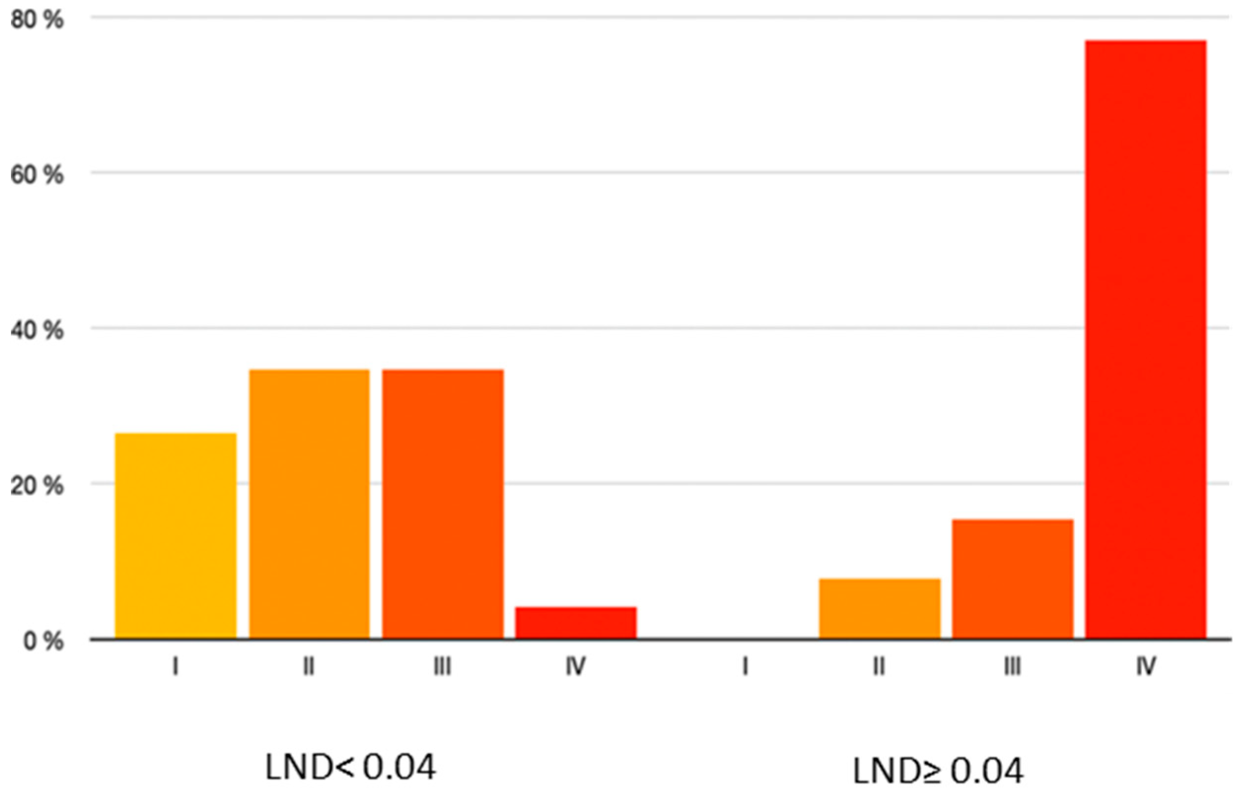
3.7. Pathologic Stage

Regarding the pathologic stage according to AJCC, a predominance of stage IV (76.92%) was observed among patients with an LND ≥ 0.04 compared to patients with LND < 0.04, where the distribution of patients among the different pathologic stages was more consistent (Figure 8).

4. Discussion

LND has been shown to be a valid predictor of survival in the case of some solid tumors, including breast cancer, colon cancer and pancreatic cancer [12]. Some studies suggest that LND may have a greater prognostic significance than conventional pathologic staging N in OSCC [12,14,17] and specifically in LSCC [18]. LND can be used as a predictor of survival as well as an indicator for the use of adjuvant therapy in LSCC [18,19].
The purpose of this study was to investigate the value of LND in the prognosis of patients diagnosed with LSCC, and to compare it with other conventional risk factors. In our study, we established the cut-off point for LND at 0.04 because it was referred to in the literature and because it included patients with pN0 and bilateral dissections.
The results of the present study show a strong correlation between LND and the presence of tumor disease in the lymph nodes (p < 0.001). This seems logical, as lymphatic tumor invasion is likely to increase the risk of developing cervical metastases and, subsequently, affect LND. This finding is in accordance with findings reported in other publications, including tumor locations other than LSCC [19]. The correlations observed with T stage (p 0.006) and with other pathological risk factors, such as pathological tumor staging according to AJCC (p < 0.001) and perineural tumor invasion (p 0.005), can be considered a sign of increased tumor aggressiveness, and support the validity of using LND to establish the prognosis of patients with LSCC.
The main objective of this work was to correlate LND with OS and DFS in LSCC. Although it was not possible for a multivariable analysis to be performed due to the limited number of patients and, although not reaching statistical significance, Kaplan–Meier curves for OS (Figure 3) and DFS (Figure 5) suggest a worse outcome for the group with LND ≥ 0.04 (OS = 56.67 ± 10.53 months; DFS = 45.22 ± 10.59 months) with respect to the group with LND < 0.04 (OS = 76.97 ± 5.27 months; DFS = 76.13 ± 5.72 months), suggesting that higher LND may be associated with reduced survival. Survival rates in our patients were similar to those published in the scientific literature.
Iftikhar et al. [20] studied a cohort similar to that of this work, with 130 patients with LSCC, and established a cut-off point of 0.012 for LND. In their study, the mean OS of those patients with LND > 0.012 was 38.1 months vs. 52.1 months in those with LND < 0.012. The mean DFS in the lower LND group was 53.6 months vs. 39.2 months in the higher LND group. In both cases, the association between LND and survival is significant. Lieng et al. [18] included 72 patients with LSCC and N+ in their cohort and set the cut-off point for LND at 0.143. They found prognostic significance of LND in terms of OS (mean 82.3 months in the high-risk LND group vs. 14.7 months in the low-risk group). Patel et al. [12], in the largest study of LND in OSCC conducted to date (4000 patients stratified according to an LND of 0.07), demonstrated the prognostic reliability of LND in terms of OS and DFS. In their study they reported a 5-year OS rate of 49% in the low LND group vs. 35% in the high LND group. The 5-year DFS rate was 55% in patients with low LND vs. 38% in those with high LND. Yamagata et al. [21] included 95 patients in their study with OSCC stratified into two risk groups based on an LND cut-off point of 0.04. In their analysis, they demonstrated that LND is a prognostic factor for overall survival (5-year OS rate of 90.5% for patients with low LND and 68.8% for patients with high LND).
As for secondary objectives, this study demonstrates a significant association between LND and perineural invasion as well as pathologic staging according to AJCC. No significance was reached in the relationship between LND and pathologic margin status and DOI, although in the latter it was very close to significance. Iftikhar et al. [20] demonstrated in their study the prognostic validity of the variables’ pathological margins, DOI and pathological staging. The same variables proved to be reliable predictors of survival in the study carried out by Patel [12].

4.1. Significance and Practical Application of Results

The main objective of this study was to determine the importance of LND in relation to LSCC so, according to this, decisions about adjuvant post-surgical treatments could be established and the need for more frequent postoperative clinical controls could be assessed. Although the results obtained did not reach significance, there is a high and strong association in the relationship between LND and overall survival and disease-free survival. These things considered, the association with other poor prognostic variables suggests the validation of LND as a prognostic factor in LSCC.

4.2. Limitations of the Study

The main limitation of this study is the retrospective design of the study. For this reason, the benefits of prospective follow-up are lost, and it significantly limits control over possible sources of bias.
In this sense, the fact of including only patients for whom resection specimens negative margins for tumor infiltration were obtained may partially limit the conclusions, since patients with affected margins are excluded, given that this could condition their survival.
Similarly, the estimation of LND may be influenced by patient selection bias, since cervical lymphadenectomy is not routinely performed on all patients, but it is decided on a case-by-case basis based on the preoperative evaluation of each patient. The total number of lymph nodes obtained from the cervical dissection was also not taken into account as an inclusion/exclusion criteria in the study, which could have caused a statistical bias by including suboptimal cervical dissections that condition the prognosis of the patients.
Regarding the variables reported in the study, it is possible that the results were affected by possible confusion factors such as the presence of comorbidities, functional status, the use of systemic therapies and subsequent local treatments. Likewise, the lack of information on the number of cycles and radiation doses used may be a limitation of the results.
Finally, another limitation of the study is the fact that the number of patients that can be obtained from a single institution in a short period of time is relatively small, which affects the statistical analysis of the data as demonstrated by the large dispersion of the results that comprise each group. In addition, it was not possible for multivariable analysis to be performed due to the limited number of patients. Further studies with larger sample sizes are required to establish the relationship between variables reported in the present work and LND.

4.3. Relation to Similar Publications and Comparison between Terms of Agreement and Disagreement

Most of the published studies on LND in carcinoma of the oral cavity include patients with OSCC. To our knowledge, the only multicenter studies performed to date, which provide the most consistent results, include cohorts of patients diagnosed with OSCC on whom cervical dissection was routinely performed in conjunction with tumor excision [12,17]. In both studies, LND is shown to be superior to the conventional N-staging system in predicting survival [12,17].
The main area of disagreement in the literature involves the establishment of the optimal cut-off point for stratifying patients according to LND. Multiple studies have used minimal p-values to identify a cut-off point for LND, leading to several threshold values ranging from 4.8% to 20% [18]. The largest study conducted to date by Patel [12] applied time-dependent curve analysis for disease-specific survival (DSS) in selecting the cut-off point for LND, setting the cut-off point at 0.07. This study demonstrated in multivariate analysis that a DSS > 0.07 was an independent prognostic factor associated with worse OS and DFS. However, the study did not provide further information on the methods used to achieve their sample size and, in their analysis, they also achieved significance for staging based on N [12,20]. Sayed [22] analyzed 1408 patients using a cut-off point of 0.088 based on time-dependent curve analysis for DFS, and reported that it was significant compared to N [12,20].
Spoerl [17] analyzed in their multicenter study a cohort of 717 patients and categorized LND based on the median value (0.055). LND was also shown to be an independent prognostic factor in OS and DFS by uni- and multivariate analysis. Likewise, Gil [14] used a cut-off point of 0.06 based on the median of the 386 patients included in their cohort, and concluded that LND reliably stratifies the risk of disease recurrence and survival in these patients. Therefore, the use of mean or median values to establish the cut-off point of the LND is not a reliable approach, since its value can deviate widely with small changes in the data or in the sample size [18,20].
Along a similar approach, Moratin [19] analyzed the comparability and universality of LND in the different anatomic locations included in OSCC, highlighting the need to develop specific treatment protocols for each anatomic sublocation, given the different treatment failure rates and clinical outcomes. In their study, they demonstrated that there is probably no universal cut-off point for LND, and that its assessment cannot constitute the only factor in the prognostic evaluation of patients with OSCC.
Another area of disagreement in the literature is the inclusion or non-inclusion of pN0 patients in the analysis of the LND cut-off point. Lieng [18], in accordance with other previous studies, discussed in their study the need to exclude those patients with negative nodes from the LND analysis, as their inclusion could influence the estimation of the LND cut-off inappropriately. In their study, they confirmed the statistical significance of the association between LND > 14.3% and worse OS and DFS. Other studies, including both multicenter studies reviewed, confirm the statistical significance of the LND value taking into account pN0 patients, as they consider that LND is derived from N staging and therefore N0 lymph node metastasis should be taken into account when establishing the LND cut-off point [12,17,20].

4.4. Recommendations and Guidelines for Future Research

In consideration of the disagreement shown in the literature, future research in this field should focus on establishing a consensus regarding the LND cut-off point in LSCC and outlining the requirements for establishing whether LND is representative or not. In this context, the estimation of LND is highly dependent on the total number of dissected lymph nodes or lymph node output. Several studies have demonstrated a high prognostic value of this variable in terms of long-term survival, even in the absence of lymph node metastases [17,21]. The AJCC establishes the threshold for selective cervical dissection as at least 10 lymph nodes and, in the case of radical dissection, at least 15 nodes [1]. However, several publications suggest that this threshold may be too low and some authors recommend dissections >18 nodes [17,21,22,23], making it necessary to review the quality recommendations for cervical lymph node dissections, especially regarding the estimated LND. In this context, a meta-analysis of current publications reviewing LND could provide further evidence on the optimal lymph node output for LND-based lymph node staging.
The main role described for LND is the selection of patients who would benefit from the application of adjuvant treatment, which entails the addition of chemotherapy to radiotherapy. Bernier [4] defined the currently accepted indications for adjuvant treatment intensification as positive surgical margins and extracapsular extension. Due to the significant morbidity associated with this treatment approach, future multicenter studies including larger cohorts may provide more accurate results to support prospective analyses incorporating LND in the decision on the adjuvant treatment.
Furthermore, it is necessary to further investigate the relevance of other pathological characteristics of the tumor that may predict the need for more aggressive adjuvant treatment, such as the size and volume of the affected lymph nodes, the location of lymph node involvement, the presence of hidden micrometastases discovered by molecular analysis or the determination of ENE, which may affect the results reported [12].

5. Conclusions

This study shows a substantial relationship between lymph node density (LND) and overall survival (OS) and disease-free survival (DFS) in lingual squamous cell carcinoma (LSCC). LND is not significantly associated with pathological margins and depth of tumour invasion (DOI), but is significantly associated with perineural infiltration. LND influences the pathological stage of LSCC but future studies should be performed to establish a consensus cut-off point for LND in lingual squamous cell carcinoma.

Author Contributions

Conceptualization, I.N.C. and C.N.C.; methodology, R.A.-C.; software, J.J.Z.-L.; validation, J.L.C.C., J.L.d.C.P.d.V. and F.A.S.; formal analysis, A.L.L. and G.D.O.; investigation, R.L.M.; resources, Á.S.U.; data curation, J.G.M.-M.; writing—original draft preparation, C.N.C.; writing—review and editing, I.N.C.; visualization, J.L.C.C.; supervision, I.N.C.; project administration, I.N.C. All authors have read and agreed to the published version of the manuscript.

Funding

This research received no external funding.

Institutional Review Board Statement

The study was conducted in accordance with the Declaration of Helsinki, and approved by the Institutional Ethics Committee of Hospital General Universitario Gregorio Marañón (protocol maxilohgugm 1/2021).

Informed Consent Statement

Not applicable.

Data Availability Statement

The data presented in this study are available on request from the corresponding author. The data are not publicly available due to data protections regulations.

Conflicts of Interest

The authors declare no conflict of interest.

References

  1. Amin, M.B.; Edge, S.B.; Greee, F.L. AJCC Cancer Staging Manual; Springer: New York, NY, USA, 2018. [Google Scholar]
  2. Rudoltz, M.S.; Benammar, A.; Mohiuddin, M. Does pathologic node status affect local control in patients with carcinoma of the head and neck treated with radical surgery and postoperative radiotherapy? Int. J. Radiat. Oncol. Biol. Phys. 1995, 31, 503–508. [Google Scholar] [CrossRef] [PubMed]
  3. Lydiatt, W.M.; Patel, S.G.; O’Sullivan, B.; Brandwein, M.S.; Ridge, J.A.; Migliacci, J.C.; Loomis, A.M.; Shah, J.P. Head and neck cancers-major changes in the American Joint Committee on cancer eighth edition cancer staging manual: Head and Neck Cancers-Major 8th Edition Changes. CA Cancer J. Clin. Marzo De 2017, 67, 122–137. [Google Scholar] [CrossRef]
  4. Bernier, J.; Cooper, J.S.; Pajak, T.F.; van Glabbeke, M.; Bourhis, J.; Forastiere, A.; Ozsahin, E.M.; Jacobs, J.R.; Jassem, J.; Ang, K.K.; et al. Defi-ning risk levels in locally advanced head and neck cancers: A comparative analysis of concurrent postoperative radiation plus chemotherapy trials of the EORTC (#22931) and RTOG (#9501). Head Neck Oct. De 2005, 27, 843–850. [Google Scholar]
  5. Parsons, J.T.; Mendenhall, W.M.; Stringer, S.P.; Cassisi, N.J.; Million, R.R. An analysis of factors influencing the outcome of postoperative irradiation for squamous cell carci-noma of the oral cavity. Int. J. Radiat. Oncol. Biol. Phys. 1997, 39, 137–148. [Google Scholar] [CrossRef] [PubMed]
  6. Gavilán, J.; Prim, M.P.; De Diego, J.I.; Hardisson, D.; Pozuelo, A. Postoperative radiothe-rapy in patients with positive nodes after functional neck dissection. Ann. Otol. Rhinol. Laryngol. 2000, 109, 844–848. [Google Scholar] [CrossRef]
  7. Shingaki, S.; Takada, M.; Sasai, K.; Bibi, R.; Kobayashi, T.; Nomura, T.; Saito, C. Impact of lymph node metastasis on the pattern of failure and survival in oral carcinomas. Am. J. Surg. 2003, 185, 278–284. [Google Scholar] [CrossRef]
  8. Ghadjar, P.; Simcock, M.; Zimmermann, F.; Betz, M.; Bodis, S.; Bernier, J.; Studer, G.; Ae-bersold, D.M. Swiss Group for Clinical Cancer Research (SAKK). Predictors of severe late radiotherapy-related toxicity after hyperfractionated radiotherapy with or without concomitant cisplatin in locally advanced head and neck cancer. Secondary re-trospective analysis of a randomized phase III trial (SAKK 10/94). Radiother. Oncol. 2012, 104, 213–218. [Google Scholar] [CrossRef]
  9. Langendijk, J.A.; Doornaert, P.; Verdonck-de Leeuw, I.M.; Leemans, C.R.; Aaronson, N.K.; Slotman, B.J. Impact of late treatment-related toxicity on quality of life among patients with head and neck cancer treated with radiotherapy. J. Clin. Oncol. 2008, 26, 3770–3776. [Google Scholar] [CrossRef]
  10. Mortensen, H.R.; Overgaard, J.; Specht, L.; Overgaard, M.; Johansen, J.; Evensen, J.F.; An-dersen, L.J.; Andersen, E.; Grau, C. Prevalence and peak incidence of acute and late normal tissue morbidity in the DAHANCA 6&7 randomised trial with accelerated radiotherapy for head and neck cancer. Radiother. Oncol. 2012, 103, 69–75. [Google Scholar] [CrossRef]
  11. Oskam, I.M.; Verdonck-de Leeuw, I.M.; Aaronson, N.K.; Kuik, D.J.; de Bree, R.; Doornaert, P.; Langendijk, J.A.; Leemans, C.R. Quality of life as predictor of survival: A prospective study on patients treated with combined surgery and radiotherapy for advanced oral and oropharyngeal cancer. Radiother. Oncol. 2010, 97, 258–262. [Google Scholar] [CrossRef]
  12. Patel, S.G.; Amit, M.; Yen, T.C.; Liao, C.T.; Chaturvedi, P.; Agarwal, J.P.; Kowalski, L.P.; Ebrahimi, A.; Clark, J.R.; Cernea, C.R.; et al. International Consortium for Outcome Research (ICOR) in Head and Neck Cancer. Lymph node density in oral cavity cancer: Results of the International Consortium for Outcomes Research. Br. J. Cancer 2013, 109, 2087–2095. [Google Scholar] [CrossRef] [PubMed]
  13. Subramaniam, N.; Balasubramanian, D.; Kumar, N.; Murthy, S.; Vijayan, S.N.; Nambiar, A.; Vidhyadharan, S.; Thankappan, K.; Iyer, S. Lymph node staging systems in oral squa-mous cell carcinoma: A comparative analysis. Oral. Oncol. 2019, 97, 92–98. [Google Scholar] [CrossRef] [PubMed]
  14. Gil, Z.; Carlson, D.L.; Boyle, J.O.; Kraus, D.H.; Shah, J.P.; Shaha, A.R.; Singh, B.; Wong, R.J.; Patel, S.G. Lymph node density is a significant predictor of outcome in patients with oral cancer. Cancer 2009, 115, 5700–5710. [Google Scholar] [CrossRef]
  15. Stein, J.P.; Cai, J.; Groshen, S.; Skinner, D.G. Risk factors for patients with pelvic lymph node metastases following radical cystectomy with en bloc pelvic lymphadenectomy: Concept of lymph node density. J. Urol. 2003, 170, 35–41. [Google Scholar] [CrossRef] [PubMed]
  16. Amar, A.; Rapoport, A.; Curioni, O.A.; Dedivitis, R.A.; Cernea, C.R.; Brandão, L.G. The den-sity of metastatic lymph node as prognostic factor in squamous cell carcinoma of the tongue and floor of the mouth. Braz. J. Otorhinolaryngol. 2012, 78, 86–90, (In English and Portuguese). [Google Scholar] [CrossRef] [PubMed]
  17. Spoerl, S.; Gerken, M.; Mamilos, A.; Fischer, R.; Wolf, S.; Nieberle, F.; Klingelhöffer, C.; Meier, J.K.; Spoerl, S.; Ettl, T.; et al. Lymph node ratio as a predictor for outcome in oral squamous cell carcinoma: A multicenter popu-lation-based cohort study. Clin. Oral. Investig. 2021, 25, 1705–1713. [Google Scholar] [CrossRef] [PubMed]
  18. Lieng, H.; Gebski, V.J.; Morgan, G.J.; Veness, M.J. Important prognostic significance of lymph node density in patients with node positive oral tongue cancer. ANZ J. Surg. 2016, 86, 681–686. [Google Scholar] [CrossRef]
  19. Moratin, J.; Metzger, K.; Kansy, K.; Ristow, O.; Engel, M.; Hoffmann, J.; Flechtenmacher, C.; Freier, K.; Freudlsperger, C.; Horn, D. The prognostic significance of the lymph node ratio in oral cancer differs for anatomical subsites. Int. J. Oral. Maxillofac. Surg. 2020, 49, 558–563. [Google Scholar] [CrossRef]
  20. Iftikhar, H.; Rozi, S.; Zahid, N.; Awan, M.S.; Nathani, K.R. Lymph node ratio as a prognos-tic marker of oral tongue squamous cell carcinoma: A cohort study. Ann. R. Coll. Surg. Engl. 2020, 102, 726–732. [Google Scholar] [CrossRef]
  21. Yamagata, K.; Fukuzawa, S.; Kanno, N.; Uchida, F.; Yanagawa, T.; Bukawa, H. Is Lymph Node Ratio a Prognostic Factor for Patients With Oral Squamous Cell Carcinoma? J. Oral. Maxillofac. Surg. 2019, 77, 1510–1519. [Google Scholar] [CrossRef] [PubMed]
  22. Sayed, S.I.; Sharma, S.; Rane, P.; Vaishampayan, S.; Talole, S.; Chaturvedi, P.; Chaukar, D.; Deshmukh, A.; Agarwal, J.P.; D’cruz, A.K. Can metastatic lymph node ratio (LNR) predict survival in oral cavity cancer patients? J. Surg. Oncol. 2013, 108, 256–263. [Google Scholar] [CrossRef] [PubMed]
  23. Ebrahimi, A.; Zhang, W.J.; Gao, K.; Clark, J.R. Nodal yield and survival in oral squamous cancer: Defining the standard of care. Cancer 2011, 117, 2917–2925. [Google Scholar] [CrossRef] [PubMed]
Figure 1. Flowchart. Sample size.
Applsci 13 05611 g001
Figure 2. Receiver operating characteristic curve analysis of lymph node density ratio cut-off values for predicting overall survival in squamous cell carcinoma of the oral cavity.
Applsci 13 05611 g002
Figure 3. Overall survival depending on LND.
Applsci 13 05611 g003
Figure 4. Kaplan–Meier for overall survival depending on LND.
Applsci 13 05611 g004
Figure 5. Disease-free survival depending on LND.
Applsci 13 05611 g005
Figure 6. Kaplan–Meier for disease-free survival depending on LND. Red line: LND ≥ 0.04; Blue line: LND < 0.04.
Applsci 13 05611 g006
Figure 7. Rate of perineural infiltration depending on the LND.
Applsci 13 05611 g007
Figure 8. Pathologic stage (AJCC) depending on the LND.
Applsci 13 05611 g008
Table 1. Distribution of demographic characteristics in the total population and according to LND.
VariableCategoriesTotal Population (n = 62)LND < 0.04LND ≥ 0.04
* Age (years) 60.86 ± 14.9062.14 ± 14.8257.57 ± 14.75
Age (years)<40
40–60
4 (6.5%)
25 (40.3%)
3 (75%)
19 (76%)
1 (25%)
6 (24%)
>6033 (53.2%)27 (81.8%)6 (18.2%)
GenderMale
Female
38 (61.29%)
24 (38.7%)
30 (78.9%)
19 (79.2%)
8 (21.1%)
5 (20.8%)
SmokerNo
Yes
28 (45.2%)
34 (54.8%)
22 (78.6%)
27 (79.4%)
6 (21.4%)
7 (20.6%)
DrinkerNo
Yes
43 (69.4%)
19 (30.6%)
33 (76.7%)
16 (84.2%)
10 (23.3%)
3 (15.8%)
LND = Lymph node density; * variable age is presented as mean ± SEM.
Table 2. Distribution of clinical variables in the total population and according to the LND.
VariableCategoriesTotal Population (n = 62)LND < 0.04LND ≥ 0.04
Clinical T125 (40.3%)21 (84%)3 (16%)
235 (56.5%)26 (74.3%)9 (25.7%)
31 (1.6%)1 (100%)0 (0.0%)
41 (1.6%)0 (0.0%)1 (100%)
Tumor locationLateral border55 (88.7%)43 (78.2%)12 (21.8%)
Dorsal1 (1.6%)1 (100%)0 (0.0%)
Ventral6 (3.2%)5 (83.3%)1 (16.7%)
SideRight24 (38.7%)18 (75%)6 (25%)
Left38 (61.3%)31 (81.6%)7 (18.4%)
Diagnostic CTNo Yes1 (1.6%)1 (100%)0 (0.0%)
61 (98.4%)48 (78.7%)13 (21.3%)
Diagnostic MRINo Yes46 (74.2%)35 (76.1%)11 (23.9%)
16 (25.8%)14 (87.5%)2 (12.5%)
Radiologic TNo valuable17 (27.4%)14 (82.4%)3 (17.6%)
117 (27.4%)17 (100%)0 (0.0%)
220 (32.3%)13 (65%)7 (35%)
35 (8.1%)2 (40%)3 (60%)
4a2 (3.2%)2 (100%)0 (0.0%)
r-DOINo valuable32 (51.6%)26 (81.2%)6 (18.8%)
<5 cm3 (4.8%)3 (100%)0 (0.0%)
5–10 cm16 (25.8%)12 (75%)4 (25%)
>10 cm9 (14.5%)6 (66.7%)3 (33.3%)
LOST2 (3.2%)2 (100%)0 (0.0%)
Clinical StageI21 (33.9%)20 (95.2%)1 (4.8%)
II27 (43.6%)20 (74.1%)7 (25.9%)
III10 (16.1%)6 (60%)4 (40%)
IVa4 (6.5%)3 (75%)1 (25%)
ReconstructionDirect closure26 (41.9%)23 (88.5%)3 (11.5%)
Local flap16 (25.8%)13 (81.3%)3 (18.7%)
Microsurgery20 (32.3%)13 (65%)7 (35%)
Neck dissectionUnilateral32 (51.6%)29 (90.6%)3 (9.4%)
Bilateral30 (48.4%)20 (66.7%)10 (33.3%)
Type of neck dissectionSelective27 (43.5%)23 (85.2%)4 (14.8%)
Functional35 (56.5%)26 (74.3%)9 (25.7%)
Postoperative radiotherapyNo Yes31 (50%)29 (93.5%)2 (6.5%)
31 (50%)20 (64.5%)11 (35.5%)
Postoperative chemotherapyNo Yes57 (91.9%)45 (78.9%)12 (21.1%)
5 (8.1%)4 (80%)1 (20%)
Table 3. Distribution of histological variables in the total population and according to the LND.
VariableCategoriesTotal Population (n = 62)LND < 0.04LND ≥ 0.04
DifferentiationWell4 (6.5%)3 (75%%)1 (25%)
Moderate48 (77.4%)38 (79.2%)10 (20.8%)
Poor10 (16.1%)8 (80%)2 (20%)
Margin StatusNear13 (21%)9 (69.2%)4 (30.8%)
Clear49 (79%)40 (81.6%)9 (18.4%)
Pathologic T115 (24.2%)14 (93.3%)1 (6.7%)
223 (37.1%)22 (95.7%)2 (4.3%)
323 (37.1%)14 (60.9%)9 (39.1%)
41 (1.6%)0 (0.0%)1 (100%)
DOINo valuable2 (3.2%)1 (50%)1 (50%)
<5 cm15 (24.2%)14 (93.3%)1 (6.7%)
5–10 cm24 (38.7%)21 (87.5%)3 (12.5%)
>10 cm21 (33.9%)13 (61.9%)8 (38.1%)
Positive neckNo Yes42 (67.7%)
20 (32.3%)
42 (100%)
7 (35%)
0 (0.0%)
13 (65%)
Histological neckpN042 (67.7%)42 (100%)0 (0.0%)
pN+ ipsilateral13 (21%)6 (46.2%)7 (53.8%)
pN+ contralateral2 (3.2%)0 (0.0%)2 (100%)
pN+ bilateral5 (8.1%)0 (0.0%)4 (100%)
Extracapsular extensionNo Yes58 (93.6%)47 (81%)11 (19%)
4 (6.5%)2 (50%)2 (50%)
Pathologic NN042 (67.7%)42 (100%)0 (0.0%)
N18 (12.9%)5 (62.5%)3 (37.5%)
N2a1 (1.6%)0 (0.0%)1 (100%)
N2b5 (8.1%)1 (20%)4 (80%)
N2c4 (6.5%)0 (0.0%)4 (100%)
N3a0 (0.0%)0 (0.0%)0 (0.0%)
N3b2 (3.2%)1(50%)1 (50%)
Perineural invasionNo Yes26 (41.9%)25 (96.2%)1 (3.8%)
36 (58.1%)24 (66.7%)12 (33.3%)
Lymphovascular invasionNo Yes55 (88.7%)45 (81.8%)10 (18.2%)
7 (11.3%)4 (57.1%)3 (42.9%)
Pathological StageI13 (21.0%)13 (100%)0 (0.0%)
II18 (29.0%)17 (94.4%)1 (5.6%)
III19 (30.6%)17 (89.5%)2 (10.5%)
IV12 (19.4%)2 (16.7%)10 (83.3%)
Dissection > 18 lymph nodesNo Yes25 (40.3%)20 (80%)5 (20%)
37 (59.7%)29 (78.4%)8 (21.6%)
Table 4. Distribution of clinical evolution in the total population and according to the LND.
VariableCategoryTotal Population (n = 62)LND < 0.04LND ≥ 0.04
Loco- regional recurrenceNo Yes45 (69.35%)38 (84.4%)7 (15.6%)
17 (27.42%)11 (64.7%)6 (35.3%)
Distant diseaseNo Yes51 (82.26%)41 (80.4%)10 (19.6%)
11 (17.74%)8 (72.7%)3 (27.3%)
ExitusNo Yes47 (75.8%)39 (83%)8 (17%)
15 (24.2%)10 (66.7%)5 (33.3%)
Table 5. Estimated overall survival in months depending on LND.
LNDMean ± Standard Error of Mean (S.E.M.)Log Rank (Mantel-Cox) (p-Value)
<0.0476.97 ± 5.27 months
≥0.0456.67 ± 10.53 months0.147
Table 6. Estimated disease-free survival depending on LND.
LNDMean ± Standard Error of Mean (S.E.M.)Log Rank (Mantel-Cox) (p-Value)
<0.0476.13 ± 5.72 months
≥0.0445.22 ± 10.59 months0.099
Disclaimer/Publisher’s Note: The statements, opinions and data contained in all publications are solely those of the individual author(s) and contributor(s) and not of MDPI and/or the editor(s). MDPI and/or the editor(s) disclaim responsibility for any injury to people or property resulting from any ideas, methods, instructions or products referred to in the content.

