

The Effect of Particle Type and Size on CoCr Surface Properties by Fine-Particle Shot Peening

1
Biological Engineering Program, Faculty of Engineering, King Mongkut’s University of Technology Thonburi (KMUTT), Bangkok 10140, Thailand
2
Department of Mechanical Engineering, Faculty of Engineering, King Mongkut’s University of Technology Thonburi (KMUTT), Bangkok 10140, Thailand
3
Department of Orthopaedics, Faculty of Medicine Ramathibodi Hospital, Mahidol University, Bangkok 10400, Thailand
*
Author to whom correspondence should be addressed.
Appl. Sci. 2023, 13(9), 5814; https://0-doi-org.brum.beds.ac.uk/10.3390/app13095814
Submission received: 30 March 2023
/
Revised: 5 May 2023
/
Accepted: 5 May 2023
/
Published: 8 May 2023
(This article belongs to the Topic Surface Treatments for Protecting from Fracture and Fatigue Damage II)
Abstract
:Cobalt–chromium (CoCr) alloy is widely used for medical implants such as for dental or joint replacements because of its strength and high corrosion resistance. By throwing a spherical media against a material surface, fine-particle shot peening can modify surface properties and, as a result, has been widely used as a low-cost and simple method to increase a metal’s wear resistance. However, no recent literature has reported the effect of particle type and size on the surface properties of CoCr alloys. This study examined two different particle types (ceramic (alumina–zirconia composites) and silica (SiO2)) and three different particle sizes to determine their effects on CoCr’s surface properties after fine-particle shot peening. The surface properties, including morphology, roughness, hardness, residual stress, and cytotoxicity, were tested to evaluate the effect of the process. The larger size and higher hardness of the particle (ceramic) changed the surface microstructure more than particles with smaller sizes and lower hardness (silica). The results of the cytotoxicity test showed that the fine-particle shot peening on the CoCr material did not affect cell viability, an important fact when considering its potential use as a surface material for medical implants. The results showed that fine-particle shot peening on CoCr material can improve several surface properties and that the larger ceramic particle offers the best results.
1. Introduction
The need for research on cobalt–chromium (CoCr) alloys stems from their widespread use in the manufacture of medical implants, particularly for dental and orthopedic applications [1]. While titanium-based alloys such as titanium–niobium are commonly used due to their biocompatibility and mechanical properties, they may not be suitable for all applications. For example, CoCr alloys have been found to have better wear resistance and higher strength compared to titanium-based alloys [2]. Additionally, CoCr alloys have been shown to have favorable tribological properties for use in joint replacements [3]. Therefore, research on CoCr alloys is important for optimizing their properties and understanding their behavior in various medical applications, particularly in cases where the superior mechanical properties of CoCr alloys may be required.
Cobalt–chromium (CoCr) alloys are widely used because of their excellent mechanical properties, high corrosion resistance, and high specific strength [4,5,6,7]. The main problems with medical devices that use the metal alloy are the wear of the material, the material debris that can affect human tissue, and the resulting decrease in the longevity of the implant [8]. Therefore, increased strength and scratch resistance can improve both the longevity of the medical implant and patient outcomes.
Shot peening has been widely used as a low-cost and simple method for increasing the life and wear resistance of metals [9]. Compressive stress and microstructure changes improve the surface-strengthening layer of the metal [10,11]. The small spherical shots for the process are made of hardened cast steel, glass, or ceramic beads. After impacting the component’s surface with shots at a relatively high impingement velocity, an inhomogeneous plastic deformation zone appears on the surface layer [12]. The study of shot peening strengthening usually focuses on mechanical properties and changes in the deformation layer rather than particle size and type.
The peening parameter can affect material properties including particle size, particle type, distance between nozzle and material surface, peening time, or peening pressure [13,14]. The hardness of the material determines what type of particle can be used for the peening process. The hardness of the particle should be higher than the surface material in order to change the surface microstructure and improve its surface properties. The hardness of the CoCr alloy is quite high; therefore, the particle to be used in the peening process for CoCr alloy must have a hardness high enough to modify the surface.
Research on shot peening of CoCr alloys has been carried out in several studies. For example, a study by Kim et al. (2017) investigated the effect of shot-blasting parameters, such as blast pressure and distance, on the surface roughness and microstructure of the CoCr alloy [15]. The study showed that increasing the blast pressure and decreasing the blast distance resulted in a higher surface roughness and more homogenous microstructure. However, another study by Maeno et al. (2018) found that shot blasting can cause surface damage and residual stresses in the CoCr alloy, which can reduce its fatigue strength [16]. This research highlights the need for careful optimization of shot-blasting parameters to avoid such problems. Therefore, this study of CoCr alloy shot peening is relevant, as it can provide insights into how to optimize this alloy’s surface properties for various applications while simultaneously avoiding potential problems that can arise from the process.
There is no recent literature that has reported the effect of particle type and size on CoCr alloy surface properties, including surface morphology, surface roughness, or surface hardness, by using the fine-particle shot peening technique. This study investigates the effect of particle type and size on the surface properties of the CoCr alloy using the fine-particle shot peening technique. While there have been previous studies on shot peening of CoCr alloys, they have mainly focused on optimizing the shot peening parameters such as peening intensity, shot size, and coverage, without considering the effect of particle type and size on the surface properties. Therefore, the proposed study aims to fill this knowledge gap by comparing the effect of different particle types and sizes on surface morphology, surface roughness, and surface hardness of the CoCr alloy. The results of this study can provide valuable insights into the optimization of shot peening parameters for the CoCr alloy and improve our understanding of how particle type and size can influence its surface properties.
2. Materials and Methods
2.1. Materials
Cobalt–Chromium Alloy
The material used for the substrates were CoCr alloys cast from cold-drawn process with the chemical compositions shown in Table 1. The material was prepared into a cylindrical shape (1 cm in diameter and 2 cm in length), and ultrasonic baths were used to clean the surface.
The properties of the CoCr alloy used in this study were a melting point of 1300 degrees Celsius, density of 8.3 g/cm3, and hardness of 343.85 HV1.961.
2.2. Particle
The spherical media was thrown against the surface material to change the surface microstructure and improve the mechanical properties of the surface material. There were two different particle types in this study: ceramic and silica. Ceramic particles are solid particles made from alumina/zirconia composites materials, while silica particles are a specific type of ceramic particle composed of silicon dioxide (SiO2) molecules.
The ceramic particle represents the higher-hardness material: the hardness was 700 HV, and the density was 3.85 g/cm3. In comparison, the hardness of the silica particle was 500–550 HV, with 2.52 g/cm3 in density. There were three different particle sizes for each of the ceramic and silica particle groups. The size of each ceramic and silica particle used is shown in Table 2.
2.3. Experimental Methods
Fine-Particle Shot Peening (FPSP)
A peening machine (MY-30AP, Sinto brand, Nagoya, Japan) was used for the fine-particle shot peening process. The pressure of shooting was set at 0.5 MPa. The distance between the nozzle and material surface was 20 cm, with the angle between the nozzle and material surface at 90 degrees. The environment was controlled, including room temperature and atmospheric pressure. The media type, particle size, and peening conditions are also shown in Table 3. The number of samples was three in each group, including an un-peened sample and peened sample (total 21 samples).
The main objectives in this study were to compare the effect of the particle type and size on the surface properties of the materials. The process started by pumping media to the nozzle with a pressure of 0.5 MPa, and the media were shot from the nozzle onto the material surface from a distance of 20 cm and an angle of 90 degrees. The media were shot until 100% of the material surface was covered.
2.4. Surface Observation
Surface morphology plays a vital role in a material’s properties and performance and can affect various applications [17]. The physical structure and appearance of a material’s surface, including its texture, roughness, and topography, can be quantified using various techniques such as scanning electron microscopy (SEM), atomic force microscopy (AFM), and profilometry [18]. Profilometry measures surface roughness and other surface features, while SEM and AFM provide high-resolution images of the surface topography. Surface morphology can be modified using various techniques and processes such as polishing, etching, and surface treatments to optimize its properties and performance [19]. In biomedical implants, for example, surface roughness plays a crucial role in the osseointegration process, and its optimization can enhance the implant’s biocompatibility [20].
For this study, surface morphology was investigated by optical microscope (DP21, Olympus Corporation, Tokyo, Japan) with a magnification of 10 times on the top surface. BF/DF stands for brightfield/darkfield, which are two contrasting illumination techniques used in optical microscopy to visualize different types of samples. For studying the effect of particle type and size on CoCr alloy surface properties using the fine-particle shot peening technique, it is recommended to use the darkfield (DF) mode of the optical microscope. The fine-particle shot peening technique involves bombarding the surface of the CoCr alloy with small, high-velocity ceramic particles to induce compressive stresses and improve the surface properties. During the process, the surface of the CoCr alloy can become rough, and the fine ceramic particles can become embedded in the surface. Darkfield illumination is useful for visualizing the embedded ceramic particles in the CoCr alloy surface because it enhances the contrast between the particles and the surrounding matrix. The dark background in darkfield mode helps to highlight the particles and minimize background interference, which can be useful for characterizing the size, distribution, and orientation of the particles.
2.5. Microstructure Analysis
To investigate the microstructure of a material, cross-sectional samples are often prepared and examined under a microscope to determine the internal structure and properties. This technique provides insights into the mechanisms responsible for the material’s behavior and properties, including the deformation processes that occur within the material.
For example, a recent study investigated the microstructure of a Ti-6Al-4V alloy by preparing cross-sectional samples and examining them using scanning electron microscopy (SEM). The study revealed that the microstructure of the alloy consisted of α and β phases and that there were secondary phases present, such as titanium carbide and titanium diboride. The authors concluded that the microstructure was responsible for the high strength and toughness of the material and that the secondary phases played a key role in enhancing its wear resistance [21].
Another study investigated the microstructure of a 7050–aluminum alloy after different heat treatments. Cross-sectional samples were prepared and examined using transmission electron microscopy (TEM), revealing the presence of various phases and substructures within the material. The authors concluded that the microstructure and properties of the alloy were highly dependent on the heat-treatment process [22].
For this study, the electron probe microanalysis (EPMA) 1600 model (Shimadzu brand, Kyoto, Japan), including secondary electron and backscatter electron techniques, was used for evaluating the surface morphology of the material.
2.6. Surface Roughness
Surface roughness refers to the irregularities and deviations on the surface of a material at a small scale that affect its texture and visual appearance. It is an important surface property that can affect the functionality and performance of a material in various applications including implants. Surface roughness can be quantified using parameters such as Ra, Rq, and Rz, which represent different aspects of the surface profile. In the context of implant materials, achieving an appropriate level of surface roughness is critical for promoting cell adhesion and osseointegration [23].
The roughness of the surface material was measured by a surface-roughness tester (Kosaka Laboratory Ltd., Tokyo, Japan) following JIS B0601 (2001) [24]. The length of measurement was 4.00 mm, with 0.2 mm/s of driving speed.
2.7. Hardness
Surface hardness refers to the resistance of a material to deformation, indentation, or penetration by a harder object. In the case of CoCr alloy, the surface hardness is an important factor, as it affects the wear resistance of the material. The hardness of the CoCr alloy used in this study was found to be around 400 HV. This value is comparable to the surface hardness reported in previous studies on CoCr alloys used for orthopedic implants [25,26].
The hardness provides wear resistance and structural strength to the material. The surface hardness was measured by a Micro Vickers hardness tester (Shimadzu brand, HMV-G series) at the top of material surface. The experiment used 1.961 N (approximately 200.2 g) in pressure and hold for 10 s.
2.8. Residual Element
The Energy Dispersive X-ray Spectrometer (EDS) by Bruker xflash 6130 connecting to Nova NanoSEM 450 was used to investigate the residual elements. The magnification of investigation was 1000 times, with 15.0 kV of Bias voltage. Elements were analyzed using Bruker Esprit 1.9 software (Bruker, Billerica, MA, USA).
2.9. Cytotoxicity Test
An MTT cytotoxicity test (ISO 10993-5:2009) was used to measure cellular metabolic activity as an indicator of cell viability, proliferation, and cytotoxicity. Preincubate L929 (Areolar fibroblast, mouse (Mus musculus)) from American Type Culture Collection (ATCC), Lot No. 70026472, at concentration of 1 × 10⁵ cells/mL was evaluated in culture medium for 24 h at 37 degrees Celsius, 95% relative humidity, and 5.0% CO2.
First, 22.637 g of CoCr was used for cytotoxicity testing, including an un-peened specimen, a specimen that was peened with silica, and a specimen that was peened with ceramic. Workpieces were cleaned with 70% alcohol for 2 min. The specimen was seeded in a 113.185 mL culture medium and incubated for 24 h at 37 degrees Celsius, 95% relative humidity, and 5.0% CO2. After the incubation period, the final concentration of the specimen solution was 20% and diluted to 2, 0.2, and 0.02% for the experiment.
Both negative and positive control groups were used to compare the result with the specimen solution. The negative control group used Thermanox plastic coverslips (NuncTM, Naperville, IL, USA) with a 6 cm2/mL extraction ratio, and the positive control group used 0.1% ZDEC polyurethane film with a 6 cm2/mL extraction ratio.
The specimen solutions and control groups were incubated for 24 h at 37 degrees Celsius, 95% relative humidity, and 5.0% CO2. Next, 50 μL of 0.1% MTT in MEM without supplements and without phenol red was added and incubated for 2 h at 37 degrees Celsius, 95% relative humidity, and 5.0% CO2. The MTT was then removed, and 100 μL of isopropanol was added. The absorbance at 570 nanometers was measured by microplate reader.
3. Results
3.1. Surface Observation
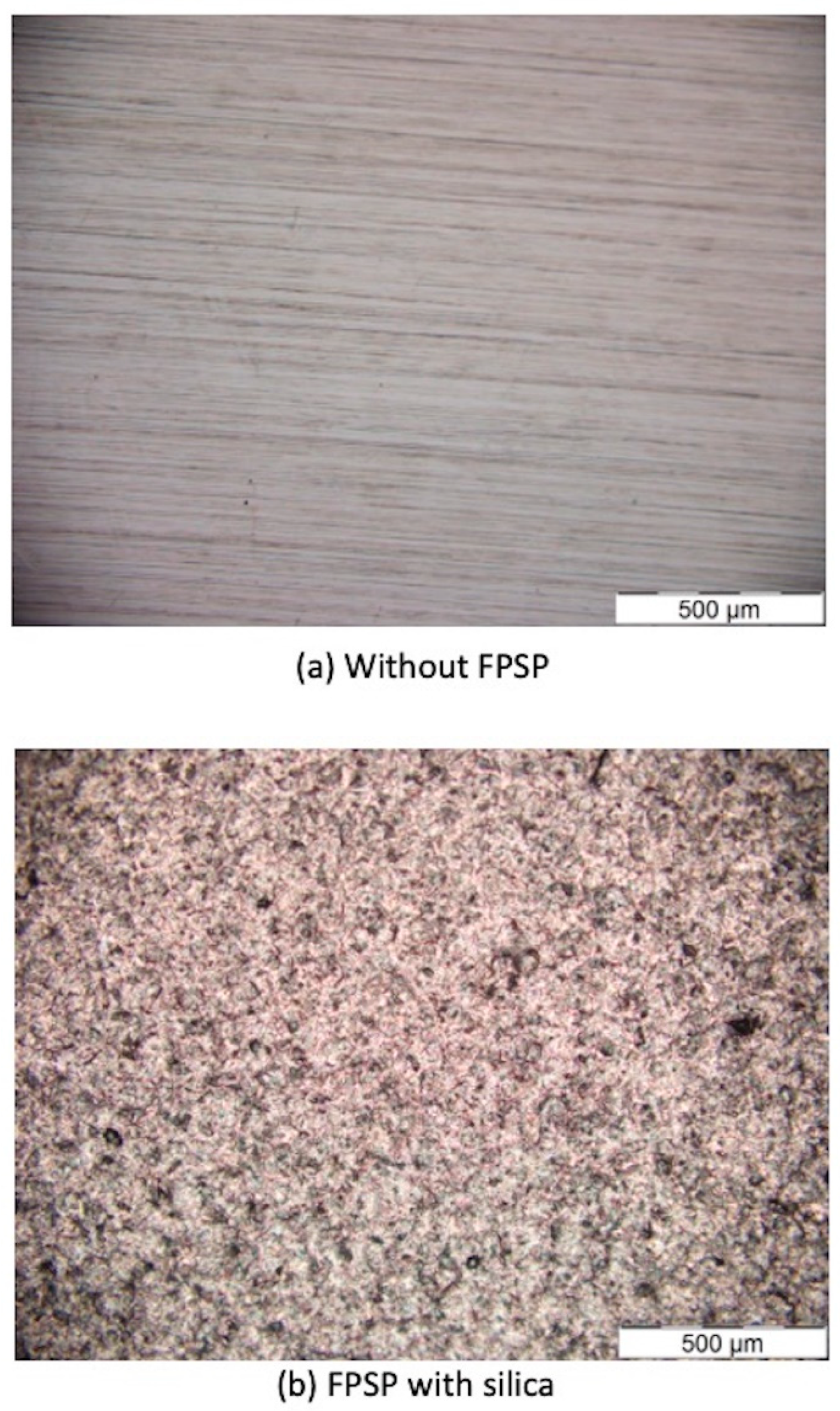
The surface morphology was evaluated by optical microscope. The results showed that there were significant differences in the surface morphologies between the CoCr alloys with and without the fine-particle shot peening process, as shown in Figure 1. The fine-particle shot peening can change surface microstructure by creating many small dimples on the surface of the materials, which represent the plastic deformation of the surface materials.
There were two media types, namely ceremic beads and silica beads, and it was found that ceramic beads generated multiple small dimples that were clearer and deeper than those of the silica beads (Figure 1). Not only did the surface morphology of the CoCr alloy change after the fine-particle shot peening process, but there was a significant difference in effect between the two particle types. The hardness of the ceramic particle was higher than the silica particle. These results showed that particles with greater hardness can create clearer, deeper, and smaller dimples on surface materials, and therefore, particle type affects the change in surface morphology.
In order to compare different particle sizes, this study used three distinct sizes for both the silica and ceramic particles. The size of the media is shown in Table 2. The results showed that the larger particle size creates bigger and clearer dimples than the smaller particle size (Figure 2). However, the observation by optical microscope only proved that fine-particle shot peening can change the surface morphology of CoCr alloy but cannot explain the change of material properties.
3.2. Microstructure Analysis
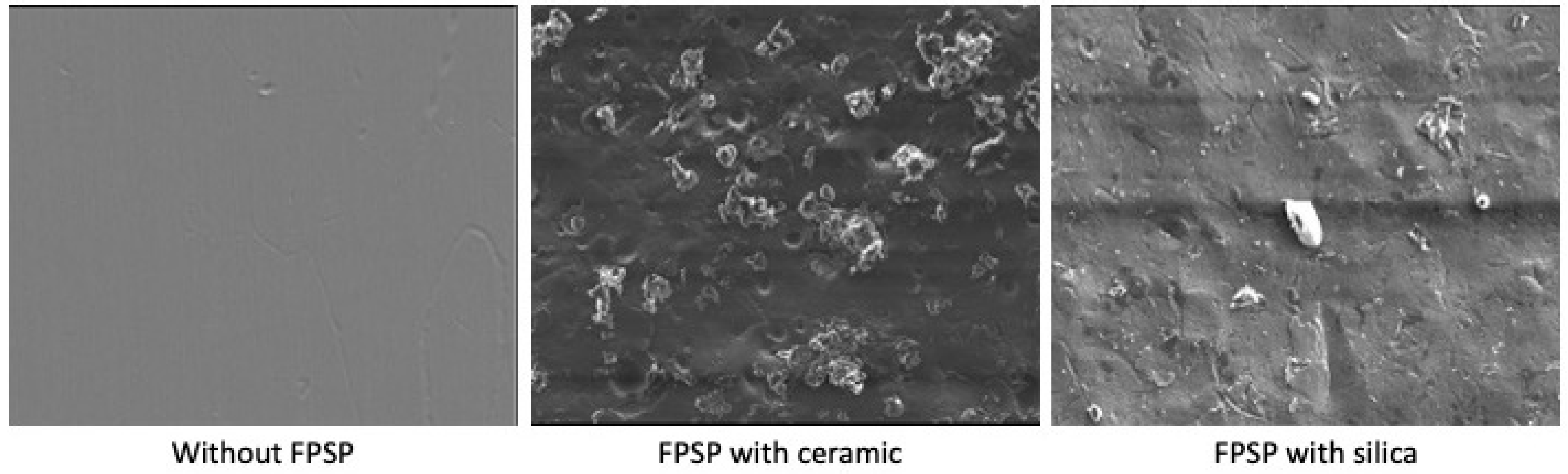
The microstructure of the surface material pre- and post surface modification were evaluated by electron probe microanalysis, including secondary electron (Figure 3), which can evaluate the surface morphology compared between pre- and post-peening process and backscatter electron (Figure 4) techniques, which show the change of surface morphology. (The light areas are the dimples made by the peening process.) The results showed that the fine-particle shot peening process can change the surface microstructure by creating surface dimples with both ceramic and silica particles.
It was also demonstrated that ceramic particles can create deeper and clearer surface dimples than silica particles. This difference may be affected by the hardness of the particle. In general, the higher the hardness value of a particle, the greater the change on the surface microstructure.
3.3. Surface Roughness
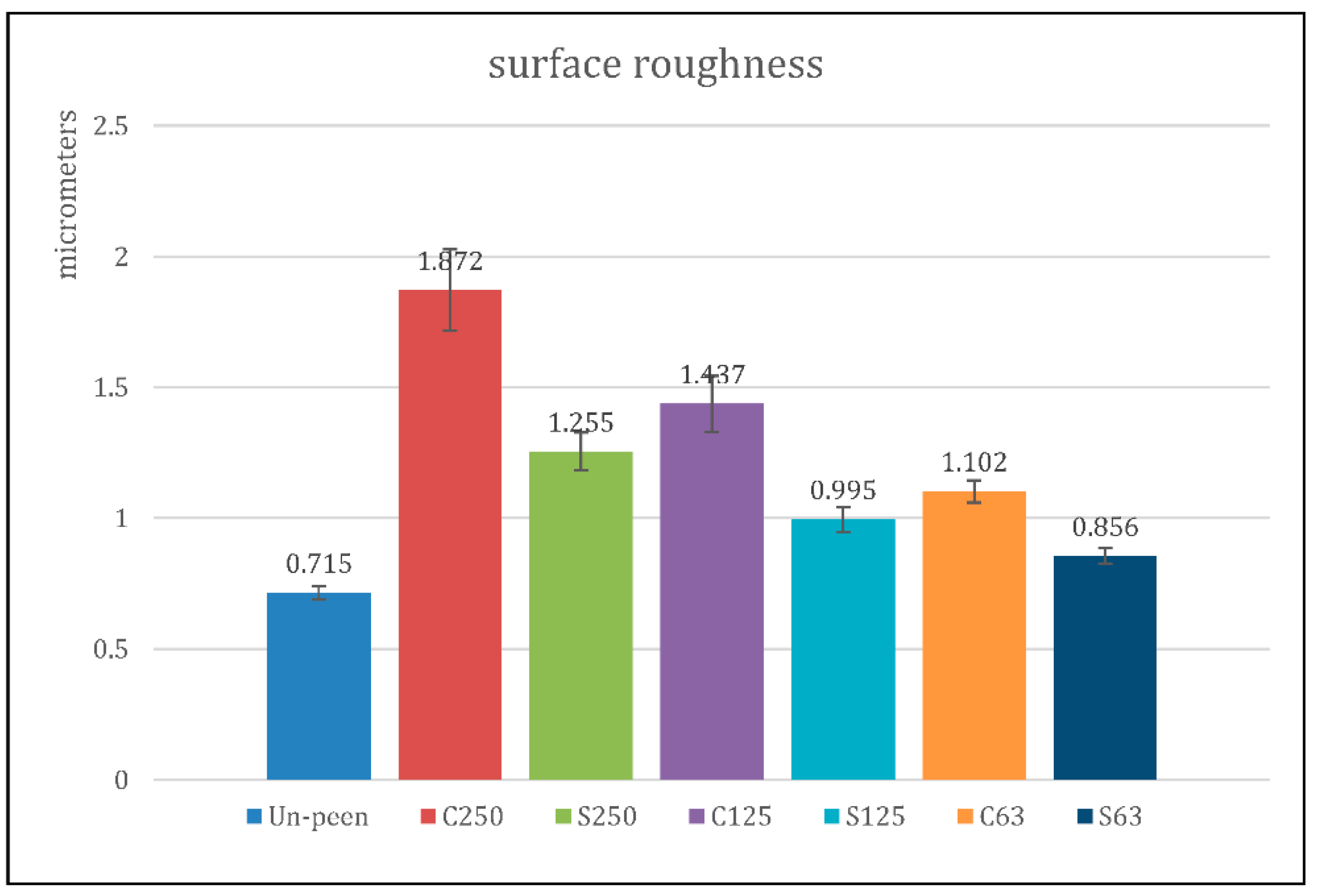
The surface-roughness tester was used to measure the mean surface roughness (Ra) of the material where the length of measurement was 4.00 mm and 0.2 mm/s of driving speed. The result is shown in Figure 5.
The results showed that fine-particle shot peening can change the surface roughness of materials and that increasing the particle size can generate higher mean surface roughness. Furthermore, the ceramic particle that represented the higher-hardness particle group can create higher mean surface roughness than the silica particle.
3.4. Hardness Analysis
The hardness of the surface material was measured by the Micro Vickers hardness tester on the top of the surface. The results showed that the fine-particle shot peening process can significantly increase surface hardness when compared with an un-peened workpiece, as shown in Figure 6.
It was observed that the ceramic particle can create higher surface hardness than the silica particle in all sizes of media. The greater hardness of the ceramic particle created surface morphology changes, including plastic deformation, more than the lower-hardness silica particle. Therefore, these results demonstrate that the hardness of the particle affects the surface properties of the materials when using the fine-particle shot peening process. In addition, the results show that different media sizes create different degrees of surface hardness after the peening process. The larger particle sizes of both ceramic and silica particles created higher material surface hardness.
From the results of the fine-particle shot peening process, the best media parameter that can create the highest surface hardness was the media with the higher hardness and larger particle size.
3.5. Residual Element
The residual element was evaluated by electron probe microanalysis to analyze elements on the surface of the material before and after performing the fine-particle shot peening process. This analysis was important, as it helped us identify whether there were any residual elements from the process left on the CoCr surface.
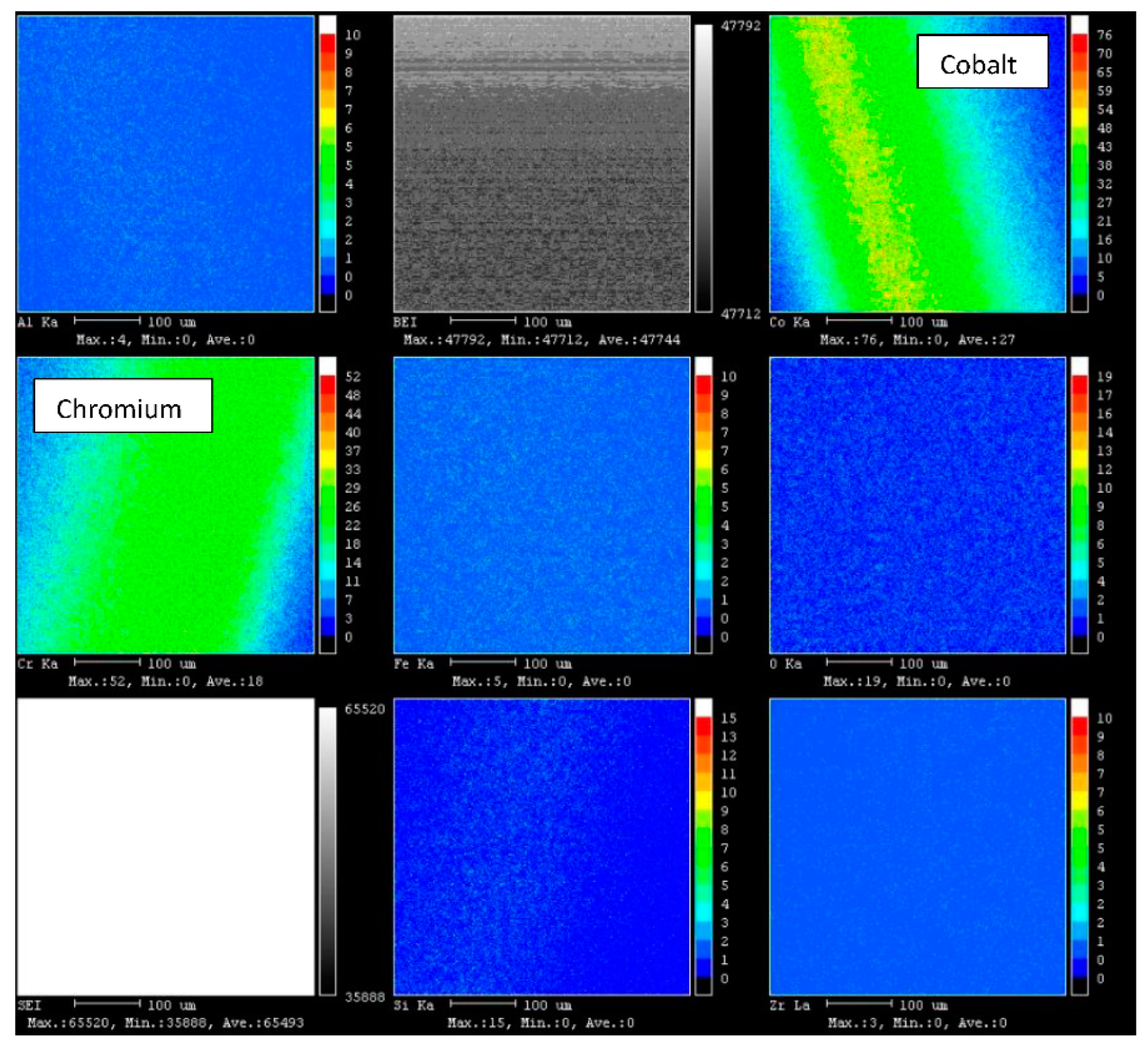
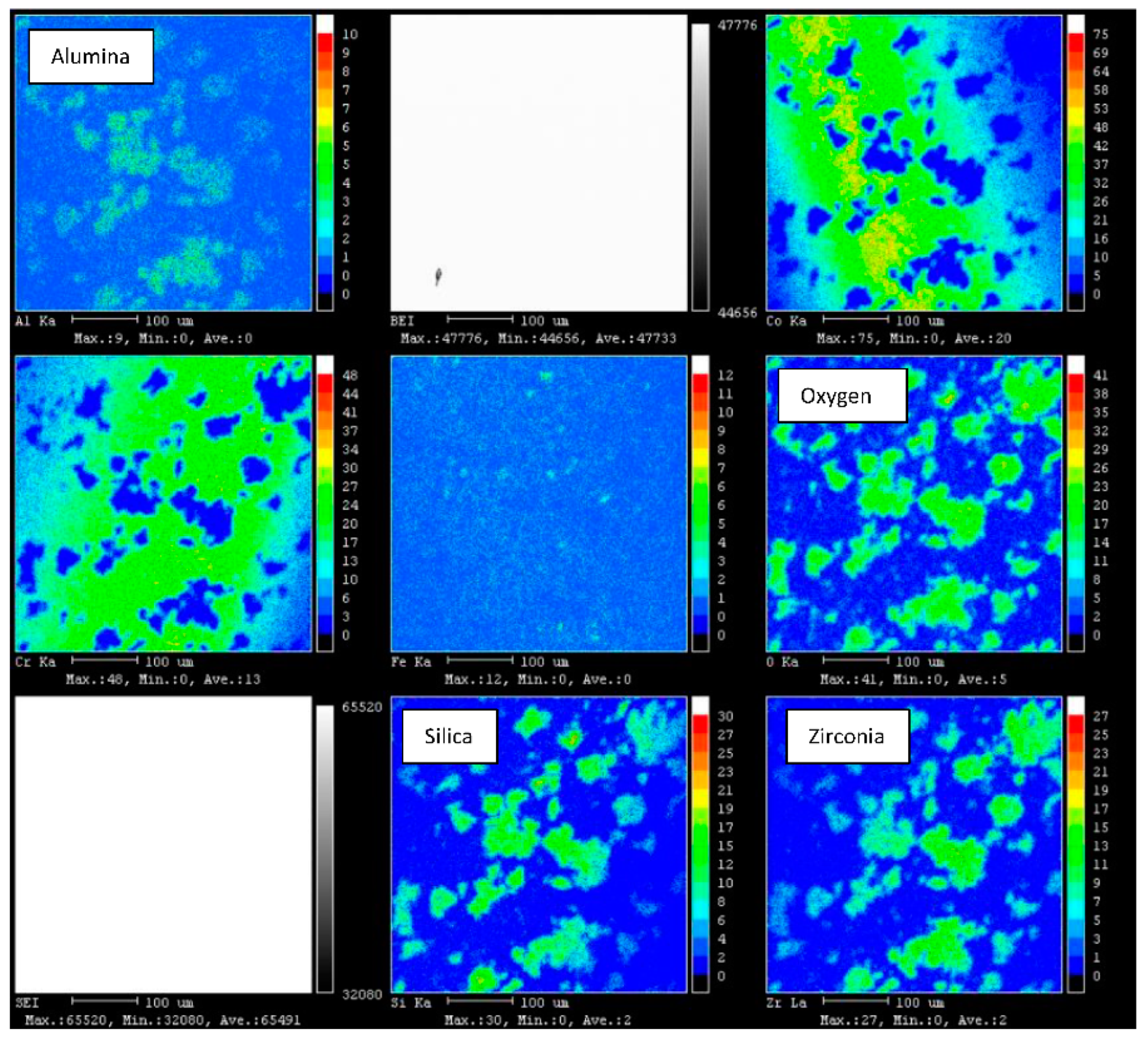
Using X-ray mapping and characteristics, results from the electron probe microanalysis showed that there were some residual elements from the particle that was used in the peening process. In a conventional workpiece (Figure 7) that did not experience the fine-particle shot peening process, we found that there were only elements of cobalt and chromium. By comparison, we detected elements of each particle type, i.e., ceramic and silica, on the workpieces used for those respective peening processes. The workpiece that used ceramic beads contained elements of ceramic, including alumina, zirconia, oxygen, and silica, and the workpiece that used silica beads contained silica and oxygen (Figure 8 and Figure 9). Therefore, we concluded that the fine-particle shot peening process leaves residual elements on the surface of the material. Residual elements must be taken into consideration, especially for medical device applications, in which specific elements could create reactions in the human body.
3.6. Cytotoxicity Test
The cytotoxicity test was evaluated with mouse fibroblast (L929) by MTT cytotoxicity test.
The result showed that the percentages of cell viability of the peening samples were nearly the same as the negative control group in all different concentrations of samples using both silica and ceramic, as shown in Table 3.
4. Discussion
The main objective of this study is to examine the effect of particle types and sizes on the surface properties of the CoCr, including surface morphology, surface roughness, surface hardness, and any residual elements, for the purposes of the improving medical devices that use CoCr materials such as dental or joint replacement implants. The results are as follows:
For the surface morphology, the results showed that the fine-particle shot peening process can change the surface morphology by creating multiple dimples on the surface material. The ceramic particle, with its higher hardness, can create clearer surface dimples than the silica particle, and for the effect of particle type, the results showed that the larger particle can create larger and clearer dimples on the CoCr surface when using both ceramic and silica particles. The results were in line with the previous study by Ongtrakulkij G. et al., which reported that a higher-hardness particle can change surface morphology more clearly and deeply than a lower-hardness particle [14].
For the surface roughness, the ceramic particle can increase the surface roughness more than the silica particle when comparing the same-sized particles. The surface dimples can enhance the lubrication properties by increasing the formation of a fluid film that decreases the friction force between the motion surface. Recent literature that has studied lubricant film formation by using artificial hip joints with micro-dimples on the surface showed that the prostheses with micro-dimples significantly increased the lubricant film thickness. Using the tribology test results, Taposh Roy et al. reported that the micro-dimpled, ceramic-on-ceramic interface of hip joint arthroplasty can reduce the coefficient of friction when compared with a non-dimpled surface [27]. From the results of recent literature, it can be determined that a surface with appropriate micro-dimpling can increase the lubrication properties and decrease the friction coefficient, ultimately resulting in a decrease to the wear-and-tear process of the motion surface [28,29].
The surface hardness is the most important factor affecting a surface material’s strength and wear process. The hardness can be defined as a material’s resistance to permanent deformation, and hardness can be used to determine resistance to the general wear-and-tear process, in particular scratch resistance. High-hardness materials can resist scratching that makes the surface irregular and increases the sharpness of the surface, which in turn increases the abrasive wear at the motion surface. As a result, high-hardness materials may be the best option for medical implants such as joint replacements, dental implants, or other orthopedics due to their wear- and scratch-resistance properties.
In recent literature, it has been reported that the fine-particle shot peening process can increase the surface hardness of the material [30]. In this study, the results showed that fine-particle shot peening can significantly increase the surface hardness of CoCr material using both silica and ceramic particles. Due to its higher hardness, the ceramic particle can increase the surface hardness more than the silica particle, and in different particle sizes, the larger particle size can create higher surface hardness than the smaller particle size. In summary, the particle that can increase greater surface hardness is a ceramic particle of a large particle size (250–425 μm).
One of the most important concerns with regard to the fine-particle shot peening process on a surface material is the residual element of any particle that was used for surface modification. Residual elements left on a surface material that will be used for a medical implant may affect the cells of the human body. In this study, the results also showed that there were some residual elements of the particle on the surface material when using either ceramic or silica particles.
The MTT assay determines the functional state of mitochondria, including cell viability, and is the most widely used cell viability assay [31]. From the cytotoxicity test results, the materials that modified the surface by fine-particle shot peening did not increase the cytotoxicity to the cell. The cell viability of the samples that modified the surface were not significantly different from the negative control. When compared to the un-peened sample, both the sample peened with silica and the one peened with ceramic showed no significant cell viability differences. The results prove that the fine-particle shot peening process on CoCr material did not affect cell viability, and therefore, this technique may be applied to the surface material of medical implants for use in the human body.
5. Conclusions
The study investigated the effect of particle type and size on the surface properties of a CoCr alloy, including surface morphology, surface roughness, and surface hardness, by using the fine-particle shot peening technique. The results indicated that the use of different types and sizes of peening media, such as silica and ceramic particles, can significantly alter the surface morphology of the CoCr alloy, inducing features such as surface roughness, texture, and defects. The study also showed that the surface roughness and hardness of the CoCr alloy can be improved by optimizing the peening parameters such as the peening pressure and duration. Furthermore, the Micro Vickers hardness test revealed that the CoCr alloy’s surface hardness increased significantly after fine-particle shot peening, indicating the potential of this technique for enhancing the material’s mechanical properties. Overall, the findings suggest that the fine-particle shot peening technique can be an effective surface-treatment method for improving the performance and durability of the CoCr alloy and other metallic materials in various engineering applications. Further research can explore the effect of different process parameters and peening media on the CoCr alloy’s surface properties and investigate the underlying mechanisms of the peening-induced changes in the material microstructure and properties.
Optimizing these parameters can lead to desirable surface properties such as improved hardness, wear resistance, and fatigue life. The authors have conducted a series of experiments varying these parameters to identify the most favorable process conditions for their specific application. In summary, the most favorable process conditions for fine-particle shot peening of the CoCr alloy can vary depending on the specific goals and constraints of the study. It is essential to conduct systematic experiments to identify the optimal process conditions for a particular application.
Therefore, the surface properties of the CoCr alloy can be improved by the fine-particle shot peening process in optimized conditions. The higher-hardness particle can change the surface properties more than the lower one, and the larger particle size can change the surface properties more than the smaller particle size. The results prove that the fine-particle shot peening process can change the surface microstructure and improve the surface properties of CoCr. For future direction, the fine-particle shot peening process may be the process of choice for improving the surface properties of medical implants such as dental implants or orthopedics implants (the bearing surface of the total hip replacement), which can increase the strength and reduce the wear process, resulting in increasing the longevity of the implant.
Author Contributions
All authors conceptualized and designed the study. A.K. and C.J. performed data collection and prepared the manuscript draft; C.J. and P.S.-n. helped critically revise the manuscript for important intellectual content. All authors have read and agreed to the published version of the manuscript.
Funding
This research and innovation activity is funded by National Research Council of Thailand (NRCT).
Institutional Review Board Statement
Not applicable.
Informed Consent Statement
Not applicable.
Data Availability Statement
The original contributions presented in the study are included in the article. Further inquiries can be directed to the corresponding author.
Acknowledgments
Thailand Science Research and Innovation (TSRI) Basic Research Fund: Fiscal year 2023 under project number FRB660073/0164. National Research Council Thailand (NRCT).
Conflicts of Interest
The authors declare that the research was conducted in the absence of any commercial or financial relationships that could be construed as a potential conflict of interest.
References
- Biemond, J.E.; Wolke, J.G.C. Cobalt-chromium alloys in fixed prosthodontics. Dent. Clin. 2017, 61, 797–814. [Google Scholar]
- Fischer, A.; Shadanbaz, S.; Choudhury, N.R.; Dias, G.J. Wear and corrosion of cobalt and titanium alloys in orthopedic implant applications. J. Mech. Behav. Biomed. Mater. 2016, 63, 245–262. [Google Scholar]
- Panteli, M.; Mäckler, T.; Tsouknidas, A. The tribology of CoCrMo alloy in simulated body fluids: A review of current literature. J. Mech. Behav. Biomed. Mater. 2017, 68, 45–57. [Google Scholar]
- Navarro, M.; Michiardi, A.; Castaño, O.; Planell, J.A. Biomaterials in orthopaedics. J. R. Soc. Interface 2008, 5, 1137–1158. [Google Scholar] [CrossRef]
- Hodgson, A.W.E.; Kurz, S.; Virtanen, S.; Fervel, V.; Olsson, C.O.A.; Mischler, S. Passive and transpassive behaviour of CoCrMo in simulated biological solutions. Electrochim. Acta 2014, 49, 2167–2178. [Google Scholar] [CrossRef]
- Xiang, D.D.; Wang, P.; Tan, X.P.; Chandra, S.; Wang, C.; Nai, M.L.S.; Tor, S.B.; Liu, W.Q.; Liu, E. Anisotropic microstructure and mechanical properties of additively manufactured Co–Cr–Mo alloy using selective electron beam melting for orthopedic implants. Mater. Sci. Eng. A 2019, 765, 138270. [Google Scholar] [CrossRef]
- Igual, M.A.; Casabán, J.L. Influence of electrochemical potential on the tribocorrosion behaviour of high carbon CoCrMo biomedical alloy in simulated body fluids by electrochemical impedance spectroscopy. Electrochim. Acta 2010, 55, 5428–5439. [Google Scholar] [CrossRef]
- Sansone, V.; Pagani, D.; Melato, M. The effects on bone cells of metal ions released from orthopaedic implants. A review. Clin. Cases Miner. Bone Metab. 2013, 10, 34–40. [Google Scholar] [CrossRef] [PubMed]
- Yuki, K.; Masaki, O.; Hiroaki, H. Modelling of Particle Behaviour in Shot Peening Process. J. Mech. Eng. Autom. 2014, 4, 83–91. [Google Scholar]
- Xu, C.; Sheng, G.; Wang, H.; Jiao, Y.; Yuan, X. Effect of high energy shot peening on the microstructure and mechanical properties of Mg/Ti joints. J. Alloy. Compd. 2017, 695, 1383–1391. [Google Scholar] [CrossRef]
- Zhang, J.; Li, X.; Yang, B.; Wang, H.; Zhang, J. Effect of micro-shot peening on fatigue properties of precipitate strengthened Cu-Ni-Si alloy in air and in salt atmosphere. Surf. Coat. Technol. 2019, 359, 16–23. [Google Scholar] [CrossRef]
- Kovacı, H.; Bozkurt, Y.B.; Yetim, A.F.; Aslan, M.; Qelik, A. The effect of surface plastic deformation produced by shot peening on corrosion behavior of a low-alloy steel. Surf. Coat. Technol. 2019, 360, 78–86. [Google Scholar] [CrossRef]
- Maryam, J.; David, P.F. Effects of shot peening parameters on gradient microstructure and mechanical properties of TRC AZ31. Mater. Charact. 2019, 148, 9–16. [Google Scholar]
- Ongtrakulkij, G.; Khantachawana, A.; Kondoh, K. Effects of media parameters on enhance ability of hardness and residual stress of Ti6Al4V by fine shot peening. Surf. Interfaces 2020, 18, 100424. [Google Scholar] [CrossRef]
- Kim, H.J.; Kim, K.B.; Kim, S.H. Effects of Shot-Blasting Parameters on the Surface Roughness and Microstructure of CoCr Alloy. Materials 2017, 10, 758. [Google Scholar]
- Maeno, S.; Tsukamoto, M.; Shoji, H.; Takamura, M.; Sasaki, K. Evaluation of the effect of shot blasting on surface damage and residual stress of CoCrMo alloy. J. Mech. Behav. Biomed. Mater. 2018, 77, 318–325. [Google Scholar]
- Wu, W. Surface morphology and nanomechanics of hydrogels. Nanomechanics Mater. Struct. 2019, 247–277. [Google Scholar]
- Kumar, N.; Gaur, V.; Kant, R. Surface morphology and roughness measurements of surfaces by atomic force microscopy. Microsc. Sci. Technol. 2018, 1295–1302. [Google Scholar]
- Koshy, P.; Lian, K.S.; Lim, C.S.; Murali, M.R. Surface morphology and topography of ceramic dental implants: A systematic review. Dent. Mater. J. 2018, 37, 339–346. [Google Scholar]
- Hu, P.; Huang, Y.; He, S.; Lu, J. Osseointegration of biomedical implants: A review. Chin. J. Traumatol. 2018, 21, 189–194. [Google Scholar]
- Jinpeng, S.; Huang, C.; Zou, B.; Liu, H.; Wang, J. Microstructure and mechanical properties of TiB2–TiC–WC composite ceramic tool materials. Mater. Des. 2012, 36, 69–74. [Google Scholar]
- Liao, Y.; Yan, H.; Xia, W.; Chen, J.; Su, B.; Li, X.; Zhao, L. Effect of Heat Treatment on the Microstructure and Properties of High Strain Rate Rolled 7050 Aluminum Alloy. Met. Mater. Int. 2021, 28, 1014–1025. [Google Scholar] [CrossRef]
- Wennerberg, A.; Albrektsson, T. Effects of titanium surface topography on bone integration: A systematic review. Clin. Oral Implant. Res. 2009, 20 (Suppl. S4), 172–184. [Google Scholar] [CrossRef] [PubMed]
- J.I.S. B0601; Geometrical Product Specifications-Surface Texture: Profile Method—Terms, Definitions and Surface Texture Parameters, Surface Roughnes. Japanese Standards Association: Tokyo, Japan, 2001.
- Jung, J.Y.; Koh, Y.H.; Song, Y.H.; Kim, Y.M. Surface modification of CoCrMo alloy by plasma nitriding for orthopedic implants. Appl. Surf. Sci. 2015, 347, 164–169. [Google Scholar]
- Tsukanaka, M.; Fujibayashi, S.; Takemoto, M.; Nakamura, T. In vitro degradation behavior and cytocompatibility of surface-modified Co–Cr–Mo alloys for orthopedic implants. J. Biomed. Mater. Res. Part B Appl. Biomater. 2012, 100, 1778–1787. [Google Scholar]
- Taposh, R.; Dipankar, C.; Azuddin, B.M.; Belinda, P.M. Fabrication and characterization of micro-dimple array on Al2O3 surfaces by using a micro-tooling. Ceram. Int. 2014, 40, 2381–2388. [Google Scholar]
- Dipankar, C.; David, R.; Shinya, S.; Pavel, H.; Martin, V.; Min, Z. Enhanced lubricant film formation through micro-dimpled hard-on-hard artificial hip joint: An in-situ observation of dimple shape effects. J. Mech. Behav. Biomed. Mater. 2018, 81, 120–129. [Google Scholar]
- Han, J.; Fang, L.; Sun, J.; Wang, Y.; Ge, S.; Zhu, H. Hydrodynamic Lubrication of Surfaces with Asymmetric Microdimple. Tribol. Trans. 2011, 54, 607–615. [Google Scholar] [CrossRef]
- Fu, P.; Chu, R.; Xu, Z.; Ding, G.; Jiang, C. Relation of hardness with FWHM and residual stress of GCr15 steel after shot peening. Appl. Surf. Sci. 2018, 431, 165–169. [Google Scholar] [CrossRef]
- Edmondson, J.M.; Armstrang, L.S.; Martiner, A.O. A rapid and simple MTT-based spectrophotometric assay for determining drug sensitivity in monolayer cultures. J. Tissue Cult. Methods 1998, 11, 15–17. [Google Scholar] [CrossRef]
Figure 1.
Surface morphology evaluated by optical microscope: (a) without peening process, (b) with fine-particle shot peening (FPSP) with silica, and (c) FPSP with ceramic.
Figure 1.
Surface morphology evaluated by optical microscope: (a) without peening process, (b) with fine-particle shot peening (FPSP) with silica, and (c) FPSP with ceramic.


Figure 2.
Surface morphology evaluated by optical microscope and comparing different sizes and types of particles.
Figure 2.
Surface morphology evaluated by optical microscope and comparing different sizes and types of particles.

Figure 3.
Surface morphology evaluated by electron probe microanalysis (secondary electron) and comparing different types of particles.
Figure 3.
Surface morphology evaluated by electron probe microanalysis (secondary electron) and comparing different types of particles.

Figure 4.
Surface morphology evaluated by electron probe microanalysis (backscatter electron) and comparing different types of particles.
Figure 4.
Surface morphology evaluated by electron probe microanalysis (backscatter electron) and comparing different types of particles.

Figure 5.
Mean surface roughness (Ra) compared between pre- and post-peening process in various types and sizes of particle.
Figure 5.
Mean surface roughness (Ra) compared between pre- and post-peening process in various types and sizes of particle.

Figure 6.
Surface hardness compared between pre- and post-peening process in various types and sizes of particle.
Figure 6.
Surface hardness compared between pre- and post-peening process in various types and sizes of particle.

Figure 7.
Residual elements of un-peened sample showing the elements of cobalt and chromium on the surface.
Figure 7.
Residual elements of un-peened sample showing the elements of cobalt and chromium on the surface.

Figure 8.
Residual elements of sample peened with ceramic particles showing the elements of alumina, zirconia, silica, and oxygen on the surface.
Figure 8.
Residual elements of sample peened with ceramic particles showing the elements of alumina, zirconia, silica, and oxygen on the surface.

Figure 9.
Residual elements of sample that was peened with silica particles (SiO2) showing the elements of silica and oxygen on the surface.
Figure 9.
Residual elements of sample that was peened with silica particles (SiO2) showing the elements of silica and oxygen on the surface.


Table 1.
Chemical composition of cobalt–chromium alloy.
| Cobalt (wt%) | Chromium (wt%) | Mo (wt%) | C (wt%) |
|---|---|---|---|
| 60–62 | 29–30 | 5–6 | 0.55–0.65 |
Table 2.
Peening conditions.
| Code Number | Particle Type | Particle Size (μm) | Pressure (MPa) | Nozzle Distance (mm) |
|---|---|---|---|---|
| S63 | Silica | 63–106 | 0.5 | 20 |
| S125 | Silica | 125–180 | 0.5 | 20 |
| S250 | Silica | 250–355 | 0.5 | 20 |
| C63 | Ceramic bead | 63–125 | 0.5 | 20 |
| C125 | Ceramic bead | 125–250 | 0.5 | 20 |
| C250 | Ceramic bead | 250–425 | 0.5 | 20 |
Table 3.
Cytotoxicity test results evaluated by mouse fibroblast (L929).
| Sample | % Cell Viability ± Uncertainly Value | |||||
|---|---|---|---|---|---|---|
| Concentration (%) | Negative Control | Positive Control | ||||
| 0.02 | 0.2 | 2 | 20 | |||
| Un-peened | 95.13 ± 4.18 | 92.79 ± 1.39 | 91.81 ± 2.34 | 90.84 ± 1.70 | 97.66 | 9.16 |
| Ceramic group | 98.68 ± 6.11 | 94.72 ± 3.70 | 93.40 ± 3.36 | 90.57 ± 1.74 | 98.30 | 9.81 |
| Silica group | 101.95 ± 4.68 | 98.05 ± 1.96 | 97.83 ± 4.30 | 94.79 ± 3.98 | 98.70 | 9.11 |
Disclaimer/Publisher’s Note: The statements, opinions and data contained in all publications are solely those of the individual author(s) and contributor(s) and not of MDPI and/or the editor(s). MDPI and/or the editor(s) disclaim responsibility for any injury to people or property resulting from any ideas, methods, instructions or products referred to in the content. |
© 2023 by the authors. Licensee MDPI, Basel, Switzerland. This article is an open access article distributed under the terms and conditions of the Creative Commons Attribution (CC BY) license (https://creativecommons.org/licenses/by/4.0/).
Share and Cite
MDPI and ACS Style
Jarungvittayakon, C.; Khantachawana, A.; Sa-ngasoongsong, P. The Effect of Particle Type and Size on CoCr Surface Properties by Fine-Particle Shot Peening. Appl. Sci. 2023, 13, 5814. https://0-doi-org.brum.beds.ac.uk/10.3390/app13095814
AMA Style
Jarungvittayakon C, Khantachawana A, Sa-ngasoongsong P. The Effect of Particle Type and Size on CoCr Surface Properties by Fine-Particle Shot Peening. Applied Sciences. 2023; 13(9):5814. https://0-doi-org.brum.beds.ac.uk/10.3390/app13095814
Chicago/Turabian StyleJarungvittayakon, Chavarat, Anak Khantachawana, and Paphon Sa-ngasoongsong. 2023. "The Effect of Particle Type and Size on CoCr Surface Properties by Fine-Particle Shot Peening" Applied Sciences 13, no. 9: 5814. https://0-doi-org.brum.beds.ac.uk/10.3390/app13095814
Note that from the first issue of 2016, this journal uses article numbers instead of page numbers. See further details here.