Morphology and Properties of Electrospun PCL and Its Composites for Medical Applications: A Mini Review

 , , and
Abstract
:1. Introduction
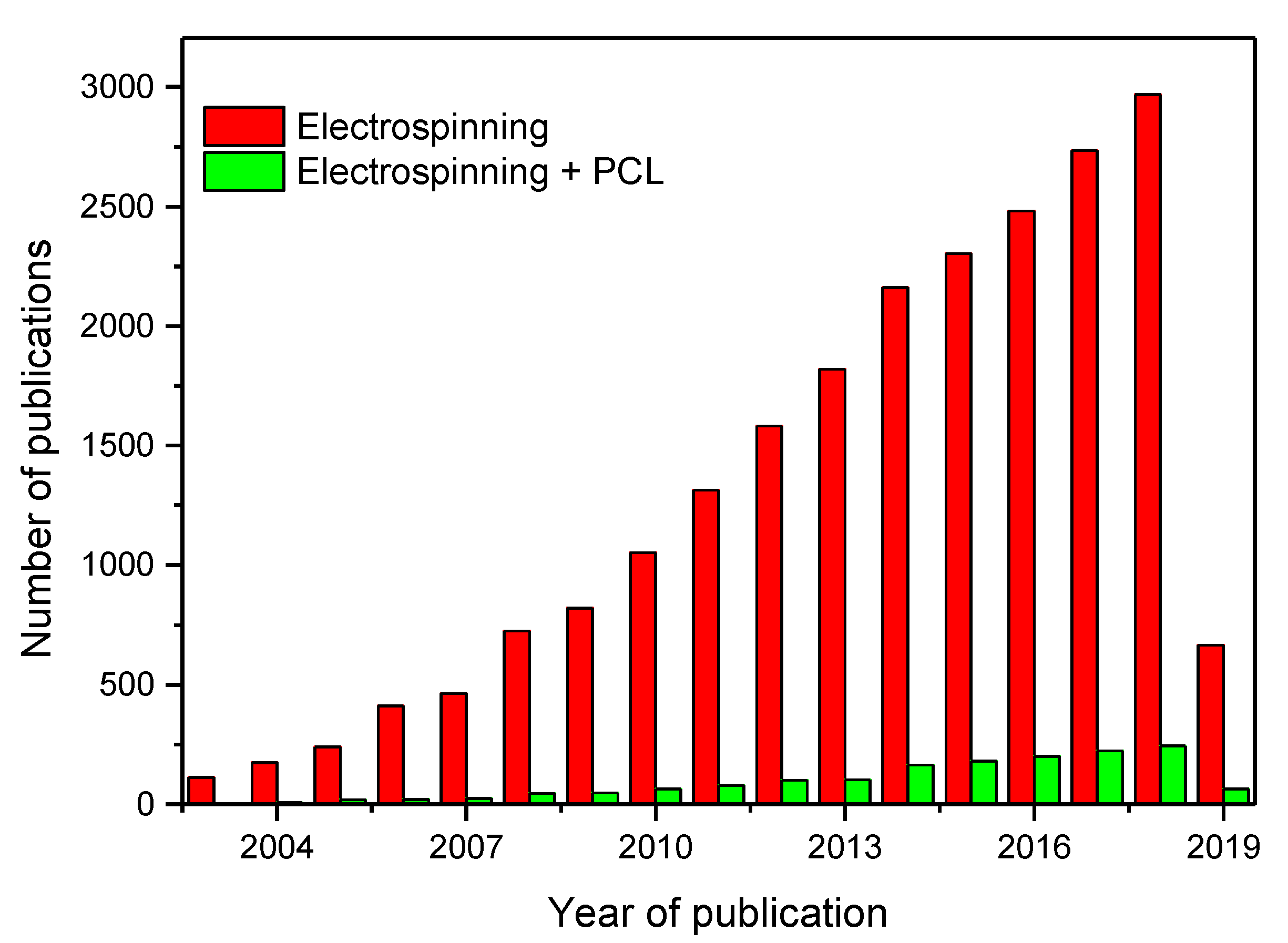
2. History of Electrospinning
Electrospun Nanoparticles
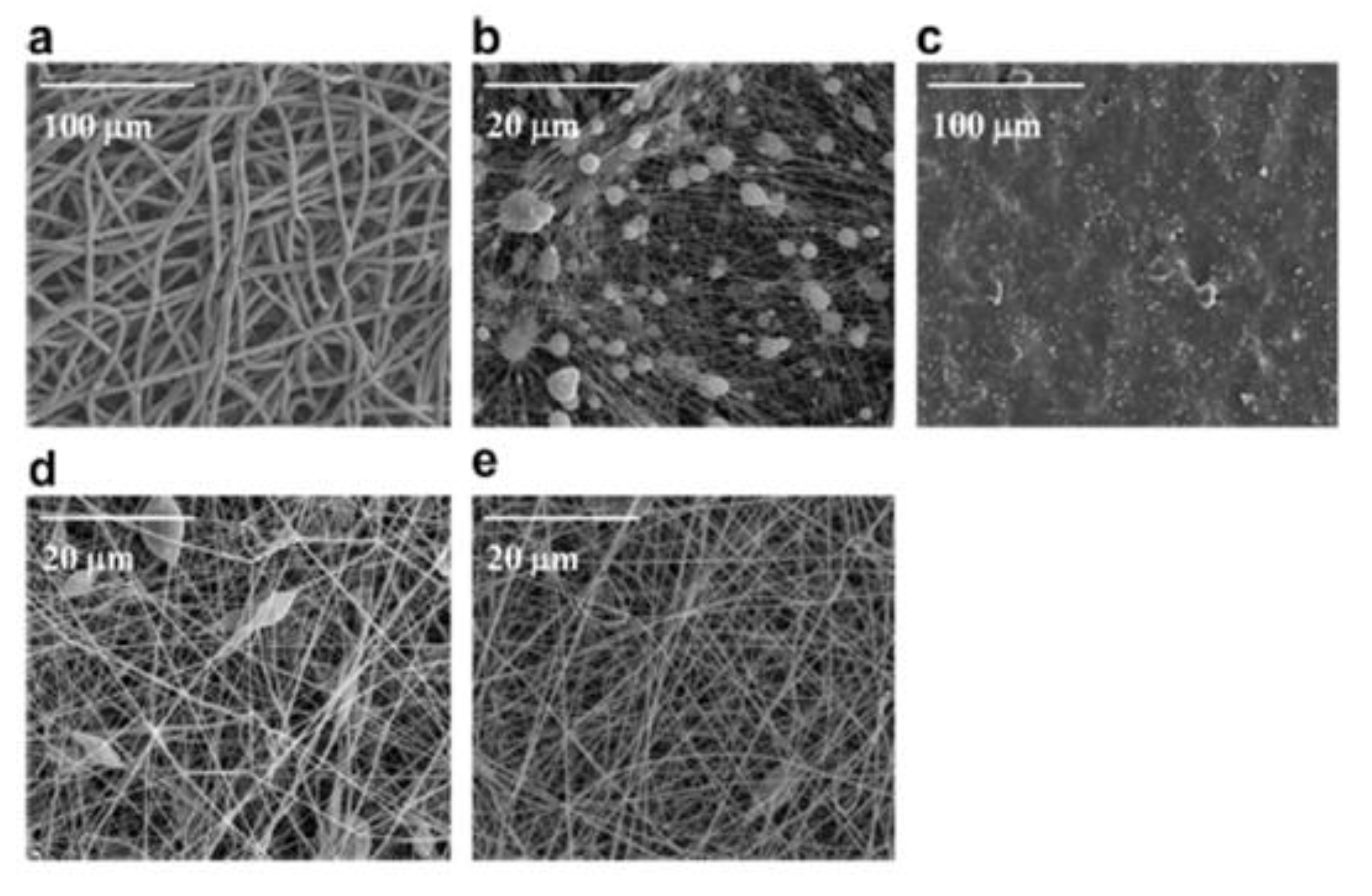
3. Solvent Effect and Resultant Morphology of PCL
4. Morphology of Electrospun PCL Blends and Composites
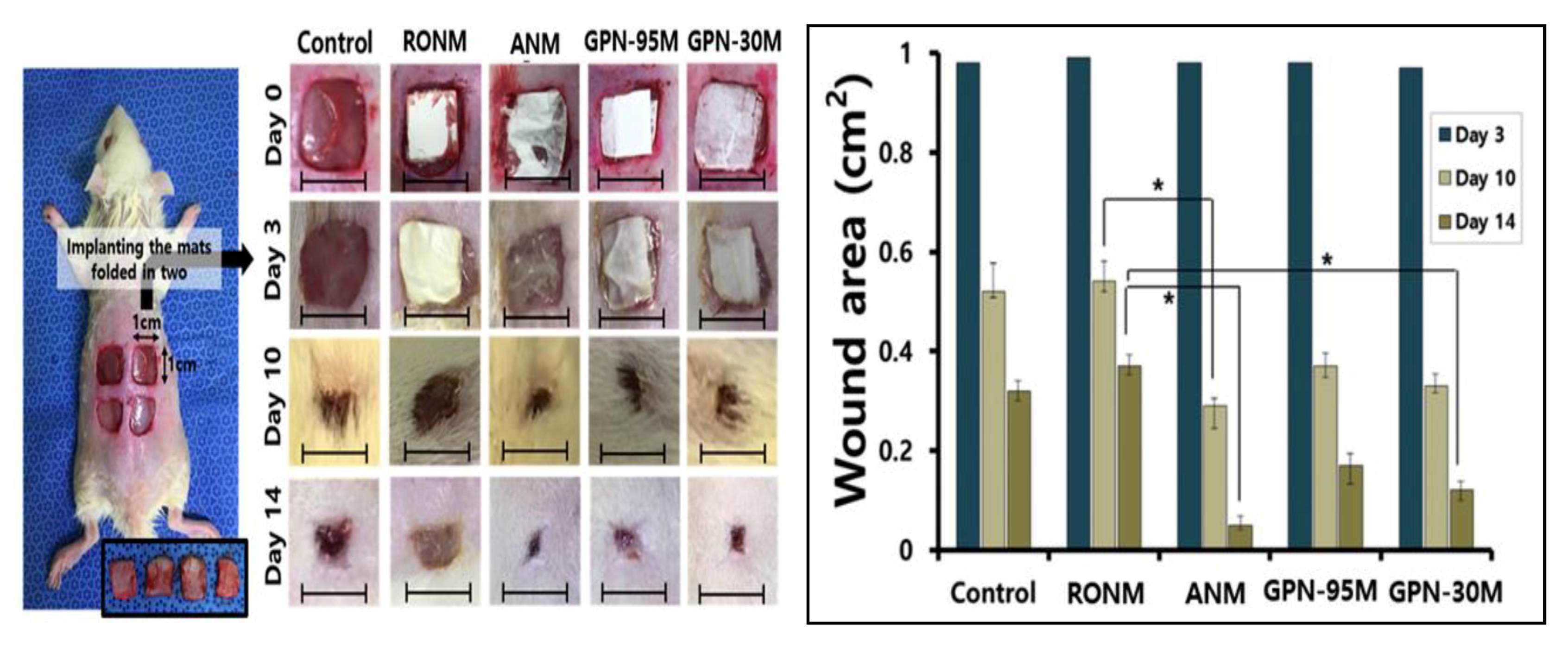
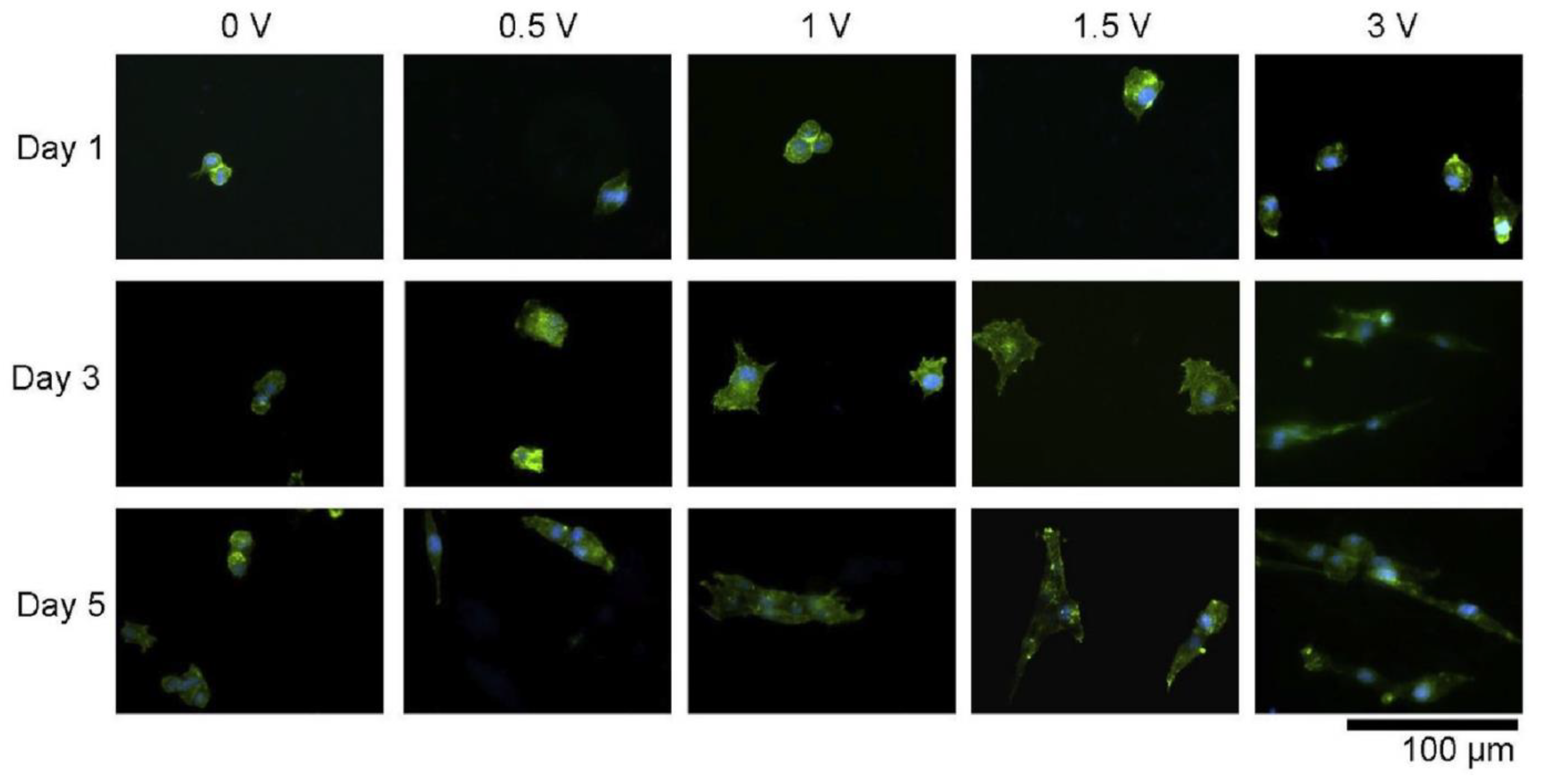
5. Biomedical Indispensability of the PCL Electrospun Nanocomposites
6. Mechanical Properties of Electrospun PCL and Its Composites
7. Conclusions and Future Remarks
Author Contributions
Funding
Acknowledgments
Conflicts of Interest
References
- Schueren, L.V.D.; Schoenmaker, B.D.; Kalaoglu, O.I.; Clerk, K.D. An alternative solvent system for the steady state electrospinning of polycarbonate. Eur. Polym. J. 2011, 47, 1256–1263. [Google Scholar] [CrossRef]
- Agarwal, S.; Wendorff, J.H.; Greiner, A. Use of electrospinning technique for biomedical applications. Polymer 2008, 49, 5603–5621. [Google Scholar] [CrossRef] [Green Version]
- Neppalli, R.; Marega, C.; Marigo, A.; Bajgai, M.P.; Kim, H.Y.; Causin, V. Poly(epsilon-caprolactone) filled with electrospun nylon fibres: A model for a facile composite fabrication. Eur. Polym. J. 2010, 46, 968–976. [Google Scholar] [CrossRef]
- Venugopal, J.; Zhang, Y.Z.; Ramakrishna, S. Fabrication of modified and functionalized polycarbonate nanofiber scaffolds for vascular tissue engineering. Nanotechnology 2005, 16, 2138–2142. [Google Scholar] [CrossRef] [PubMed]
- Jiang, S.; Chen, Y.; Duan, G.; Mei, C.; Greiner, A.; Agarwal, S. Electrospun nanofiber reinforced composites: A review. Polym. Chem. 2018. [Google Scholar] [CrossRef]
- Lee, K.H.; Kim, H.Y.; Khil, M.S.; Ra, Y.M.; Lee, D.R. Characterization of nanostructured poly(epsilon-caprolactone) nonwoven mats via electrospinning. Polymer 2003, 44, 1208–1294. [Google Scholar] [CrossRef]
- Moghe, A.K.; Hufenus, R.; Hudson, S.M.; Gupta, B.S. Effect of addition of a fugitive salt on electrospinnability of poly(epsilon-caprolactone). Polymer 2009, 50, 3311–3318. [Google Scholar] [CrossRef]
- Li, W.; Tuli, R.; Okafor, C.; Derfoul, A.; Danielson, K.G.; Hall, D.J. A three dimensional nanofibrous scaffold for cartilage tissue engineering using human mesenchymal stem cells. Biomaterials 2005, 26, 599–609. [Google Scholar] [CrossRef]
- Shin, M.; Ishii, O.; Sueda, T.; Vacanti, J.P. Contractile cardiac grafts using a novel nanofibrous mesh. Biomaterials 2004, 25, 3717–3723. [Google Scholar] [CrossRef]
- Gaumer, J.; Prasad, A.; Lee, D.; Lannutti, J. Structure-function relationship and source-to-ground distance in electrospun polycaprolactone. Acta Biomater. 2009, 5, 1552–1561. [Google Scholar] [CrossRef] [PubMed]
- Del Gaudio, C.; Bianco, A.; Folin, M.; Baiguera, S.; Grigioni, M. Structural characterization and cell response evaluation of electrospun PCL membranes: Micrometric versus submicrometric fibers. J. Biomed. Mater. Res. A 2009, 89A, 1028–1039. [Google Scholar] [CrossRef] [PubMed]
- Yu, H.; Jang, J.; Kim, T.; Lee, H.; Kim, H. Apatite-mineralized polycarbonate nanofibrous web as a bone tissue regeneration substrate. J. Biomed. Mater. Res. A 2009, 88A, 747–754. [Google Scholar] [CrossRef] [PubMed]
- Lie, X.; Xie, J.; Yuan, X.; Xia, Y. Coating electrospun poly(ɛ-caprolactone) fibers with gelatin and calcium phosphate and their use as biomimetric scaffolds for bone tissue engineering. Langmuir 2008, 24, 14145–14150. [Google Scholar] [CrossRef] [PubMed]
- Lowery, J.L.; Datta, N.; Rutledge, G.C. Effect of fiber diameter, pore size and seeding method on growth of human dermal fibroblasts in electrospun poly(epsilon-caprolactone) fibrous mats. Biomaterials 2010, 31, 491–504. [Google Scholar] [CrossRef] [PubMed]
- Prabhakaran, M.P.; Venugopal, J.; Chan, C.K.; Ramakrishna, S. Surface modified electrospun nanofibrous scaffolds for nerve tissue engineering. Nanotechnology 2008, 19, 1725–1731. [Google Scholar] [CrossRef] [PubMed]
- Formhals, A. Process and Apparatus for Preparing Artificial Thread. U.S. Patent 1,975,504, 2 October 1934. [Google Scholar]
- Feltz, K.P.; Growney Kalaf, E.A.; Chen, C.C.; Martin, R.S.; Sell, S.A. A review of electrospinning manipulation techniques to direct fier deposition and maximize pore size. De Gruyter Open 2017, 1, 46–61. [Google Scholar]
- Rojas-Mercado, A.S.; Moreno-Cortez, I.E.; Lucio-Porto, R.L.; López Pavón, L.L. Encapsulation and immobilization of ficin extract in electrospun polymeric nanofibers. Int. J. Biol. Macromol. 2018, 118, 2287–2295. [Google Scholar] [CrossRef]
- Kalantari, K.; Afifi, A.M.; Jahangirian, H.; Webster, T.J. Biomedical applications of chitosan electrospun nanofibers as a green Polymer. Carbohydr. Polym. 2019, 207, 588–600. [Google Scholar] [CrossRef]
- Wang, S.; Hu, F.; Li, J.; Zhang, S.; Shen, M.; Huang, M.; Shi, X. Design of electrospun nanofibrous mats for osteogenic differentiation of mesenchymal stem cells. Nanotechnol. Biol. Med. 2018, 14, 2505–2520. [Google Scholar] [CrossRef]
- Wang, C.; Sun, S.; Zhang, L.; Yin, J.; Jiao, T.; Zhang, L.; Xu, Y.; Zhou, J.; Peng, Q. Facile preparation and catalytic performance characterization of AuNPs-loaded hierarchical electrospun composite fiers by solvent vapor annealing treatment. Colloids Surf. A. 2019, 561, 283–291. [Google Scholar] [CrossRef]
- Yun, B.J.; Kwon, J.E.; Lee, K.; Koh, W.-G. Highly sensitive metal-enhanced florescence biosensor prepared on electrospun fiers decorated with silica-coated silver nanoparticles. Sens. Actuators B Chem. 2019, 284, 140–147. [Google Scholar] [CrossRef]
- Hivechi, A.; Bahrami, S.H.; Siegel, R.A. Investigation of morphological, mechanical and biological properties of cellulose nanocrystal reinforced electrospun gelatin nano fibers. Int. J. Biol. Macromol. 2019, 124, 411–417. [Google Scholar] [CrossRef]
- Ray, S.S.; Chen, S.-S.; Li, C.W.; Nguyen, N.C.; Nguyen, H.T. A comprehensive review: Electrospinning technique for fabrication and surface modification of membranes for water treatment application. RSC Adv. 2016, 6, 85495–85514. [Google Scholar] [CrossRef]
- Li, J.-J.; Yang, Y.-Y.; Yu, D.-G.; Du, Q.; Yang, X.-L. Fast dissolving drug delivery membrane based on the ultra-thin shell of electrospun core-shell nanofibers. Eur. J. Pharm. Sci. 2018, 122, 195–204. [Google Scholar] [CrossRef] [PubMed]
- Sukigara, S.; Gandhi, M.; Ayutsede, J.; Micklus, M.; Ko, F. Regeneration of Bombyx mori silk by electrospinning-part 1: Processing parameters and geometric properties. Polymer 2003, 44, 5721–5727. [Google Scholar] [CrossRef]
- Fadaie, M.; Mirzaei, E.; Geramizade, B.; Asvar, Z. Incorporation of nanofibrillated chitosan into electrospun PCL nanofibers makes scaffolds with enhanced mechanical and biological properties. Carbohydr. Polym. 2018, 199, 628–640. [Google Scholar] [CrossRef] [PubMed]
- Alali, K.T.; Liu, J.; Aljebawi, K.; Liu, Q.; Chen, R.; Yu, J.; Zhang, M.; Wang, J. 3D hybrid Ni-Multiwall carbon nanotubes/carbon nanofibers for detecting sarin nerve agent at room temperature. J. Alloys Compd. 2019, 780, 680–689. [Google Scholar] [CrossRef]
- Yang, G.; Li, X.; He, Y.; Ma, J.; Ni, G.; Zhou, S. From nano to micro to macro: Electrospun hierarchically structured polymeric fibers for biomedical applications. Prog. Polym. Sci. 2018, 81, 80–113. [Google Scholar] [CrossRef]
- Kiran, A.S.K.; Kumar, T.S.S.; Perumal, G.; Sanghavi, R.; Doble, M.; Ramakrishna, S. Dual nanofibrous bioactive coating and antimicrobial surface treatment for infection resistant titanium implants. Prog. Org. Coat. 2018, 121, 112–119. [Google Scholar] [CrossRef]
- Quiros, J.; Boltes, K.; Rosal, R. Biactive applications for electrospun fibers. Polym. Rev. 2016, 56, 631–667. [Google Scholar] [CrossRef]
- Hou, L.; Zhang, X.; Mikael, P.E.; Lin, L.; Dong, W.; Zheng, Y.; Simmons, T.J.; Zhang, F.; Linhardt, R.J. Biodegradable and bioactive PCL-PGS core-shell fibers for tissue engineering. ACS Omega 2017, 2, 6321–6328. [Google Scholar] [CrossRef] [PubMed]
- Xie, J.; Li, X.; Xia, Y. Putting electrospun nanofibers to work for biomedical research. Macomol. Rapid Commun. 2008, 29, 1775–1792. [Google Scholar] [CrossRef]
- Deliormanli, A.M.; Konyali, R. Preparation of mineralization of 13–93 bioactive glass-containing electrospun poly-epsilo-caprolactone composite nanofibrous mats. J. Thermoplast. Compos. Mater. 2018, 32, 1–20. [Google Scholar]
- Miguel, P.S.; Ribeiro, M.P.; Coutinho, P.; Correia, I.J. Electrospun polycaprolactone/aloe vera chitosan nanofibrous asymmetric membranes aimed for wound healing applications. Polymers 2017, 9, 183. [Google Scholar] [CrossRef]
- Ramirea-Cedillo, E.; Ortega-Lara, W.; Rocha-Pizana, M.R.; Gutierrez-Uribe, J.A.; Elias-Zuniga, A.; Rodriguez, C.A. Electrospun polycaprolactone fibrous membranes containing Ag, TiO2 and Na2Ti6O13 particles for potential use in bone regeneration. Membranes 2019, 9, 12. [Google Scholar]
- Li, W.; Shi, L.; Zhang, X.; Liu, K.; Ullah, I.; Cheng, P. Electrospinning of polycaprolactone nanofibers using H2O as benign additive in polycaprolactone/glacial acetic solution. J. Appl. Polym. Sci. 2017, 143, 45578. [Google Scholar]
- Ekram, B.; Abdel-Hady, B.M.; El-Kady, A.M.; Amr, S.M.; Waley, A.I.; Gurguis, O.W. Optimum parameters for the production of nanoscale electrospun polycaprolactone to be used as a biomedical material. Adv. Nat. Sci. Nanosci. Nanotechnol. 2017, 8, 045018. [Google Scholar] [CrossRef]
- Croisier, F.; Duwez, A.-S.; Jérôme, C.; Léonard, A.F.; van der Werf, K.O.; Dijkstra, P.J.; Bennink, M.L. Mechanical testing of electrospun PCL fibers. Acta Biomater. 2012, 8, 218–224. [Google Scholar] [CrossRef]
- Baker, S.R.; Banerjee, S.; Bonin, K.; Guthold, M. Determining the mechanical properties of electrospun poly-ε-caprolactone (PCL) nanofibers using AFM and a novel fiber anchoring technique. Mater. Sci. Eng. C 2016, 59, 203–212. [Google Scholar] [CrossRef]
- Balakrishnan, P.B.; Gardella, L.; Forouharshad, M.; Pellegrino, T.; Monticelli, O. Star poly(ɛ-caprolactone)-based electrospun fibers as biocompatible scaffold for doxorubicin with prolonged drug release activity. Colloids Surf. B 2018, 161, 488–496. [Google Scholar] [CrossRef]
- Katsogiannis, K.A.G.; Vladisavljević, G.T.; Georgiadou, S. Porous electrospun polycarbonate (PCL) fibres by phase separation. Eur. Polym. J. 2015, 69, 284–295. [Google Scholar] [CrossRef]
- Wang, X.; Zhao, H.; Turng, L.-S.; Li, Q. Crystalline morphology of electrospun poly(ε-caprolactone) (PCL) nanofibers. Ind. Eng. Chem. Res. 2013, 52, 4939–4949. [Google Scholar] [CrossRef]
- Qin, X.; Wu, D. Effect of different solvents on poly(caprolactone) (PCL) electrospun nonwoven membranes. J. Therm. Anal. Calorim. 2012, 107, 1007–1013. [Google Scholar] [CrossRef]
- Hassan, M.I.; Sun, T.; Sultana, N. Fabrication of nanohydroxyapatite/poly(carbonate) composite microfibers using electrospinning technique for tissue engineering applications. J. Nanomater. 2014, 2014, 1–7. [Google Scholar]
- Surucu, S.; Sasmazel, H.T. Development of core-shell coaxially electrospun composite PCL/chitosan scaffold. Int. J. Macromol. 2016, 92, 321–328. [Google Scholar] [CrossRef]
- Shalumon, K.T.; Anulekha, K.H.; Girish, C.M.; Prasanth, R.; Nair, S.V.; Jayakumar, R. Single step electrospinning of chitosan/polycaprolactone nanofibers using formic acid/acetone solvent mixture. Carbohydr. Polym. 2010, 80, 413–419. [Google Scholar] [CrossRef]
- Hanas, T.; Sampath Kumar, T.S.; Perumal, G.; Doble, M.; Ramakrishna, S. Electrospun PCL/HA coated friction stir processed AZ31/HA composites for degradable implant applications. J. Mater. Process Technol. 2018, 252, 398–406. [Google Scholar]
- Ke, R.; Yi, N.; Tao, S.; Wen, Y.; Hongyu, Z. Electrospun PCL/gelatin composite nanofiber structures for effective guided bone regeneration membranes. Mater. Sci. Eng. C 2017, 78, 324–332. [Google Scholar]
- Dulnik, J.; Denis, P.; Sajkiewicz, P.; Kolbuk, D.; Choińska, E. Biodegradation of biocomponent PCL/gelatin and PCL/collagen nanofibers electrospun from alternative solvent system. Polym. Degrad. Stab. 2016, 130, 10–21. [Google Scholar] [CrossRef]
- Gong, M.; Chi, C.; Ye, J.; Liao, M.; Xie, W.; Wu, C.; Shi, R.; Zhang, L. Icariin-loaded electrospun PCL/gelatin nanofiber membrane as potential artificial periosteum. Colloids Surf. B Bionterfaces 2018, 170, 201–209. [Google Scholar] [CrossRef] [PubMed]
- Hivechi, A.; Bahrami, S.H.; Siegel, R.A. Drug release and biodegradability of electrospun cellulose nanocrystal reinforced polycaprolactone. Mater. Sci. Eng. C 2019, 94, 929–937. [Google Scholar] [CrossRef]
- Houshyar, S.; Kumar, G.S.; Rifai, A.; Tran, N.; Nayak, R.; Shanks, R.A.; Padhye, R.; Fox, K.; Bhattacharyya, A. Nanodiamond/poly-ε-caprolactone nanofibrous scaffold for wound management. Mater. Sci. Eng. C 2019, 100, 378–387. [Google Scholar] [CrossRef] [PubMed]
- Aydogdu, M.O.; Ekren, N.; Suleymanoglu, M.; Erdem-Kuruca, S.; Lin, C.-C.; Bulbul, E.; Erdol, M.N.; Oktar, F.N.; Terzi, U.K.; Kilic, O.; et al. Novel electrospun polycaprolactone/graphene oxide/Fe3O4 nanocomposites for biomedical applications. Colloids Surf. B 2018, 172, 718–727. [Google Scholar] [CrossRef]
- Zhang, X.; Wang, C.; Liao, M.; Dai, L.; Tang, Y.; Zhang, H.; Coates, P.; Sefat, F.; Zheng, L.; Song, J.; et al. Aligned electrospun cellulose scaffolds coated with rhBMP-2 for both in vitro and in vivo bone tissue engineering. Carbohydr. Polym. 2019, 213, 27–38. [Google Scholar] [CrossRef] [PubMed] [Green Version]
- Mahboobeh, A.-S.; Mohammad, M.Y.; Masoud, T.-M.; Masoumeh, D. Controlled release of lawsone from polycaprolactone/gelatin electrospun nano fibers for skin tissue regeneration. Int. J. Biol. Macromol. 2019, 124, 478–491. [Google Scholar]
- Wang, Y.; Cui, W.; Chouc, J.; Wen, S.; Sun, Y.; Zhang, H. Electrospun nanosilicates-based organic/inorganic nanofibers for potential bone tissue engineering. Colloids Surf. B 2018, 172, 90–97. [Google Scholar] [CrossRef]
- Nazeer, M.A.; Yilgor, E.; Yilgor, I. Electrospun polycaprolactone/silk fibroin nanofibrous bioactive scaffolds for tissue engineering applications. Polymer 2019, 168, 86–94. [Google Scholar] [CrossRef]
- Lee, J.M.; Chae, T.; Sheikh, F.A.; Ju, H.W.; Moon, B.M.; Park, H.J.; Park, Y.R.; Park, C.H. Three dimensional poly(ε-caprolactone) and silk fibroin nanocomposite fibrous matrix for artificial dermis. Mater. Sci. Eng. C 2016, 68, 758–767. [Google Scholar] [CrossRef]
- Chaparro, F.J.; Presley, K.F.; Coutinho da Silva, M.A.; Lannutti, J.J. Sintered electrospun polycaprolactone for controlled model drug delivery. Mater. Sci. Eng. C 2019, 99, 112–120. [Google Scholar] [CrossRef]
- Heidari, M.; Bahrami, H.; Ranjbar-Mohammadi, M. Fabrication, optimization and characterization of electrospun poly(caprolactone)/gelatin/graphene nano fibrous mats. Mater. Sci. Eng. C 2017, 78, 218–229. [Google Scholar] [CrossRef] [PubMed]
- Rad, Z.P.; Mokhtari, J.; Abbasi, M. Fabrication and characterization of PCL/zein/gum arabic electrospun nanocomposite scaffold for skin tissue engineering. Mater. Sci. Eng. C 2018, 93, 356–366. [Google Scholar]
- Sepahvandi, A.; Eskandari, M.; Moztarzadeh, F. Fabrication and characterization of SrAl2O4: Eu2+ Dy3+/CS-PCL electrospun nanocomposite scaffold for retinal tissue regeneration. Mater. Sci. Eng. C 2016, 66, 306–314. [Google Scholar] [CrossRef] [PubMed]
- Shafiei, S.S.; Shavandi, M.; Ahangari, G.; Shokrolahi, F. Electrospun layered double hydroxide/poly (ε-caprolactone) nanocomposite scaffolds for adipogenic differentiation of adipose-derived mesenchymal stem cells. Appl. Clay Sci. 2016, 127, 52–63. [Google Scholar] [CrossRef]
- Perumal, G.; Ramasamy, B.; Nandkumar, A.M.; Doble, M. Nanostructure coated AZ31 magnesium cylindrical mesh cage for potential long bone segmental defect repair applications. Colloids Surf. B 2018, 172, 690–698. [Google Scholar] [CrossRef] [PubMed]
- Prado-Prone, G.; Silva-Bermudez, P.; Almaguer-Flores, A.; García-Macedo, J.A.; García, V.I.; Rodil, S.E.; Ibarra, C.; Velasquillo, C. Enhanced antibacterial nanocomposite mats by coaxial electrospinning of polycaprolactone fibers loaded with Zn-based nanoparticles. Nanomed. NBM 2018, 14, 1695–1706. [Google Scholar] [CrossRef]
- Aguirre-Chagala, Y.E.; Altuzar, V.; León-Sarabia, E.; Tinoco-Magaña, J.C.; Yañez-Limón, J.M.; Mendoza-Barrera, C. Physicochemical properties of polycaprolactone/collagen/elastin nano fibers fabricated by electrospinning. Mater. Sci. Eng. C 2017, 76, 897–907. [Google Scholar] [CrossRef]
- Gönen, S.O.; Taygun, M.E.; Aktürk, A.; Küçükbayrak, S. Fabrication of nanocomposite mat through incorporating bioactive glass particles into gelatin/poly(ε-caprolactone) nano fibers by using Box-Behnken design. Mater. Sci. Eng. C 2016, 67, 684–693. [Google Scholar] [CrossRef]
- Shoba, E.; Lakra, R.; Kiran, M.S.; Korrapati, P.S. Strategic design of cardiac mimetic core-shell nanofibrous scaffold impregnated with Salvianolic acid B and Magnesium L-ascorbic acid 2-phosphate for myoblast differentiation. Mater. Sci. Eng. C 2018, 90, 131–147. [Google Scholar] [CrossRef]
- Gönen, S.O.; Taygun, M.E.; Küçükbayrak, S. Fabrication of bioactive glass containing nanocomposite fiber mats for bone tissue engineering applications. Compos. Struct. 2016, 138, 96–106. [Google Scholar] [CrossRef]
- Kim, J.I.; Kim, C.S. Harnessing nanotopography of PCL/collagen nanocomposite membrane and changes in cell morphology coordinated with wound healing activity. Mater. Sci. Eng. C 2018, 91, 824–837. [Google Scholar] [CrossRef]
- Dumitriu, R.P.; Mitchell, G.R.; Davis, F.J.; Vasile, C. Functionalized Coatings by Electrospinning for Anti-oxidant Food Packaging. Procedia Manuf. 2017, 12, 59–65. [Google Scholar] [CrossRef]
- Wu, G.; Deng, X.; Song, J.; Chen, F. Enhanced biological properties of biomimetic apatite fabricated polycaprolactone/chitosan nanofibrous bio-composite for tendon and ligament regeneration. J. Photochem. Photobiol. B 2018, 178, 27–32. [Google Scholar] [CrossRef]
- Hu, W.-W.; Ting, J.-C. Gene immobilization on alginate/polycaprolactone fibers through electrophoretic deposition to promote in situ transfection efficiency and biocompatibility. Int. J. Biol. Macromol. 2019, 121, 1337–1345. [Google Scholar] [CrossRef] [PubMed]
- Hu, W.-W.; Hu, Z.-C. The control of alginate degradation to dynamically manipulate scaffold composition for in situ transfection application. Int. J. Biol. Macromol. 2018, 117, 1169–1178. [Google Scholar] [CrossRef] [PubMed]
- Hu, W.-W.; Wu, Y.-C.; Hu, Z.-C. The development of an alginate/polycaprolactone composite scaffold for in situ transfection application. Carbohydr. Polym. 2018, 183, 29–36. [Google Scholar] [CrossRef] [PubMed]
- Marrella, A.; Tedeschi, G.; Giannoni, P.; Lagazzo, A.; Sbrana, F.; Barberis, F.; Quarto, R.; Puglisi, F.; Scaglione, S. “Green-reduced” graphene oxide induces in vitro an enhanced biomimetic mineralization of polycaprolactone electrospun meshes. Mater. Sci. Eng. C 2018, 93, 1044–1053. [Google Scholar] [CrossRef]
- McInnes, S.J.; Macdonald, T.J.; Parkin, I.P.P.; Nann, T.; Voelcker, N.H. Electrospun Composites of Polycaprolactone and Porous Silicon Nanoparticles for the Tunable Delivery of Small Therapeutic Molecules. Nanomaterials 2018, 8, 205. [Google Scholar] [CrossRef] [PubMed]
- Zanetti, M.; Mazon, L.R.; de Meneses, A.C.; Silva, L.L.; de Araújo, P.H.H.; Fiori, M.A.; de Oliveira, D. Encapsulation of geranyl cinnamate in polycaprolactone nanoparticles. Mater. Sci. Eng. C 2019, 97, 198–207. [Google Scholar] [CrossRef] [PubMed]
- Kaffashi, B.; Davoodi, S.; Oliaei, E. Poly(e-caprolactone)/triclosan loaded polylactic acid nanoparticles composite: A long-term antibacterial bionanocomposite with sustained release. Int. J. Pharm. 2016, 508, 10–26. [Google Scholar] [CrossRef] [PubMed]
- Repanas, A.; Bader, A.; Klett, A.; Ngezahayo, A.; Glasmacher, B. The effect of dipyridamole embedded in a drug delivery system made by electrospun nanofibers on aortic endothelial cells. J. Drug Deliv. Sci. Technol. 2016, 35, 343–352. [Google Scholar] [CrossRef]
- Wu, B.; Yang, Y.; Shi, J.; Chang, S.; Li, S.; Lu, W.F.; Trau, D.; Fuh, J.Y.H. A biologically inspired hierarchical PCL/F127 scaffold for esophagus tissue repair. Mater. Lett. 2019, 243, 132–135. [Google Scholar] [CrossRef]
- Tiwari, A.P.; Joshi, M.K.; Kim, J.I.; Unnithan, A.R.; Lee, J.; Park, C.H.; Kim, C.S. Bimodal fibrous structures for tissue engineering: Fabrication, characterization and in vitro biocompatibility. J. Colloid Interface Sci. 2016, 476, 29–34. [Google Scholar] [CrossRef] [PubMed]
- Nadim, A.; Khorasani, S.N.; Kharaziha, M.; Davoodi, S.M. Design and characterization of dexamethasone-loaded poly (glycerolsebacate)-poly caprolactone/gelatin scaffold by coaxial electro spinning for soft tissue engineering. Mater. Sci. Eng. C 2017, 78, 47–58. [Google Scholar] [CrossRef] [PubMed]
- Chou, S.-F.; Woodrow, K.A. Relationships between mechanical properties and drug release from electrospun fibers of PCL and PLGA blends. J. Mech. Behav. Biomed. Mater. 2017, 65, 724–733. [Google Scholar] [CrossRef]
- Holmes, B.; Fang, X.; Zarate, A.; Keidar, M.; Zhang, L.G. Enhanced human bone marrow mesenchymal stem cell chondrogenic differentiation in electrospun constructs with carbon nanomaterials. Carbon 2016, 97, 1–13. [Google Scholar] [CrossRef] [Green Version]
- Deng, S.; Ma, J.; Guo, Y.; Chen, F.; Fu, Q. One-step modification and nanofibrillation of microfibrillated cellulose for simultaneously reinforcing and toughening of poly(ε-caprolactone). Compos. Sci. Technol. 2018, 157, 168–177. [Google Scholar] [CrossRef]
- Chlanda, A.; Kijeńska, E.; Rinoldi, C.; Tarnowski, M.; Wierzchoń, T.; Swieszkowski, W. Structure and physico-mechanical properties of low temperature plasma treated electrospun nanofibrous scaffolds examined with atomic force microscopy. Micron 2018, 107, 79–84. [Google Scholar] [CrossRef] [PubMed]
- Daelemans, L.; Steyaert, I.; Schoolaert, E.; Goudenhooft, C.; Rahier, H.; De Clerck, K. Nanostructured Hydrogels by Blend Electrospinning of Polycaprolactone/Gelatin Nanofibers. Nanomaterials 2018, 8, 551. [Google Scholar] [CrossRef] [PubMed]


| Solvent System | Morphological Results | Refs. |
|---|---|---|
| 1:1 tetrahydrofurane (THF)/N,N-dimethylformamide (DMF) (w/w) at concentration of 15 wt% | SEM images of PCL fibers showed a smooth bead-less fibrous structures | [39] |
| 1,1,1,3,3,3-hexafluoro-2-propanol to a concentration (10% w/vol) | Anchoring of individual PCL fibers was confirmed by optical microscopy images of the fibers before and after manipulations. | [40] |
| 15 wt% of PCL dissolved in N,N-dimethylformamide (DMF) and dichloromethane (DCM) solvent mixture at 70:30 ratio: DMF:DCM | PCL mat with an average diameter of ~1.5 µm were obtained. | [41] |
| PCL was electrospun with good solvents such as chloroform (CF), dichloromethane (DCM), tetrahydrofuran (THF) and formic acid (FA) were used with poor solvent dimethylsulfoxide (DMSO) | Porous structure consist of bead-free fibers having average diameters of 1470 to 2270 nm was obtained using 12.5% v/v PCL in CF/DMSO | [42] |
| PCL pellets was dissolved in a solvent mixture of chloroform and dimethylformamide (7:3 v:v) | Aligned and randomly oriented PCL nanofibers were collected with different collectors | [43] |
| PCL was dissolved in N,N-dimethylformamide (DMF), 1-methyl-2-pyrrolidone (NMP), tetrahydrofuran (THF), chloroform (CF) and dimethylsulfoxide (DMSO) | When NMP, AC and DMF were employed as the solvents for PCL electrospinning, PCL fibers showed smaller fiber diameters than those of DCM, CF and THF | [44] |
| PCL was dissolved in acetic acid | Optical microscopy images showed coarse features of the fibers. A 17 wt% concentration showed large beads with diameters of about 15 µm randomly spread. An increase in concentration (viz 19 wt%) resulted in large beads only | [37] |
| Formic acid/acetic acid (FA/AA) and formic acid/acetone (FA/A) for electrospinning of PCL | Finer fibers were obtained for formic acid/acetic acid when compared with those produced by formic acid/acetone solvent system. Optimum conditions for PCL nanofibers electrospinning were produced for 70:30 FA/AA solvent ratio with 15% PCL concentration | [38] |
| PCL Blend or Composite System | Preparation (Solvent System) | Morphological Results | Application | Refs. |
|---|---|---|---|---|
| Chitosan (CS)/PCL | Both CS and PCL were dissolved in formic acid/acetone mixture (70:30). All CS/PCL solutions having different concentrations were electrospun at room temperature | PCL was kept at 6% and CS was concentration was varied from 0.5% to 2%. In the concentration of 0.5% three concentrations of CS/PCL i.e., 1:3, 1:1 and 3:1 were electrospun. The 1:3 composition showed fine nanofibers with uneven morphology. The increase in CS concentration to 1:1 and 3:1 resulted in highly beaded fibers. Furthermore, CS was kept at a fixed concentration of 1% and PCL was varied from 4% to 10%. At 4% PCL, both 1:3 and 1:1 compositions of CS/PCL gave a beaded fibrous structure. The 3:1 concentration resulted in beaded and irregular fibers | Electrospun scaffold would be applied for biomedical applications | [47] |
| PCL/hydroxyapatite (HA)/AZ31/HA | PCL/HA was dispersed in chloroform-methanol (3:1 v/v) mixture. HA nanoparticles were added to AZ31 (2.9% Al, 0.88% Zn, 0.001% Fe, 0.02% Mn and the remaining was Mg) by friction stir processing to fabricate AZ31/HA metal composite. The HA/AZ31 was treated with nitric acid (HNO3) and coated with PCL/HA by electrospinning | SEM images showed cell adhesion and proliferation. | Degradable implant applications | [48] |
| PCL/gelatin | Electrospun nanofibrous membranes with different compositions of PCL and gelatin were prepared first. Then membranes were cross-linked by genipin | SEM and AFM images showed that the nanofibers possessed uniform and smooth structures in two (2D) and three (3D) dimension. The average diameters of the nanofibers were in the region of 200–600 nm. The addition of gelatin to PCL improved adhesion and proliferation | Guided bone regeneration (Bone tissue engineering) | [49] |
| PCL/gelatin and PCL/collagen nanofibers | Two polymer solutions with different solvents were prepared. One of the solutions was prepared with hexafluoroisopropanol (HFIP) and the other solution with acetic acid (AA)and formic acid (FA) mixture at 9:1 ratio (AA:FA). Polymer concentration was kept at 5% w/w for solutions prepared with HFIP and 15% w/w for AA/FA mixture. PCL:gelatin ratios were kept 9:1, 8:2 and 7:3, while PCL:collagen was kept at 9:1. All materials were electrospun at the same temperature range (22–24 ℃) and 50–55% humidity | SEM images showed that the electrospun fibers had similar morphology irrespective of the solvent used even after 90 days of biodegradation. It was further observed from SEM analysis that the nanofibers electrospun from AA/FA biopolymer were present in the form strings more exposed to leaching than those nanofibers electrospun from perfluorinated alcohols | Tissue engineering | [50] |
| PCL/gelatin | PCL/gelatin solution was fabricated by dissolving PCL and gelatin in trifluoroethanol (TFE). Polymer concentration was kept at 6 wt.% and the composition of PCL:gelatin was 8:2 | SEM images showed smooth and uniform distribution of nanofibers with interconnected pores with no aggregation | Artificial periosteum | [51] |
| Electrospun PCL/Fiber Nanocomposites | ||||
|---|---|---|---|---|
| Nanofiller(s) | System | Biomedical Application | Average Diameter (nm) | Refs. |
| Processed PCL | Mono | Fast dissolving drug delivery | 0.34–1.56 × 10⁻³ | [25] |
| Chitosan | Binary | Tissue engineering | 400–4 × 10⁻³ | [27] |
| Hydroxyapatite (Ti coated) | Binary | Drug delivery, bone tissue engineering | 100 | [30] |
| AZ31/hydroxyapatite | Ternary | Biomineralization (temporary implants) | 600 | [48] |
| Cellulose nanocrystals | Binary | Drug delivery | 233 | [52] |
| Nanodiamond | Binary | Proliferation of epithelial cells (wound healing) | 300–600 | [53] |
| Gelatin/Lawsone | Ternary | Wound healing, cell proliferation | 238–297 | [56] |
| Nanosilicates | Binary | Bone tissue engineering | 241–321 | [57] |
| Silk fibroin | Binary | Cell proliferation, tissue engineering | 217–25 | [58,59] |
| Encapsulated PCL | Mono | Drug delivery | 804 ± 390 | [60] |
| Gelatin/graphene | Ternary | Cell proliferation | 185 | [61] |
| Zein/gum Arabic | Ternary | Skin tissue regeneration, antibacterial activity | 367–645 | [62] |
| Chitosan/SrAl2O4:Eu2+ Dy3+ | Ternary | Retinal tissue engineering | 50 | [63] |
| Layered double hydroxide | Binary | Tissue engineering, adipogenic differentiation | 0.1–1.2 × 10⁻³ | [64] |
| Pluronic F127/nano hydroxyapatite | Ternary | Metallic implants (oesteo-integration) | 534 | [65] |
| ZnO | Binary | Antibacterial properties | 1.019–0.511 × 10⁻³ | [66] |
| Collagen/elastin | Ternary | Tissue engineering | 310–693 | [67] |
| Gelatin | Binary | Tissue engineering | 584 ± 337 | [67,68] |
| Bioactive glass | Binary | Osteogenic, angiogenic and antibacterial potential | 346–532 | [69] |
| Collagen | Binary | Neovascularization and reepithelization | 2.6–4.9 × 10⁻³ | [71] |
| Vitamin E | Binary | Antioxidant properties | - | [72] |
| Chitosan/hydroxyapatite | Ternary | Tendon and ligament regeneration | 200 | [73] |
| Alginate | Binary | Gene immobilization and transfection | - | [73,74,75] |
| Reduced graphene oxide | Binary | Biomineralization, osteogenic and cell proliferation | 380–410 | [76] |
| Silicon nanoparticles | Binary | Camptothecin delivery | 161 ± 58 | [77] |
| Geranyl cinnamate | Binary | Drug delivery | 186.8 ± 6.2 | [78] |
| Triclosan | Binary | Drug delivery, antibacterial activity | 40–60 × 10−3 | [79] |
| Dipyridamole | Binary | Endothelial cell growth | 604–816 × 10−3 | [80] |
| F127 | Binary | Esophageal tissue repair | 36 × 10−3 | [81] |
| Human serum albumin | Binary | Tissue regeneration | 356 ± 70 | [82] |
| Solvent System | PCL/Filler Nanocomposites | Mechanical Properties and Observations | Refs. |
|---|---|---|---|
| Dimethylformamide/chloroform | PCL/Chitosan nanofibrils | Ultimate tensile strength and Young’s modulus show the increasing trend with the increase in the chitosan nanofibrils content to the highest values of 32.9 MPa ± 4.4 and 6.03 MPa ± 0.65 at 10%, respectively. The increase in Young’s modulus is due to the interaction between the inter-molecular chains of CN and PCL. Elongation at break presented a general decrease with the incorporation of chitosan nanofibrils reaching the minimal value of 44 MPa ± 15 at 7.5%. This observable decrease is explained by the stiffening effect arising from the intrinsic structure of CN. | [27] |
| Acetic acid | PCL/Cellulose nanocrystals | The Young’s modulus increased significantly from 23 MPa ± 3.9 to 43 ± 2.3 MPa pristine PCL to 1% of cellulose nanocrystals (CNCs) but decreased to 39.0 ± 5.9, 39.6 ± 1 and 27.8 ± 3.9 for 1.5%, 2.5 and 4% contents, respectively. This is due to the agglomeration of the CNCs at higher contents within the PCL nanofiber creating the stress abetting areas. However, the tensile strength and strain showed a general increase with increasing the concentration of CNCs probably due to the existence of the amorphous region. | [52] |
| Dimethylformamide | PCL/Graphene oxide/Fe3O4 | Both the elongation at break and tensile strength increase gradually with the increase of graphene oxide (GO) whilst PCL/Fe3O4 ratio remains constant at 10:1. The increase of these mechanical properties is due to the uniform distribution of GO in the composites of PCL/Fe3O4, and also the strong interfacial adhesion between the components of the electrospun mats. | [54] |
| Dimethylformamide | PCL/Lawsome/Gelatin | PCL/Gel shows highest tensile strength of 2.14 ± 0.3 MPa, Young’s modulus of 2.12 ± 0.9 MPa and strain of 37% ± 6.6%. Addition of lawsome notably reduced tensile strength to 1.7 ± 0.9, 1.217 ± 1.4 and 0.84 ± 0.8 MPa for 0.5, 1 and 1.5% concentrations, respectively, and Young’s modulus also decreased from 1.9 ± 1.1 MPa and 1.38 ± 0.6 MPa for 0.5% and 1.5%. The sample presented a drastic decrease of tensile strength by 91% from dry to wet condition accordingly. The diminishing of the mechanical properties is due to the plasticization arising from the incorporation of lawsome nanofiller. | [56] |
| 2,2,2-Trifloroethanol | PCL/Nanoclay | Of all mechanical properties presented, modulus, tensile and strain, there was a discernible increase when the nanoclay is introduced at 1% and 5%. This could be on the account of the increase in crystallinity of the PCL fibers in the presence of nanoclay. However, a further increase of nanoclay to 10% diminishes the reinforcing affects and leads to a decrease of mechanical properties. Thus, the improvement of these mechanical properties is solely dependent on the concentration of the nanoclay in question. | [57] |
| Formic acid | PCL/Silk fibroin | Elastic modulus increased from pure PCL fiber of 21.6 MPa ± 1.7 to 49.3 MPa ± 6 and 98.1 MPa ± 23.7 at 20 wt% and 40 wt%, respectively. Elongation at break decreased by roughly 50% from for both nanocomposites with the incorporation of silk fibroin. But the tensile strength remained virtually unchanged with the addition of the silk fibroin. | [58] |
| Acetic acid/formic | PCL/Zein/Gum Arabic | Elongation at break and tensile strength of the electrospun fibers increase due to the moisture quantity of gum Arabic (GA) leading to the additional elasticity. Also, the presence of hydroxyl groups in GA and PCL causes the hydrogen bonding in which the tensile strength is likely enhanced. | [62] |
| Dimethylformamide/chloroform | PCL/Chitosan/SrAl2O4:Eu2+ Dy3+ | An increase in the nanophosphor concentration has considerably increased the tensile strength due to the reduction of ductility of the PCL nanocomposites. The observed flexibility at particularly 30% nanophosphor content displays the strain capability analogous to that of retinal tissue. | [63] |
| Dimethylformamide | PCL/Layered double hydroxide | The tensile strength and tensile strain increase with the incorporation of nanoclay at 0.1% and 1%, but manifested a dramatic decrease at 10%. This is largely due to the diameter distribution and presence of stress accelerators emanating from overloaded PCL fiber. Young’s modulus presented the gradual decreasing trend with the increase of the nanoclay until to 2.38 MPa at 10% content. | [64] |
| Dioxane/isopropanol | PCL/Collagen | The decrease of elastic modulus from 3.04 GPa to 0.8 GPa was observed on the account of average diameters of fibers and the thickness of the mats also considered to be influencing the elasticity of the treated PCL/collagen samples. | [67] |
| Acetic acid/dimethylsulphoxide | PCL/Chitosan | Young’s modulus shows a favourable increase of about 215.5 MPa, which is in analogous range with the tendon rigidity at the acceptable ligament regeneration load of 250 MPa. | [72] |
| Chloroform | PCL/Graphene/Nanotube | There is a significant increase of the Young’s modulus with the incorporation of graphene/nanotube at 0.5% and 1% because of the uniform dispersion of the PCL fiber. | [86] |
| Dimethylformamide | PCL/Cellulose nanofibers | Elongation at break and the tensile strength increase with increasing the cellulose nanofibers content, reaching the maximum at 1%, after which it decreases with further increase at 5% due to the self-agglomeration of the aggregates at higher concentration by virtue of hydrophobicity of the matrix. | [87] |
| Acetic acid | PCL/F127 | Young’s modulus of E-jetted scaffold, hierarchical scaffold and TIPS PLLC show a comparable values of 6.1 MPa ± 0.8, 6.6 MPa ± 0.2 and 6.67 MPa, respectively. Accordingly, the ultimate tensile strength displayed 0.7 MPa ± 0.3, 0.5 MPa ± 0.1 and 1.8 MPa ± 0.3. | [82] |
| Acetic acid/formic acid | PCL | Elastic modulus is of pristine PCL and the plasma treated PCL remain unchanged at 6 MPa. | [88] |
| Acetic acid/formic acid | PCL/Gelatin | Loss modulus increased with the addition of gelatin into the PCL because of the weak interaction and poor affinity between two components of the nanocomposites. | [89] |
| Electrospun PCL/Blend Fiber Nanocomposites | |||
| Melt electrospinning | PCL/LATC30 | Loss and storage moduli increase with the incorporation of LAT30 into the PCL matrix due to the long relaxation time and improved elasticity, which control the movement of polymer chains in the blend mats. | [80] |
| Chloroform/methanol | PCL/PGS/Gelatin | Elongation at break increases from 36.8 MPa ± 4 to 102.0 MPa ± 4 in dry and wet conditions, respectively. Also, the tensile strength shows an increase by 0.74 MPa ± 0.24 and 1.61 MPa ± 0.1 for wet and dry conditions accordingly. The elastic modulus displays a decrease by nearly 81% from dry to wet medium. The introduction of less rigid gelatin accounts for the modulus strength close to intrinsic myocardium. | [84] |
| Hexafluoroisopropanol | PCL/PLGA/Tenofovir | The tensile strength and Young’s modulus showed infinitesimal changes with the addition of tenofovir. The incorporation of tenofovir has insignificant effect on the general mechanical properties of the electrospun PCL/PLGA blend. | [85] |
© 2019 by the authors. Licensee MDPI, Basel, Switzerland. This article is an open access article distributed under the terms and conditions of the Creative Commons Attribution (CC BY) license (http://creativecommons.org/licenses/by/4.0/).
Share and Cite
Mochane, M.J.; Motsoeneng, T.S.; Sadiku, E.R.; Mokhena, T.C.; Sefadi, J.S. Morphology and Properties of Electrospun PCL and Its Composites for Medical Applications: A Mini Review. Appl. Sci. 2019, 9, 2205. https://0-doi-org.brum.beds.ac.uk/10.3390/app9112205
Mochane MJ, Motsoeneng TS, Sadiku ER, Mokhena TC, Sefadi JS. Morphology and Properties of Electrospun PCL and Its Composites for Medical Applications: A Mini Review. Applied Sciences. 2019; 9(11):2205. https://0-doi-org.brum.beds.ac.uk/10.3390/app9112205
Chicago/Turabian StyleMochane, Mokgaotsa Jonas, Teboho Simon Motsoeneng, Emmanuel Rotimi Sadiku, Teboho Clement Mokhena, and Jeremia Shale Sefadi. 2019. "Morphology and Properties of Electrospun PCL and Its Composites for Medical Applications: A Mini Review" Applied Sciences 9, no. 11: 2205. https://0-doi-org.brum.beds.ac.uk/10.3390/app9112205