Research Progress on Conducting Polymer-Based Biomedical Applications


1
School of Materials Science and Engineering, Georgia Institute of Technology, Atlanta, GA 30332, USA
2
Department of Materials Science and Engineering, Hongik University, Sejong 30016, Korea
3
Department of Polymer Engineering, Graduate School, Chonnam National University, Gwangju 61186, Korea
4
School of Polymer Science and Engineering, Chonnam National University, Gwangju 61186, Korea
5
Alan G. MacDiarmid Energy Research Institute, Chonnam National University, Gwangju 61186, Korea
*
Authors to whom correspondence should be addressed.
Appl. Sci. 2019, 9(6), 1070; https://0-doi-org.brum.beds.ac.uk/10.3390/app9061070
Submission received: 23 February 2019
/
Revised: 6 March 2019
/
Accepted: 10 March 2019
/
Published: 14 March 2019
(This article belongs to the Special Issue Recent Trends in Polymer Nanoscience and Nanotechnology)
Abstract
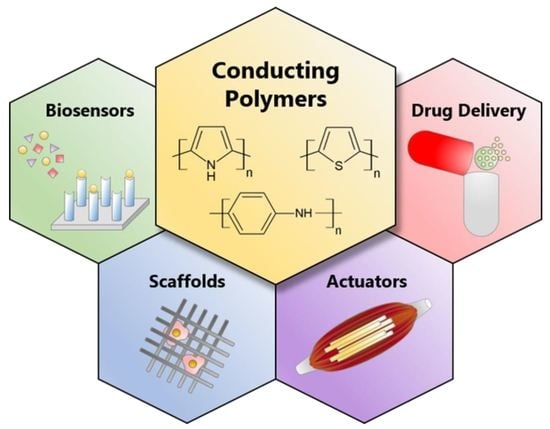
:Conducting polymers (CPs) have attracted significant attention in a variety of research fields, particularly in biomedical engineering, because of the ease in controlling their morphology, their high chemical and environmental stability, and their biocompatibility, as well as their unique optical and electrical properties. In particular, the electrical properties of CPs can be simply tuned over the full range from insulator to metal via a doping process, such as chemical, electrochemical, charge injection, and photo-doping. Over the past few decades, remarkable progress has been made in biomedical research including biosensors, tissue engineering, artificial muscles, and drug delivery, as CPs have been utilized as a key component in these fields. In this article, we review CPs from the perspective of biomedical engineering. Specifically, representative biomedical applications of CPs are briefly summarized: biosensors, tissue engineering, artificial muscles, and drug delivery. The motivation for use of and the main function of CPs in these fields above are discussed. Finally, we highlight the technical and scientific challenges regarding electrical conductivity, biodegradability, hydrophilicity, and the loading capacity of biomolecules that are faced by CPs for future work. This is followed by several strategies to overcome these drawbacks.
1. Introduction
Biomaterials generally indicate natural and synthetic substances that interact compatibly with biological systems [1]. The field of biomedical applications is defined by biosensors, tissue engineering, artificial muscles, drug delivery, etc., according to the component of the living organism with which the biomaterials interact for the purpose of medical diagnosis and treatment. Biomedical engineering involves both biology and engineering in an effort to apply engineering fundamentals and materials to medicine and healthcare. The current level of biomedical engineering has been reached primarily by diversifying biomaterials based on specific uses. While, biomaterials were generally acquired from the natural environment before the 20th century, a new variety of materials—such as ceramics, metals, carbons, synthetic polymers, alloys and composites have been extensively utilized as biomaterials since the early 20th century. These novel materials exhibit many advantages such as enhanced mechanical properties, chemical and biological performance, high reproducibility, and improved functionality [2,3,4,5,6,7]. Furthermore, their properties could be tuned to a target application by controlling the precursor materials and synthesis parameters. In particular, synthetic polymers have been considered promising biomaterials because they are compatible with biological environments; biocompatibility is one of the key aspects required in biomedical applications [8]. In addition to biocompatibility, polymeric materials possess some advantages, including chemical stability, good processability, and low production cost, allowing them to be widely used in various fields of engineering [9,10]. However, there are limits in certain biomedical applications where the semiconducting or metallic properties of biomaterials are essential, as conventional polymers are insulators.
As a new class of polymers, namely, conducting polymers (CPs) emerged, the limitations began to collapse. CPs are polymers with π-conjugated backbones that show electrical conductivity when given charges by redox reactions. CPs have attracted an immense amount of attention since the initial discovery of metallic polyacetylene in the late 1970s, as it was revealed that CPs can exhibit electrical conductivity in a broad range via a doping process using a relevant dopant [11]. They not only have electrical and optical characteristics similar to those of metals and semiconductors, but also exhibit the advantages of conventional polymers mentioned above. Furthermore, the level and duration of electrical stimulation can be externally controlled with CPs, which is beneficial for biomedical applications [12]. Therefore, CPs have various abilities that are useful for medical diagnoses and treatments of damaged body parts, including (i) ease of functionalization with bioactive molecules, (ii) alteration of a cellular response to electrical or optical or physical properties, (iii) charge transfer from an electrode to ions in living tissue, (iv) dimensional changes upon oxidation and reduction, and (v) entrapment and the release of drugs and biomolecules [13,14,15]. Additionally, their biocompatibility in vivo as well as in vitro is another great merit for their utilization in biomedical engineering [16,17,18,19].
Biocompatibility refers to the ability of a biomaterial to function in medical treatment by deriving a beneficial cellular or tissue response from a living host organism while not inciting any undesirable effect in the recipient of the therapy [20]. It is a crucial characteristic of a biomaterial that it is determined by a variety of factors including: physical, chemical, biological, medical, and design elements [21]. Since biomaterials must coexist with the tissue of the host; biocompatible materials need to possess long-term storage capability, and resistance to corrosion and chemical components of biological fluids. In addition, they should not cause any response that is inflammatory, toxic, carcinogenic, or allergic [22].
The biocompatibility of a material is measured through various tests concerning the interaction of the material with physiological fluids and cells, and stimulation of an immune response, both in vitro and in vivo [21]. In vitro studies are conducted to roughly assess a possibility of a material of interest harming relevant cell types. Assays composed of MTT (3-(4,5-dimethylthiazol-2-yl)-2,5-diphenyltetrazolium bromide) or its variants are commonly used to test cellular metabolic activity since high rates of MTT reduction is expected from rapidly dividing cells [23]. In addition, DNA synthesis, cell proliferation, cell membrane integrity, and toxicity based on cell death or tissue injury are also measures tested by in vitro studies [24,25,26].
There may be effects of a biomaterial not only from direct contact with cells but also from indirect exposure since it can generate diffusible components including residual solvents, monomers and breakdown products [23]. On top of the in vitro tests, in vivo studies are also essential to fully assess the biocompatibility of a material, as the cell-based assays used in vitro do not concern the rest of the human body. A material that is not directly cytotoxic can cause a destructive reaction. For example, Yeo at al. discovered that a UV-cross-linkable chitosan depressed cell viability in a moderate degree in cell culture but caused vigorous peritoneal adhesions in the peritoneal cavity [27].
The evaluation of biocompatibility can vary depending on the applications of biomaterials. In drug delivery, a material that does not cause any tissue injury may still damage a recipient of the treatment from drug delivery, as well as unpredicted complications including intravascular coagulation and embolism [26,28,29]. Therefore, it is important to consider the effect of drug delivery systems on an entire human body to eliminate any potential risk. The biocompatibility in tissue engineering refers to the ability of a scaffold to perform as a substrate to promote cell growth. In this context, biocompatible materials for tissue engineering should be able to support appropriate cellular activity such as the facilitation of molecular and mechanical signaling systems for tissue regeneration [30].
The biocompatibility of conducting polymers is affected by their polymer compositions including chemical structures, functional groups, morphologies, and synthetic processes [31]. Diverse conducting polymers such as polypyrrole (PPy) and polyaniline (PANI) have exhibited a great cellular response and support of the growth of various cell types, which is an essential aspect of biocompatibility in biomedical applications [32,33,34,35,36]. Furthermore, the biocompatibility of conducting polymers can be readily improved by introducing biocompatible molecules, segments and side chains to the polymers [37]. An in vivo study by Ramanaviciene et al. exhibited great biocompatibility of chemically synthesized PPy particles in mice over six weeks of treatment without any negative effects on cell viability and proliferation [18]. PANI has also been reported to show great biocompatibility in terms of dermal irritation and sensitization by Humpolicek et al. [38].
In spite of the attractive features of a CP mentioned above, a few challenges that hamper their widespread adoption in biomedical applications still remain. This article seeks to review recent research efforts and progress in the use of CPs for biomedical applications. In particular, this review focuses on the motivation and function of the CPs in the research fields of biosensors, tissue engineering, artificial muscles, and drug delivery. Furthermore, the challenges that the CPs are currently facing in the research fields above will be discussed regarding the charge carrier mobility, biodegradability, hydrophilicity, and loading capacity of biomolecules. Finally, a summary of representative approaches that could resolve the challenges is presented.
2. Conducting Polymers (CPs)
CPs have π-conjugated backbones in their structure. sp2-hybridized atoms with alternating single and double bonds enable the CPs to exhibit electrical conductivity. For this reason, they are also called conjugated polymers in their neutral states before they are processed to obtain permanent charges. Polyacetylene (PA) was first recognized as a CP in the 1970s and has been widely exploited ever since [39,40]. However, this noncyclic polyene is unstable in air and is therefore difficult to process [41]. As a result, solution-processible aromatic CPs, such as PPy, PANI, polythiophene (PT), and poly(3,4-ethyelenedioxythiophene) (PEDOT), have become a popular alternative [42]. Their phenyl or pentyl groups offer excellent thermal stability and conductivity [43,44]. In Table 1, the structures and applications of the aforementioned aromatic CPs are shown.
Two commonly used methods to synthesize the CPs are chemical and electrochemical redox polymerization. The former usually produces powders, whereas a thin film is obtained via the latter.
Although the chemical synthetic route enables the mass production of CPs, its process is typically more complicated [41]. It is also worth noting that further modification of bulk CPs after polymerization is necessary to obtain the required electronic properties. On the other hand, the processes of polymerization and doping are more controllable, and thin films are readily obtainable in the case of the electrochemical polymerization technique. However, this method is not suitable for large production quantities [45].
The charges in the CPs are controlled via doping where they are oxidized (p-doping) or reduced (n-doping) while retaining neutrality via counter ions from doping. Figure 1 depicts the p-doping of heterocyclic polymers. Charge carriers injected in the form of charged polarons (radical ions) or bipolarons (dications or dianions) migrate along the conjugated CP backbone, from which electrical conductivity is generated [41]. Owing to their unique electrical properties, the CPs have been of interest in a variety of scientific applications, including capacitors, field emission display, surface protection, and biomedical engineering [42].
3. Biomedical Applications
3.1. Biosensors
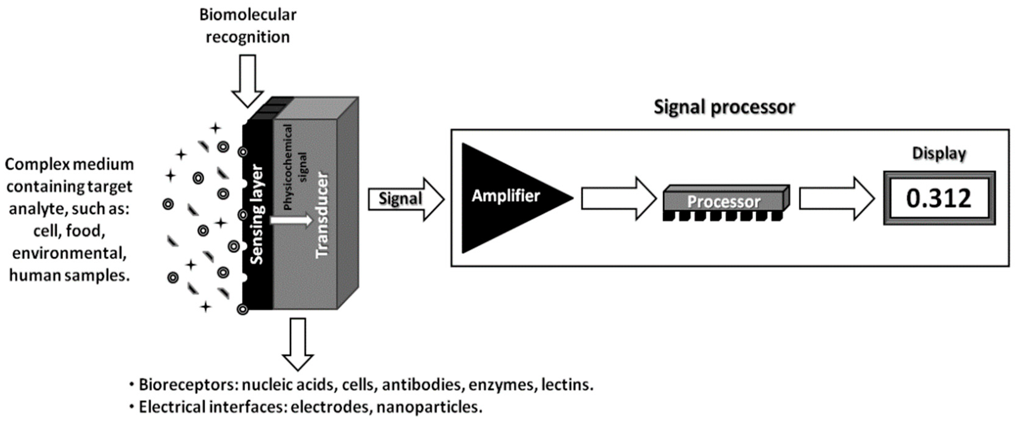
A biosensor is an analytical device that generates electrical signals according to biological responses from an analyte. As the biological species is monitored by a sensing element in a biosensor, measurable electrical signals proportional to the concentration of the analyte are generated. As shown in Figure 2, a biosensor is mainly composed of a bioreceptor, transducer, and electronic module [55]. Various sensing elements, such as antibodies, cells, enzymes, and tissues, can be used as bioreceptors that interact with specific analytes. The extent of the interaction caught on the bioreceptors is transformed by the transducer into an electrochemical, optical, or thermal signal [56,57,58]. Once the transduced signals are delivered to the electronics, they are often magnified by an amplifier. Subsequently, a signal processor displays the amplified signals in a user-friendly way. Biosensors can detect various signals such as current, potential, change in conductivity, and light absorbance [59].
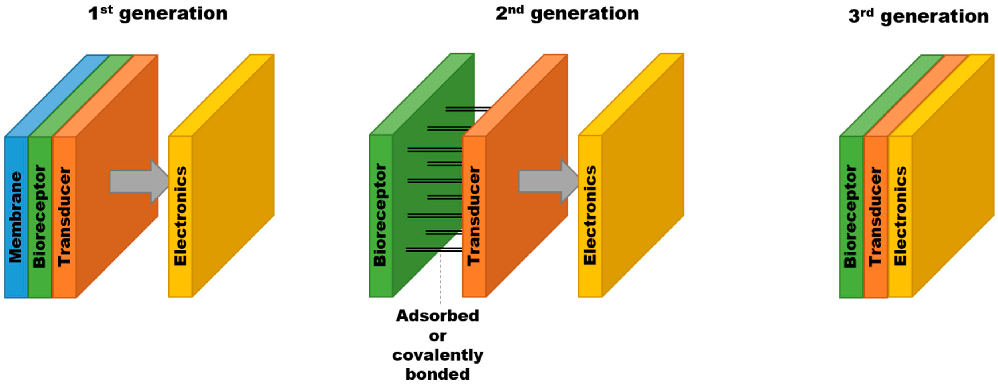
Biosensors have evolved through three generations depending on the level of integration of a bioreceptor and a transducer as illustrated in Figure 3 [60]. The first generation involves the bioreceptor immobilized in a dialysis membrane, which is attached to the surface of the transducer. In the second generation, the bioreceptor is either adsorbed or covalently connected to the transducer without the need for the membrane. The third generation includes the bioreceptor directly bound to the transducer and the electronics, allowing their complete integration as a biosensor unit.
In biosensors, the CPs are used as transducers owing to their great electrical conductivity as well as their great sensitivity and versatility of available monomer types. Furthermore, biomaterials for a receptor can be easily integrated with the CPs, thereby enabling the facile assembly of biosensors. Furthermore, the CPs are cost effective and can be adapted to a variety of fabrication techniques to form biosensors. PPy, PT, polycarbazole (PCz), poly(o-phenylenediamine) (POPD), PA, and polyfuran (PFu) are commonly used as biosensors.
The CPs have been exploited in diverse applications for biosensors including medical purposes and environmental monitoring. Extensively studied blood glucose biosensors are a good example of the medical applications [61,62,63]. They use enzymes to oxidize glucose in a number of steps and the concentration of glucose is detected by a transducer composed of the CPs. PPy, PANI and polyaminobenzoic acid (PAB) are typical CPs used in glucose biosensors [64,65]. Cholesterol biosensors monitor the level of cholesterol in blood, which is considered an important measure in clinical diagnostics. PPy can be used as a transducer in cholesterol biosensors to immobilize enzyme cholesterol oxidase by physical adsorption. In cholesterol biosensors, the oxidation of cholesterol in PPy films results in amperometric responses [66]. Biosensors are also capable of monitoring environmental hazards. In gas sensors for environmental monitoring, the CPs exhibit a fast change in electrical conductivity resulting from the reduction of the polymers when organic vapor or moisture is detected [67,68,69]. PANI films have been reported to be effective in ammonia gas sensors, which is beneficial for food-packaging applications [70,71]. In a humidity sensor, films of CPs, such as poly(3,4-ethylenedioxythiophene)/polystyrene sulfonate (PEDOT/PSS) and poly(anilinesulfonic acid) have been shown to be capable of detecting a change in a wide range of humidity [72,73].
The main challenge of biosensors is to combine biologically reactive elements such as antibodies, cells, and enzymes with the CPs in order to function as an electrical signal generator. The CPs are great matrices for the immobilization of the bioreceptors due to their biocompatibility, sensitivity, and durability. In addition, they are flexible to endure the embedment of the biomolecules [74,75]. Such immobilization can be done by physical adsorption, chemical adsorption, covalent bonding, crosslinking, or entrapment [76,77,78,79,80,81].
Physical adsorption takes advantage of physical interactions such as Van der Waals and electrostatic forces between bioreceptors and CPs. The anionic bioreceptors are attached to the cationic CPs through electrostatic forces. It is worth noting that physical adsorption does not require the functionalization of monomers. However, the leaching of the bioreceptors may occur over time due to the weak forces of physical adsorption. In this context, chemical adsorption that involves covalent binding between bioreceptors and the CPs is introduced. Despite its simplicity, the precise control of biomolecule orientation on the surface film remains a challenge. Such random orientations may block active sites on the surface of the CPs. In this regard, a targeted coupling strategy where crosslinking agents and functional groups, such as –NH2, –COOH, and –SH, are selectively introduced onto biomolecules. Such targeted coupling strategies enable the precise control of the biomolecule’s orientation. However, it is difficult for the biomolecules to maintain enzyme activity due to the harsh treatment by toxic chemicals involved in the crosslinking process [82,83,84,85].
Entrapment techniques where monomers, dopants, and biomolecules are mixed together in a solution are usually performed in electrochemical polymerization. In this strategy, polymerization of the CPs and immobilization of the biomolecules occur simultaneously. However, it is not cost effective because a high concentration of biomolecules is required during the process. The procedure of entrapment causes sensing elements to become less accessible to the analytes. Furthermore, the quaternary structure of proteins is damaged by the hydrophobic CPs, and therefore their biological activity as sensing elements decreases [86,87].
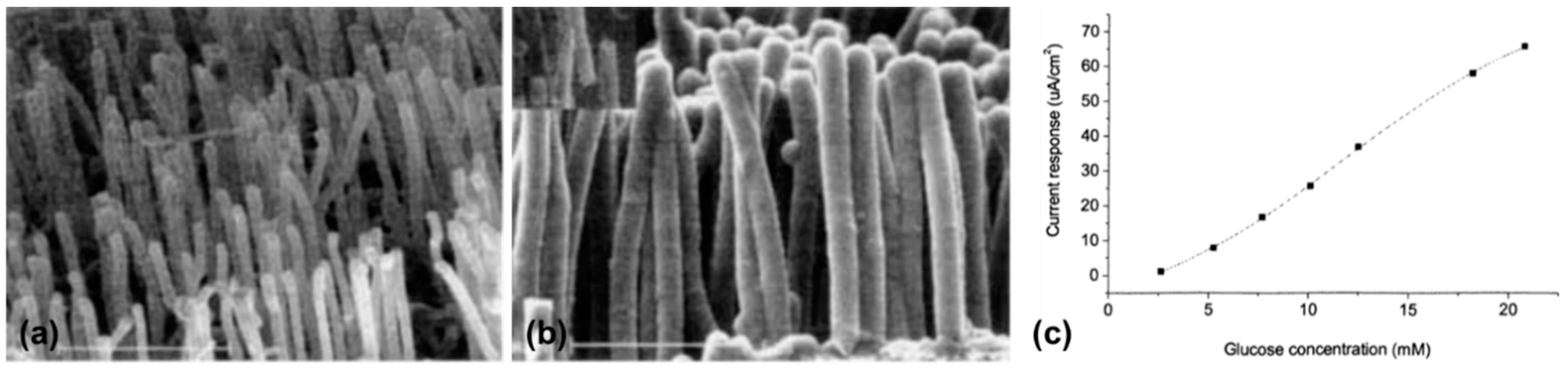
Despite the advantages of low cost and easy production, the CPs in biosensors have limitations—in that the CPs are more vulnerable to surrounding chemical conditions including oxygen, acids, and bases. In addition, the charge carrier mobility is not satisfactory compared to other conducting materials such as carbon nanotubes and silicon nanowires [88]. Gao et al. suggested coaxial nanowire composites composed of PPy and carbon nanotubes in an effort to exploit complimentary attributes, such as the flexibility and light weight of the CPs and the high electrical conductivity of carbon nanotubes [89]. Specifically, PPy was electrodeposited onto an array of carbon nanotubes, resulting in individually coated nanotubes with a great electroactive surface area [90]. Glucose oxidase was immobilized by the oxidation of pyrrole to form a glucose biosensor.
In Figure 4a,b, the scanning electron micrographs of the carbon nanotubes are compared before and after the electrodeposition of PPy. The layer of evenly coated PPy, thinner than 100 nm, was found in the array of carbon nanotubes with a spacing between 100 nm and 400 nm. Figure 4c illustrates the electrical current according to the concentration of glucose. The response increases linearly with the detected amount of glucose up to 20 mM, which is higher than the requirement for practical glucose biosensors, typically 15 mM.
3.2. Scaffolds for Tissue Engineering
The action potential generated at the synapse plays a key role in neural communication and electrical conductivity, and is known to help the proliferation and differentiation of cells [91,92]. A scaffold is a structural element utilized in tissue engineering that serves as a template for the regeneration of functional tissues and organs. Materials for scaffolds in tissue engineering require the following characteristics: hydrophilicity, biocompatibility, biodegradability, and mechanical properties resembling those of tissues. Therefore, the CPs are promising candidates for scaffolds for tissue engineering to regenerate or replace damaged organs such as skin, tissue, and spinal cord [93,94,95,96,97].
In early studies, pure CPs, such as PPy, PANI, and PT, that offer cytocompatibility were employed for scaffolds, as they are capable of adhesion, proliferation, and differentiation of diverse cell types [98,99,100,101]. These polymers can be synthesized in the form of films on the surface of an electrode either chemically or electrochemically [41]. To adjust the mechanical properties, it is common to blend the CPs with other degradable and more flexible polymers, including poly(lactic acid) (PLA), poly(lactic-co-glycolic acid) (PLGA), and polycaprolactone (PCL). For example, PPy/polycaprolactone films are prepared by merging polycaprolactone films in a mixture of polystyrene sulfonic acid and pyrrole in water, followed by the addition of ferric chloride, as an oxidant. As a result, conductive PPy-coated polycaprolactone films are formed, which display a similar resistivity as the native cardiac tissues [102]. Polyurethane is biocompatible and has suitable mechanical and physical characteristics for scaffolds, and therefore can be used to form composite films when blended with the CPs [103]. Conducting composite films composed of polyurethane and PPy are fabricated by the polymerization of pyrrole in an emulsion mixture of polyurethane and subsequent pressing into the form of films. It should be noted that the stiffness is tunable depending on the ratio of PPy to polyurethane [104].
Conducting copolymer films are another type of modification to enable the CPs to be more widely used in tissue engineering. Since not all the CPs are natively biodegradable, their use in vivo can be limited [105]. The addition of aniline to form a copolymer with the CPs is an effective way to provide the CPs with biodegradability. Copolymers of aniline and pyrrole functionalized with hydrolyzable groups have been reported to show a similar electroactivity to the CPs while also being biodegradable [106,107].
Nanofibrous polymeric scaffolds have been extensively researched to mimic the natural extracellular matrix (ECM). The CP nanofibers can be synthesized by electrospinning, phase separation, and molecular assembly [108,109,110]. In the process of electrospinning, the CPs are mixed with biodegradable polymers, then electrospun into the form of nanofibers. They possess a high surface area, porosity, and tunable diameters in the range of a few nanometers [111,112,113]. Scaffolds fabricated by the electrospinning of conductive nanofibers using PANI blended with PLA or gelatin have been developed and showed an electrical conductivity of 4.2 × 10⁻³ S cm−1, which is substantial enough to be exploited for cardiac tissue engineering [114,115].
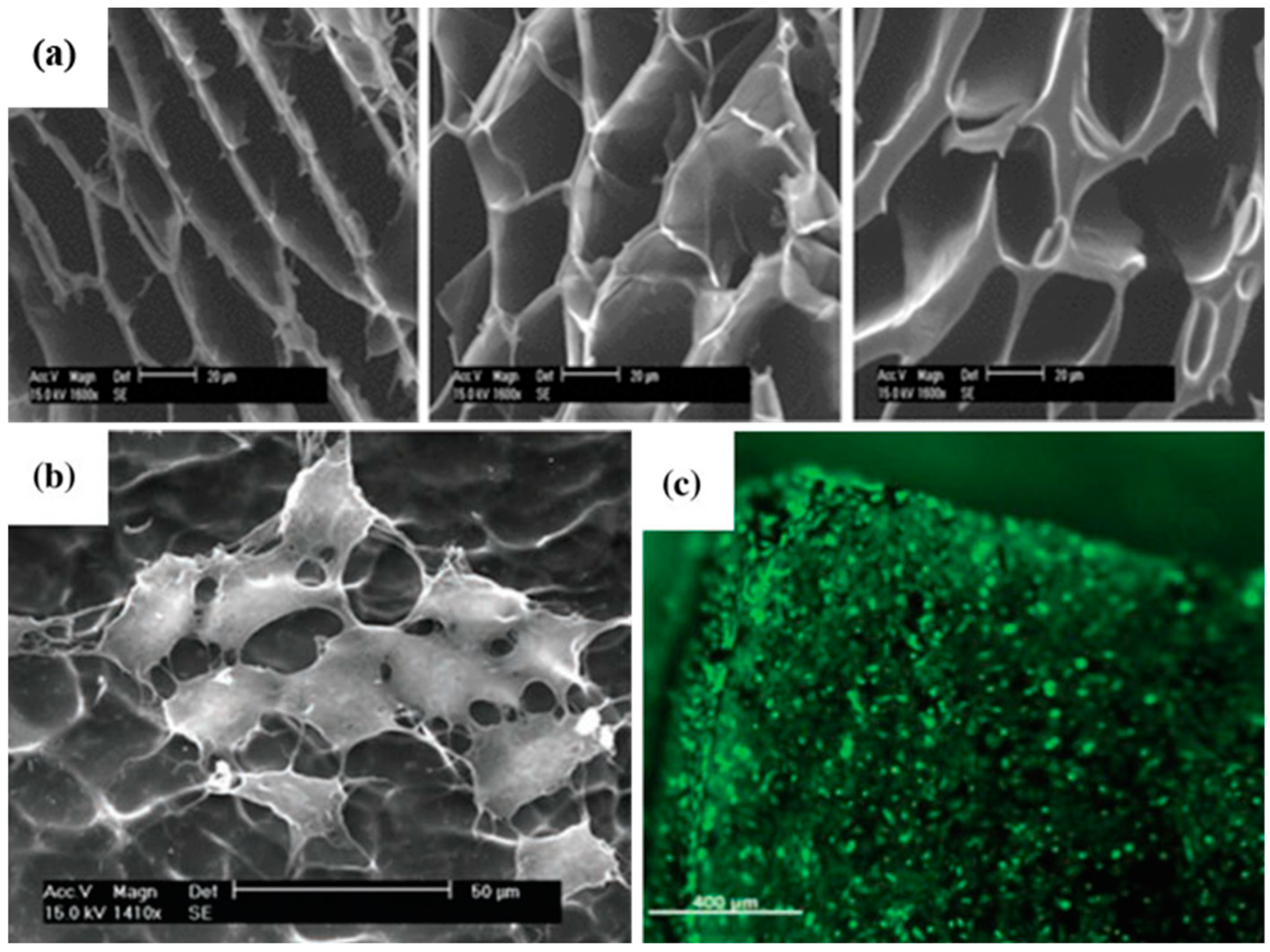
Electroconductive hydrogels are hybrid materials comprised of CPs blended with conventional polymers that provide aqueous gel networks. These hydrogels exhibit rubber-like flexibility, tunability, and biocompatibility, which are essential requirements for the use in scaffolds [116,117,118,119]. An electroconductive hydrogel made of poly(ethylene dioxythiophene) and poly(acrylic acid) (PAA) has been developed by free radical polymerization since they display distinguished electroactivity, mechanical properties, and swelling [120]. Despite their excellent electrical conductivity, physiological pH of living tissue (7.4) diminishes electrical conductivity of electroconductive hydrogels, as the charges on the CP backbone return to the neutral states [121]. Also, hydration in physiological media allows physically trapped CPs to escape the network, resulting in a further decrease in electrical conductivity [122]. Single component CP hydrogels fabricated from CPs as a main matrix can be a solution to the limitations of electroconductive hydrogels. However, it is challenging to form such hydrogels since conjugated polymers lack flexibility, water solubility, and functional side chains [123]. Therefore, only a few studies have been reported on the single component hydrogels. Nevertheless, there are potential candidates for these hydrogels, and polythiophenes are one example owing to a variety of water-soluble polythiophenes that have been developed [124]. Mawad et al. demonstrated the fabrication of single component hydrogels composed of poly (3-thiophene acetic acid) (PTAA), which is based on polythiophenes [125]. 1,1′-carbonyldiimidazole (CDI) was utilized as a crosslinking agent to form the hydrogels since it is soluble in water and washable from the network. The crosslinking density was determined by the ratio of CDI to the monomer of PTAA. In Figure 5a, scanning electron micrographs of the hydrogels with different crosslinking densities are shown. All of the hydrogels exhibited porous networks with varying wall structures and pore size distributions. A well-established morphology of C2C12 myoblast cells 72 h after the incubation on the surface of the hydrogels is shown in Figure 5b,c, which indicates that the hydrogels assisted the adhesion and proliferation of the cells. Depending on the crosslinking density, the swelling ratio and porosity as well as mechanical properties could be tuned. Excellent swelling ratio up to ~850% was observed when the crosslinking density was low. The compressive modulus was also affected by the crosslinking density and measured to be 17.88 kPa with a high crosslinking density, which is comparable to muscle tissue. Furthermore, great electrical conductivity of ~10−2 S cm−1 was obtained in physiological conditions.
Tissue engineering usually concerns three approaches: (1) utilization of biomolecules to help host cells regenerate tissue, (2) introduction of repair cells directly to the defective area, and (3) growth of cells on a scaffold in bioengineered atmospheres that would help the regeneration of tissue [126]. Tissue scaffolds should mimic the chemical, mechanical, and topographical properties of the ECM composed of proteins and polysaccharides [127]. Therefore, a controllable geometry, high degree of porosity with appropriate pore size, great surface-to-volume ratio, and a high degree of pore interconnection are required for tissue scaffolds [128]. Nanofabrication technologies that are able to synthesize nanostructures satisfying such factors include electrospinning, chemical etching, and photolithography. Electrospinning is commonly used to generate ECM-like nanofibrous scaffolds. PPy, PANI, and PT have been proven to be compatible with cells and tissues in in vivo and in vitro systems [126].
3.3. Actuators for Artificial Muscles
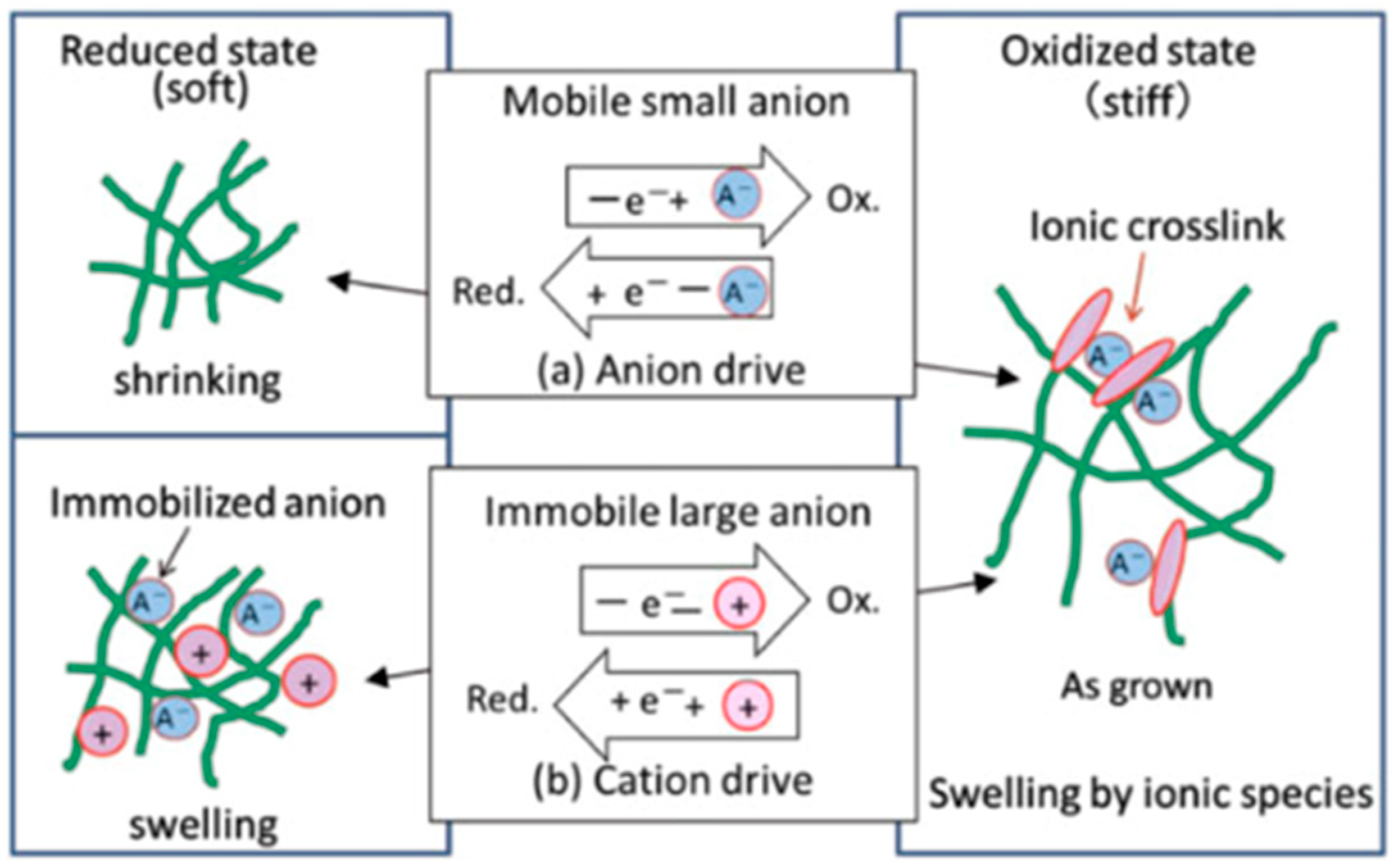
The volume change of the CPs due to the electrochemomechanical reactions pave a way towards their utilization as actuators for artificial muscle applications [129]. The volumetric expansion of a CP in electrolyte is controlled by tuning the applied voltage. It is notable that the oxidized state is stiffer than the reduced state due to the ionic crosslink at the polaron site and delocalization of π-electron [130]. The size of anions used for the process determines whether the electrochemomechanical reaction occurs via anion drive or cation drive, as illustrated in Figure 6. Small anions, such as ClO4− and bis-(trifluoromethanesulfonyl)imide (TSFI−), come out of the bulk CPs during reduction, and thereby the reduced state is shrunk, which is called anion drive (Figure 6a). On the other hand, when large anions, such as dodecylbenzenesulfonic (DBS−) acid, are used, long alkyl chains of the anions cause entanglement and immobilization in the network. The reduction of the CPs then occurs via the cation drive, where cations inserted from the electrolyte solution result in a swollen polymer network by the volume of additional cations (Figure 6b) [131].
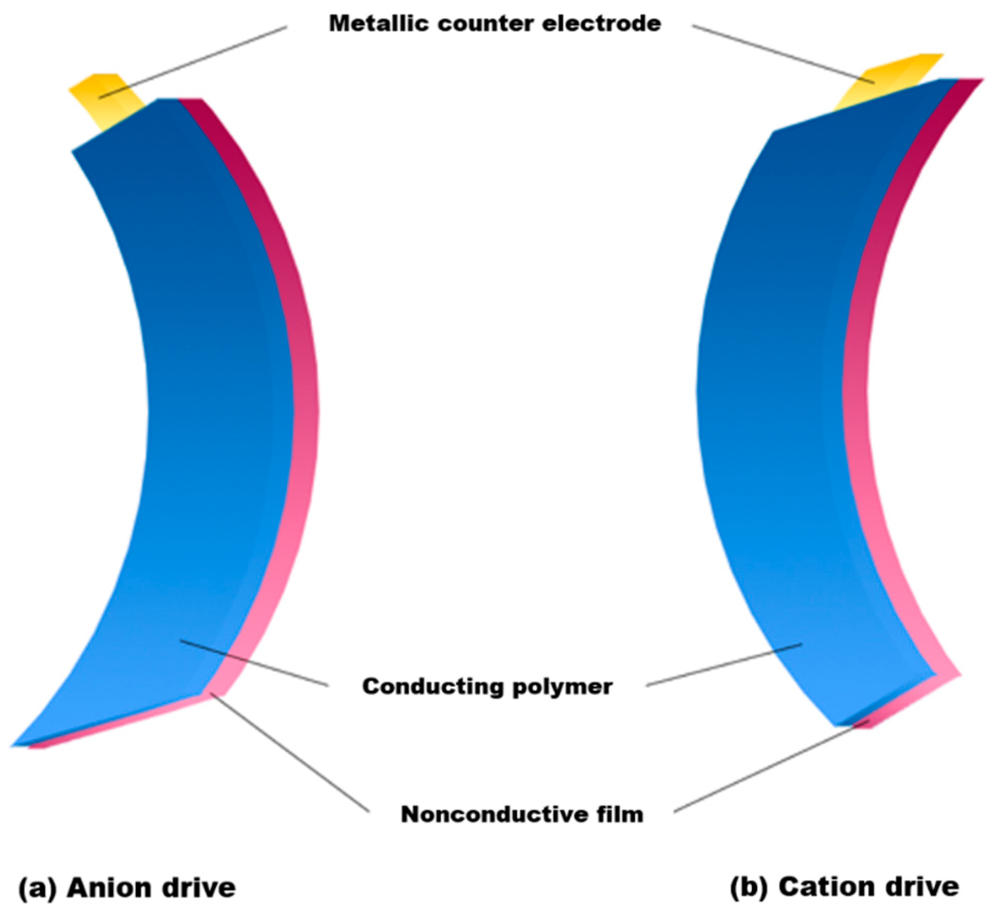
In actuators, a linear or angular movement can be derived from the volumetric change of the CPs. One of the most widely used configurations is a bilayer structure composed of a CP film grown on a metallic electrode with a nonconductive film. The device exhibits macroscopic bending behaviors shown in Figure 7 due to swelling or shrinking induced by a mechanical stress gradient across their interface resulting from electrochemical reactions. Specifically, the anion-driven device bends towards the CP-convex form upon oxidation via swelling of the CP. On the other hand, the cation drive results in bending the device in a CP-concave manner, as they shrink by oxidation. It is yet unavoidable to use a metallic counter electrode in a bilayer actuator. However, it often results in deterioration of the actuating film caused by corrosion of the metal electrode exposed to a harsh environment (i.e., a sudden change in pH and chemical) [129].
In an effort to exclude the metallic counter electrode, a trilayer actuator was developed in which two CP films were attached to double-sided tape [132]. When the trilayer structure is immersed in an electrolyte (Figure 8), two opposite volume changes are produced as one of the CP films undergoes anion-driven swelling, whereas the other film experiences cation-driven shrinking [129]. The main advantage of the CPs, as actuators for artificial muscles, is their low operating voltage (as low as 2~10 V) [133]. In addition, they provide higher strains in a more cost-effective way than carbon nanotubes [134]. However, the electromechanical coupling, the conversion efficiency from electrical to mechanical energy, is not as high as that in carbon nanotubes. Therefore, the application of the CPs in a large-scale actuator has been hampered [135,136].
3.4. Drug Delivery
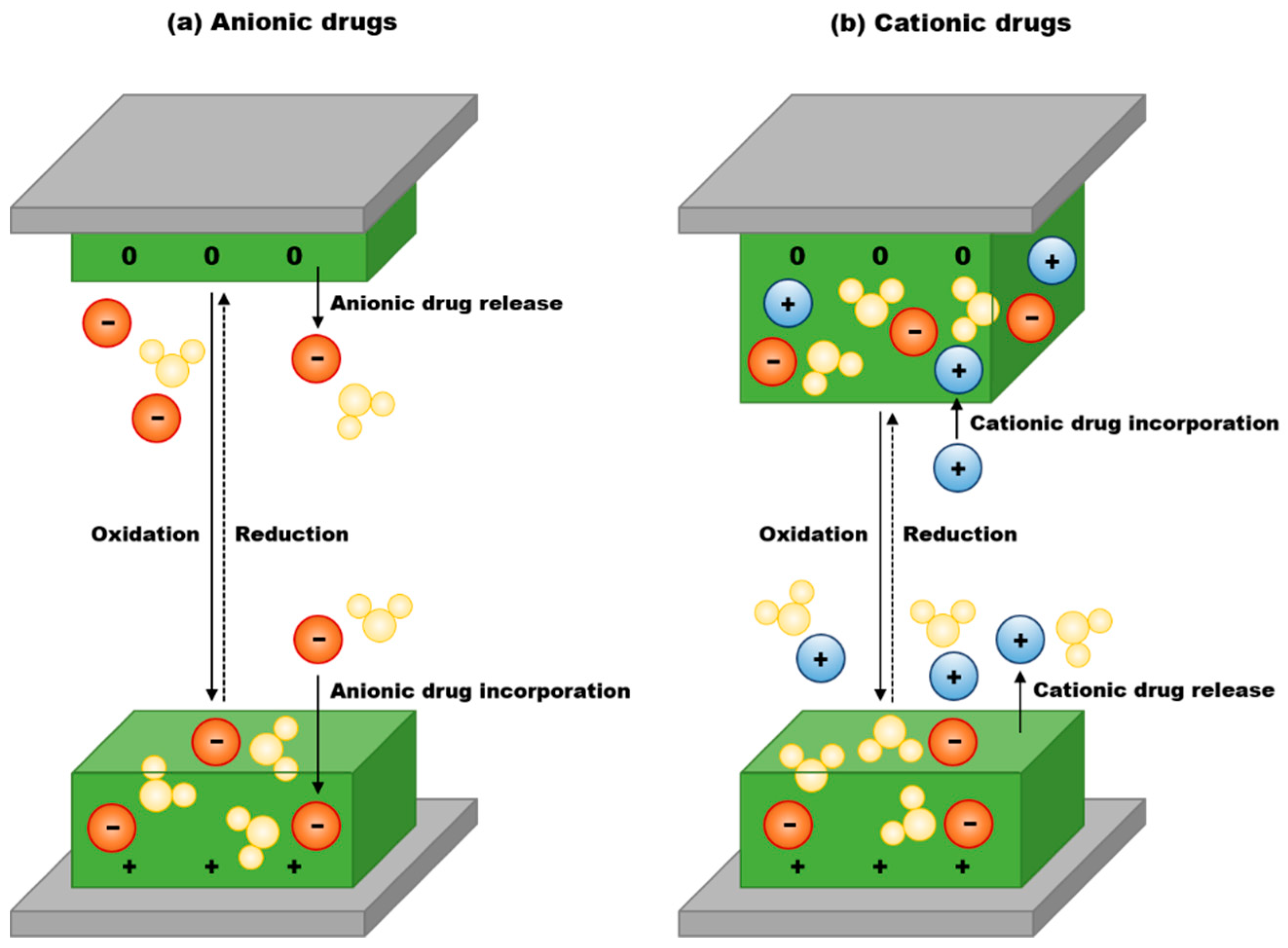
The CPs in drug-delivery applications utilize their electrical stimulation to release several therapeutic proteins and drugs. PPy is one of the most widely studied CPs for drug delivery due to their controllable and reversible redox reactions that enable the delivery of drugs [41]. The rate of drug release in the CPs can be precisely controlled via alteration of their redox state, resulting in changes in polymer charge, conductivity, and volume. Specifically, as illustrated in Figure 9, the mechanism of drug release by the CPs is similar to that of bioactuators. Anionic drugs that are incorporated using small anions leach out as the volume of the CP decreases upon reduction, whereas cationic drugs prepared with immobile anions are released by cation-driven actuation when the CPs are oxidized [137].
Drugs of interest can be incorporated during the polymerization of the CPs. For anionic drugs, the incorporation into the CPs occurs by anions that balance out the positive charges induced by oxidation. In contrast, cationic drugs are incorporated into the CPs with the help of cations [138]. The incorporation of drug molecules into CP matrices during polymerization can be hindered due to the unfavorable interaction among drug molecules and polymers. Moreover, the drug molecules weaken the adherence of the CPs to the electrode, which is crucial to provide electrical stimuli for drug release [137]. In order to resolve such challenge, drug molecules can be incorporated after synthesizing the CPs. A buffer layer of pristine CPs between the electrode and CP/bioactive drug mixture can improve adherence preventing detachment of the CP/bioactive drug layer [138].
A drug molecule for the use in the CP-based drug delivery system needs to meet the following requirements; (1) it should not be electroactive at the voltages applied to the systems in order to maintain its biological activity, (2) the pKa needs to be moderated to precisely control the loading and releasing of the drug, (3) short half-lives are preferred to reduce the risk of accumulation, and (4) it should not be toxic to local tissues that may be subject to higher concentrations of the drug [137].
4. Current Stage and Challenges
To date, significant progress has been achieved in exploiting the CPs in the field of biomedical engineering owing to their electrical conductivity and biocompatibility. Nevertheless, some drawbacks still limit the utilization of these polymer materials in biomedical engineering.
Biodegradability is one of the critical issues of typical CPs that requires improvement, especially in tissue engineering, where scaffolds are expected to eventually decompose. A scaffold is an artificial support device implanted temporarily in order to boost the differentiation and proliferation of cells until new tissues are formed. Therefore, the CPs used as scaffolds must degrade after they achieve their role [105]. In an effort to ensure biodegradability of a scaffold, the CPs have been blended with other degradable polymers. For example, PLA, a common biodegradable polymer, was mixed with conductive PPy nanoparticles to provide both conductivity and degradability [41].
Armelin et al. reported on the fabrication of stable freestanding nanomembranes by combining a PT derivative that has carboxylate substituents in the 3-position of the heterocyclic ring and a biodegradable polyester [138]. The nanomembranes were fabricated by spin-coating mixtures of a PT derivative, poly(3-thiophene methyl acetate) (P3TMA) and polybutylene succinate (PBS) for use as bioactive platforms for tissue engineering [139]. Although it is possible to provide the CPs with biodegradability, there is a tradeoff between the electrical conductivity and biodegradability, as the overall structure is modified in such a way that the conductive properties are hindered.
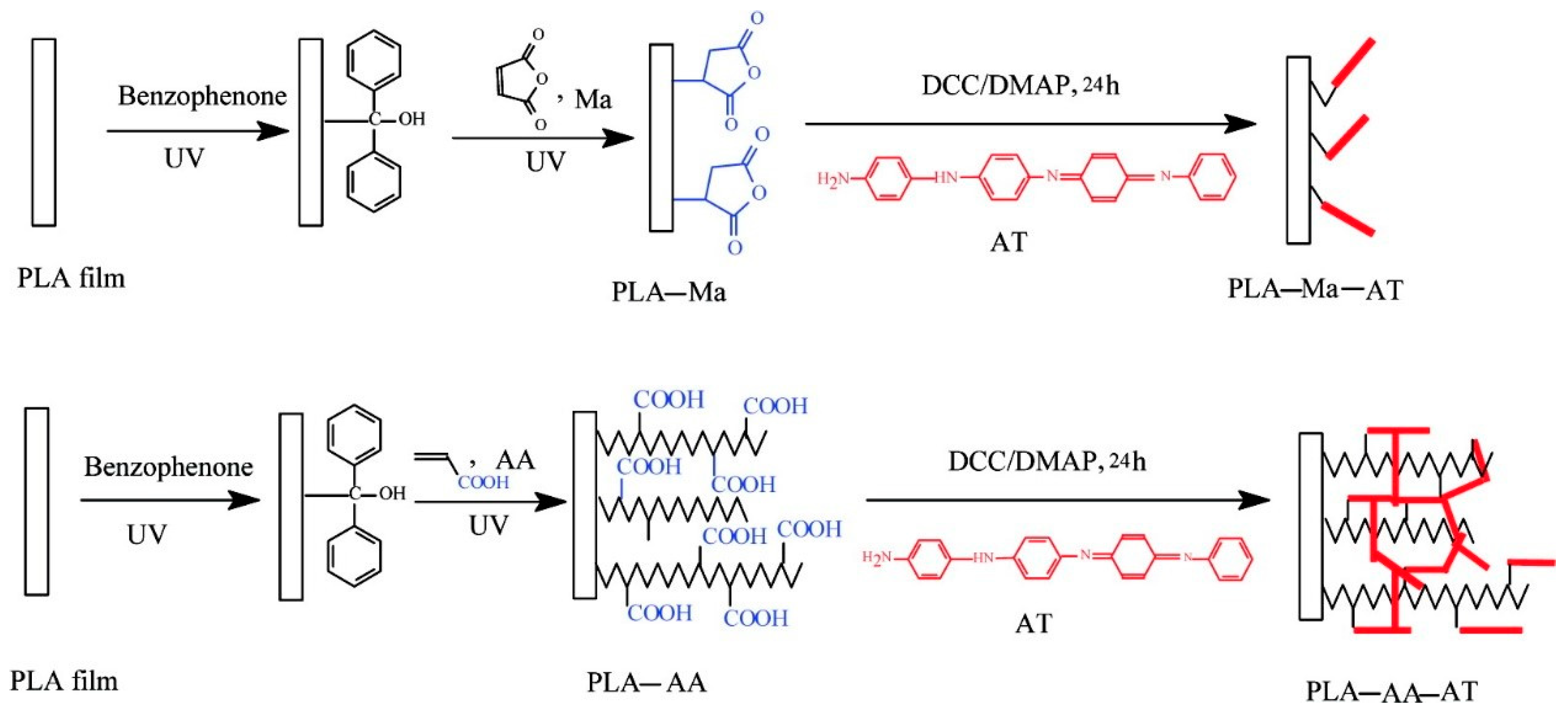
Hydrophobicity of the CPs is also an obstacle that must be overcome in order to apply the CPs to scaffolds and biosensors. Despite their excellent mechanical properties and biocompatibility, their lack of hydrophilicity hampers the attachment of cells onto their surfaces [140,141]. The surface properties can be modified via functionalization. For example, Guo et al. proposed a two-step method, as shown in Figure 10, for the functionalization and surface modification of PLA to promote hydrophilicity as well as to introduce conductive aniline oligomers onto the surface of PLA. It resulted in improved cell adhesion and controllable cell behaviors such as spreading, proliferation, and differentiation in a later stage. Specifically, carboxyl acid and anhydride functionalities were introduced onto a PLA film using photo-crosslinking. Subsequently, aniline tetramers were coupled with the functionalized PLA films. The surface modification enhanced both hydrophilicity and electroactivity at the surface [142].
Actuators based on the CPs are electrically controllable with low voltage and biocompatible with liquid electrolytes such as bodily fluids, which makes the CPs attractive materials for the application. However, the mobility of charge carriers in the CPs needs improvement to provide faster responses in the actuators. By blending the CPs with nanofibers and nanotubes, the charge-carrier mobility can increase due to their porous structures, but making the blended materials biocompatible is another issue [143].
Finally, for drug delivery, there is a limit to the amount of therapeutic substances that can be delivered using the CPs. Increasing porosity and surface area is one way to increase the drug-loading capacity. Films of template-grown CP can enhance the porosity and increase the surface area by forming nanostructures. According to Sharma et al., PPy scaffolds created with the help of a 3-dimensional colloidal crystal template made of monodisperse poly(methyl methacrylate) (PMMA) exhibited a high surface area [144]. Nanofibers can also be used as a template to fabricate the CPs with a high porosity and surface area. Abidian et al. constructed poly(3,4-ethylenedioxythiophene) (PEDOT) nanotubes using nanofibers of poly(lactide-co-glycolide) (PLGA) as a template for a precisely controlled release of dexamethasone [145]. PEDOT was electrochemically deposited around the electrospun nanofibers of PLGA. Subsequently, PLGA can be dissolved, resulting in hollow nanotubes of PEDOT. Figure 11a compares the release of dexamethasone from the PLGA nanofiber and the PEDOT-coated PLGA nanofiber. The layer of PEDOT around the PLGA nanofibers slows down the release of dexamethasone. Furthermore, the drug release can be controlled more precisely by actively actuating the PEDOT nanotubes loaded with the drug with an electric field. As depicted in Figure 11b, upon the application of a positive voltage, PEDOT shrinks as electrons are inserted into the chains and counterions are discharged into the solution. This contraction of the nanotubes allows the release of the drug at the ends of the nanotubes. In Figure 11c, the cumulative mass releases of dexamethasone from the PLGA nanofibers and the PEDOT-coated PLGA nanofibers with and without electrical stimulation are shown. By the electrochemical deposition of PEDOT around the PLGA nanofibers, a substantial decrease in the amount of dexamethasone released was observed. In addition, when the electrical stimulation of 1 V was applied at specific points, the amount of the drug release increased significantly. SEM images of the PEDOT nanotubes after dissolution of the PLGA nanofibers in the core in Figure 11d–f reveal the hollow morphology of PEDOT as well as the well-defined internal and external texture. The diameters of the nanotubes varied from 100 to 600 nm, whereas the average wall thickness of PEDOT ranged from 50 to 100 nm.
5. Summary
In summary, we first reviewed the motivation and function of the CPs in biomedical applications including biosensors, tissue engineering, artificial muscles, and drug delivery. The CPs have been widely developed and studied in biomedical engineering over the past few decades; they can provide unique optical and electrical properties along with the advantages of ease of controlling their morphology, high chemical and environmental stability, and biocompatibility. The most commonly used CPs are PPy, PANI, PT, and PEDOT, which are simply synthesized via chemical or electrochemical redox polymerizations. In biosensors, the CPs act as a transducer that turns chemical reactions into electrical signals. As a scaffold in tissue engineering, they serve to promote the proliferation and differentiation of cells. Furthermore, these polymers are utilized not only as actuators for artificial muscles, because of their volume change properties upon redox reactions, but also as a drug carrier in the process of drug delivery, because of their ability to release drugs upon electrical stimulation.
However, the use of the CPs in biomedical engineering has been limited by a lack of biodegradability, low hydrophilicity, low charge-carrier mobility, and a small loading capacity of biomolecules. Nevertheless, ongoing studies have been investigating a wide range of possibilities for resolving these issues. As a response to low biodegradability, it has been strongly suggested that the CPs should be blended with biodegradable polymers because the blend scaffolds are expected to degrade faster than pure CP ones. To enhance the hydrophilicity of the CPs, surface modification and functionalization have been attempted using the chemicals and small molecules that possess hydrophilic properties. Hybridization with other conducting materials, such as carbon nanotubes and nanofibers, have proven to be an effective strategy that supplements the low charge-carrier mobility of the CPs. Finally, it has been reported that the limit in drug-loading capacity can be overcome by forming nanostructures with higher porosity and surface area.
In spite of significant advances in biomedical engineering by the use of the CPs as a biomaterial, there are still many challenges to be surmounted, including poor biodegradability, low hydrophilicity, low charge-carrier mobility, and small loading capacity of biomolecules. However, we believe that the CPs will become more promising materials for biomedical research in the near future, as a variety of ways to overcome the weaknesses of the CPs are continuously being explored.
Author Contributions
Writing—Original Draft Preparation, Y.P., J.J., and M.C.; Writing—Review & Editing Y.P., J.J., and M.C.
Funding
This work was supported by the National Research Foundation of Korea (NRF) with a grant funded by the Korean government (Ministry of Science, ICT & Future Planning, MSIP) (NRF-2017R1C1B5017856 and NRF-2017R1C1B1004605).
Conflicts of Interest
The authors declare no conflict of interest.
References
- Ratner, B.D.; Hoffman, A.S.; Schoen, F.J.; Lemons, J.E. Biomaterials science: A multidisciplinary endeavor. In Biomaterials Science: An Introduction to Materials in Medicine, 2nd ed.; Elsevier Academic Press: San Diego, CA, USA, 2004; pp. 1–9. [Google Scholar]
- Zheng, J.; Tang, M.; Hu, Y.-Y. Lithium ion pathway within Li7La3Zr2O12-polyethylene oxide composite electrolytes. Angew. Chem. Int. Ed. 2016, 55, 12538–12542. [Google Scholar] [CrossRef] [PubMed]
- Han, W.; Zhao, G.; Zhang, X.; Zhou, S.; Wang, P.; An, Y.; Xu, B. Graphene oxide grafted carbon fiber reinforced siliconborocarbonitride ceramics with enhanced thermal stability. Carbon 2015, 95, 157–165. [Google Scholar] [CrossRef]
- Chen, H.; Ginzburg, V.V.; Yang, J.; Yang, Y.; Liu, W.; Huang, Y.; Du, L.; Chen, B. Thermal conductivity of polymer-based composites: Fundamentals and applications. Prog. Polym. Sci. 2016, 59, 41–85. [Google Scholar] [CrossRef]
- Nie, H.; Wang, C.-H. Fabrication and characterization of PLGA/HAp composite scaffolds for delivery of BMP-2 plasmid DNA. J. Control. Release 2007, 120, 111–121. [Google Scholar] [CrossRef]
- Gao, C.; Peng, S.; Feng, P.; Shuai, C. Bone biomaterials and interactions with stem cells. Bone Res. 2017, 5, 17059. [Google Scholar] [CrossRef] [PubMed] [Green Version]
- Prakash, J.; Pivin, J.C.; Swart, H.C. Noble metal nanoparticles embedding into polymeric materials: From fundamentals to applications. Adv. Colloid Interface Sci. 2015, 226, 187–202. [Google Scholar] [CrossRef] [PubMed]
- Williams, D.F. On the nature of biomaterials. Biomaterials 2009, 30, 5897–5909. [Google Scholar] [CrossRef]
- Leja, K.; Lewandowicz, G. Polymer Biodegradation and Biodegradable Polymers-a Review. Pol. J. Environ. Stud. 2010, 19, 255–266. [Google Scholar]
- Li, Y. Challenges and Issues of Using Polymers as Structural Materials in MEMS: A Review. J. Microelectromech. Syst. 2018, 27, 581–598. [Google Scholar] [CrossRef]
- Fincher, C.R.; Ozaki, M.; Heeger, A.J.; MacDiarmid, A.G. Donor and acceptor states in lightly doped polyacetylene, (CH)x. Phys. Rev. B 1979, 19, 4140–4148. [Google Scholar] [CrossRef]
- Rivers, T.J.; Hudson, T.W.; Schmidt, C.E. Synthesis of a Novel, Biodegradable Electrically Conducting Polymer for Biomedical Applications. Adv. Funct. Mater. 2002, 12, 33–37. [Google Scholar] [CrossRef]
- Fattahi, P.; Yang, G.; Kim, G.; Abidian, M.R. A review of organic and inorganic biomaterials for neural interfaces. Adv. Mater. 2014, 26, 1846–1885. [Google Scholar] [CrossRef]
- Smela, E. Conjugated polymer actuators for biomedical applications. Adv. Mater. 2003, 15, 481–494. [Google Scholar] [CrossRef]
- Nambiar, S.; Yeow, J.T.W. Conductive polymer-based sensors for biomedical applications. Biosens. Bioelectron. 2011, 26, 1825–1832. [Google Scholar] [CrossRef]
- Wang, C.H.; Dong, Y.Q.; Sengothi, K.; Tan, K.L.; Kang, E.T. In-vivo tissue response to polyaniline. Synth. Metals 1999, 102, 1313–1314. [Google Scholar] [CrossRef]
- Wang, X.; Gu, X.; Yuan, C.; Chen, S.; Zhang, P.; Zhang, T.; Yao, J.; Chen, F.; Chen, G. Evaluation of biocompatibility of polypyrrole in vitro and in vivo. J. Biomed. Mater. Res. Part A 2004, 68A, 411–422. [Google Scholar] [CrossRef]
- Ramanaviciene, A.; Kausaite, A.; Tautkus, S.; Ramanavicius, A. Biocompatibility of polypyrrole particles: An in-vivo study in mice. J. Pharm. Pharm. 2007, 59, 311–315. [Google Scholar] [CrossRef]
- Zhao, H.; Zhu, B.; Sekine, J.; Luo, S.-C.; Yu, H.-H. Oligoethylene-Glycol-Functionalized Polyoxythiophenes for Cell Engineering: Syntheses, Characterizations, and Cell Compatibilities. ACS Appl. Mater. Interfaces 2012, 4, 680–686. [Google Scholar] [CrossRef]
- Black, J. Biological Performance of Materials: Fundamentals of Biocompatibility, 4th ed.; CRC Press: Boca Raton, FL, USA, 2005; pp. 3–16. [Google Scholar]
- Ahmed, M.H.; Byrne, J.A.; Keyes, T.E.; Ahmed, W.; Elhissi, A.; Jackson, M.J.; Ahmed, E. Characteristics and applications of titanium oxide as a biomaterial for medical implants. In The Design and Manufacture of Medical Devices; Davim, J.P., Ed.; Woodhead Publishing: Sawston, UK, 2012; pp. 1–57. [Google Scholar]
- Heimann, R.B. Materials science of crystalline bioceramics: A review of basic properties and applications. CMU J. 2002, 1, 23–46. [Google Scholar]
- Kohane, D.S.; Langer, R. Biocompatibility and drug delivery systems. Chem. Sci. 2010, 1, 441–446. [Google Scholar] [CrossRef]
- Slater, T.F.; Sawyer, B.; Sträuli, U. Studies on succinate-tetrazolium reductase systems: III. Points of coupling of four different tetrazolium salts. Biochim. Biophys. Acta 1963, 77, 383–393. [Google Scholar] [CrossRef]
- Haining, W.N.; Anderson, D.G.; Little, S.R.; von Berwelt-Baildon, M.S.; Cardoso, A.A.; Alves, P.; Kosmatopoulos, K.; Nadler, L.M.; Langer, R.; Kohane, D.S. pH-triggered microparticles for peptide vaccination. J. Immunol. 2004, 173, 2578. [Google Scholar] [CrossRef]
- Epstein-Barash, H.; Shichor, I.; Kwon, A.H.; Hall, S.; Lawlor, M.W.; Langer, R.; Kohane, D.S. Prolonged duration local anesthesia with minimal toxicity. Proc. Natl. Acad. Sci. USA 2009, 106, 7125–7130. [Google Scholar] [CrossRef] [Green Version]
- Yeo, Y.; Burdick, J.A.; Highley, C.B.; Marini, R.; Langer, R.; Kohane, D.S. Peritoneal application of chitosan and UV-cross-linkable chitosan. J. Biomed. Mater. Res. Part A 2006, 78A, 668–675. [Google Scholar] [CrossRef]
- Hudson, S.P.; Padera, R.F.; Langer, R.; Kohane, D.S. The biocompatibility of mesoporous silicates. Biomaterials 2008, 29, 4045–4055. [Google Scholar] [CrossRef] [Green Version]
- Kohane, D.S.; Plesnila, N.; Thomas, S.S.; Le, D.; Langer, R.; Moskowitz, M.A. Lipid–sugar particles for intracranial drug delivery: Safety and biocompatibility. Brain Res. 2002, 946, 206–213. [Google Scholar] [CrossRef]
- Goonoo, N.; Bhaw-Luximon, A.; Bowlin, G.L.; Jhurry, D. An assessment of biopolymer- and synthetic polymer-based scaffolds for bone and vascular tissue engineering. Polym. Int. 2013, 62, 523–533. [Google Scholar] [CrossRef]
- Kulkarni, A.A.; Rao, P.S. Synthesis of polymeric nanomaterials for biomedical applications. In Nanomaterials in Tissue Engineering; Gaharwar, A.K., Sant, S., Hancock, M.J., Hacking, S.A., Eds.; Woodhead Publishing: Sawston, UK, 2013; pp. 27–63. [Google Scholar]
- Lakard, B.; Ploux, L.; Anselme, K.; Lallemand, F.; Lakard, S.; Nardin, M.; Hihn, J.Y. Effect of ultrasounds on the electrochemical synthesis of polypyrrole, application to the adhesion and growth of biological cells. Bioelectrochemistry 2009, 75, 148–157. [Google Scholar] [CrossRef]
- Ghasemi-Mobarakeh, L.; Prabhakaran, M.P.; Morshed, M.; Nasr-Esfahani, M.H.; Baharvand, H.; Kiani, S.; Al-Deyab, S.S.; Ramakrishna, S. Application of conductive polymers, scaffolds and electrical stimulation for nerve tissue engineering. J. Tissue Eng. Regen. Med. 2011, 5, e17–e35. [Google Scholar] [CrossRef]
- Zhang, Q.; Yan, Y.; Li, S.; Feng, T. The synthesis and characterization of a novel biodegradable and electroactive polyphosphazene for nerve regeneration. Mater. Sci. Eng. C 2010, 30, 160–166. [Google Scholar] [CrossRef]
- Huang, L.; Hu, J.; Lang, L.; Wang, X.; Zhang, P.; Jing, X.; Wang, X.; Chen, X.; Lelkes, P.I.; MacDiarmid, A.G.; et al. Synthesis and characterization of electroactive and biodegradable ABA block copolymer of polylactide and aniline pentamer. Biomaterials 2007, 28, 1741–1751. [Google Scholar] [CrossRef]
- Cui, X.; Lee, V.A.; Raphael, Y.; Wiler, J.A.; Hetke, J.F.; Anderson, D.J.; Martin, D.C. Surface modification of neural recording electrodes with conducting polymer/biomolecule blends. J. Biomed. Mater. Res. 2001, 56, 261–272. [Google Scholar] [CrossRef]
- Williams, R.L.; Doherty, P.J. A preliminary assessment of poly(pyrrole) in nerve guide studies. J. Mater. Sci. Mater. Med. 1994, 5, 429–433. [Google Scholar] [CrossRef]
- Humpolicek, P.; Kasparkova, V.; Saha, P.; Stejskal, J. Biocompatibility of polyaniline. Synth. Metals 2012, 162, 722–727. [Google Scholar] [CrossRef]
- Shirakawa, H.; Louis, E.J.; MacDiarmid, A.G.; Chiang, C.K.; Heeger, A.J. Synthesis of electrically conducting organic polymers: Halogen derivatives of polyacetylene, (CH)4. J. Chem. Soc. Chem. Commun. 1977, 578–580. [Google Scholar] [CrossRef]
- Heeger, A.J. Semiconducting and Metallic Polymers: The Fourth Generation of Polymeric Materials (Nobel Lecture). Angew. Chem. Int. Ed. 2001, 40, 2591–2611. [Google Scholar] [CrossRef]
- Guimard, N.K.; Gomez, N.; Schmidt, C.E. Conducting polymers in biomedical engineering. Prog. Polym. Sci. 2007, 32, 876–921. [Google Scholar] [CrossRef]
- Jang, J. Conducting Polymer Nanomaterials and Their Applications. In Emissive Materials Nanomaterials; Abe, A., Dusˇek, K., Kobayashi, S., Eds.; Springer: Berlin/Heidelberg, Germany, 2006; pp. 189–260. [Google Scholar]
- Hong, S.Y.; Marynick, D.S. Understanding the conformational stability and electronic structures of modified polymers based on polythiophene. Macromolecules 1992, 25, 4652–4657. [Google Scholar] [CrossRef] [Green Version]
- Kundu, K.; Giri, D. Evolution of the electronic structure of cyclic polythiophene upon bipolaron doping. J. Chem. Phys. 1996, 105, 11075–11080. [Google Scholar] [CrossRef] [Green Version]
- Li, X.; Wang, Y.; Yang, X.; Chen, J.; Fu, H.; Cheng, T.; Wang, Y. Conducting polymers in environmental analysis. TrAC Trends Anal. Chem. 2012, 39, 163–179. [Google Scholar] [CrossRef]
- Chronakis, I.S.; Grapenson, S.; Jakob, A. Conductive polypyrrole nanofibers via electrospinning: Electrical and morphological properties. Polymer 2006, 47, 1597–1603. [Google Scholar] [CrossRef]
- Aoki, T.; Tanino, M.; Sanui, K.; Ogata, N.; Kumakura, K. Secretory function of adrenal chromaffin cells cultured on polypyrrole films. Biomaterials 1996, 17, 1971–1974. [Google Scholar] [CrossRef]
- Lee, J.-W.; Serna, F.; Nickels, J.; Schmidt, C.E. Carboxylic Acid-Functionalized Conductive Polypyrrole as a Bioactive Platform for Cell Adhesion. Biomacromolecules 2006, 7, 1692–1695. [Google Scholar] [CrossRef] [Green Version]
- Castano, H.; O’Rear, E.A.; McFetridge, P.S.; Sikavitsas, V.I. Polypyrrole Thin Films Formed by Admicellar Polymerization Support the Osteogenic Differentiation of Mesenchymal Stem Cells. Macromol. Biosci. 2004, 4, 785–794. [Google Scholar] [CrossRef]
- Lai, J.; Yi, Y.; Zhu, P.; Shen, J.; Wu, K.; Zhang, L.; Liu, J. Polyaniline-based glucose biosensor: A review. J. Electroanal. Chem. 2016, 782, 138–153. [Google Scholar] [CrossRef]
- Kim, Y.; Cook, S.; Tuladhar, S.M.; Choulis, S.A.; Nelson, J.; Durrant, J.R.; Bradley, D.D.C.; Giles, M.; McCulloch, I.; Ha, C.-S.; et al. A strong regioregularity effect in self-organizing conjugated polymer films and high-efficiency polythiophene:fullerene solar cells. Nat. Mater. 2006, 5, 197–203. [Google Scholar] [CrossRef]
- Oh, J.-W.; Choi, J.; Luong, B.; Kim, N. Effects of polythiophene as a photosensitizer on dynamic and steady-state photorefractive performance in polymeric composites. Macromol. Res. 2010, 18, 8–13. [Google Scholar] [CrossRef]
- Snook, G.A.; Kao, P.; Best, A.S. Conducting-polymer-based supercapacitor devices and electrodes. J. Power Sources 2011, 196, 1–12. [Google Scholar] [CrossRef]
- Peramo, A.; Urbanchek, M.G.; Spanninga, S.A.; Povlich, L.K.; Cederna, P.; Martin, D.C. In Situ Polymerization of a Conductive Polymer in Acellular Muscle Tissue Constructs. Tissue Eng. Part A 2008, 14, 423–432. [Google Scholar] [CrossRef]
- Andrade, C.; Oliveira, M.D.; Faulin, T.; Hering, V.; Abdalla, D.S.P. Biosensors for detection of low-density lipoprotein and its modified forms. In Biosensors for Health, Environment and Biosecurity; Serra, P.A., Ed.; Intech: Rijeka, Croatia, 2011; pp. 215–240. [Google Scholar]
- Borisov, S.M.; Wolfbeis, O.S. Optical Biosensors. Chem. Rev. 2008, 108, 423–461. [Google Scholar] [CrossRef]
- Grieshaber, D.; MacKenzie, R.; Vörös, J.; Reimhult, E. Electrochemical Biosensors—Sensor Principles and Architectures. Sensors 2008, 8, 1400–1458. [Google Scholar] [CrossRef]
- Ramanathan, K.; Danielsson, B. Principles and applications of thermal biosensors. Biosens. Bioelectron. 2001, 16, 417–423. [Google Scholar] [CrossRef]
- Gerard, M.; Chaubey, A.; Malhotra, B.D. Application of conducting polymers to biosensors. Biosens. Bioelectron. 2002, 17, 345–359. [Google Scholar] [CrossRef]
- Arshak, K.; Velusamy, V.; Korostynska, O.; Oliwa-Stasiak, K.; Adley, C. Conducting Polymers and Their Applications to Biosensors: Emphasizing on Foodborne Pathogen Detection. IEEE Sens. J. 2009, 9, 1942–1951. [Google Scholar] [CrossRef]
- Newman, J.D.; Turner, A.P.F. Home blood glucose biosensors: A commercial perspective. Biosens. Bioelectron. 2005, 20, 2435–2453. [Google Scholar] [CrossRef]
- Wang, J. Glucose Biosensors: 40 Years of Advances and Challenges. Electroanalysis 2001, 13, 983–988. [Google Scholar] [CrossRef]
- Fogh-Andersen, N.; D’Orazio, P. Proposal for standardizing direct-reading biosensors for blood glucose. Clin. Chem. 1998, 44, 655–659. [Google Scholar]
- Adeloju, S.B.; Moline, A.N. Fabrication of ultra-thin polypyrrole–glucose oxidase film from supporting electrolyte-free monomer solution for potentiometric biosensing of glucose. Biosens. Bioelectron. 2001, 16, 133–139. [Google Scholar] [CrossRef]
- Das, P.; Das, M.; Chinnadayyala, S.R.; Singha, I.M.; Goswami, P. Recent advances on developing 3rd generation enzyme electrode for biosensor applications. Biosens. Bioelectron. 2016, 79, 386–397. [Google Scholar] [CrossRef]
- Kumar, A.; Rajesh; Chaubey, A.; Grover, S.K.; Malhotra, B.D. Immobilization of cholesterol oxidase and potassium ferricyanide on dodecylbenzene sulfonate ion-doped polypyrrole film. J. Appl. Polym. Sci. 2001, 82, 3486–3491. [Google Scholar] [CrossRef]
- Bartlett, P.N.; Ling-Chung, S.K. Conducting polymer gas sensors part II: Response of polypyrrole to methanol vapour. Sens. Actuators 1989, 19, 141–150. [Google Scholar] [CrossRef]
- Dobay, R.; Harsányi, G.; Visy, C. Conducting polymer based electrochemical sensors on thick film substrate. Electroanalysis 1999, 11, 804–808. [Google Scholar] [CrossRef]
- Krutovertsev, S.A.; Sorokin, S.I.; Zorin, A.V.; Letuchy, Y.A.; Antonova, O.Y. Polymer film-based sensors for ammonia detection. Sens. Actuators B 1992, 7, 492–494. [Google Scholar] [CrossRef]
- Lepsenyi, I.; Reichardt, A.; Inzelt, G.; Harsanyi, G. Highly sensitive and selective polymer based gas sensors. In Proceedings of the 12th European Microelectronics and Packaging Conference—IMAPS Europe, Harrogate, UK, 7–9 June 1999; pp. 301–305. [Google Scholar]
- Matindoust, S.; Farzi, A.; Nejad, M.B.; Abadi, M.H.S.; Zou, Z.; Zheng, L.-R. Ammonia gas sensor based on flexible polyaniline films for rapid detection of spoilage in protein-rich foods. J. Mater. Sci. Mater. Electron. 2017, 28, 7760–7768. [Google Scholar] [CrossRef]
- Nohria, R.; Khillan, R.K.; Su, Y.; Dikshit, R.; Lvov, Y.; Varahramyan, K. Humidity sensor based on ultrathin polyaniline film deposited using layer-by-layer nano-assembly. Sens. Actuators B 2006, 114, 218–222. [Google Scholar] [CrossRef]
- Wei, Q.; Mukaida, M.; Ding, W.; Ishida, T. Humidity control in a closed system utilizing conducting polymers. RSC Adv. 2018, 8, 12540–12546. [Google Scholar] [CrossRef] [Green Version]
- Tully, E.; Higson, S.P.; O’Kennedy, R. The development of a ‘labeless’ immunosensor for the detection of Listeria monocytogenes cell surface protein, Internalin B. Biosens. Bioelectron. 2008, 23, 906–912. [Google Scholar] [CrossRef]
- Malhotra, B.D.; Chaubey, A.; Singh, S. Prospects of conducting polymers in biosensors. Anal. Chim. Acta 2006, 578, 59–74. [Google Scholar] [CrossRef]
- Ansari, S.A.; Husain, Q. Potential applications of enzymes immobilized on/in nano materials: A review. Biotechnol. Adv. 2012, 30, 512–523. [Google Scholar] [CrossRef]
- Jayasree, R.; Chandrasekar, R.; Cindrella, L. Synthesis and characterization of polypyrrole-platinum composite for use as electrode material. Polym. Compos. 2012, 33, 1652–1657. [Google Scholar] [CrossRef]
- Molino, P.J.; Higgins, M.J.; Innis, P.C.; Kapsa, R.M.I.; Wallace, G.G. Fibronectin and Bovine Serum Albumin Adsorption and Conformational Dynamics on Inherently Conducting Polymers: A QCM-D Study. Langmuir 2012, 28, 8433–8445. [Google Scholar] [CrossRef]
- Sadekar, A.G.; Mohite, D.; Mulik, S.; Chandrasekaran, N.; Sotiriou-Leventis, C.; Leventis, N. Robust PEDOT films by covalent bonding to substrates using in tandem sol-gel, surface initiated free-radical and redox polymerization. J. Mater. Chem. 2012, 22, 100–108. [Google Scholar] [CrossRef]
- Karim, F.; Fakhruddin, A.N.M. Recent advances in the development of biosensor for phenol: A review. Rev. Environ. Sci. Biotechnol. 2012, 11, 261–274. [Google Scholar] [CrossRef]
- Jiao, Y.; Liu, Z.; Shao, X.; Zhou, C. Protein adsorption and cytocompatibility of poly(L-lactic acid) surfaces modified with biomacromolecules. J. Appl. Polym. Sci. 2012, 125, E501–E510. [Google Scholar] [CrossRef]
- Chaubey, A.; Gerard, M.; Singhal, R.; Singh, V.S.; Malhotra, B.D. Immobilization of lactate dehydrogenase on electrochemically prepared polypyrrole–polyvinylsulphonate composite films for application to lactate biosensors. Electrochim. Acta 2001, 46, 723–729. [Google Scholar] [CrossRef]
- Ramanathan, K.; Pandey, S.S.; Kumar, R.; Gulati, A.; Murthy, A.S.N.; Malhotra, B.D. Covalent immobilization of glucose oxidase to poly(O-amino benzoic acid) for application to glucose biosensor. J. Appl. Polym. Sci. 2000, 78, 662–667. [Google Scholar] [CrossRef]
- Pang, X.; Imin, P.; Zhitomirsky, I.; Adronov, A. Amperometric Detection of Glucose Using a Conjugated Polyelectrolyte Complex with Single-Walled Carbon Nanotubes. Macromolecules 2010, 43, 10376–10381. [Google Scholar] [CrossRef]
- He, S.; Buelt, A.A.; Hanley, J.M.; Morgan, B.P.; Tennyson, A.G.; Smith, R.C. Sterically Encumbered Bipyridyl-Derivatized Conjugated Polymers and Metallopolymers Incorporating Phenylenevinylene, Phenyleneethynylene, and Fluorenylene Segments. Macromolecules 2012, 45, 6344–6352. [Google Scholar] [CrossRef]
- Mousty, C.; Galland, B.; Cosnier, S. Electrogeneration of a hydrophilic cross-linked polypyrrole film for enzyme electrode fabrication. Application to the amperometric detection of glucose. Electroanalysis 2001, 13, 186–190. [Google Scholar] [CrossRef]
- Fabiano, S.; Tran-Minh, C.; Piro, B.; Anh Dang, L.; Chau Pham, M.; Vittori, O. Poly 3,4-ethylenedioxythiophene as an entrapment support for amperometric enzyme sensor. Mater. Sci. Eng. C 2002, 21, 61–67. [Google Scholar] [CrossRef] [Green Version]
- Xia, L.; Wei, Z.; Wan, M. Conducting polymer nanostructures and their application in biosensors. J. Colloid Interface Sci. 2010, 341, 1–11. [Google Scholar] [CrossRef]
- Gao, M.; Dai, L.; Wallace, G.G. Biosensors Based on Aligned Carbon Nanotubes Coated with Inherently Conducting Polymers. Electroanalysis 2003, 15, 1089–1094. [Google Scholar] [CrossRef]
- Gao, M.; Huang, S.; Dai, L.; Wallace, G.; Gao, R.; Wang, Z. Aligned coaxial nanowires of carbon nanotubes sheathed with conducting polymers. Angew. Chem. Int. Ed. 2000, 39, 3664–3667. [Google Scholar] [CrossRef]
- Pedrotty, D.M.; Koh, J.; Davis, B.H.; Taylor, D.A.; Wolf, P.; Niklason, L.E. Engineering skeletal myoblasts: Roles of three-dimensional culture and electrical stimulation. Am. J. Physiol. Heart Circ. Physiol. 2005, 288, H1620–H1626. [Google Scholar] [CrossRef]
- Kawahara, Y.; Yamaoka, K.; Iwata, M.; Fujimura, M.; Kajiume, T.; Magaki, T.; Takeda, M.; Ide, T.; Kataoka, K.; Asashima, M.; et al. Novel Electrical Stimulation Sets the Cultured Myoblast Contractile Function to ‘On’. Pathobiology 2006, 73, 288–294. [Google Scholar] [CrossRef]
- Barbier-Chassefiere, V.; Garcia-Filipe, S.; Yue, X.L.; Kerros, M.E.; Petit, E.; Kern, P.; Saffar, J.L.; Papy-Garcia, D.; Caruelle, J.P.; Barritault, D. Matrix therapy in regenerative medicine, a new approach to chronic wound healing. J. Biomed. Mater. Res. A 2009, 90, 641–647. [Google Scholar] [CrossRef]
- Bach, A.D.; Beier, J.P.; Stern-Staeter, J.; Horch, R.E. Skeletal muscle tissue engineering. J. Cell. Mol. Med. 2004, 8, 413–422. [Google Scholar] [CrossRef]
- Novikov, L.N.; Novikova, L.N.; Mosahebi, A.; Wiberg, M.; Terenghi, G.; Kellerth, J.-O. A novel biodegradable implant for neuronal rescue and regeneration after spinal cord injury. Biomaterials 2002, 23, 3369–3376. [Google Scholar] [CrossRef]
- Wei, Q.; Becherer, T.; Angioletti-Uberti, S.; Dzubiella, J.; Wischke, C.; Neffe, A.T.; Lendlein, A.; Ballauff, M.; Haag, R. Protein interactions with polymer coatings and biomaterials. Angew. Chem. Int. Ed. 2014, 53, 8004–8031. [Google Scholar] [CrossRef]
- Wei, Q.; Haag, R. Universal polymer coatings and their representative biomedical applications. Mater. Horiz. 2015, 2, 567–577. [Google Scholar] [CrossRef]
- Zhao, F.; Shi, Y.; Pan, L.; Yu, G. Multifunctional Nanostructured Conductive Polymer Gels: Synthesis, Properties, and Applications. Acc. Chem. Res. 2017, 50, 1734–1743. [Google Scholar] [CrossRef]
- Balint, R.; Cassidy, N.J.; Cartmell, S.H. Conductive polymers: Towards a smart biomaterial for tissue engineering. Acta Biomater. 2014, 10, 2341–2353. [Google Scholar] [CrossRef] [Green Version]
- Liu, S.; Wang, J.; Zhang, D.; Zhang, P.; Ou, J.; Liu, B.; Yang, S. Investigation on cell biocompatible behaviors of polyaniline film fabricated via electroless surface polymerization. Appl. Surf. Sci. 2010, 256, 3427–3431. [Google Scholar] [CrossRef]
- Zhang, Z.; Roy, R.; Dugré, F.J.; Tessier, D.; Dao, L.H. In vitro biocompatibility study of electrically conductive polypyrrole-coated polyester fabrics. J. Biomed. Mater. Res. 2001, 57, 63–71. [Google Scholar] [CrossRef]
- Spearman, B.S.; Hodge, A.J.; Porter, J.L.; Hardy, J.G.; Davis, Z.D.; Xu, T.; Zhang, X.; Schmidt, C.E.; Hamilton, M.C.; Lipke, E.A. Conductive interpenetrating networks of polypyrrole and polycaprolactone encourage electrophysiological development of cardiac cells. Acta Biomater. 2015, 28, 109–120. [Google Scholar] [CrossRef] [Green Version]
- Chattopadhyay, D.K.; Raju, K.V.S.N. Structural engineering of polyurethane coatings for high performance applications. Prog. Polym. Sci. 2007, 32, 352–418. [Google Scholar] [CrossRef]
- Broda, C.R.; Lee, J.Y.; Sirivisoot, S.; Schmidt, C.E.; Harrison, B.S. A chemically polymerized electrically conducting composite of polypyrrole nanoparticles and polyurethane for tissue engineering. J. Biomed. Mater. Res. Part A 2011, 98A, 509–516. [Google Scholar] [CrossRef]
- Guo, B.; Glavas, L.; Albertsson, A.-C. Biodegradable and electrically conducting polymers for biomedical applications. Prog. Polym. Sci. 2013, 38, 1263–1286. [Google Scholar] [CrossRef]
- Zelikin, A.N.; Lynn, D.M.; Farhadi, J.; Martin, I.; Shastri, V.; Langer, R. Erodible Conducting Polymers for Potential Biomedical Applications. Angew. Chem. Int. Ed. 2002, 41, 141–144. [Google Scholar] [CrossRef]
- Guimard, N.K.E.; Sessler, J.L.; Schmidt, C.E. Toward a Biocompatible and Biodegradable Copolymer Incorporating Electroactive Oligothiophene Units. Macromolecules 2009, 42, 502–511. [Google Scholar] [CrossRef] [Green Version]
- Ma, P.X. Biomimetic materials for tissue engineering. Adv. Drug Deliv. Rev. 2008, 60, 184–198. [Google Scholar] [CrossRef] [Green Version]
- Holzwarth, J.M.; Ma, P.X. Biomimetic nanofibrous scaffolds for bone tissue engineering. Biomater. 2011, 32, 9622–9629. [Google Scholar] [CrossRef] [Green Version]
- Arioz, I.; Erol, O.; Bakan, G.; Dikecoglu, F.B.; Topal, A.E.; Urel, M.; Dana, A.; Tekinay, A.B.; Guler, M.O. Biocompatible Electroactive Tetra(aniline)-Conjugated Peptide Nanofibers for Neural Differentiation. Acs Appl. Mater. Interfaces 2018, 10, 308–317. [Google Scholar] [CrossRef]
- Jiang, T.; Carbone, E.J.; Lo, K.W.H.; Laurencin, C.T. Electrospinning of polymer nanofibers for tissue regeneration. Prog. Polym. Sci. 2015, 46, 1–24. [Google Scholar] [CrossRef] [Green Version]
- Sun, B.; Long, Y.Z.; Zhang, H.D.; Li, M.M.; Duvail, J.L.; Jiang, X.Y.; Yin, H.L. Advances in three-dimensional nanofibrous macrostructures via electrospinning. Prog. Polym. Sci. 2014, 39, 862–890. [Google Scholar] [CrossRef]
- Zhang, J.; Qiu, K.; Sun, B.; Fang, J.; Zhang, K.; Ei-Hamshary, H.; Al-Deyab, S.S.; Mo, X. The aligned core–sheath nanofibers with electrical conductivity for neural tissue engineering. J. Mater. Chem. B 2014, 2, 7945–7954. [Google Scholar] [CrossRef]
- Ostrovidov, S.; Ebrahimi, M.; Bae, H.; Nguyen, H.K.; Salehi, S.; Kim, S.B.; Kumatani, A.; Matsue, T.; Shi, X.; Nakajima, K.; et al. Gelatin–Polyaniline Composite Nanofibers Enhanced Excitation–Contraction Coupling System Maturation in Myotubes. ACS Appl. Mater. Interfaces 2017, 9, 42444–42458. [Google Scholar] [CrossRef]
- Wang, L.; Wu, Y.; Hu, T.; Guo, B.; Ma, P.X. Electrospun conductive nanofibrous scaffolds for engineering cardiac tissue and 3D bioactuators. Acta Biomater. 2017, 59, 68–81. [Google Scholar] [CrossRef]
- Lau, H.K.; Kiick, K.L. Opportunities for Multicomponent Hybrid Hydrogels in Biomedical Applications. Biomacromolecules 2015, 16, 28–42. [Google Scholar] [CrossRef]
- Li, G.; Wu, J.; Wang, B.; Yan, S.; Zhang, K.; Ding, J.; Yin, J. Self-Healing Supramolecular Self-Assembled Hydrogels Based on Poly(l-glutamic acid). Biomacromolecules 2015, 16, 3508–3518. [Google Scholar] [CrossRef]
- Ryu, J.H.; Lee, Y.; Kong, W.H.; Kim, T.G.; Park, T.G.; Lee, H. Catechol-Functionalized Chitosan/Pluronic Hydrogels for Tissue Adhesives and Hemostatic Materials. Biomacromolecules 2011, 12, 2653–2659. [Google Scholar] [CrossRef]
- Guo, B.-L.; Gao, Q.-Y. Preparation and properties of a pH/temperature-responsive carboxymethyl chitosan/poly(N-isopropylacrylamide)semi-IPN hydrogel for oral delivery of drugs. Carbohydr. Res. 2007, 342, 2416–2422. [Google Scholar] [CrossRef]
- Mawad, D.; Artzy-Schnirman, A.; Tonkin, J.; Ramos, J.; Inal, S.; Mahat, M.M.; Darwish, N.; Zwi-Dantsis, L.; Malliaras, G.G.; Gooding, J.J.; et al. Electroconductive Hydrogel Based on Functional Poly(Ethylenedioxy Thiophene). Chem. Mater. 2016, 28, 6080–6088. [Google Scholar] [CrossRef] [Green Version]
- Tang, Q.; Wu, J.; Sun, H.; Lin, J.; Fan, S.; Hu, D. Polyaniline/polyacrylamide conducting composite hydrogel with a porous structure. Carbohydr. Polym. 2008, 74, 215–219. [Google Scholar] [CrossRef]
- Lin, J.; Tang, Q.; Wu, J.; Li, Q. A multifunctional hydrogel with high-conductivity, pH-responsive, and release properties from polyacrylate/polyptrrole. J. Appl. Polym. Sci. 2010, 116, 1376–1383. [Google Scholar] [CrossRef]
- Mawad, D.; Lauto, A.; Wallace, G.G. Conductive polymer hydrogels. In Polymeric Hydrogels as Smart Biomaterials; Kalia, S., Ed.; Springer International Publishing: Cham, Switzerland, 2016; pp. 19–44. [Google Scholar]
- Das, S.; Chatterjee, D.P.; Ghosh, R.; Nandi, A.K. Water soluble polythiophenes: Preparation and applications. RSC Adv. 2015, 5, 20160–20177. [Google Scholar] [CrossRef]
- Mawad, D.; Stewart, E.; Officer, D.L.; Romeo, T.; Wagner, P.; Wagner, K.; Wallace, G.G. A Single Component Conducting Polymer Hydrogel as a Scaffold for Tissue Engineering. Adv. Funct. Mater. 2012, 22, 2692–2699. [Google Scholar] [CrossRef]
- Bendrea, A.-D.; Cianga, L.; Cianga, I. Review paper: Progress in the Field of Conducting Polymers for Tissue Engineering Applications. J. Biomater. Appl. 2011, 26, 3–84. [Google Scholar] [CrossRef]
- Hardy, J.G.; Lee, J.Y.; Schmidt, C.E. Biomimetic conducting polymer-based tissue scaffolds. Curr. Opin. Biotechnol. 2013, 24, 847–854. [Google Scholar] [CrossRef]
- Kim, B.-S.; Park, I.-K.; Hoshiba, T.; Jiang, H.-L.; Choi, Y.-J.; Akaike, T.; Cho, C.-S. Design of artificial extracellular matrices for tissue engineering. Prog. Polym. Sci. 2011, 36, 238–268. [Google Scholar] [CrossRef]
- Arias-Pardilla, J.; Otero, T.F.; Martínez, J.G.; Ismail, Y.A. Biomimetic sensing-actuators based on conducting polymers. In Fundaments and Applications of Conducting Polymers; de Jesus Motheo, A., Ed.; Intech: Rijeka, Croatia, 2012. [Google Scholar]
- Fukushima, T.; Asaka, K.; Kosaka, A.; Aida, T. Fully Plastic Actuator through Layer-by-Layer Casting with Ionic-Liquid-Based Bucky Gel. Angew. Chem. Int. Ed. 2005, 44, 2410–2413. [Google Scholar] [CrossRef]
- Kaneto, K. Conducting Polymers. In Soft Actuators: Materials, Modeling, Applications, and Future Perspectives; Asaka, K., Okuzaki, H., Eds.; Springer: Tokyo, Japan, 2014; pp. 95–109. [Google Scholar]
- García-Córdova, F.; Valero, L.; Ismail, Y.A.; Otero, T.F. Biomimetic polypyrrole based all three-in-one triple layer sensing actuators exchanging cations. J. Mater. Chem. 2011, 21, 17265–17272. [Google Scholar] [CrossRef]
- Madden, J.D.; Cush, R.A.; Kanigan, T.S.; Hunter, I.W. Fast contracting polypyrrole actuators. Synth. Metals 2000, 113, 185–192. [Google Scholar] [CrossRef]
- Mirfakhrai, T.; Madden, J.D.W.; Baughman, R.H. Polymer artificial muscles. Mater. Today 2007, 10, 30–38. [Google Scholar] [CrossRef]
- Madden, J.D.W.; Schmid, B.; Hechinger, M.; Lafontaine, S.R.; Madden, P.G.A.; Hover, F.S.; Kimball, R.; Hunter, I.W. Application of polypyrrole actuators: Feasibility of variable camber foils. IEEE J. Ocean. Eng. 2004, 29, 738–749. [Google Scholar] [CrossRef]
- Madden, J.D.; Madden, P.G.; Hunter, I.W. Conducting polymer actuators as engineering materials. In Proceedings of the SPIE’s 9th Annual International Symposium on Smart Structures and Materials, San Diego, CA, USA, 8–12 March 2015; SPIE: Bellingham, WA, USA; p. 15. [Google Scholar]
- Svirskis, D.; Travas-Sejdic, J.; Rodgers, A.; Garg, S. Electrochemically controlled drug delivery based on intrinsically conducting polymers. J. Control. Release 2010, 146, 6–15. [Google Scholar] [CrossRef]
- Thompson, B.C.; Moulton, S.E.; Ding, J.; Richardson, R.; Cameron, A.; O’Leary, S.; Wallace, G.G.; Clark, G.M. Optimising the incorporation and release of a neurotrophic factor using conducting polypyrrole. J. Control. Release 2006, 116, 285–294. [Google Scholar] [CrossRef]
- Armelin, E.; Gomes, A.L.; Perez-Madrigal, M.M.; Puiggali, J.; Franco, L.; del Valle, L.J.; Rodriguez-Galan, A.; Campos, J.S.d.C.; Ferrer-Anglada, N.; Aleman, C. Biodegradable free-standing nanomembranes of conducting polymer:polyester blends as bioactive platforms for tissue engineering. J. Mater. Chem. 2012, 22, 585–594. [Google Scholar] [CrossRef]
- Rasal, R.M.; Janorkar, A.V.; Hirt, D.E. Poly(lactic acid) modifications. Prog. Polym. Sci. 2010, 35, 338–356. [Google Scholar] [CrossRef]
- Yan-Peng, J.; Fu-Zhai, C. Surface modification of polyester biomaterials for tissue engineering. Biomed. Mater. 2007, 2, R24. [Google Scholar]
- Guo, B.; Finne-Wistrand, A.; Albertsson, A.-C. Electroactive hydrophilic polylactide surface by covalent modification with tetraaniline. Macromolecules 2012, 45, 652–659. [Google Scholar] [CrossRef]
- Ates, M. A review study of (bio)sensor systems based on conducting polymers. Mater. Sci. Eng. C 2013, 33, 1853–1859. [Google Scholar] [CrossRef]
- Sharma, M.; Waterhouse, G.I.N.; Loader, S.W.C.; Garg, S.; Svirskis, D. High surface area polypyrrole scaffolds for tunable drug delivery. Int. J. Pharm. 2013, 443, 163–168. [Google Scholar] [CrossRef]
- Abidian, M.R.; Kim, D.H.; Martin, D.C. Conducting-polymer nanotubes for controlled drug release. Adv. Mater. 2006, 18, 405–409. [Google Scholar] [CrossRef]
Figure 1.
p-doping of heterocyclic polymers (X = S, N, O): a polaron and a bipolaron are subsequently introduced upon oxidation.
Figure 1.
p-doping of heterocyclic polymers (X = S, N, O): a polaron and a bipolaron are subsequently introduced upon oxidation.

Figure 2.
A schematic of a biosensor. Adapted with permission from [55]. Copyright 2011 Intech.
Figure 2.
A schematic of a biosensor. Adapted with permission from [55]. Copyright 2011 Intech.

Figure 3.
Three generations of biosensors.

Figure 4.
Scanning electron micrographs of an array of pure carbon nanotubes before the electrodeposition of polypyrrole (a) and the aligned coaxial nanowires after polypyrrole was deposited (the inset shows a single nanotube) (b). Anodic current response of the glucose sensor composed of polypyrrole, carbon nanotubes and glucose oxidase, depending on the concentration of glucose detected (c). Adapted with permission from [89]. Copyright 2003 John Wiley and Sons.

Figure 5.
Scanning electron micrographs of the poly (3-thiophene acetic acid) (PTAA) hydrogels with different crosslinking densities: low (left), middle (middle), and high (right) (a). Morphology of C2C12 myoblast cells at 72 h of post-seeding observed by SEM (b) and by a fluorescence assay (c). Adapted with permission from [125]. Copyright 2012 John Wiley and Sons.

Figure 6.
Electrochemomechanical reactions using small anions result in the shrinking of CPs upon reduction by anion drive (a), whereas large anions cause CPs to swell when reduced by cation drive (b). Reprinted with permission from [131]. Copyright 2014 Springer.
Figure 6.
Electrochemomechanical reactions using small anions result in the shrinking of CPs upon reduction by anion drive (a), whereas large anions cause CPs to swell when reduced by cation drive (b). Reprinted with permission from [131]. Copyright 2014 Springer.

Figure 7.
A bilayer actuator bends upon oxidation of the CP: anion-driven swelling results in the CP layer on the concave side (a), whereas cation-driven shrinking results in the CP layer on the convex side when the CP is reduced (b).
Figure 7.
A bilayer actuator bends upon oxidation of the CP: anion-driven swelling results in the CP layer on the concave side (a), whereas cation-driven shrinking results in the CP layer on the convex side when the CP is reduced (b).

Figure 8.
Bending of a trilayer actuator is caused by one CP film that swells by anion drive and the other CP film that shrinks by cation drive. Republished with permission of Royal Society of Chemistry, from Biomimetic polypyrrole based all three-in-one triple layer sensing actuators exchanging cations, García-Córdova et al., J. Mater. Chem., 21, 2011 [132]; permission conveyed through Copyright Clearance Center, Inc.

Figure 9.
Release of anionic drugs occurs by anion driven shrinking when a CP is oxidized (a) whereas cationic drugs are released during reduction of a CP by cation drive (b).
Figure 9.
Release of anionic drugs occurs by anion driven shrinking when a CP is oxidized (a) whereas cationic drugs are released during reduction of a CP by cation drive (b).

Figure 10.
Photografting of poly(lactic acid) (PLA) films with maleic anhydride (MA) (top) and acrylic acid (AA) (bottom), followed by subsequent coupling reactions. Adapted with permission from [142]. Copyright 2012 American Chemical Society.
Figure 10.
Photografting of poly(lactic acid) (PLA) films with maleic anhydride (MA) (top) and acrylic acid (AA) (bottom), followed by subsequent coupling reactions. Adapted with permission from [142]. Copyright 2012 American Chemical Society.

Figure 11.
A schematic of the release of dexamethasone from the PLGA nanofiber and the PEDOT-coated PLGA nanofiber (a). A mechanism of drug release resulting from contraction of the PEDOT nanotube by a positive applied voltage (b). Cumulative mass of dexamethasone released over time from the PLGA nanofibers (black), the PEDOT/PLGA nanofibers with no electrical stimulation (red), and the PEDOT/PLGA nanofibers when 1 V was applied at five specific points (c). Scanning electron micrographs of the PEDOT nanotubes after removal of PLGA (d–f). Adapted with permission from [145]. Copyright 2006 John Wiley and Sons.


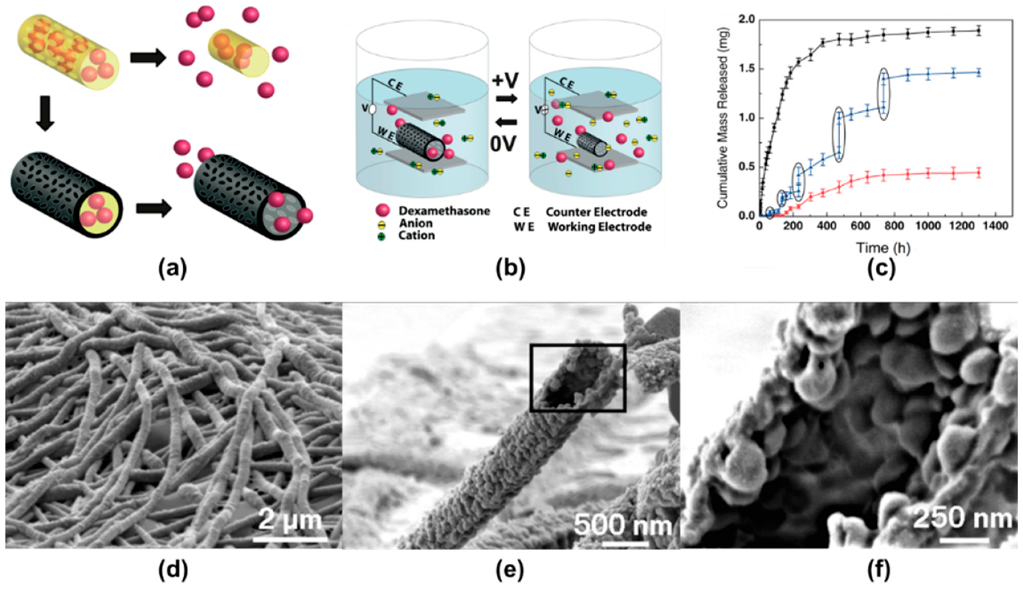
Table 1.
Chemical structures, applications of common aromatic Conducting polymers (CPs): Polypyrrole (PPy), Polyaniline (PANI), Polythiophene (PT), and Poly(3,4-ethyelenedioxythiophene) (PEDOT).
Table 1.
Chemical structures, applications of common aromatic Conducting polymers (CPs): Polypyrrole (PPy), Polyaniline (PANI), Polythiophene (PT), and Poly(3,4-ethyelenedioxythiophene) (PEDOT).
| Polymer | Chemical Structure | Applications | References |
|---|---|---|---|
| Polypyrrole (PPy) |  | Fuel cells, corrosion protection, computer displays, microsurgical tools, biosensors | [33,46,47,48,49] |
| Polyaniline (PANI) |  | Biosensors, neural probes, drug delivery, tissue engineering | [38,50] |
| Polythiophene (PT) |  | Biosensors, solar cells, tissue engineering, photosensitizers, supercapacitors | [41,51,52,53] |
| Poly(3,4-ethyelenedioxythiophene) (PEDOT) |  | Neural electrodes, nerve grafts, heart muscle patches | [54] |
© 2019 by the authors. Licensee MDPI, Basel, Switzerland. This article is an open access article distributed under the terms and conditions of the Creative Commons Attribution (CC BY) license (http://creativecommons.org/licenses/by/4.0/).
Share and Cite
MDPI and ACS Style
Park, Y.; Jung, J.; Chang, M. Research Progress on Conducting Polymer-Based Biomedical Applications. Appl. Sci. 2019, 9, 1070. https://0-doi-org.brum.beds.ac.uk/10.3390/app9061070
AMA Style
Park Y, Jung J, Chang M. Research Progress on Conducting Polymer-Based Biomedical Applications. Applied Sciences. 2019; 9(6):1070. https://0-doi-org.brum.beds.ac.uk/10.3390/app9061070
Chicago/Turabian StylePark, Yohan, Jaehan Jung, and Mincheol Chang. 2019. "Research Progress on Conducting Polymer-Based Biomedical Applications" Applied Sciences 9, no. 6: 1070. https://0-doi-org.brum.beds.ac.uk/10.3390/app9061070
Note that from the first issue of 2016, this journal uses article numbers instead of page numbers. See further details here.