Article

Phytomedicine-Based Potent Antioxidant, Fisetin Protects CNS-Insult LPS-Induced Oxidative Stress-Mediated Neurodegeneration and Memory Impairment

Division of Applied Life Science (BK 21), College of Natural Sciences, Gyeongsang National University, Jinju 52828, Korea
*
Author to whom correspondence should be addressed.
These authors contributed equally to this work.
Submission received: 16 May 2019 / Revised: 5 June 2019 / Accepted: 11 June 2019 / Published: 14 June 2019

Abstract

:
Phytomedicine based natural flavonoids have potent antioxidant, anti-inflammatory, and neuroprotective activities against neurodegenerative diseases. The aim of the present study is to investigate the potent neuroprotective and antioxidant potential effects of fisetin (natural flavonoid) against central nervous system (CNS)-insult, lipopolysaccharide (LPS)-induced reactive oxygen species (ROS), neuroinflammation, neurodegeneration, and synaptic/memory deficits in adult mice. The mice were injected intraperitoneally (i.p.) with LPS (250 μg/kg/day for 1 week) and a fisetin dosage regimen (20 mg/kg/day i.p. for 2 weeks, 1 week pre-treated to LPS and 1 week co-treated with LPS). Behavioral tests, and biochemical and immunofluorescence assays were applied. Our results revealed that fisetin markedly abrogated the LPS-induced elevated ROS/oxidative stress and activated phosphorylated c-JUN N-terminal Kinase (p-JNK) in the adult mouse hippocampus. Fisetin significantly alleviated LPS-induced activated gliosis. Moreover, fisetin treatment inhibited LPS-induced activation of the inflammatory Toll-like Receptors (TLR4)/cluster of differentiation 14 (CD14)/phospho-nuclear factor kappa (NF-κB) signaling and attenuated other inflammatory mediators (tumor necrosis factor-α (TNF-α), interleukin-1 β (IL1-β), and cyclooxygenase (COX-2). Furthermore, immunoblotting and immunohistochemical results revealed that fisetin significantly reversed LPS-induced apoptotic neurodegeneration. Fisetin improved the hippocampal-dependent synaptic and memory functions in LPS-treated adult mice. In summary, our results strongly recommend that fisetin, a natural potent antioxidant, and neuroprotective phytomedicine, represents a promising, valuable, and therapeutic candidate for the prevention and treatment of neurodegenerative diseases.

1. Introduction

Neuroinflammation is considered to be a key event in the neurodegeneration process inherent to various aging pathologies. Within the central nervous system (CNS), microglia are considered a key player in the immune system that trigger various neuroinflammatory responses [1]. Literature reviews suggest that microglial activation plays a pivotal role in various stress conditions including oxidation, neuroinflammation, and neurodegenerative diseases [2,3]. Under varying environmental conditions such as infections and the introduction of toxins and endogenous proteins, microglia become over-activated and release reactive oxygen species (ROS) that cause neurotoxicity and initiate an immune response via toll-like receptors (TLRs), whose activation communicates downstream inflammatory mediators [4,5]. The elevated levels of oxidative stress resulting from an increased level of ROS production, a decrease in the antioxidant system, or both, play a critical role in the aging process and in the development of degenerative diseases [6]. The inflammatory agent lipopolysaccharide (LPS) is an endotoxin derived from the outer membrane of Gram-negative bacteria and is a strong activator of host defense responses. Recent studies suggested that the systemic administration of LPS causes inflammatory processes in the body, which causes deleterious effects in the brain [7,8,9,10,11,12,13]. LPS administration induces memory impairments and neuroinflammation via activated TLR4/NFκB signaling and regulates various downstream proinflammatory mediators, such as nitric oxide (NO), prostaglandin E2 (PGE2), and cyclooxygenase (COX)2, and proinflammatory cytokines, including interleukin-1 (IL-1), IL-6, and tumor necrosis factor-α (TNF-α) [14]. Recent studies have suggested that among the mammalian TLRs families, TLR4 plays an important role in inflammation pathogenesis. TLR4 acts as a primary initiator of innate immune responses to pathogens by activating a cascade of pro-inflammatory events. The LPS administration triggers the activation of TLR4 via downstream signaling factors, such as adaptor myeloid differentiation protein 88 (MyD88), leading to the activation of nuclear factor-κB (NF-κB) and ultimately inducing the expression of inflammation-related genes. The systematic LPS administration impaired learning and memory performance, increased amyloid beta (Aβ)-burden, and diminished synaptic and memory functions [13,15,16].
The epidemiological comprehensive surveys reported that over the past few years, it has become a primary focus of researchers to search for potential effective therapeutic agents from natural sources to combat inflammatory and neurodegenerative diseases. Among the natural sources, phytomedicine derived flavonoids are a primary focus because of their potent biological and therapeutic activities which are beneficial for health improvements [17,18,19,20]. Flavonoids are polyphenolic compounds abundantly found in foods and vegetables and widely used as nutritional supplements for the treatment of diabetes, obesity, and cardiovascular and neurological disorders [17,21,22,23,24]. Fisetin (3, 7, 3,4-tetrahydroxyflavone) (Figure 1), is a well-known and potent flavonoid, which is commonly found in many fruits and vegetables such as apples, grapes, kiwis, strawberries, onions, persimmons, and cucumbers. Several studies have suggested that fisetin is a biochemically and physiologically active flavonoid based phytomedicine, showing various strong pharmacological activities against cancer, oxidative stress, inflammatory bowel disease, and cognitive/ynaptic dysfunctions [25,26,27,28,29,30]. Similarly, we also recently reported the neuroprotective effect of fisetin against Aβ-induced neurotoxicity and cognitive/synaptic dysfunctions [31]. Therefore, the present study was designed to investigate the potential antioxidant and neuroprotective effects of phytomedicine-based small flavonoid molecules such as fisetin against the LPS-induced neurotoxicity in the adult brain with particular and major focus to the hippocampus.

2. Materials and Methods

2.1. Chemicals

Fisetin, LPS, and dimethyl sulfoxide (DMSO) were purchased from Sigma-Aldrich Chemical Co. (St. Louis, MO, USA).

2.2. Animals Used in the Experiment

Animals used for the experimental purpose were wild-type C57BL/6N mice (28–32 g, 10 weeks old) purchased from Samtako Bio (Osan, S. Korea). The mice were kept for 1 week in the university animal housing for acclimatization under a 12-hr/12-hr light/dark cycle at a controlled temperature (23 °C) with 60 ± 10% humidity and were provided with food and water ad libitum. For animal care and treatment, we have followed the guidelines (Approval ID: 125) issued by the ethics committee (IACUC), Division of Applied Life Sciences, Gyeongsang National University, South Korea. Efforts were made to minimize the suffering of the animals.

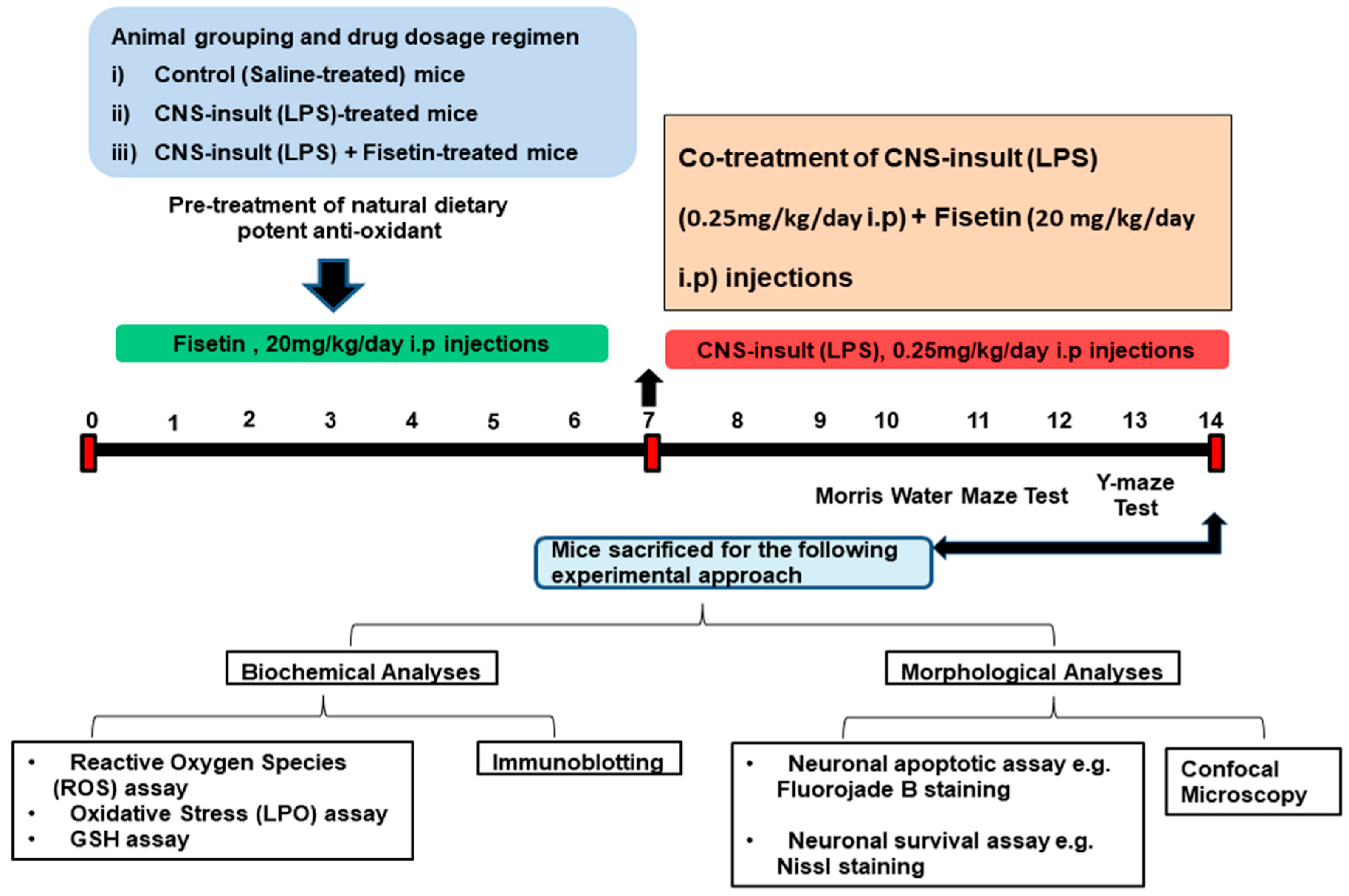
2.3. Study Designing, Animals Grouping, and Experimental Approach

In order to accomplish our hypothesis, we designed the following studies (Figure 2) as per our previous reports [9,12,31], and designed fundamental studies particularly for the optimization of the fisetin dosage regimen. To evade ambiguous sex-dependent differences, we involved only male mice in the study. Adult male mice (13 mice per group) were randomly divided into the following 3 groups: (i) mice treated with saline as a vehicle for 14 days (control (C)); (ii) mice treated with LPS injected at a dose of 250 µg/kg for 7 days; and (iii) mice treated with LPS (250 µg/kg) for 7 days and fisetin (20 mg/kg) for 14 days (7 days prior to LPS and 7 days with LPS; LPS+Fis). Fisetin was first dissolved in 0.1% (v/v) DMSO and then was diluted with saline such that the total administered volume was in 0.9% saline. The LPS, fisetin, and saline were administered intraperitoneally (i.p.) to the mice.

2.4. Behavioral Studies

To know the beneficial effect of phytomedicine-based fisetin on behavioral performance, we designed a behavioral study (13 mice/group) through a Morris water maze (MWM) and a Y-maze test.
The MWM test is a parameter task to evaluate memory functions. The experimental apparatus consisted of a circular water tank (100 cm in diameter, 40 cm in height) containing water (23 ± 1 °C) to a depth of 15.5 cm, which was rendered opaque by adding white paint. A transparent escape platform (10 cm in diameter, 20 cm in height) was hidden 1 cm below the water surface and placed at the midpoint of one quadrant. The MWM test was started on day 7 and completed on day 14 of the experimental schedule (Figure 2). Each mouse received 3–4 training periods each day for 4 consecutive days using a single hidden platform in one quadrant with three rotating starting quadrants. Latency to escape from the water maze (finding the submerged escape platform) was calculated for each trial. The probe test was performed by removing the platform and allowing each mouse to swim freely for 60 s. The number of crossing and time spent in the target quadrant (where the platform was located during hidden platform training) was measured. The time spent in the target quadrant was considered to represent the degree of memory consolidation that has taken place after learning. All data were recorded using video-tracking software (SMART, Panlab Harvard Apparatus Bioscience Company, Holliston, MA, USA).
The Y-maze was constructed from painted black wood. Each arm of the maze was 50 cm long, 20 cm high, and 10 cm wide at the bottom and top. The Y-maze was initiated on day 12 and completed on day 14 of the experimental schedule (Figure 2). Each mouse was placed at the center of the apparatus and allowed to move freely through the maze for three 8-min sessions. The series of arm entries was visually observed. The spontaneous alternation was defined as the successive entry of the mice into the three arms in overlapping triplet sets. Alteration behavior (%) was calculated as follows: (successive triplet sets (entries into three different arms consecutively)/total number of arm entries-2) × 100.

2.5. Protein Extraction from Mice Brains for Biochemical Analyses

At the completion of behavioral testing, mice brains were immediately removed, and hippocampal tissue was carefully separated, frozen on dry ice, and stored at −80 °C until processing. Further, the hippocampal tissue was homogenized in 0.2 M Phosphate Buffer saline (PBS) with a phosphatase inhibitor and protease inhibitor cocktail and then centrifuged at 10,000× g at 4 °C for 25 min. The supernatants were collected and stored at −80 °C until processing for biochemical analyses.

2.6. Western Blot Analysis

The protein concentrations were measured through a BioRad protein assay kit (BioRad Laboratories, CA, USA). Equal amounts of protein (20–30 µg) underwent electrophoresis using 4%–12% BoltTM Mini Gels (Novex, Life Technologies, Kiryat Shmona, Israel). The membranes were blocked in 5% (w/v) skim milk to reduce non-specific binding and incubated with primary antibodies (Table 1) overnight at 4 °C at a 1:1000 dilution. After undergoing a reaction with a horseradish peroxidase-conjugated secondary antibody, as appropriate, the proteins were detected using an Electrochemiluminescence (ECL) detection reagent according to the manufacturer’s instructions (Amersham Pharmacia Biotech, Uppsala, Sweden). Then, X-ray films were scanned, and the optical densities of the bands were analyzed through densitometry using the computer-based Sigma Gel program, version 1.0 (SPSS, Chicago, IL, USA).

2.7. Antibodies Used in the Western Blotting

The primary antibodies used in the Western blot analysis are explained in Table 1. The secondary antibodies used in our experiments, goat anti-mouse IgG, goat anti-rabbit IgG, and rabbit anti-goat IgG were purchased from Santa Cruz Biotechnology.

2.8. Tissue Sample Preparation for Morphological Analysis

At the completion of behavioral testing, mice were perfused transcardially with 4% ice-cold paraformaldehyde, and brains were post-fixed for 72 h in 4% paraformaldehyde and transferred to 20% sucrose for 72 h. Then, brains were frozen in O.C.T. compound (A.O, USA), and 14-µm coronal sections were cut using a CM 3050C cryostat (Leica, Wetzlar, Germany). The sections were thaw-mounted on Probe-on Plus charged slides (Fisher, Rock-ford, IL, USA).

2.9. Immunofluorescence Staining

The morphological evaluations were performed as previously described with some modifications [13,21]. The prepared tissue slides were washed twice for 10 min in 0.01 M PBS, followed by incubation for 1 h in a blocking solution containing 2% normal serum according to the antibody treatment and 0.3% Triton X-100 in PBS. After blocking, the slides were incubated overnight at 4 °C in the primary antibodies (mouse polyclonal phosphorylated c-JUN N-terminal Kinase (p-JNK), rabbit polyclonal anti-p-NFκB, mouse polyclonal TNF-α, rabbit polyclonal anti- ionized calcium-binding adaptor molecule 1 (Iba-1), mouse monoclonal post synaptic density-95 (PSD-95) from Santa Cruz Biotechnology and mouse monoclonal 8-Oxoguanine (8-OxoG) from Millipore) and diluted 1:100 in blocking solution. After incubation with primary antibodies, the sections were incubated for 2 h in the secondary tetramethylrhodamine (TRITC)/fluorescein isothiocyanate (FITC)-labeled antibodies (1:50) (Santa Cruz Biotechnology, Dallas, Texas, USA). After secondary antibody incubation, tissue slides were washed twice for 5 min. Slides were mounted with 4′, 6′-diamidino-2-phenylindole (DAPI) and Prolong Antifade Reagent (Molecular Probe, Eugene, OR, USA). Then, slides were examined using a confocal laser-scanning microscope (Flouview FV 1000, Olympus, Tokyo, Japan).

2.10. Fluoro-Jade B (FJB) Staining

FJB staining was performed as previously described [13] with some modifications. After air-drying the tissue slides overnight, the slides were immersed in a solution of 1% sodium hydroxide and 80% ethanol for 5 min. Then, slides were immersed in 70% alcohol and distilled water for 2 min each. Tissue slides were transferred to a solution of 0.06% potassium permanganate for 10 min, rinsed with distilled water and then immersed in a solution of 0.1% acetic acid and 0.01% FJB for 20 min. The slides were washed with distilled water and allowed to dry for 10 min. Glass coverslips were mounted using Dibutylphthalate Polystyrene Xylene (DPX) non-fluorescent mounting medium, and images were assessed with a confocal laser scanning microscope (Flouview FV 1000 MPE, Olympus, Tokyo, Japan). The images were proceeded for quantification using the computer-based ImageJ program.

2.11. Cresyl Violet (Nissl) Staining

Cresyl violet (Nissl) staining was performed to evaluate the histological examination and extent of neuronal cell death or neuronal survival. Slides containing 14-µm sections of tissue were washed twice for 15 min in 0.01 M PBS and stained with a 0.5% cresyl violet solution (containing a few drops of glacial acetic acid) for 10–15 min. Then, sections were washed with distilled water and dehydrated in a graded ethanol series (70%, 95%, and 100%), placed in xylene and coverslipped using mounting medium, and finally, slides were examined with fluorescent light microscopy. The results were analyzed using the computer-based ImageJ program.

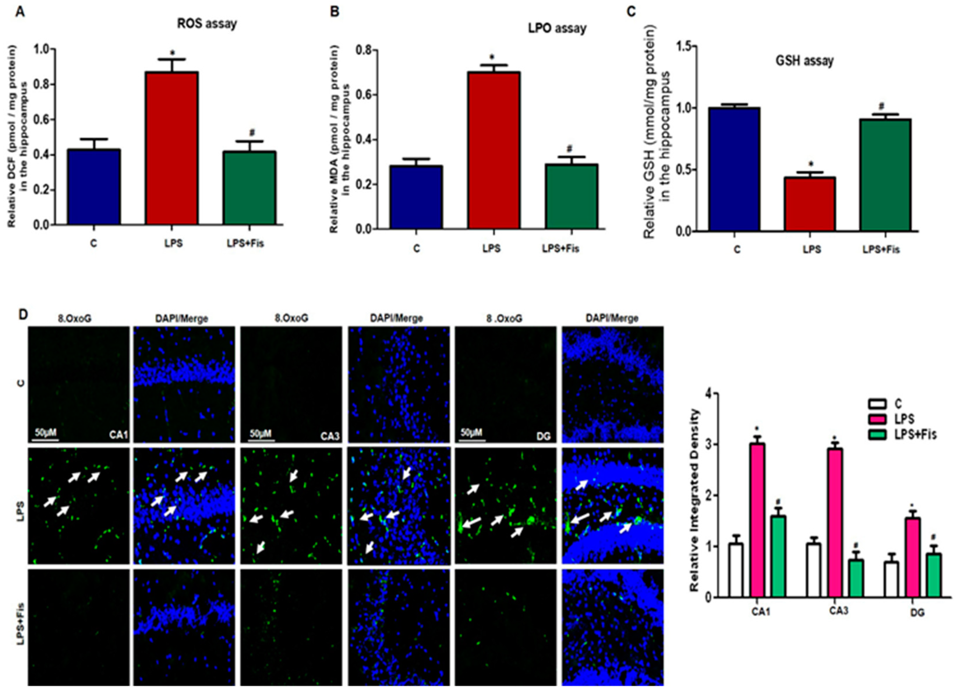
2.12. ROS Assay in Mouse Hippocampus Homogenates

The ROS assay was based on the oxidation of 2′ 7′-dichlorodihydrofluorescein diacetate (DCFH-DA) to 2′ 7′-dichlorofluorescein (DCF). Subsequently, brain homogenates were diluted with ice-cold Lock’s buffer at a 1:20 ratio to make a final concentration of 2.5 mg tissue/500 mL. The reaction mixture of Lock’s buffer (1 mL, pH = 7.4), 0.2 mL homogenate, and 10 mL of the stock solution of DCFH-DA (5 mM) was incubated at room temperature for 15 min to convert DCFH-DA to the fluorescent product DCF. The conversion of DCFH-DA to DCF was performed using a spectrofluorimeter with excitation at 484 nm and emission at 530 nm. For background fluorescence assessment (conversion of DCFH-DA in the absence of homogenate), we ran parallel blanks. The quantification analysis of ROS is expressed as pmol DCF formed/mg protein.

2.13. Lipid Peroxidation (LPO) Analysis in Mouse Hippocampus Homogenates

The LPO levels were determined in the hippocampus (n = 8 mice per group) homogenates through analyzing the malondialdehyde (MDA) level, a biomarker of LPO, by using the commercial lipid peroxidation kit (catalog # K739-100) from Biovision Incorporated, A 95035 USA. The assay was performed according to the provided protocol.

2.14. Glutathione (GSH) Analysis in Mouse Hippocampus Homogenates

The GSH levels in the hippocampus (n = 8 mice per group) homogenates were assessed by using the commercially available glutathione assay kit (BioVision’s catalog #K264-100) according to the provided protocol.

2.15. Statistical Analysis

Western blot bands were scanned and analyzed through densitometry using the Sigma Gel System (SPSS Inc., Chicago, IL). Density values are expressed as the mean ± standard error of the mean (SEM). ImageJ software was used for immunohistological quantitative analysis. The data are the mean ± SEM. Statistical analysis was performed through one-way ANOVA followed by post-hoc analysis. Statistical calculations and graphs were made through Prism 5 software (Graph-Pad Prism 5 Software, San Diego, USA). P-values less than 0.05 were considered to be statistically significant. * p < 0.05 control versus LPS, # p < 0.05, LPS versus LPS + fisetin.

3. Results

3.1. Effect of Fisetin Dosage Regimen on LPS-Induced Oxidative Stress in the Mouse Brain

A recently reported study showed that LPS exposure mediates ROS accumulation, which in turn enhances the expression level of pro-inflammatory mediators in addition to playing the main role in various neurological disorders [29]. Herein, consistently, we also observed that systemic LPS administration enhanced the production and accumulation of ROS and oxidative stress (p < 0.05). Further, we found that fisetin treatments significantly (p < 0.05) attenuated the upregulated levels of ROS and LPO compared to the LPS-only treated group (Figure 3A,B). We next performed the GSH assay to study alterations in oxidative stress levels. We observed reduced levels of GSH (p < 0.05) in the LPS-injected mice brain than that of the control saline-injected mice. Fisetin treatment to the LPS-treated group escalated the GSH expression level as compared to the LPS-only treated group (p < 0.05) (Figure 3C). We also performed immunofluorescence analysis to evaluate the expression of 8-OxoG, a predominant parameter of oxidative stress and potentially expressed in the degenerated brain [27]. Interestingly, our immunofluorescence results indicated that LPS significantly enhances the immunofluorescence reactivity of 8-OxoG in the Cornu Ammonis 1 (CA1) (molecular layer and pyramidal cells) and Cornu Ammonis 3 (CA3) (molecular layer and pyramidal cells) regions as well as in the Dentate gyrus (DG) (hilum and granular cells) region of the hippocampus (p < 0.05). Fisetin dosage significantly reversed the increased expression levels of 8-OxoG in the fisetin-treated group relative to LPS-only treated group (p < 0.05) (Figure 3D). These results suggest that the natural-based phytomedicine flavonoid, fisetin, has the potential effect to mitigate the systemic LPS-induced accumulated ROS and LPO in the hippocampus of adult mice brains.

3.2. Effect of Fisetin Dosage on LPS-Induced Activation of p-JNK Expressions in the Mouse Hippocampus

Various studies have demonstrated that LPS treatment exaggerates the expression of the stress associated kinases. Among them, p-JNK is the main marker which promotes neuroinflammation and neurodegeneration [9,31]. Recently, a well-known study demonstrated that LPS induced the activation of p-JNK, which led to neuroinflammation and neuronal cell death [31]. In our study, we also investigated the LPS-induced activation of p-JNK expression through Western blot analysis. Our results revealed that fisetin treatments significantly reversed the upregulated expression level of p-JNK proteins in the LPS-treated group (p < 0.05) relative to the LPS-only treated group (p < 0.05) (Figure 4A). To confirm the Western blot results, we performed immunofluorescence staining of p-JNK. Our immunofluorescence results confirmed that LPS-treatment increased levels of p-JNK (p < 0.05) in the DG (hilum and granular cells) and CA3 (molecular layer and pyramidal cells) regions of the hippocampus compared to saline-treated controls (Figure 4B), while fisetin supplementation for two weeks significantly (p < 0.05) inhibited the higher expression and immunofluorescence reactivity of p-JNK in the DG (hilum and granular cells) and CA3 (molecular layer and pyramidal cells) of the LPS-treated group compared to the LPS-only treated group (Figure 4B).

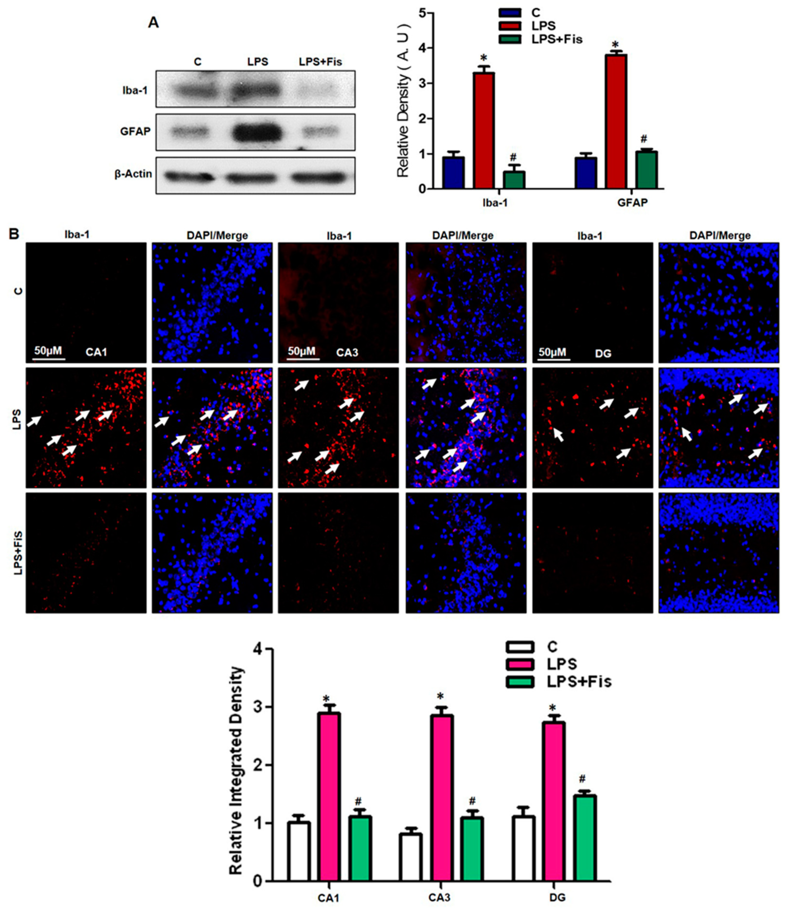
3.3. Effect of Fisetin on LPS-Induced Activation of Microglia and Astrocytes in the Adult Mouse Hippocampus

Prominent in vitro and in vivo studies have demonstrated that LPS exposure induces glial activation, neuroinflammation, and neurodegeneration. Activated gliosis and neuroinflammation are crucial factors involved in the pathogenesis of several neurodegenerative diseases such as Alzheimer’s disease (AD) and Parkinson’s disease (PD). The LPS-injection significantly induces astrocyte activation in an inflammatory rodent model [7,12,13,32]. Therefore, in our study, we also investigated the LPS-induced glial activation in the adult mouse hippocampus. Our Western blot results showed that LPS injection significantly induced the activation of both astrocytes and microglia in the LPS-treated group compared to the saline-treated control group (p < 0.05) (Figure 5A). Interestingly, fisetin dosage (20 mg/kg/day for 2 weeks, 1 week prior LPS and 1 week co-treated with LPS) suppressed the activation of both Iba-1 and GFAP protein expression levels compared to the LPS-only treated group (p < 0.05). Further, we also performed immunofluorescence analysis for Iba-1 reactivity in the adult mouse hippocampus. Our morphological results demonstrated that LPS treatment significantly increases the immunofluorescence reactivity of activated Iba-1 in the CA1 (molecular layer and pyramidal cells) and CA3 (molecular layer and pyramidal cells) regions as well as in the DG (hilum and granular cells) regions of the hippocampus (p < 0.05). Fisetin treatment significantly reduced the amount of activated Iba-1 in LPS-treated mice compared to mice treated with LPS alone (p < 0.05) (Figure 5B). Therefore, these results suggest that fisetin acts as a potent antioxidant and might possess potent anti-inflammatory activity through the suppression of activated astrocytes and microglia in the adult mouse hippocampus.

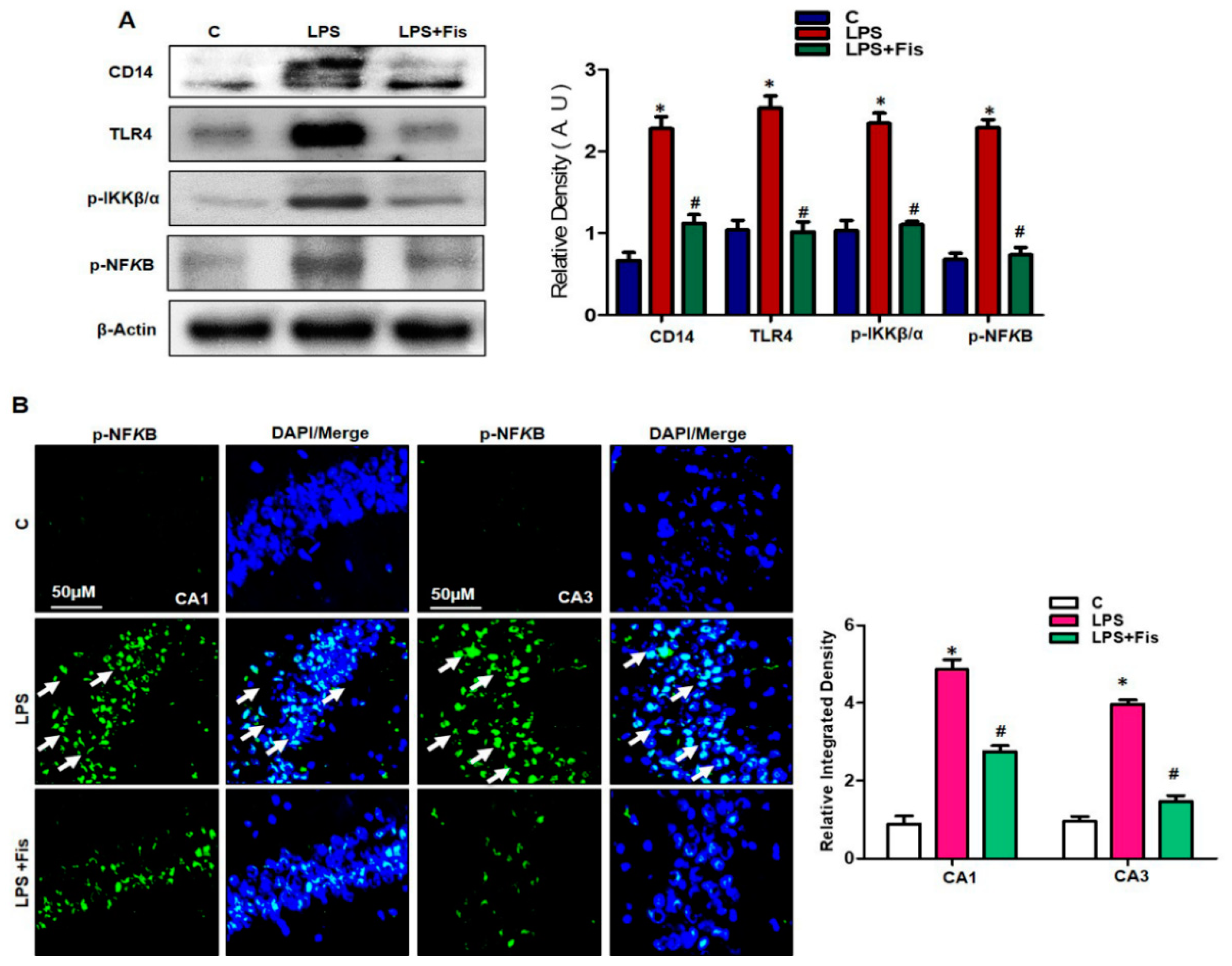
3.4. Effect of Fisetin on LPS-Induced Activation of TLR4/NFκB Signaling and Inflammatory Mediators in the Adult Mouse Hippocampus

It has recently been reported that peripheral systemic LPS administration significantly mediates the activation of the TLR4/NFκB pathway in LPS-induced neuroinflammation and developed AD-like pathologies [9]. Therefore, in our study, we also investigated the anti-inflammatory activity of fisetin against LPS-induced activation of inflammatory signaling (TLR4/p-NFκB). Interestingly, our results showed that LPS treatment for seven days increased the TLR4, CD14, inhibitor of nuclear factor kappa-B kinase subunit beta (IKKB-β), and p-NFκB protein expression levels in the LPS-treated group (p < 0.05), while fisetin (20 mg/kg) treatment, a natural polyphenol, could significantly reverse their higher expression levels in the LPS-treated group compared to the LPS-only treated group (p < 0.05) (Figure 6A). Recently, Badshah et al. 2015 reported that LPS administration significantly activated NFκB signaling, ultimately enhancing neuroinflammation [10]. Therefore, we further performed an immunofluorescence assay to determine the immunoreactivity of p-NFκB protein in the hippocampal regions of adult mice. The immunofluorescence results showed increased reactivity of p-NFκB in LPS-treated mice compared to the control group (p < 0.05), while fisetin (20 mg/kg) treatments for 2 weeks (1 week prior to LPS and 1 week co-treated with LPS) significantly reversed the LPS-induced enhancements of immunofluorescence reactivity in the CA1 (molecular layer and pyramidal cells) and CA3 (molecular layer and pyramidal cells) regions of the hippocampus compared to the LPS-only treated group (p < 0.05) (Figure 6B). These results suggest that fisetin has the ability to suppress the LPS-induced activation of inflammatory signaling cascades and shows effective anti-inflammatory activity in the adult mouse brain.

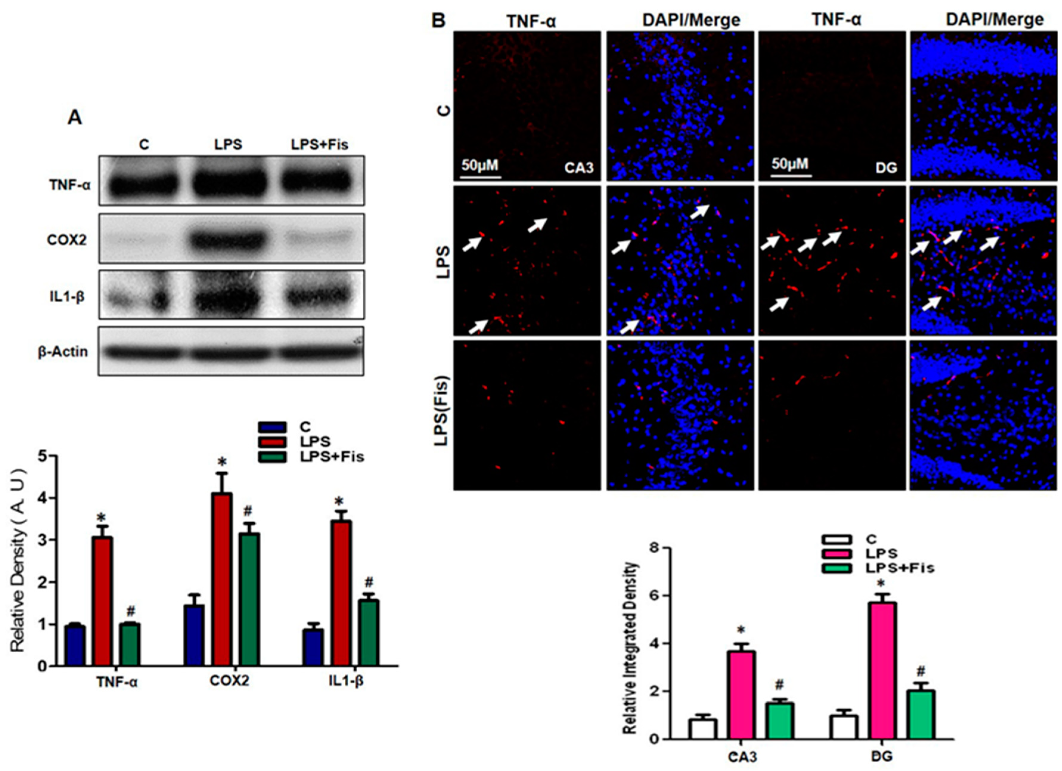
3.5. Effect of Fisetin on the LPS-Induced Upregulation of Inflammatory Mediators in the Adult Mouse Hippocampus

Several studies suggest that the activation of gliosis, the elevation in pro-inflammatory cytokines and chemokines and the generation of ROS are responsible for immune response dysregulation, which contributes to neurodegeneration, neuroinflammation, and AD pathogenesis [7,8,9,10,11,12,13]. Therefore, in this study, we also investigated the LPS-induced activation of inflammatory mediators in the hippocampal region of adult mice brains. The Western blot results showed that there was a significant increase in the protein expression levels of the inflammatory markers TNF-α (p < 0.05), COX2 (p < 0.05), and IL1-β (p < 0.05) in LPS-treated group compared to the saline-treated control group. Interestingly, fisetin (20 mg/kg, i.p. for 2 weeks; 1 week prior to LPS and 1 week co-treated with LPS) supplementation reversed the elevated expression levels in the LPS-treated group compared to LPS-only treated group (p < 0.05) (Figure 7A). Further, to verify the anti-inflammatory effect of fisetin, we performed an immunofluorescence assay of the key cytokine TNF-α. Our morphological analyses of immunofluorescence results demonstrated that fisetin supplementation significantly attenuated the increased immunoreactivity of TNF-α protein in the CA3 (molecular layer and pyramidal cells) and DG (hilum and granular cells) regions of hippocampus in the LPS-treated group (p < 0.05) relative to the LPS-only treated group (p < 0.05) (Figure 7B). These findings suggest that fisetin treatment (20 mg/kg, i.p. for 2 weeks; 1 week prior to LPS and 1 week co-treated with LPS) significantly suppressed the LPS-induced activation of inflammatory mediators in the adult mouse hippocampus.

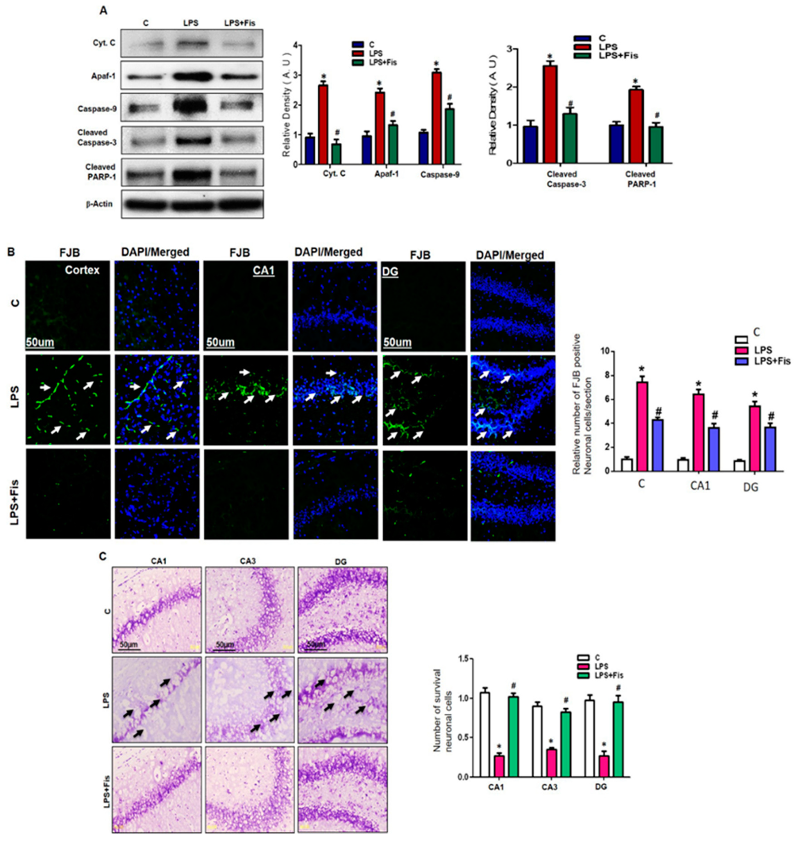
3.6. Effect of Fisetin on LPS-Induced Apoptotic Neurodegeneration in the Adult Mouse Brain

To investigate the neuroprotective property of fisetin against LPS-mediated apoptotic neurodegeneration in the hippocampus of the adult mouse, we examined several apoptotic markers. Recently, mounting studies have shown that LPS administration significantly activates apoptotic neurodegeneration in adult mice. Badshah et al. recently showed that LPS-induced activation of neuroinflammatory and mitochondrial apoptotic pathways through the upregulation of several apoptotic markers such as Bax, cytochrome C, caspase-9, and caspase-3 in an adult mouse model [7,8,9,10,11,12]. Therefore, we examined the protein expression levels of cytochrome C, Apoptotic protease activating factor 1 (Apaf-1), caspase-9, caspase-3, and Poly (ADP-ribose) polymerase-1 (PARP-1) through Western blot analysis in the hippocampi of adult mice. Our Western blot results demonstrated that LPS administration upregulated the expression levels of cytochrome C (p < 0.05), Apaf-1 (p < 0.05), caspase-9 (p < 0.05), caspase-3 (p < 0.05), and PARP-1 (p < 0.05) in the LPS-treated group compared to the control group (Figure 8A). However, fisetin (20 mg/kg, i.p. for 2 weeks; 1 week prior to LPS and 1 week co-treated with LPS) treatment significantly reversed the enhanced expression levels of cytochrome C (p < 0.05), Apaf-1 (p < 0.05), caspase-9 (p < 0.05), caspase-3 (p < 0.05) and PARP-1 (p < 0.05) in the LPS-treated group relative to the LPS-only treated group (Figure 8A). Furthermore, we investigated the neuroprotective effect of fisetin through morphological analysis using FJB and Nissl staining in the cortex and hippocampus of adult mice. FJB is known to be used to evaluate the extent of neuronal cell death, and, herein, we also used it to investigate the level of apoptotic neurodegeneration in LPS and fisetin-treated mice. Our FJB results demonstrated that LPS treatment significantly increased the number of degenerative neuronal cells in the cortex (p < 0.05), CA1 (molecular layer and pyramidal cells) (p < 0.05), CA3 (molecular layer and pyramidal cells) (p < 0.05), and DG (hilum and granular cells) (p < 0.05) regions of the hippocampus, while fisetin treatment markedly decreased the LPS-induced neurodegeneration, which was evident from the reduced number of FJB-positive cells compared to the LPS-only treated group (p < 0.05) (Figure 8B). Furthermore, Nissl staining was carried out to evaluate the extent of neuronal damage in the LPS-treated and fisetin-treated adult mouse hippocampus and cortex. Our Nissl staining results demonstrated that LPS treatment significantly enhanced the number of damaged, shrunken, and fragmented neurons in the CA1 (molecular layer and pyramidal cells) (p < 0.05), CA3 (molecular layer and pyramidal cells) (p < 0.05), and DG (hilum and granular cells) (p < 0.05) regions of the hippocampus relative to the control group. Hence, fisetin treatment significantly reversed the LPS-induced neuronal damage in the LPS and fisetin-treated group (p < 0.05) and enhanced the viability of neurons relative to the LPS-only treated group (p < 0.05) (Figure 8C). These results suggest that fisetin supplementation significantly inhibited LPS-induced neurodegeneration in the adult mouse brain.

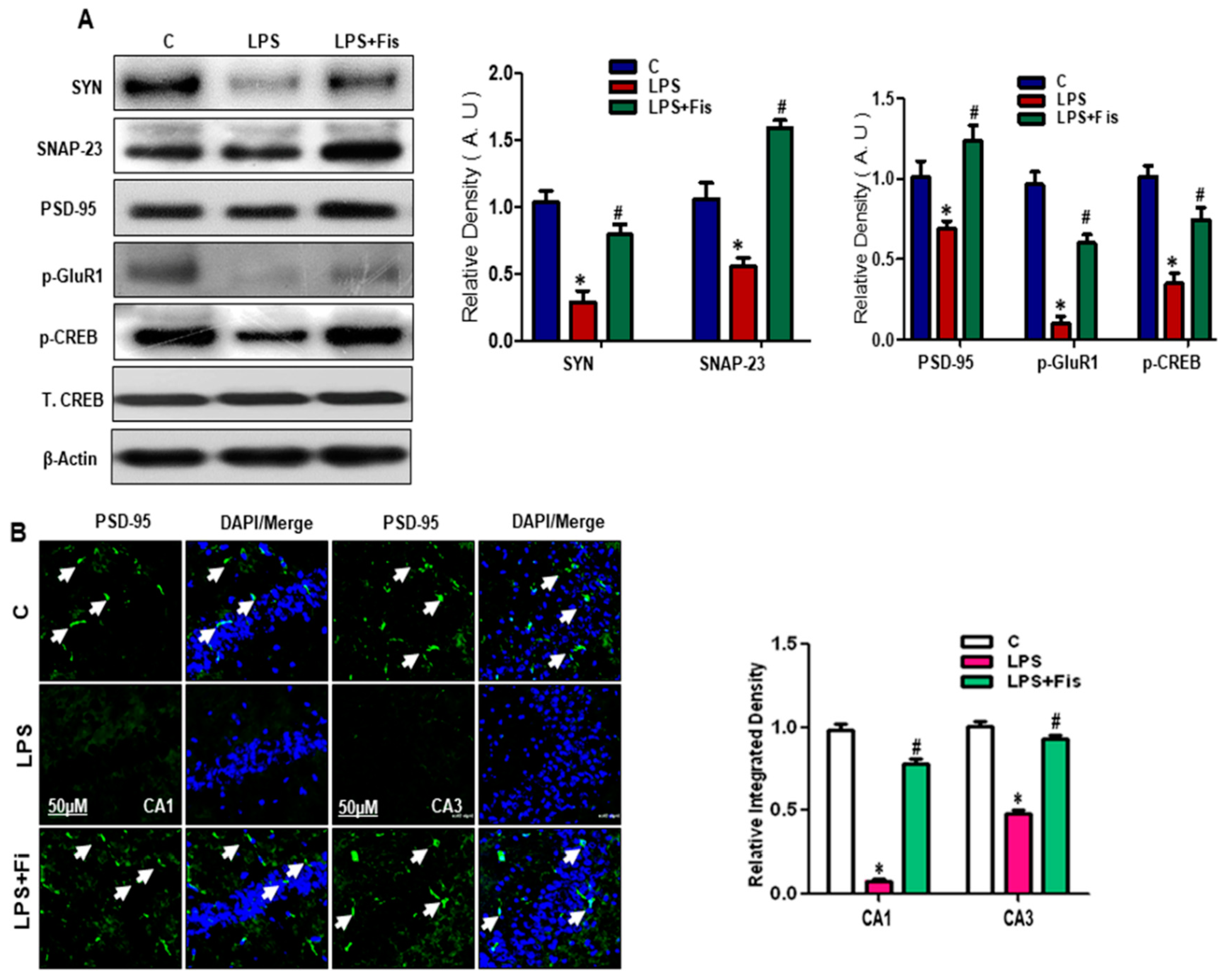
3.7. Effect of Fisetin on LPS-Induced Disruption of Pre- and Post-Synaptic and Memory Function in the Adult Mouse Hippocampus

It has been recently reported that LPS administration resulted in the disruption of synaptic and cognitive functions in mice [13]. Therefore, we also investigated the LPS-induced disruption of pre-synaptic and post-synaptic proteins in the adult mouse hippocampus. Our Western blot results showed that LPS administration significantly reduced the expression level of pre-synaptic synaptosomal-associated protein 23 (SNAP-23) (p < 0.05), SYN (p < 0.05)) and post-synaptic (PSD-95 (p < 0.05), phospho-glutamate receptor (p-GluR1) (p < 0.05), phospho- cAMP response element-binding protein (p-CREB) (p < 0.05)) proteins in the hippocampi of LPS-treated mice compared to the control group (Figure 9A). However, fisetin treatment (20 mg/kg, i.p. for 2 weeks; 1 week prior to LPS and 1 week co-treated with LPS) significantly reversed the LPS effect on the pre-synaptic (SNAP-23, SYN) (p < 0.05) and post-synaptic (PSD-95, p-GluR1, p-CREB) (p < 0.05) protein expression levels compared to LPS-only treated group (Figure 9A). Next, we performed immunofluorescence analysis of post-synaptic protein markers in the hippocampi of adult mice. Our morphological results showed reduce expression of post-synaptic (PSD-95) immunofluorescence reactivity in the LPS-treated group (p < 0.05) compared to the control group (Figure 9B). Interestingly, fisetin treatment significantly enhanced the expression levels of PSD-95 in the CA1 (molecular layer and pyramidal cells) (p < 0.05) and CA3 (molecular layer and pyramidal cells) (p < 0.05) regions of the hippocampus, compared to the LPS-only treated group (Figure 9B). These results suggest that fisetin treatment (20 mg/kg, i.p. for 2 weeks; 1 week prior to LPS and 1 week co-treated with LPS) significantly improved synaptic functions associated with the pre- and post-synaptic proteins in the hippocampi of adult mice.

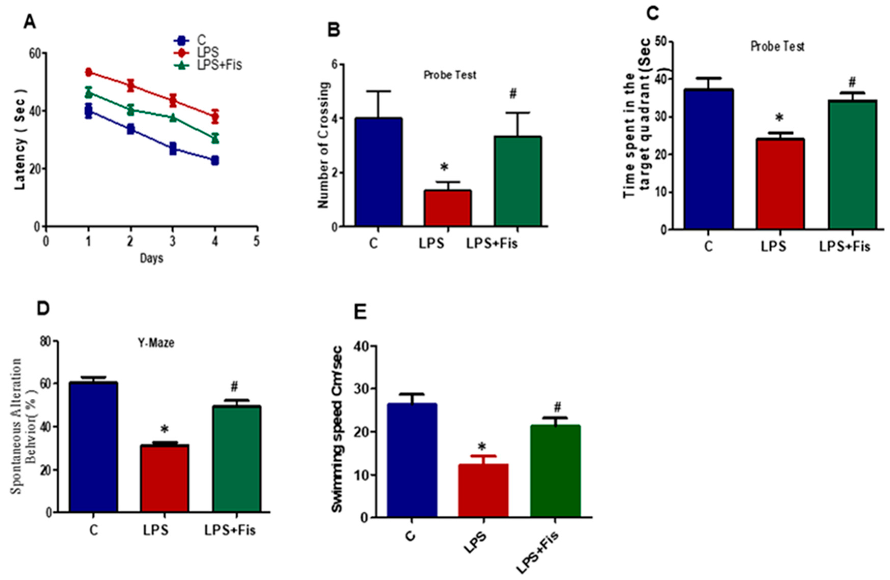
3.8. Effect of Fisetin on CNS-Insult, LPS-Induced Memory Dysfunction
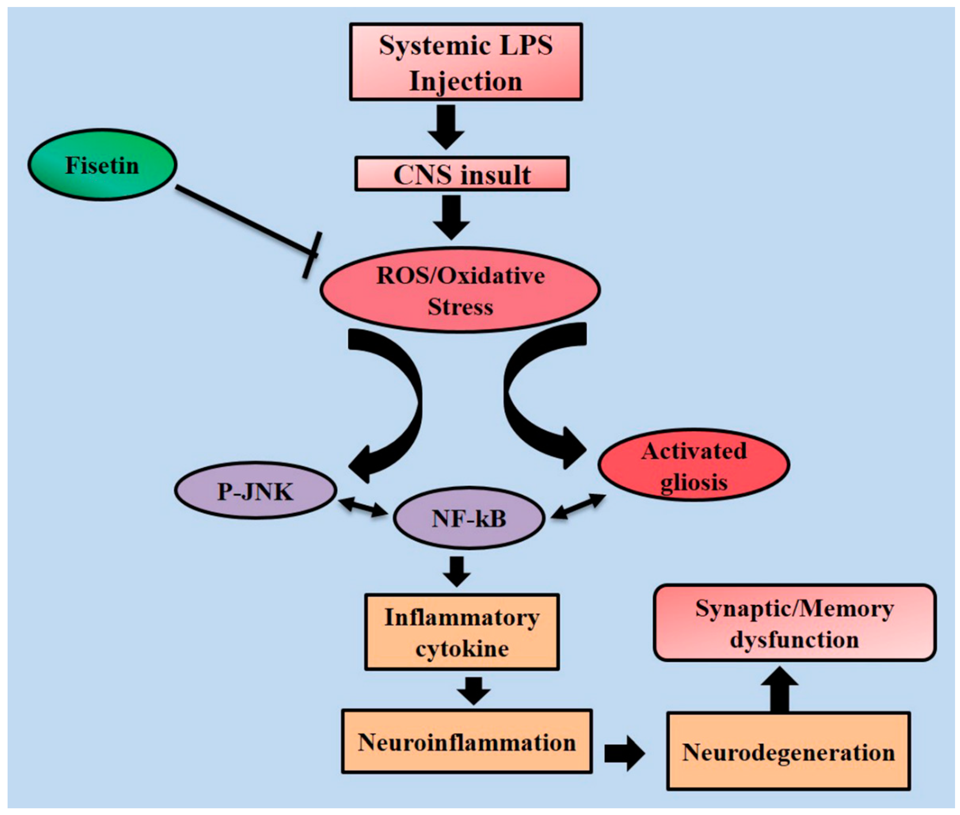
After one week of fisetin dosage (20 mg/kg, i.p.) and the start of LPS on day eight, the behavioral study via MWM test was started. Initially, for four days, we trained all animals in the MWM task where they were required to find a submerged hidden platform and we analyzed the time required to reach the hidden platform. We observed that LPS-injected animals took a long time to find the hidden platform compared to the control mice (p < 0.05). However, the fisetin treatment reversed the LPS effect and significantly improved the performance of the mice, as revealed by the mice taking less time to reach the hidden platform compared to the LPS-injected mice (p < 0.05) (Figure 10A). After a training session and training latency with a one-day interval, we performed a probe test. The probe test results also revealed that fisetin reversed the LPS effect, showing a decrease in the number of mice that crossed over the hidden platform (p < 0.05) and spent less time in the target quadrant (p < 0.05). However, the results revealed a significant increase in the number of platform crossings (p < 0.05) and an increase in the time spent in the target quadrant (p < 0.05) in which the hidden platform was previously located in the training session (Figure 10B,C). Next, we examined the mean swim speeds during training days to observe the motor ability among the saline, LPS alone, and LPS+Fisetin-treated group. We found a significantly (p < 0.05) lower swimming speed in the LPS-treated group compared to the saline-treated group. This represented the motor problem in animals which may add to the latency differences. However, the Fisetin treatment regulated the mean swim speeds in the LPS+Fisetin-treated mice group (p < 0.05) (Figure 10E).
Next, in order to analyze the short term spatial working memory, we designed and evaluated spontaneous alternation behavior percentage (%) through the Y-maze task. On day 12, we trained the mice until day 13. Following training, we observed the final performance of the mice on day 14. Our results revealed that LPS-injected mice showed a less spontaneous alternation behavior percentage (%) than that of the control saline-treated (p < 0.05) mice. Fisetin treatment improved and restored the special memory (increased the spontaneous alternation behavior percentage (%)), of the LPS-injected mice (p < 0.05) (Figure 10D). All of these behavioral results showed that fisetin treatment to LPS-injected mice reversed the LPS effect and significantly improved memory (Figure 10A–D).

4. Discussion

This study was designed to investigate the therapeutic efficacy of the natural based flavonoid fisetin against LPS-induced oxidative stress, neuroinflammation, synaptic loss, and neurodegeneration in the adult mouse brain. We have shown in our study that fisetin treatment for two weeks markedly inhibited the LPS-induced increased levels of in vivo ROS and LPO, and activation of an inflammatory cascade through the suppression of activated gliosis and TLR4/CD14/NFκB signaling pathways and apoptotic neurodegeneration in the hippocampus of adult mice. Recently reported literature supports the notion that LPS induces oxidative stress, neuroinflammation, neurodegeneration, and synaptic dysfunction [7,13]. It has been evident from well-reported studies that bacterial LPS exacerbates chronic inflammation, beta-amyloid accumulation, memory defects, and neurodegeneration leading to early stages of AD [33,34]. Several in vitro and in vivo studies have demonstrated that the systemic administration of LPS causes the activation of ROS and activated glial cells (astrocytes and microglial cells), the elevation of cytokines including TNF-α, intracellular adhesion molecule-1, and IL-6, as well as the increase of inflammatory proteins, such as inducible nitric oxide synthase and cyclooxygenase-2, which are factors known to be responsible for neuroinflammatory disorders and lead to memory impairments [35,36].
It has been recently demonstrated that elevated levels of thiobarbituric acid-reactive substance (TBARS) as well as oxidative DNA damage, measured by 8-OxoG, and reduced levels of the protein responsible for removing damaged DNA, 8-OxoG DNA glycosylase 1 (OGG1), were detected in the brains of patients with AD [7,37]. LPS, a major constituent of Gram-negative bacteria, can trigger a variety of inflammatory reactions, including the release of proinflammatory cytokines, which in turn causes the release of ROS from mitochondria as well as NO and other cell mediators from monocytes and macrophages. The increased level of ROS is responsible for mediating various pathological events such as the peroxidation of lipids, DNA, and various proteins [38]. Similarly, our current study demonstrated that fisetin dosage (20 mg/kg, i.p. for 2 weeks; 1 week prior to LPS and 1 week co-treated with LPS) markedly overcomes the LPS-induced upregulation of ROS, lipid peroxidation, and 8-OxoG, a key biomarker of oxidative stress detected in the brains of AD patients [13]. Well-reported in vitro and in vivo evidences suggest that fisetin treatments show potent antioxidant activity and attenuate Aβ1-42-induced cognitive and synaptic dysfunction, neuroinflammation, and neurodegeneration [28,31]. Increased production of inflammatory mediators can induce severe neurodegenerative diseases, such as AD, PD, cerebral ischemia, multiple sclerosis, and trauma [34]. The stress kinase JNK is an important mediator of activated gliosis and is responsible for the release of pro-inflammatory cytokines such as IL-1β and TNF-α. Previously, it has been reported that LPS and oxidative stress together enhance apoptotic neurodegeneration and inflammatory responses [36,39]. Furthermore, recent reports have revealed that LPS treatment increases the phosphorylation of JNK, and the excessive production of proinflammatory cytokines related to neuroinflammation and apoptotic neurodegeneration [40,41]. In this study, we also found that LPS (250 µg/kg) exposure enhanced the phosphorylation of p-JNK, and their downstream pro-inflammatory cytokines (i.e., TNF-α, IL-1β, and COX2), whereas fisetin (20 mg/kg) supplementation for 2 weeks could inhibit their expression. Additionally, we hypothesized that fisetin treatment would significantly inhibit TLR4 downstream signaling through the TLR4/CD14 receptor complex and inhibit LPS-induced neuroinflammation and apoptotic neurodegeneration.
The TLR4 signaling plays an important role in the brain and mediates autoimmune responses, inducing neuroinflammation and neurodegeneration diseases, such as AD [42]. Well-reported in vitro and in vivo studies have revealed that LPS-stimulated inflammatory mediator production, release of inflammatory cytokines, and suppression of NF-κB and TLR4 signaling pathways ultimately results in chronic inflammation and neurodegeneration [43]. Recently, evidence has suggested that the activation of TLR4 evokes proinflammatory cytokines through the signaling of different adaptor proteins, such as TIRAP and MyD88, and activates NFκB and activator protein (AP)-1, which mediate the activation of other inflammatory genes [44,45]. Interestingly, our data showed that dietary flavonoid fisetin dosage (20 mg/kg, i.p. for 2 weeks; 1 week prior to LPS and 1 week co-treated with LPS) significantly protected and inhibited the LPS-induced suppression of the TLR4/CD14 signaling pathway and its downstream inflammatory mediators in the mouse hippocampus. Previously, studies have shown that the interaction of CD14, a glycosylphosphatidylinositol-anchored monocytic antigen, with TLR4 is an early event of neuroinflammation signaling activation [46]. Additionally, upon LPS binding, CD14 associates with the extracellular domain of TLR4, which in turn activates the TLR4 intracellular domain-mediated signaling complex including MyD88, IRAK, and TRAF, leading to downstream signal transduction through NF-κB-mediated inflammatory cascades [47,48].
In the progression of AD pathogenesis, neuroinflammation plays a key role and has been considered a main pathological event of numerous neurological disorders. Activation of microglia and astrocytes and increased levels of inflammatory mediators such as IL1-β, TNF-α, and TGFβ have been found in aged subjects [49,50]. Previously reported studies have shown that activated microglia are involved in all degenerative conditions in the CNS [51,52]. Therefore, the suppression of microglia activation may contribute to neuronal cell survival. Our results showed that LPS exposure markedly induced the activation of gliosis, as indicated by the increased expression of both Iba-1 (activated microglia) and GFAP (astrocytes) compared to that of control saline-treated adult mice. Recently, we reported that fisetin treatments attenuated the overactivation of glial cells in adult mice [25]. It has been previously reported that microglial overactivation is considered an early pathogenic event that precedes neutrophil destruction in patients with AD [53,54]. Yang et al. recently reported that LPS exposure mediates neuroinflammation and neurodegeneration through the activation of microglia, NF-κB, and the p38/JNK pathway in both in vitro and in vivo models [55]. Well-established studies have indicated that even a single systemic injection of LPS can impair spatial memory and long-term potentiation (LTP) and decrease neurogenesis in the hippocampus [56]. Recently, reported evidence indicates that a disruption of synaptic function is a primary feature of AD and causes cognitive dysfunction and memory impairments with or without the induction of neurodegeneration [57]. Our results indicate that fisetin dosage (20 mg/kg, i.p. for 2 weeks; 1 week prior to LPS and 1 week co-treated with LPS) restored the memory and synaptic dysfunctions via elevation of the expression of pre- and post-synaptic proteins, such as Synaptophysin (SYN), SNAP-23, PSD-95, p-GluR1, and p-CREB in the hippocampus of adult mice brains.
Several well-published articles have revealed that LPS exposure mediates the activation of mitochondrial apoptotic pathways and enhances apoptotic cell death [58]. In recent years, LPS-induced apoptotic neurodegeneration has been widely investigated in the brain and multiple organs, including the lungs, liver, heart, etc. [59,60,61,62]. The apoptotic pathway involves a diverse array of stimuli that disrupts the mitochondrial membrane and causes the release of cytochrome C from the intermembrane space into the cytosol, followed by the activation of caspase-9, which in turn, cleaves caspase-3, leading to apoptotic cell death [55]. In the present study, LPS exposure markedly enhanced the release of cytochrome C from the mitochondria as well as Apaf-1 expression levels, while fisetin dosage (20 mg/kg, i.p. for 2 weeks; 1 week prior to LPS and 1 week co-treated with LPS) significantly inhibited the release of cytochrome C and Apaf-1 expression in the hippocampus of adult mice. The activation of caspase-3 plays a crucial role during the execution phase of apoptosis, which involves nuclear condensation, DNA degradation, and membrane blebbing [63]. In the present study, the decrement in the levels of cleaved caspase-3 and cleaved PARP-1 in the hippocampus of fisetin-treated mice indicates that fisetin treatments attenuated the LPS-induced neuronal apoptosis. Furthermore, the FJB and Nissl staining results revealed that fisetin treatments significantly overcame the LPS-induced apoptotic neurodegeneration in the adult mice brain. Interestingly, FJB and Nissl staining results further suggested the possible beneficial effect of fisetin on neurogenesis in the DG region of hippocampus, particularly LPS associated anxiety and depression models, where DG neurogenesis is required to overcome the detrimental effects of LPS. Previous studies [64,65,66], also reported that fisetin enhanced neurogenesis, which might be associated with the memory improving and anti-depressant effect of fisetin.

5. Conclusions

In conclusion, the results of the current study suggest that a fisetin dosage (20 mg/kg, i.p. for 2 weeks; 1 week prior to LPS and 1 week co-treated with LPS) markedly reduced oxidative stress, systemic inflammation, neurodegeneration, and synaptic and memory dysfunction induced by LPS in the adult mouse brain. Interestingly, fisetin mitigated neuronal apoptosis by regulating the intrinsic apoptotic cascades and provided potent neuroprotection in LPS-induced mouse model of neurodegeneration. The underling potent neuroprotective and anti-oxidant mechanism has been described in the schematic diagram (Figure 11). Taken together, these results suggested that fisetin is a promising neuroprotective and potent antioxidant agent that deserves further exploration as a safe, effective, and therapeutic tool against neurodegeneration and age-related complications such as AD.

Author Contributions

A.A. designed, executed the experimental work, and drafted the manuscript. T.A. analyzed the data and equally contributed to the manuscript writing. S.U.R. performed Western blot and morphological experiments. M.O.K. is a corresponding author, who reviewed and approved the manuscript and holds all responsibilities related to this manuscript. All authors reviewed the manuscript.

Funding

This research was supported by the Brain Research Program through the National Research Foundation of Korea (NRF) funded by the Ministry of Science, ICT (2016M3C7A1904391).

Acknowledgments

We are thankful to Sohail Khan for the assistance of animal handling and drug administration.

Conflicts of Interest

The authors declare no competing financial interests.

References

  1. Brown, G.C.; Neher, J.J. Inflammatory neurodegeneration and mechanisms of microglial killing of neurons. Mol. Neurobiol. 2010, 41, 242–247. [Google Scholar] [CrossRef] [PubMed]
  2. Von-Bernhardi, R.; Eugenín-von Bernhardi, L.; Eugenín, J. Microglial cell dysregulation in brain aging and neurodegeneration. Front. Aging Neurosci. 2015, 7, 124. [Google Scholar] [CrossRef] [PubMed] [Green Version]
  3. Skaper, S.D. The brain as a target for inflammatory processes and neuroprotective strategies. Ann. N. Y. Acad. Sci. 2007, 1122, 23–34. [Google Scholar] [CrossRef] [PubMed]
  4. Block, M.L.; Zecca, L.; Hong, J.S. Microglia-mediated neurotoxicity: Uncovering the molecular mechanisms. Nat. Rev. Neurosci. 2007, 8, 57–69. [Google Scholar] [CrossRef]
  5. Jeong, J.W.; Lee, H.H.; Han, M.H.; Kim, G.Y.; Kim, W.J.; Choi, Y.H. Anti-inflammatory effects of genistein via suppression of the toll-like receptor 4-mediated signaling pathway in lipopolysaccharide-stimulated BV2 microglia. Chem. Biol. Interact. 2012, 214, 30–39. [Google Scholar] [CrossRef] [PubMed]
  6. Chen, X.; Guo, C.; Kong, J. Oxidative stress in neurodegenerative diseases. Neural Regen. Res. 2012, 7, 376–385. [Google Scholar] [PubMed]
  7. Badshah, H.; Ali, T.; Rehman, S.; Amin, A.; Ullah, F.; Kim, T.H.; Kim, M.O. Protective effect of Lupeol against LPS-induced neuroinflammation via p38/JNK pathway in adult mice brain. J. Neuroimmune Pharmacol. 2016, 11, 48–60. [Google Scholar] [CrossRef]
  8. Khan, M.S.; Ali, T.; Abid, M.N.; Jo, M.H.; Khan, A.; Kim, M.W.; Yoon, G.H.; Cheon, E.W.; Rehman, S.U.; Kim, M.O. Lithium ameliorates lipopolysaccharide-induced neurotoxicity in the cortex and hippocampus of the adult rat brain. Neurochem. Int. 2017, 108, 343–354. [Google Scholar] [CrossRef]
  9. Khan, M.S.; Ali, T.; Kim, M.W.; Jo, M.H.; Jo, M.G.; Badshah, H.; Kim, M.O. Anthocyanins protect against LPS-induced oxidative stress-mediated neuroinflammation and neurodegeneration in the adult mouse cortex. Neurochem. Int. 2016, 100, 1–10. [Google Scholar] [CrossRef]
  10. Badshah, H.; Ali, T.; Kim, M.O. Osmotin attenuates LPS-induced neuroinflammation and memory impairments via the TLR4/NF-κB signalling pathway. Sci. Rep. 2016, 6, 24493. [Google Scholar] [CrossRef]
  11. Qin, L.; Wu, X.; Block, M.L.; Liu, Y.; Breese, G.R.; Hong, J.S.; Knapp, D.J.; Crews, F.T. Systemic LPS causes chronic Neuro-inflammation and progressive neurodegeneration. Glia 2007, 55, 453–462. [Google Scholar] [CrossRef] [PubMed]
  12. Khan, M.S.; Ali, T.; Kim, M.W.; Jo, M.H.; Chung, J.I.; Kim, M.O. Anthocyanins improve hippocampus memory function and prevent neurodegeneration via JNK/Akt/GSK3β signaling in LPS-treated adult mice. Mol. Neurobiol. 2019. [Google Scholar] [CrossRef]
  13. Noh, H.; Jeon, J.; Seo, H. Systemic injection of LPS induces region-specific neuroinflammation and mitochondrial dysfunction in normal mouse brain. Neurochem Int. 2014, 69, 35–40. [Google Scholar] [CrossRef]
  14. Font-Nieves, M.; Sans-Fons, M.G.; Gorina, R.; Bonfill-Teixidor, E.; Salas-Pérdomo, A.; Márquez-Kisinousky, L.; Santalucia, T.; Planas, A.M. Induction of COX2 enzyme and downregulation of COX1 expression by lipopolysaccharide (LPS) control prostaglandin E2 production in astrocytes. J. Biol. Chem. 2012, 287, 6454–6468. [Google Scholar] [CrossRef] [PubMed]
  15. Anwar, M.A.; Basith, S.; Choi, S. Negative regulatory approaches to the attenuation of Toll-like receptor signaling. Exp. Mol. Med. 2013, 45, e11. [Google Scholar] [CrossRef] [PubMed]
  16. Deng, X.; Li, M.; Ai, W.; He, L.; Lu, D.; Patrylo, P.R.; Cai, H.; Luo, X.; Li, Z.; Yan, X. Lipopolysaccharide-induced neuroinflammation is associated with Alzheimer-like amyloidogenic axonal pathology and dendritic degeneration in rats. Adv. Alzheimer Dis. 2014, 3, 78–93. [Google Scholar] [CrossRef]
  17. Ullah, R.; Khan, M.; Ali, S.S.; Saeed, K.; Kim, M.O. Natural antioxidant anthocyanins—A hidden therapeutic candidate in metabolic disorders with major focus in neurodegeneration. Nutrients 2019, 11, 1195. [Google Scholar] [CrossRef]
  18. Muhammad, T.; Ikram, M.; Ullah, R.; Rehman, U.R.; Kim, M.O. Hesperetin, a citrus flavonoid, attenuates LPS-induced neuroinflammation, apoptosis and memory impairments by modulating TLR4/NF-κB signaling. Nutrients 2019, 11, 648. [Google Scholar] [CrossRef]
  19. Ikram, M.; Muhammad, T.; Rehman, S.U.; Khan, A.; Jo, M.G.; Ali, T.; Kim, M.O. Hesperetin confers neuroprotection by regulating Nrf2/TLR4/NF-κB signaling in an Aβ mouse model. Mol. Neurobiol. 2019. [Google Scholar] [CrossRef]
  20. Costa, S.L.; Silva, V.D.; Dos Santos Souza, C.; Santos, C.C.; Paris, I.; Muñoz, P.; Segura-Aguilar, J. Impact of plant-derived flavonoids on neurodegenerative diseases. Neurotox. Res. 2016, 30, 41–52. [Google Scholar] [CrossRef]
  21. Ali, T.; Kim, T.; Rehman, S.U.; Khan, M.S.; Amin, F.U.; Khan, M.; Ikram, M.; Kim, M.O. Natural dietary supplementation of anthocyanins via PI3K/Akt/Nrf2/HO-1 pathways mitigates oxidative stress, neurodegeneration, and memory impairment in a mouse model of Alzheimer’s disease. Mol. Neurobiol. 2017, 55, 6076–6093. [Google Scholar] [CrossRef] [PubMed]
  22. Rehman, S.U.; Shah, S.A.; Ali, T.; Chung, J.I.; Kim, M.O. Anthocyanins reversed D-galactose-induced oxidative stress and neuroinflammation mediated cognitive impairment in adult rats. Mol. Neurobiol. 2017, 54, 255–271. [Google Scholar] [CrossRef] [PubMed]
  23. Badshah, H.; Ali, T.; Ahmad, A.; Kim, M.J.; Abid, N.B.; Shah, S.A.; Yoon, G.H.; Lee, H.Y.; Kim, M.O. Co-treatment with anthocyanins and vitamin C ameliorates ethanol-induced neurodegeneration via modulation of GABAB receptor signaling in the adult rat brain. CNS Neurol. Disord. Drug Targets 2015, 14, 791–803. [Google Scholar] [CrossRef] [PubMed]
  24. Ali, T.; Kim, M.J.; Rehman, S.U.; Ahmad, A.; Kim, M.O. Anthocyanin-loaded PEG-gold nanoparticles enhanced the neuroprotection of anthocyanins in an Aβ1-42 mouse model of Alzheimer’s disease. Mol. Neurobiol. 2016. [Google Scholar] [CrossRef] [PubMed]
  25. Rengarajan, T.; Yaacob, N.S. The flavonoid fisetin as an anticancer agent targeting the growth signaling pathways. Eur. J. Pharm. 2016, 789, 8–16. [Google Scholar] [CrossRef] [PubMed]
  26. Sahu, B.D.; Kumar, J.M.; Sistla, R. Fisetin a dietary flavonoid, ameliorates experimental colitis in mice: Relevance of NF-κB signaling. J. Nutr. Biochem. 2016, 28, 171–182. [Google Scholar] [CrossRef] [PubMed]
  27. Leotoing, L.; Wauquier, F.; Guicheux, G.; Miot-Noirault, E.; Wittrant, Y.; Coxam, V. The polyphenol fisetin protects bone by repressing NF-κB and MKP-1-dependent signaling pathways in osteoclasts. PLoS ONE 2013, 8, e68388. [Google Scholar] [CrossRef]
  28. Maher, P.; Dargusch, R.; Ehren, J.L.; Okada, S.; Sharma, K.; Schubert, D. Fisetin lowers methyl glyoxal dependent protein glycation and limits the complications of diabetes. PLoS ONE 2011, 6, e21226. [Google Scholar] [CrossRef]
  29. Kim, S.C.; Kang, S.H.; Jeong, S.J.; Kim, S.H.; Ko, H.S. Inhibition of c-Jun N-terminal kinase and nuclear factor kappa B pathways mediates fisetin-exerted anti-inflammatory activity in lipopolysccharide-treated RAW264.7 cells. Immunopharmacol. Immunotoxicol. 2012, 34, 645–650. [Google Scholar] [CrossRef]
  30. Sechi, M.; Syed, D.N.; Pala, N.; Mariani, A.; Marceddu, S.; Brunetti, A.; Mukhtar, H.; Sanna, V. Nanoencapsulation of dietary flavonoid fisetin: Formulation and in vitro antioxidant and α-glucosidase inhibition activities. Mater. Sci. Eng. C. Mater. Biol. Appl. 2016, 68, 594–602. [Google Scholar] [CrossRef]
  31. Ahmad, A.; Ali, T.; Park, H.Y.; Badshah, H.; Rehman, S.U.; Kim, M.O. Neuroprotective effect of fisetin against amyloid-beta-induced cognitive/synaptic dysfunction, neuroinflammation, and neurodegeneration in adult mice. Mol. Neurobiol. 2017, 54, 2269–2285. [Google Scholar] [CrossRef] [PubMed]
  32. Rehman, S.U.; Ali, T.; Alam, S.I.; Ullah, R.; Zeb, A.; Lee, K.W.; Rutten, B.P.F.; Kim, M.O. 5-Ferulic Acid Rescues LPS-Induced Neurotoxicity via Modulation of the TLR4 Receptor in the Mouse Hippocampus. Mol. Neurobiol. 2017, 56, 2774–2790. [Google Scholar] [CrossRef]
  33. Lykhmus, O.; Mishra, N.; Koval, L.; Kalashnyk, O.; Gergalova, G.; Uspenska, K.; Komisarenko, S.; Soreq, H.; Skok, M. Molecular Mechanisms Regulating LPS-Induced Inflammation in the Brain. Front. Mol. Neurosci. 2016, 8, 19. [Google Scholar] [CrossRef] [PubMed]
  34. Liraz-Zaltsman, S.; Yaka, R.; Shabashov, D.; Shohami, E.; Biegon, A. Neuroinflammation-Induced Memory Deficits Are Amenable to Treatment with D-Cycloserine. J. Mol. Neurosci. 2016, 60, 46–62. [Google Scholar] [CrossRef] [PubMed]
  35. Torika, N.; Asraf, K.; Danon, A.; Apte, R.N.; Fleisher-Berkovich, S. Telmisartan Modulates Glial Activation: In Vitro and In Vivo Studies. PLoS ONE 2016, 11, e0155823. [Google Scholar] [CrossRef]
  36. Moriyama, M.; Kurebayashi, R.; Kawabe, K.; Takano, K.; Nakamura, Y. Acetate attenuates lipopolysaccharide-induced nitric oxide production through an anti-oxidative mechanism in cultured primary rat astrocytes. Neurochem. Res. 2016, 41, 3138–3146. [Google Scholar] [CrossRef] [PubMed]
  37. Li, R.; Tong, J.; Tan, Y.; Zhu, S.; Yang, J.; Ji, M. Low molecular weight heparin prevents lipopolysaccharide induced-hippocampus-dependent cognitive impairments in mice. Int. J. Clin. Exp. Pathol. 2015, 8, 8881–8891. [Google Scholar]
  38. Santhanasabapathy, R.; Sudhandiran, G. Farnesol attenuates lipopolysaccharide-induced neurodegeneration in Swiss albino mice by regulating intrinsic apoptotic cascade. Brain Res. 2015, 16, 42–56. [Google Scholar] [CrossRef]
  39. Lee, Y.J.; Choi, D.Y.; Yun, Y.P.; Han, S.B.; Oh, K.W.; Hong, J.T. Epigallocatechin-3-gallate prevents systemic inflammation induced memory deficiency and amyloidogenesis via its anti-neuroinflammatory properties. J. Nutr. Biochem. 2013, 24, 298–310. [Google Scholar] [CrossRef]
  40. Sliwinska, A.; Kwiatkowski, D.; Czarny, P.; Toma, M.; Wigner, P.; Drzewoski, J.; Fabianowska-Majewska, K.; Szemraj, J.; Maes, M.; Galecki, P.; et al. The levels of 7,8-dihydrodeoxyguanosine (8-oxoG)and 8 oxoguanine DNA glycosylase 1 (OGG1)A potential diagnostic biomarkers of Alzheimer’s disease. J. Neurol. Sci. 2016, 368, 155–159. [Google Scholar] [CrossRef]
  41. Lovell, M.A.; Ehmann, W.D.; Butler, S.M.; Markesbery, W.R. Elevated thiobarbituric acid-reactive substances and antioxidant enzyme activity in the brain in Alzheimer’s disease. Neurology 1995, 45, 1594–1601. [Google Scholar] [CrossRef] [PubMed]
  42. Tucsek, Z.; Radnai, B.; Racz, B.; Debreceni, B.; Priber, J.K.; Dolowschiak, T.; Palkovics, T.; Gallyas, F.J.; Sumegi, B.; Veres, B. Suppressing LPS-induced early signal transduction in macrophages by a polyphenol degradation product: A critical role of MKP-1. J. Leukoc. Biol. 2011, 89, 105–111. [Google Scholar] [CrossRef] [PubMed]
  43. Mecocci, P.; MacGarvey, U.; Beal, M.F. Oxidative damage to mitochondrial DNA is increased in Alzheimer’s disease. Ann. Neurol. 1994, 36, 747–751. [Google Scholar] [CrossRef] [PubMed]
  44. Wang, T.; Lin, H.; Tu, Q.; Liu, J.; Li, X. Fisetin protects DNA against oxidative damage and its possible mechanism. Adv. Pharm. Bull. 2016, 6, 267–270. [Google Scholar] [CrossRef] [PubMed]
  45. González-Scarano, F.; Baltuch, G. Microglia as mediators of inflammatory and degenerative diseases. Annu. Rev. Neurosci. 1999, 22, 219–240. [Google Scholar] [CrossRef] [PubMed]
  46. Waetzig, V.; Czeloth, K.; Hidding, U.; Mielke, K.; Kanzow, M.; Brecht, S.; Goetz, M.; Lucius, R.; Herdegen, T.; Hanisch, U.K. c-Jun N-terminal kinases (JNKs) mediate pro-inflammatory actions of microglia. Glia 2005, 50, 235–246. [Google Scholar] [CrossRef] [PubMed]
  47. Yang, J.; Liu, R.; Lu, F.; Xu, F.; Zheng, J.; Li, Z.; Cui, W.; Wang, C.; Zhang, J.; Xu, S.; et al. Fast Green FCF Attenuates Lipopolysaccharide-Induced Depressive-Like Behavior and Downregulates TLR4/Myd88/NF-κB Signal Pathway in the Mouse Hippocampus. Front. Pharmacol. 2019, 10, 501. [Google Scholar] [CrossRef] [PubMed]
  48. Guo, J.; Wang, L.; Wang, L.; Qian, S.; Zhang, D.; Fang, J.; Pan, J. Berberine protects human umbilical vein endothelial cells against LPS-induced apoptosis by blocking JNK-mediated signaling. Evid. Based Complement. Alternat. Med. 2016, 69, 839–856. [Google Scholar] [CrossRef]
  49. Wang, G.; Liu, L.; Zhang, Y.; Han, D.; Liu, J.; Xu, J.; Xie, X.; Wu, Y.; Zhang, D.; Ke, R.; et al. Activation of PPARγ attenuates LPS-induced acute lung injury by inhibition of HMGB1-RAGE levels. Eur. J. Pharmacol. 2014, 726, 27–32. [Google Scholar] [CrossRef]
  50. Go, M.; Kou, J.; Lim, J.E.; Yang, J.; Fukuchi, K.I. Microglial response to LPS increases in wild-type mice during aging but diminishes in an Alzheimer’s mouse model: Implication of TLR4 signaling in disease progression. Biochem. Biophys. Res. Commun. 2016, 479, 331–337. [Google Scholar] [CrossRef]
  51. Cheong, M.H.; Lee, S.R.; Yoo, H.S.; Jeong, J.W.; Kim, G.Y.; Kim, W.J.; Jung, I.C.; Choi, Y.H. Anti-inflammatory effects of Polygala tenuifolia root through inhibition of NF-κB activation inlipopolysaccharide-induced BV2 microglial cells. J. Ethnopharmacol. 2011, 137, 1402–1408. [Google Scholar] [CrossRef] [PubMed]
  52. Kawai, T.; Akira, S. The role of pattern-recognition receptors in innate immunity: Update on Toll-like receptors. Nat. Immunol. 2010, 11, 373–384. [Google Scholar] [CrossRef] [PubMed]
  53. Zhu, H.T.; Bian, C.; Yuan, J.C.; Chu, W.H.; Xiang, X.; Chen, F.; Wang, C.S.; Feng, H.; Lin, J.K. Curcumin attenuates acute inflammatory injury by inhibiting the TLR4/MyD88/NF-κB signaling pathway in experimental traumatic brain injury. J. Neuroinflammation. 2014, 27, 59. [Google Scholar] [CrossRef] [PubMed]
  54. Zeng, K.W.; Zhao, M.B.; Ma, Z.Z.; Jiang, Y.; Tu, P.F. Protosappanin A inhibits oxidative and nitrative stress via interfering the interaction of transmembrane proteinCD14 with Toll-like receptor-4 in lipopolysaccharide-induced BV-2 microglia. Int. Immunopharmacol. 2012, 14, 558–569. [Google Scholar] [CrossRef]
  55. Yang, C.; Yu, L.; Kong, L.; Ma, R.; Zhang, J.; Zhu, Q.; Zhu, J.; Hao, D. Pyrroloquinoline quinone (PQQ) inhibits lipopolysaccharide induced inflammation in part via downregulated NF-κB and p38/JNK activation in microglial and attenuates microglia activation in lipopolysaccharide treatment mice. PLoS ONE 2014, 9, e109502. [Google Scholar] [CrossRef]
  56. Medvedev, A.E.; Lentschat, A.; Wahl, L.; Golenbock, D.T.; Vogel, S.N. Dysregulated LPS-induced TLR4-MyD88 complex formation and IRAK-1 activation in endotoxin tolerant human monocytes. FASEB J. 2002, 16, A287. [Google Scholar]
  57. McGeer, P.L.; McGeer, E.G. Inflammation, autotoxicity and Alzheimer disease. Neurobiol. Aging 2001, 22, 799–809. [Google Scholar] [CrossRef]
  58. Von Bernhardi, R.; Tichauer, J.E.; Eugenín, J. Aging-dependent changes of microglial cells and the irrelevance for neurodegenerative disorders. J. Neurochem. 2010, 112, 1099–1114. [Google Scholar] [CrossRef]
  59. Kreutzberg, G.W. Microglia: A sensor for pathological events in the CNS. Trends Neurosci. 1996, 19, 312–318. [Google Scholar] [CrossRef]
  60. Hanisch, U.K.; Kettenmann, H. Microglia: Active sensor and versatile effector cells in the normal and pathologic brain. Nat. Neurosci. 2007, 10, 1387–1394. [Google Scholar] [CrossRef]
  61. McGeer, P.L.; Itagaki, S.; Tago, H.; McGeer, E.G. Reactive microglia in patients with senile dementia of the Alzheimer type are positive for the histocompatibility glycoprotein HLA-DR. Neurosci. Lett. 1987, 79, 195–200. [Google Scholar] [CrossRef]
  62. Veerhuis, R.; Janssen, I.; De Groot, C.J.; Van Muiswinkel, F.L.; Hack, C.E.; Eikelenboom, P. Cytokines associated with amyloid plaques in Alzheimer’s disease brain stimulate human glial and neuronal cell cultures to secrete early complement proteins, but not C1-inhibitor. Exp. Neurol. 1999, 160, 289–299. [Google Scholar] [CrossRef] [PubMed]
  63. Valero, J.; Mastrella, G.; Neiva, I.; Sánchez, S.; Malva, J.O. Long-term effects of an acute and systemic administration of LPS on adult neurogenesis and spatial memory. Front. Neurosci. 2014, 21, 83. [Google Scholar] [CrossRef] [PubMed]
  64. Ishige, K.; Schubert, D.; Sagara, Y. Flavonoids protect neuronal cells from oxidative stress by three distinct mechanisms. Free Radic. Biol. Med. 2011, 30, 433–446. [Google Scholar] [CrossRef]
  65. Maher, P.; Akaishi, T.; Abe, K. Flavonoid fisetin promotes ERK-dependent long-term potentiation and enhances memory. Proc. Natl. Acad. Sci. USA 2006, 103, 16568. [Google Scholar] [CrossRef] [PubMed]
  66. Wang, Y.; Wang, B.; Lu, J.; Shi, H.; Gong, S.; Wang, Y.; Hamdy, R.C.; Chua, B.H.L.; Yang, L.; Xu, X. Fisetin provides antidepressant effects by activating the tropomyosin receptor kinase B signal pathway in mice. J. Neurochem. 2017, 143, 561–568. [Google Scholar] [CrossRef] [PubMed]
Figure 1. Chemical structure of phytomedicine-based fisetin.
Jcm 08 00850 g001
Figure 2. Study design, animals grouping, dosage regimen for drug and behavioral analyses as well as the biochemical and morphological experimental approach for the whole study.
Jcm 08 00850 g002
Figure 3. Effect of fisetin treatments on lipopolysaccharide (LPS)-induced oxidative stress in a mouse brain. (A) The graph represents the levels of reactive oxygen species (ROS) accumulation in the hippocampi of adult mice. The results were shown as the means ± SEM (number = 8 mice/group) for three repeated and reproducible independent experiments. (B) The graph represents the levels of malondialdehyde (MDA) in the hippocampi of adult mice. The results were shown as the means ± SEM (number = 8 mice/group) for three repeated and reproducible independent experiments. (C) The graph represents the levels of glutathione (GSH) in the hippocampi of adult mice. The results were shown as the means ± SEM (number = 8 mice/group) for three repeated and reproducible independent experiments. (D) Representative images of immunofluorescence of 8-OxoG (green, FITC; blue, 4′, 6′-diamidino-2-phenylindole (DAPI)) in the CA1 (molecular layer and pyramidal cells), CA3 (molecular layer and pyramidal cells) and DG (hilum and granular cells) regions of the hippocampi of adult mice. The results were shown as the means ± SEM (number = 5 mice/group) for three repeated and reproducible independent experiments. Magnified 10×. Scale bar = 50 μm. * shows a significant difference between control and LPS-treated groups; # shows a significant difference between LPS-treated and LPS+Fis-treated groups. Significance = p < 0.05.
Jcm 08 00850 g003
Figure 4. Effect of fisetin on the LPS-induced activation of p-JNK expression levels in the mouse hippocampus. (A) The Western blot band of p-JNK was quantified using Sigma Gel software, and the differences are represented by a histogram. β-actin was used as a loading control. The density values are expressed in arbitrary units (A.U.) as the means ± SEM (number = 8 mice/group) for three repeated and reproducible independent experiments. (B) A representative image of immunofluorescence staining of p-JNK (green, FITC; blue, DAPI) in the DG (hilum and granular cells) and CA3 (molecular layer and pyramidal cells) regions of the hippocampi of adult mice. The results were shown as the means ± SEM (number = 5 mice/group) for three repeated and reproducible independent experiments. Magnified 10×. Scale bar = 50 μm. * shows a significant difference between control and LPS-treated groups; # shows a significant difference between LPS-treated and LPS+Fis-treated groups. Significance = p < 0.05.
Jcm 08 00850 g004
Figure 5. Effect of fisetin on LPS-induced activation of microglia and astrocytes in the adult mouse hippocampus. (A) The Western blot analysis of microglia (Iba-1) and astrocyte (GFAP) antibodies were quantified using Sigma Gel software, and the differences are represented by a histogram. β-actin was used as a loading control. The density values are expressed in arbitrary units (A.U.) as the means ± SEM (number = 8 mice/group) for three repeated and reproducible independent experiments. (B) The representative image of immunofluorescent staining of Iba-1 in the CA1 (molecular layer and pyramidal cells), CA3 (molecular layer and pyramidal cells), and DG (hilum and granular cells) regions of the hippocampi of adult mice. The results were shown as the means ± SEM (number = 5 mice/group) for three repeated and reproducible independent experiments. Magnified 10×. Scale bar = 50 μm. * shows a significant difference between control and LPS-treated groups; # shows a significant difference between LPS-treated and LPS+Fis-treated groups. Significance = p < 0.05.
Jcm 08 00850 g005
Figure 6. Effect of fisetin on the LPS-induced activation of inflammatory signaling (TLR4/NFκB) in the adult mouse hippocampus. (A) The Western blot analysis of CD14, TLR4, p-IKKβ, p-NFκB, and tumor necrosis factor-α (TNF-α) in the hippocampus of mice. The bands were quantified using Sigma Gel software, and the differences are represented by a histogram. β-actin was used as a loading control. The density values are expressed in arbitrary units (A.U.) as the means ± SEM (number = 8 mice/group) for three repeated and reproducible independent experiments. (B) Representative images of immunofluorescence staining of p-NFκB in the CA1 (molecular layer and pyramidal cells) and CA3 (molecular layer and pyramidal cells) regions of the hippocampus. The results were shown as the means ± SEM (number = 5 mice/group) for three repeated and reproducible independent experiments. Magnified 10×. Scale bar = 50 μm. * shows a significant difference between control and LPS-treated groups; # shows a significant difference between LPS-treated and LPS+Fis-treated groups. Significance = p < 0.05.
Jcm 08 00850 g006
Figure 7. Effect of fisetin on the LPS-induced upregulation of inflammatory mediators in the hippocampus of the adult mouse. (A) Western blot analysis of inflammatory mediators using the antibodies to TNFα, cyclooxygenase (COX)2, and interleukin-1 (IL1)-β in the mouse hippocampus. The bands were quantified using Sigma Gel software, and the differences are represented by a histogram. β-actin was used as a loading control. The density values are expressed in arbitrary units (A.U.) as the means ± SEM (number = 8 mice/group) for three repeated and reproducible independent experiments. (B) Representative images of immunofluorescent staining of TNFα in the CA3 (molecular layer and pyramidal cells) and DG (hilum and granular cells) regions of the hippocampus of the mouse brain. The results were shown as the means ± SEM (number = 5 mice/group) for three repeated and reproducible independent experiments. Magnified 10×. Scale bar = 50 μm. * shows a significant difference between control and LPS-treated groups; # shows a significant difference between LPS-treated and LPS+Fis-treated groups. Significance = p < 0.05.
Jcm 08 00850 g007
Figure 8. Effect of fisetin on the LPS-induced apoptotic neurodegeneration in adult mice brain. (A) Western blots analysis of apoptotic markers using antibodies Cyt.C, Apaf-1, caspase-9, cleaved caspase-3, and cleaved PARP-1 in the mice hippocampus. The bands were quantified using Sigma Gel software, and the differences are represented by a histogram. β-actin was used as a loading control. The density values are expressed in arbitrary units (A.U) as the means ± SEM (number = 8 mice/group) for three repeated and reproducible independent experiments. (B) Representative images of (fluoro-jade B) FJB staining in the cortex, CA1 (molecular layer and pyramidal cells), CA3 (molecular layer and pyramidal cells), and DG (hilum and granular cells) hippocampus of the mouse brain. Magnified 10×. Scale bar = 50 μm (C) Representative images of cresyl violet staining in the CA1 (molecular layer and pyramidal cells), CA3 (molecular layer and pyramidal cells), and DG (hilum and granular cells) hippocampus of the mouse brain. The presented data is relative to the control. Magnification 20×, Scale bar = 50 µm. * shows a significant difference between the control and LPS-treated groups; # shows a significant difference between LPS-treated and LPS+Fis-treated groups. Significance = p < 0.05.
Jcm 08 00850 g008
Figure 9. Effect of fisetin on LPS-induced disruption of synaptic and memory function in the adult mice. (A) Western blot analysis of presynaptic (SNAP-23, SYN) and postsynaptic (PSD-95, p-GluR1, p-CREB) proteins markers in the mouse hippocampus. Bands were quantified using Sigma Gel software, and the differences are represented by a histogram. β-actin was used as a loading control. The density values are expressed in arbitrary units (A.U) as the means ± SEM (number = 8 mice/group) for three repeated and reproducible independent experiments. (B) Representative image of immunofluorescence images of PSD-95 in the CA1 (molecular layer and pyramidal cells) and CA3 (molecular layer and pyramidal cells) of the hippocampus of mice brains (number = 5 mice/group). Magnified 10×. Scale bar = 50 μm. * shows a significant difference between control and LPS-treated groups; # shows a significant difference between LPS-treated and LPS+Fis-treated groups. Significance = p < 0.05.
Jcm 08 00850 g009
Figure 10. Effect of fisetin on LPS-induced memory dysfunction. The behavioral studies were performed through the Morris water maze (MWM) and the Y-maze test. The mice (13 mice per group) were used for the behavioral analysis. (A) The time taken (escape latency (s)) to reach the submerged hidden platform during training. (B) The number of platform crossings during the probe test. (C) The graphs represent the time spent in the target quadrant (where the platform was located during the hidden platform training session) during the probe test. (D) The graphs represent the % of spontaneous alternation behavior in the Y-maze test. (E) Represents the mean swim speeds during training days. The graphs express the means ± SEM (n = 13 mice/group). * shows a significant difference between control and LPS-treated groups; # shows a significant difference between LPS-treated and LPS+Fis-treated groups. Significance = p < 0.05.
Jcm 08 00850 g010
Figure 11. This representative schematic diagram predicts and highlights the proposed potent neuroprotective and antioxidant therapeutic effect of fisetin, a natural dietary flavonoid which protects LPS-induced neurotoxicity in adult mice.
Jcm 08 00850 g011
Table 1. Primary antibodies detail information.
AntibodyHostApplicationManufacturerCatalog NumberConcentration
Iba-1RabbitWB/IFSanta Cruz Biotechnology, USASC: 984681:1000/1:100
GFAPMouseWB=SC: 336731:1000
p-JNKMouseWB/IF=SC: 62541:1000/1:100
TLR-4GoatWB=SC: 162401:1000
p-NF-κBMouseWB/IF=SC 80081:1000/1:100
CD14MouseWB=SC: 589511:1000
TNF-αMouseWB/IF=SC: 84361:1000/1:100
COX-2RabbitWB=SC: 79511:1000
IL-1βMouseIF=SC: 322941:100
Apaf-1MouseWB=SC: 658911:1000
Cyto. cMouseWB=SC: 131561:1000
PARP-1MouseWB=SC: 80071:1000
Caspase-3MouseWB=SC: 72721:1000
PSD-95MouseWB/IF=SC: 719331:1000/1:100
SynaptophysinRabbitWB=SC: 177501:1000
SNAP-23MouseWB=SC: 3742151:100
Caspase-9RabbitWBCell Signaling, USA9508S1:1000
p-CREB (Ser 133)RabbitWB=9198S1:1000
p-GluR1 (Ser 845)RabbitWB=8084S1:1000
p-IKKBβ/αRabbitWBAbcam, USAAb591951:1000
8-OxoGMouseIFMilliporeMAB35601:100
WB: Western blot; IF: immunofluorescence.

