Frequency of Additional Congenital Dental Anomalies in Children with Cleft Lip, Alveolar and Palate


Abstract
:1. Introduction
2. Experimental Section
3. Results
4. Discussion
5. Conclusions
Author Contributions
Funding
Conflicts of Interest
References
- Materna-Kiryluk, A. Polish Register of Congenital Developmental Defects as a Source of Data for Epidemiological, Etiological Research and Medical Care Planning (Monograph); Scientific Publisher of the Medical University of Karol Marcinkowski: Poznan, Poland, 2014. [Google Scholar]
- Russell, K.A.; McLeod, C.E. Canine Eruption in Patients with Complete Cleft Lip and Palate. Cleft Palate-Craniofac. J. 2008, 45, 73–80. [Google Scholar] [CrossRef] [PubMed]
- Tortora, C.; Meazzini, M.C.; Garattini, G.; Brusati, R. Prevalence of Abnormalities in Dental Structure, Position, and Eruption Pattern in a Population of Unilateral and Bilateral Cleft Lip and Palate Patients. Cleft Palate-Craniofac. J. 2008, 45, 154–162. [Google Scholar] [CrossRef] [PubMed]
- Westerlund, A.; Sjöström, M.; Björnström, L.; Ransjö, M. What Factors Are Associated With Impacted Canines in Cleft Patients? J. Oral Maxillofac. Surg. 2014, 72, 2109–2114. [Google Scholar] [CrossRef] [PubMed]
- Boyne, P.J.; Sands, N.R. Secondary bone grafting of residual alveolar and palatal clefts. J. Oral Surg. 1972, 30, 87–92. [Google Scholar] [PubMed]
- Sindet-Pedersen, S.; Enemark, H. Comparative study of secondary and late secondary bone-grafting in patients with residual cleft defects. Short-term evaluation. Int. J. Oral Surg. 1985, 14, 389–398. [Google Scholar] [CrossRef]
- Kleinpoort, F.; Ferchichi, H.; Belkhou, A.; Tramini, P.; Bigorre, M.; Captier, G. Early secondary bone grafting in children with alveolar cleft does not modify the risk of maxillary permanent canine impaction at the age of 10 years. J. Cranio-Maxillofac. Surg. 2017, 45, 515–519. [Google Scholar] [CrossRef] [PubMed]
- Lilja, J.; Kalaaji, A.; Friede, H.; Elander, A. Combined bone grafting and delayed closure of the hard palate in patients with unilateral cleft lip and palate: Facilitation of lateral incisor eruption and evaluation of indicators for timing of the procedure. Cleft Palate-Craniofac. J. 2000, 37, 98–105. [Google Scholar] [CrossRef]
- da Silva Filho, O.G.; Castro Machado, F.M.; Andrade, A.C.; Souza Freitas, J.A.; Bishara, S.E. Upper dental arch morphology of adult unoperated complete bilateral cleft lip and palate. Am. J. Orthod. Dentofac. Orthop. 1998, 114, 154–161. [Google Scholar] [CrossRef]
- da Silva Filho, O.G.; Ramos, A.L.; Abdo, R.C.C. The influence of unilateral cleft lip and palate on maxillary dental arch morphology. Angle Orthod. 1992, 62, 283–290. [Google Scholar]
- Vilarinho, M.A.; Lira, A.S. Palatally impactd canine: Diagnosis and treatment options. Braz. J. Oral Sci. 2010, 9, 70–76. [Google Scholar]
- Ranta, R. The development of the permanent teeth in children with complete cleft lip and palate. Proc. Finn. Dent. Soc. 1972, 68 (Suppl. S3), 6–27. [Google Scholar]
- Vichi, M.; Franchi, L. Abnormalities of the maxillary incisors in children with cleft lip and palate. ASDC J. Dent. Child. 1995, 62, 412–417. [Google Scholar] [PubMed]
- Lai, M.C.; King, N.M.; Wong, H.M. Abnormalities of Maxillary Anterior Teeth in Chinese Children with Cleft Lip and Palate. Cleft Palate-Craniofac. J. 2009, 46, 58–64. [Google Scholar] [CrossRef] [PubMed]
- Jamal, G.A.A.; Hazza’a, A.M.; Rawashdeh, M.A.A. Prevalence of dental anomalies in a population of cleft lip and palate patients. Cleft Palate-Craniofac. J. 2010, 47, 413–420. [Google Scholar] [CrossRef] [PubMed]
- Dempf, R.; Teltzrow, T.; Kramer, F.J.; Hausamen, J.E. Alveolar bone grafting in patients with complete clefts: A comparative study between secondary and tertiary bone grafting. Cleft Palate-Craniofac. J. 2002, 39, 18–25. [Google Scholar] [CrossRef]
- Akcam, M.O.; Evirgen, S.; Uslu, O.; Memikoğlu, U.T. Dental anomalies in individuals with cleft lip and/or palate. Eur. J. Orthod. 2010, 32, 207–213. [Google Scholar] [CrossRef]
- Newlands, L. Secondary alveolar bone grafting in cleft lip and palate patients. Br. J. Oral Maxillofac. Surg. 2000, 38, 488–491. [Google Scholar] [CrossRef]
- Alqerban, A. Impacted maxillary canine in unilateral cleft lip and palate: A literature review. Saudi Dent. J. 2018, 31, 84–92. [Google Scholar] [CrossRef]
- Marinho, N.; Leyendecker, A., Jr.; de Arruda, J.A.A.; Tanikawa, D.Y.S.; Calasans-Maia, M.; Bueno, D.F. Impaction of Canine Tooth after Alveolar Bone Graft in Patients with Cleft Lip and Palate: A Systematic Review. Clin. Surg. Oral Maxillofac. Surg. 2019, 4, 2415. [Google Scholar]
- Mikoya, T.; Inoue, N.; Matsuzawa, Y.; Totsuka, Y.; Kajii, T.S.; Hirosawa, T. Monocortical mandibular bone grafting for reconstruction of alveolar cleft. Cleft Palate-Craniofac. J. 2010, 47, 454–468. [Google Scholar] [CrossRef]
- Thuaksuban, N.; Nuntanaranont, T.; Pripatnanont, P. A comparison of autogenous bone graft combined with deproteinized bovine bone and autogenous bone graft alone for treatment of alveolar cleft. Int. J. Oral Maxillofac. Surg. 2010, 39, 1175–1180. [Google Scholar] [CrossRef] [PubMed]
- Sharma, S.; Rao, D.J.; Majumder, K.; Jain, H. Secondary alveolar bone grafting: Radiographic and clinical evaluation. Ann. Maxillofac. Surg. 2012, 2, 41–45. [Google Scholar] [CrossRef] [Green Version]
- Celikoglu, M.; Buyuk, S.K.; Sekerci, A.E.; Cantekin, K.; Candirli, C. Maxillary Dental Anomalies in Patients with Cleft Lip and Palate: A Cone Beam Computed Tomography Study. J. Clin. Pediatr. Dent. 2015, 39, 183–186. [Google Scholar] [CrossRef] [PubMed]
- Vellone, V.; Cirignaco, G.; Cavarretta, B.; Cascone, P. Canine Eruption After Secondary Alveolar Bone Graft in Unilateral Cleft Lip and Palate Patients. J. Craniofac. Surg. 2017, 28, 1206–1210. [Google Scholar] [CrossRef] [PubMed]
- Enemark, H.; Krantz-Simonsen, E.; Schramm, J.E. Secondary bonegrafting in unilateral cleft lip palate patients: Indications and treatment procedure. Int. J. Oral Surg. 1985, 14, 2–10. [Google Scholar] [CrossRef]
- Trindade, I.K.; Mazzottini, R.; Filho, O.G.D.S.; Trindade, I.E.K.; Deboni, M. Long-term radiographic assessment of secondary alveolar bone grafting outcomes in patients with alveolar clefts. Oral Surg. Oral Med. Oral Pathol. Oral Radiol. Endodontol. 2005, 100, 271–277. [Google Scholar] [CrossRef]
- Kumar, R.; Heggie, A.; Shand, J.; Dominguez-Gonzalez, S.; Kilpatrick, N.; Shah, J. Secondary bone grafting of alveolar clefts: A review of outcome at two centres in Australia and the UK. Br. J. Oral Maxillofac. Surg. 2017, 55, 496–499. [Google Scholar] [CrossRef]
- Matsui, K.; Echigo, S.; Kimizuka, S.; Takahashi, M.; Chiba, M. Clinical Study on Eruption of Permanent Canines after Secondary Alveolar Bone Grafting. Cleft Palate-Craniofac. J. 2005, 42, 309–313. [Google Scholar] [CrossRef]
- Khalaf, K.; Miskelly, J.; Voge, E.; Macfarlane, T.V. Prevalence of hypodontia and associated factors: A systematic review and meta-analysis. J. Orthod. 2014, 41, 299–316. [Google Scholar] [CrossRef] [Green Version]
- Jiroutova, O.; Müllerova, Z. The occurence of hypodontia in patients with cleft lip and/or palate. Acta Chir. Plast. 1994, 36, 53–56. [Google Scholar]
- Shapira, Y.; Lubit, E.; Kuftinec, M.M. Hypodontia in children with various types of clefts. Angle Orthod. 2000, 70, 16–21. [Google Scholar]
- Kalita, J.; Komorowska, A. Prevalence of hypodontia in patients with various types of clefts. Orthod. Forum. 2007, 3, 45–51. [Google Scholar]
- Tan, E.L.Y.; Kuek, M.C.; Wong, H.C.; Ong, S.A.K.; Yow, M. Secondary Dentition Characteristics in Children With Nonsyndromic Unilateral Cleft Lip and Palate: A Retrospective Study. Cleft Palate-Craniofac. J. 2018, 55, 582–589. [Google Scholar] [CrossRef] [PubMed]
- Seo, Y.-J.; Park, J.W.; Kim, Y.H.; Baek, S.-H. Associations between the risk of tooth agenesis and single-nucleotide polymorphisms of MSX1 and PAX9 genes in nonsyndromic cleft patients. Angle Orthod. 2013, 83, 1036–1042. [Google Scholar] [CrossRef] [PubMed] [Green Version]
- Oberoi, S.; Gill, P.; Chigurupati, R.; Hoffman, W.Y.; Hatcher, D.C.; Vargervik, K. Three-Dimensional Assessment of the Eruption Path of the Canine in Individuals with Bone-Grafted Alveolar Clefts Using Cone Beam Computed Tomography. Cleft Palate-Craniofac. J. 2010, 47, 507–512. [Google Scholar] [CrossRef] [PubMed]
- Enemark, H.; Jensen, J.; Bosch, C. Mandibular bone graft material for reconstruction of alveolar cleft defects: Long-term results. Cleft Palate-Craniofac. J. 2001, 38, 155–163. [Google Scholar] [CrossRef]
- da Silva, A.P.; Costa, B.; de Carvalho Carrara, C.F. Dental Anomalies of Number in The Permanent Dentition of Patients With Bilateral Cleft Lip: Radiographic Study. Cleft Palate-Craniofac. J. 2008, 45, 473–476. [Google Scholar] [CrossRef] [PubMed]


| Cleft | Unerupted Canine Angle in the Maxilla | Total | |
|---|---|---|---|
| Correct | Incorrect | ||
| Complete | 56 | 30 * | 86 |
| 65.12% | 34.88% * | ||
| Incomplete | 25 | 1 * | 26 |
| 96.15% | 3.85% * | ||
| Chi2Y = 8.120, p = 0.004 | |||
| Cleft | Unerupted Canine Angle on the Cleft Side | Total | |
|---|---|---|---|
| Correct | Incorrect | ||
| Complete | 26 | 26 * | 52 |
| 50% | 50% * | ||
| Incomplete | 13 | 1 * | 14 |
| 92.86% | 7.14% * | ||
| Chi2Y = 6.702, p = 0.010 | |||
| Analyzed Variable | Hypodontia | Total | p Value | Hyperdontia | Total | p Value | |||
|---|---|---|---|---|---|---|---|---|---|
| + | − | + | − | ||||||
| Cleft | complete | 17 | 26 | 43 | p = 0.806 | 11 | 32 | 43 | p = 0.698 |
| incomplete | 4 | 9 | 13 | 2 | 11 | 13 | |||
| Unerupted canine angle | correct | 13 | 18 | 31 | p = 0.627 | 6 | 25 | 31 | p = 0.657 |
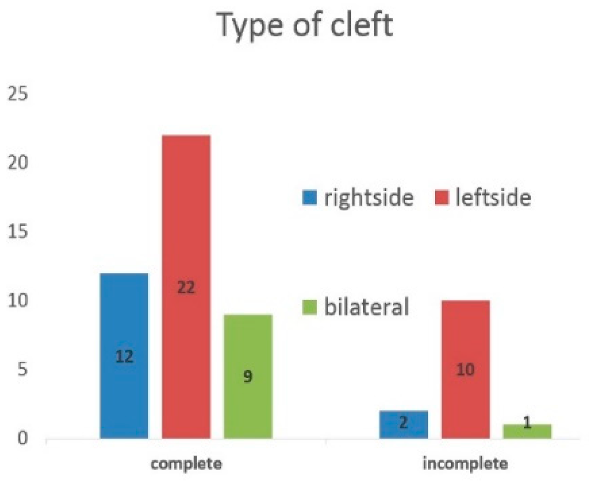
| Type of Cleft | Left Side Cleft Lip and Alveolar Cleft | Left Side Cleft Lip, Alveolar Cleft and Cleft Palate | Right Side Cleft Lip and Alveolar Cleft | Right Side Cleft Lip, Alveolar Cleft and Cleft Palate | Bilateral Cleft Lip and Alveolar Cleft | Bilateral Cleft Lip, Alveolar Cleft and Cleft Palate | Total | ||||||||
|---|---|---|---|---|---|---|---|---|---|---|---|---|---|---|---|
| Number of Persons in the Group | n = 10 | n = 22 | n = 2 | n = 12 | n = 1 | n = 9 | 56 | ||||||||
| Side of dental anomaly | right | left | right | left | right | left | right | left | right | left | right | left | |||
| Hypodontia | Maxillary lateral incisor | 1 | 5 | 8 | 1 | 1 | 4 | 1 | 1 | 1 | 2 | 25 | |||
| Second premolar | Maxillary | 2 | 3 | 1 | 1 | 7 | |||||||||
| Mandibular | 1 | 2 | 1 | 4 | |||||||||||
| Hyperdontia | Maxillary lateral incisor | 2 | 2 | 2 | 3 | 1 | 2 | 2 | 14 | ||||||
| Analyzed Variable | Canines Eruption | Total | p Value | ||
|---|---|---|---|---|---|
| + | − | ||||
| Cleft | complete | 12 | 1 | 13 | p = 0.782 |
| 92.31% | 7.69% | ||||
| incomplete | 41 | 2 | 43 | ||
| 95.35% | 4.65% | ||||
| Unerupted canine angle | correct | 31 | 0 | 31 | p = 0.166 |
| 100% | 0% | ||||
| incomplete | 22 | 3 | 25 | ||
| 88% | 12% | ||||
| Hyperdontia | − | 40 | 3 | 43 | p = 0.782 |
| 93.02% | 6.98% | ||||
| + | 13 | 0 | 13 | ||
| 100% | 0% | ||||
| Hypodontia | − | 33 | 2 | 35 | p = 0.646 |
| 94.29% | 5.71% | ||||
| + | 20 | 1 | 21 | ||
| 95.24% | 4.76% | ||||
| Expansion of the maxilla | − | 24 | 2 | 26 | p = 0.899 |
| 92.31% | 7.69% | ||||
| + | 29 | 1 | 30 | ||
| 96.67% | 3.33% | ||||
| Deciduous canine extraction | − | 35 | 2 | 37 | p = 0.542 |
| 94.59% | 5.41% | ||||
| + | 17 | 1 | 18 | ||
| 94.44% | 5.56% | ||||
| Secondary alveolar bone grafting | − | 18 | 1 | 19 | p = 0.563 |
| 94.74% | 5.26% | ||||
| + | 34 | 2 | 36 | ||
| 94.44% | 5.56% | ||||
Publisher’s Note: MDPI stays neutral with regard to jurisdictional claims in published maps and institutional affiliations. |
© 2020 by the authors. Licensee MDPI, Basel, Switzerland. This article is an open access article distributed under the terms and conditions of the Creative Commons Attribution (CC BY) license (http://creativecommons.org/licenses/by/4.0/).
Share and Cite
Pastuszak, P.; Dunin-Wilczyńska, I.; Lasota, A. Frequency of Additional Congenital Dental Anomalies in Children with Cleft Lip, Alveolar and Palate. J. Clin. Med. 2020, 9, 3813. https://0-doi-org.brum.beds.ac.uk/10.3390/jcm9123813
Pastuszak P, Dunin-Wilczyńska I, Lasota A. Frequency of Additional Congenital Dental Anomalies in Children with Cleft Lip, Alveolar and Palate. Journal of Clinical Medicine. 2020; 9(12):3813. https://0-doi-org.brum.beds.ac.uk/10.3390/jcm9123813
Chicago/Turabian StylePastuszak, Przemysław, Izabella Dunin-Wilczyńska, and Agnieszka Lasota. 2020. "Frequency of Additional Congenital Dental Anomalies in Children with Cleft Lip, Alveolar and Palate" Journal of Clinical Medicine 9, no. 12: 3813. https://0-doi-org.brum.beds.ac.uk/10.3390/jcm9123813