Article

Differential Occurrence of Cuticular Wax and Its Role in Leaf Physiological Mechanisms of Three Edible Aroids of Northeast India

by
Facundo Pieniazek
1,2,
Madhumita Dasgupta
1,
Valeria Messina
2,
Mayengbam Premi Devi
1,
Yumnam Indrani Devi
1,
Sansuta Mohanty
3,
Satyapriya Singh
3,
Bibhuti Bhusan Sahoo
4,
Potshangbam Nongdam
5,
Gobinda Chandra Acharya
3 and
Manas Ranjan Sahoo
1,3,*
1
ICAR Research Complex for North Eastern Hill Region, Imphal 795004, India
2
The National Council for Scientific and Technical Research (CONICET), Godoy Cruz 2290, Argentina
3
Central Horticultural Experiment Station, ICAR-Indian Institute of Horticultural Research, Bhubaneswar 751019, India
4
Regional Research and Technology Transfer Station (RRTTS, Bhawanipatna), Odisha University of Agriculture and Technology, Bhubaneswar 751003, India
5
Department of Biotechnology, Manipur University, Canchipur, Imphal 795003, India
*
Author to whom correspondence should be addressed.
Submission received: 21 April 2022 / Revised: 18 May 2022 / Accepted: 18 May 2022 / Published: 21 May 2022

Abstract

:
The localization of cuticular wax (CW) on the leaf epidermis and its interaction with the physiological mechanisms of three edible aroids, Alocasia, Colocasia, and Xanthosoma, were assessed. CW in the leaf tissues was visualized using scanning electron microscopy, which was higher in Colocasia (10.61 mg·dm−2) and Xanthosoma (11.36 mg·dm−2) than in Alocasia (1.36 mg·dm−2). Colocasia CW exhibited superhydrophobic properties with a higher static contact angle (CA) (>150°) than Xanthosoma (99.0°) and Alocasia (128.7°). The higher CW in Colocasia and Xanthosoma resulted in better leaf chlorophyll stability, moisture retention ability, and cellular membrane integrity compared to Alocasia. CW acted as a protecting barrier against deleterious solar radiation in terms of sun protection factor (SPF). The glossy appearance of wax crystals in the Alocasia leaf cuticles resulted in higher SPF. Overall, Colocasia CW highly influenced the qualitative and protective mechanisms of the leaf. Our study sheds light on the pivotal role of CW in the physiological properties of aroid leaves, which would be useful for the selection of wax-rich plants for augmenting future breeding strategies. The information would also be useful for further exploration of the industrial potential of superhydrophobic wax crystals obtained from edible aroids.

1. Introduction

Aroids are important minor food crops, belong to the Araceae family, cultivated widely in the tropics of the world [1,2]. Edible aroids are one of the cheapest sources of carbohydrates and dietary energy; thus, they have social and economic significance in terms of daily nutrition intake for about 400 million people around the world [3]. Despite a low share among the tuber crops, production of aroids exceeded 10.54 million tons worldwide [4].
About 105 genera and 3040 species of aroids are available globally, out of which 146 species are found in India, northeastern states in particular [5]. The ethnobotanical importance of 32 wild and cultivated aroid species of the genera Alocasia, Amorphophallus, Colocasia, Homalomena, Lasia, and Monstera have been reported in northeastern India [5]. Alocasia and Colocasia are cultivated widely in this region, whereas Amorphophallus, Homalomena, Lasia, and Monstera are limited to the marshy areas and wetlands of Assam state. In a field ethnobotanical research study, Alocasia, Colocasia, and Xanthosoma were identified as the three major cultivated edible aroids in northeast India [6]. Leaves, pseudo-stems, and corms of these three aroids are consumed as vegetables and traditional medicines by the tribal communities of the northeastern hill region of India [7]. These aroids were used as folk medicines in the ancient world [8] due to their high antioxidant, anti-inflammatory, antinociceptive, and anticarcinogenic properties [9,10]. In addition to food and medicinal uses, aroids have many possible applications as animal feed, carbohydrates, energy, and waxes for various industrial uses [11]. Aroid starch could be a substitute for 40% of biodegradable plastics [12]. Aroids contain higher cuticular wax (CW) in the leaf tissues among the terrestrial plants, which still need to be explored.
Preliminary reports have indicated the hydrophobic properties of Colocasia leaf wax [13,14]; however, information on Alocasia and Xanthosoma leaf wax is still unavailable. Plant-derived hydrophobic and nontoxic edible waxes have ample scope in the food industries, especially for food protection and preservation [15]. The wide use of carnauba, beeswax, or petroleum-based waxes in the food industries warrants studies consumer preference and health hazards [16]. Aroids are widely cultivated under harsh environments [3], and exploration of edible wax from cheaply available natural resources would add momentum to the food and postharvest industries [17].
Cuticular wax (CW) in leaf tissues acts as a protective barrier against several biotic and abiotic factors [18] such as adverse solar radiation and ultraviolet (UV) penetration, drought and chilling injury, and bacterial, fungal, and insect infestations [19]. CW in leaf cuticles forms a plant–environment interface and plays an important role in photosynthesis, osmoregulation, transpiration, and leaf gas exchange [20]. Lipophilic CW coating on the leaf cuticle enables plants to prevent the dehydration caused due to nonstomatal water loss, prevents chlorosis, maintains leaf pigmentation, and deters membrane injury caused by various environmental factors and invaders [21]. Zeisler-Diehl et al. [22] reported the biosurfactant and protective function of plant cuticular wax and its interaction with physiological properties in different crops. They also concluded that epicuticular wax does not establish the transpiration barrier, which essentially constitutes intracuticular wax. However, studies on the role of CW in the protective mechanisms against the most proficient fungal pathogen Phytophthora colocasiae Racib. (Pc) [23] in aroids are limited.
Despite being an essential biological constituent, information on the role of cuticular wax in leaf tissues of aroids is scanty. A clear understanding of the relationship between CW constituents and eco-physiological events in the leaf tissues of edible aroids needs to be established for further exploration and utilization of aroid wax. In the present study, we investigated the differential occurrence of CW and its role in the leaf physiological properties of three edible underutilized aroid species, Alocasia, Colocasia, and Xanthosoma, cultivated in the northeastern hill region of India. We observed the influence of CW on SPF, hydrophobicity, wettability, chlorophyll stability, relative water content and moisture loss, cell membrane stability, and antifungal activity against Pc in these three underutilized aroids. Understanding the physiological role of CW would be useful for the selection of wax-rich plants for crop improvement and future breeding strategies. Our study also aimed to explore the untapped potential of aroid CW coupled with SPF, hydrophobicity, wettability, and differential elemental constituents for possible industrial applications. The information on hydrophobicity, sun protecting factor, antifungal agents, etc. would be useful to harness the prospects of CW from edible aroids as a potential source of water-repellent agents, UV protection films, targeted agrochemicals emulsions, food coating, packaging material, and encapsulating agents in the food and pharmaceutical industries.

2. Materials and Methods

2.1. Collection of Leaf Samples

Fresh leaves of Alocasia macrorrhizos (L.) G. Don [A], Colocasia esculenta (L.) Schott [C], and Xanthosoma sagittifolium (L.) Schott [X] were collected from the ICAR Research Complex for the Northeastern Hill Region (ICAR RC NEHR), Imphal valley, Manipur, India, at 24°50′ N latitude, 93°55′ E longitude, and an altitude of 860 m above mean sea level. The necessary permission was obtained to collect the aroid leaves, and the voucher specimens (A1/20: Alocasia, RCMC–5/20: Colocasia, and X3/20: Xanthosoma) were submitted to ICAR RC NEHR, Manipur, India. The study complied with all local and national regulations. Aroid leaves were identified by Dr. Manas R. Sahoo, Principal Scientist (Horticulture), ICAR RC NEHR, Manipur Center, India, in consultation with Dr. Vivek Hegde, Scientist (Horticulture), Central Tuber Crops Research Institute (CTCRI), Thiruvananthapuram, Kerala, India. The leaf samples were collected during May–June 2020 at 8:00–9:00 a.m. and immediately processed for wax estimation and analyses of leaf characteristics.

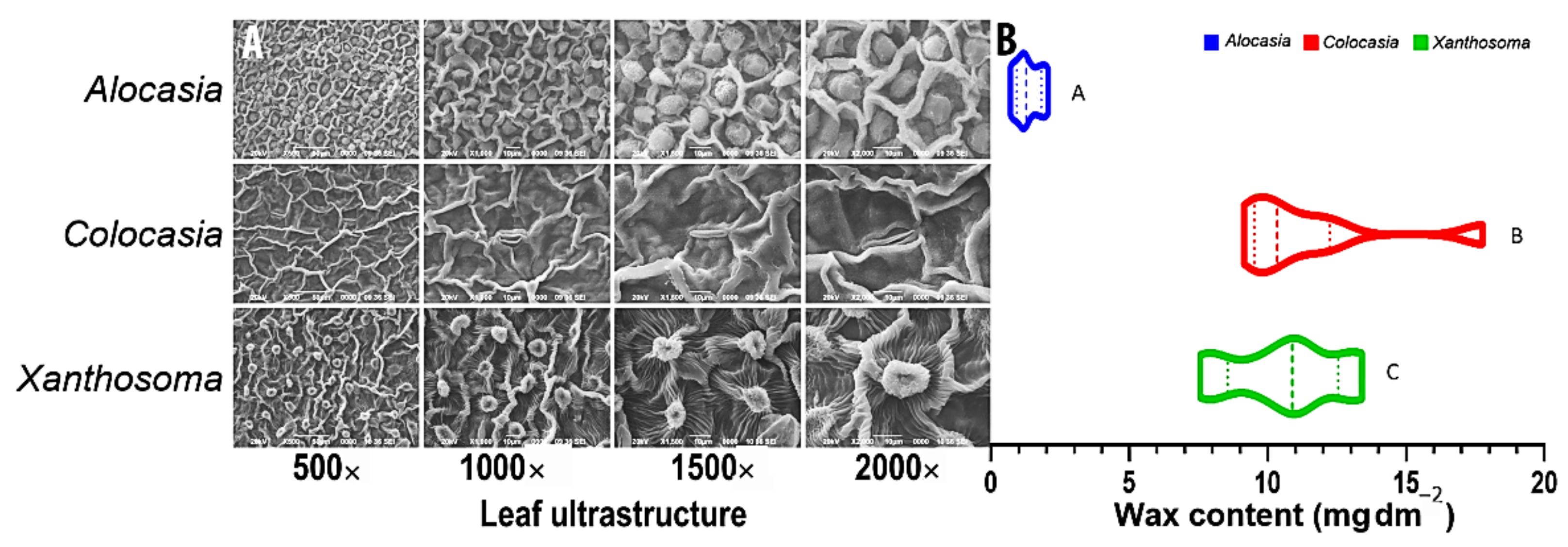
2.2. Scanning Electron Microscopy (SEM) of Aroid Leaves

Scanning electron microscopy (SEM) was performed to observe epicuticular wax microstructure on the fresh leaves of Alocasia, Colocasia, and Xanthosoma. Samples were cross-sectioned using a scalpel, mounted on the holders, and coated with gold particles as described by Pieniazek and Messina [24]. Microscopic visualization of wax crystals was performed using a scanning electron microscope (JOEL–JSM 6390LV, Japan) at magnifications of 500×, 1000×, 1500×, and 2000×. Brightness and contrast are the most important variables that need to be controlled during the acquisition of images; therefore, the values of these parameters were kept constant for each magnification during image acquisition [25]. The elemental composition of these waxes was analyzed using SEM with an energy-dispersive X-ray (EDX) system.

2.3. Extraction and Estimation of Cuticular Wax

The leaf surface area was measured using Image J software [24] before wax estimation. The wax extraction process [13] was optimized by submerging the leaves in 99.9% chloroform (HiMedia, Mumbai, India) in a glass beaker (Borosil, Mumbai, India) for 15, 30, 45, 60, 90, 120, and 180 s under a laminar airflow (Labtop Instruments Pvt. Ltd., Palghar, India). The extract was placed in the rotary evaporator R-210 (Buchi, Switzerland) until the chloroform was evaporated. The wax particles were carefully collected by scraping with a scalpel. The experiment was repeated thrice with eight determinations. The results were calculated using the following equation:
Wax   content = W w L A ,
where Ww is the weight of the wax in mg, and LA is the leaf area in cm2.
For the analysis of leaf physiological properties such as chlorophyll, chlorophyll stability index (CSI), relative water content (RWC), cell membrane injury (CMI), and Phytophthora colocasiae (Pc) infectivity studies, the leaf pieces (~5 × 5 cm) were dipped in chloroform for an optimum duration of 1 min, followed by submerging in Milli-Q water for 1 min. Leaves in triplicate were used for physiological assays.

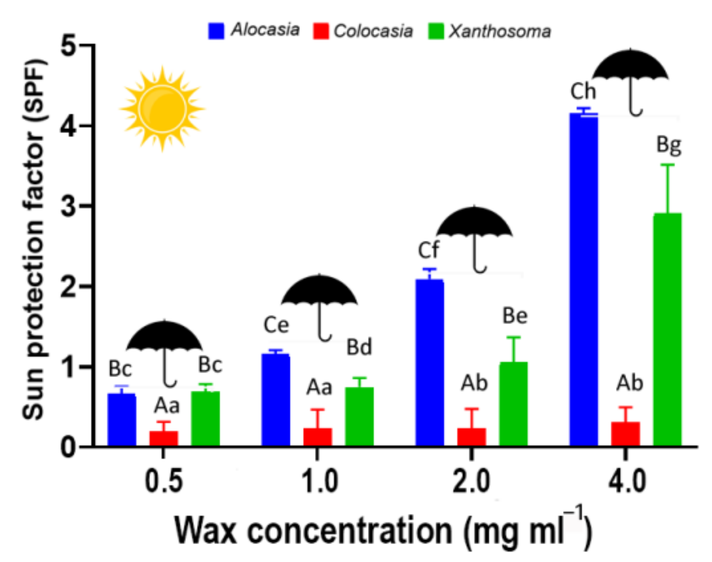
2.4. Sun Protection Factor (SPF)

The wax extracted from the three aroid species was dissolved in methanol (HiMedia, Mumbai, India) at different concentrations (4.0 mg·mL−1, 2.0 mg·mL−1, 1.0 mg·mL−1, and 0.5 mg·mL−1). The absorbance of different wax concentrations was recorded in a UV/Vis spectrophotometer (Eppendorf, Hamburg, Germany) every 5 nm at 290 to 320 nm [26]. SPF was calculated using the following equation:
SPF = CF   × 290 320   EE λ ×   I λ ×   Abs λ ,
where Abs is the absorbance of the samples, CF is a correction factor (=10), and EE λ × I λ is the product of the erithermal efficiency spectrum and the solar simulator intensity spectrum, which was tabulated following the methodology of Sayre et al. [27].

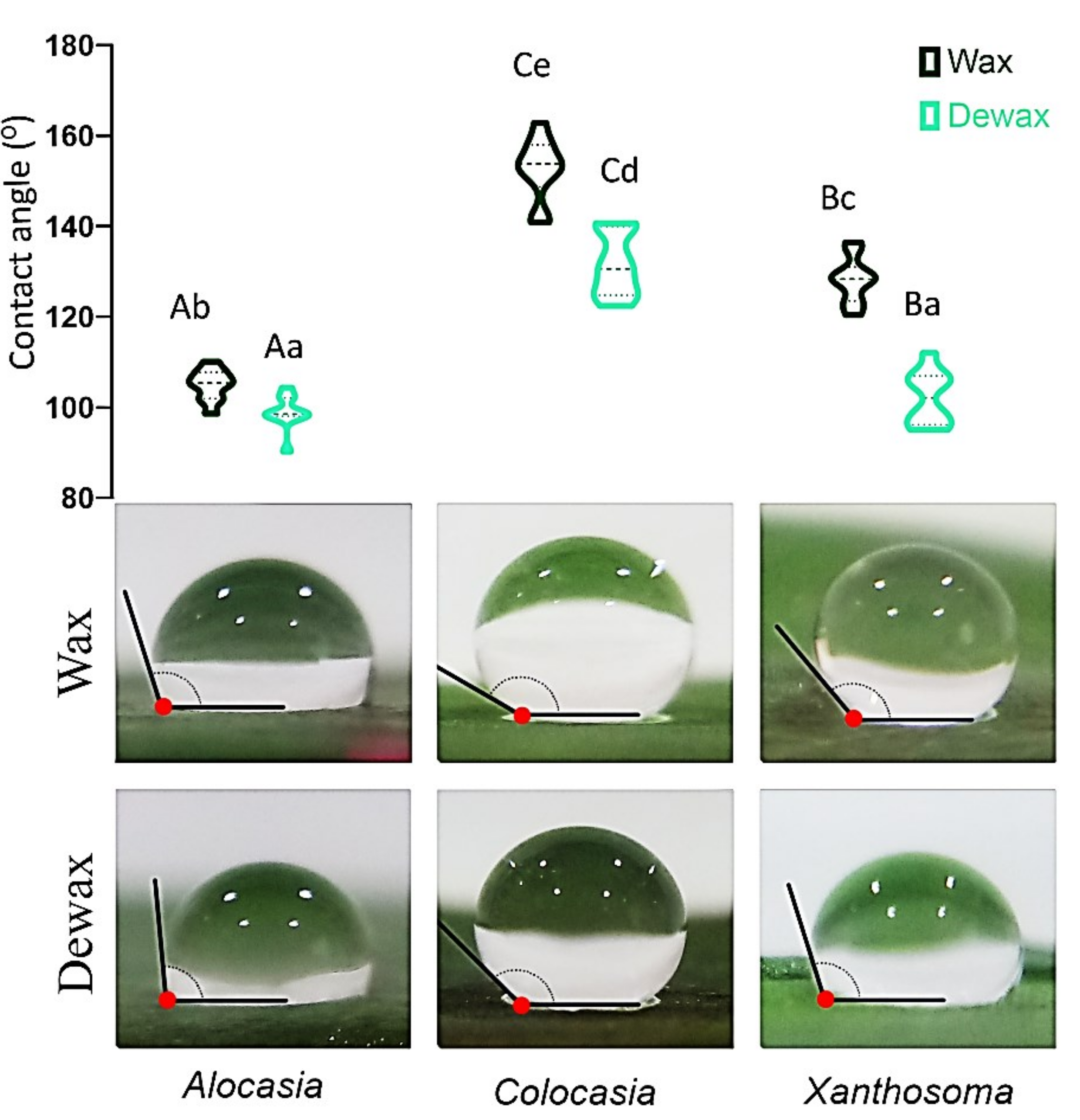
2.5. Contact Angle and Wettability

After wax extraction, fresh and dewaxed leaves were flattened upon a white surface with transparent tape. A drop of Milli-Q water (10 µL) (Merck, Mumbai, India) was placed on the surface of the leaves with and without wax. A digital camera with a macro lens was placed perpendicularly to the sample to capture the image. The contact angle was estimated using Image J software [24]. The experiment was repeated thrice with three replications.
In order to observe the wettability, the extracted wax was dissolved in chloroform (HiMedia, Mumbai, India) at different concentrations (100 mg·mL−1, 75 mg·mL−1, 50 mg·mL−1, 25 mg·mL−1, and 0 mg·mL−1). About 0.25 mL of each concentration was poured on 3 × 3 cm2 filter paper pieces and allowed to evaporate the chloroform under a laminar hood (Labtop Instruments Pvt. Ltd., Palghar, India). The filter paper without wax coating (0 mg·mL−1) was used as a control. Once the chloroform was completely evaporated, a 10 µL Milli-Q water (Merck, Mumbai, India) droplet was placed on the center of the wax-coated filter paper pieces. The time until the water droplet was completely absorbed was measured using a stopwatch. The live video of the absorbed water droplet on the wax-coated paper pieces was recorded by a video camera at 1080 × 720 pixels. (https://mfr.osf.io/render?url=https://osf.io/kby54/?direct%26mode=render%26action=download%26mode=render; accessed on 9 March 2022).

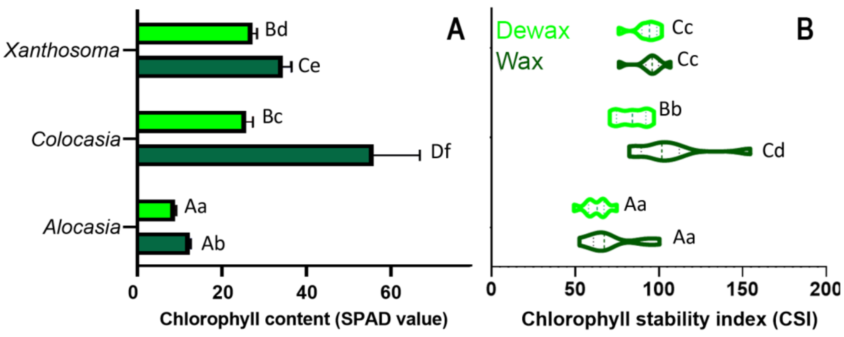
2.6. Chlorophyll Content (Ch) and Chlorophyll Stability Index (CSI)

Fresh waxed and dewaxed leaf samples of Alocasia, Colocasia, and Xanthosoma were collected and cut into 5 × 5 cm2 pieces. The leaf pieces were submerged in a water bath containing Milli-Q water (Merck, Mumbai, India) at 56 °C for 30 min to determine the pigment stability against the control sample submerged in Milli-Q water at room temperature (25 °C). The chlorophyll content (Ch) of the control leaf samples (ChC) and the hot water-treated samples (ChT) was measured using an SPAD-502 portable leaf greenness meter (Minolta Corp, Romsey, NJ, USA). Eight measurements each were recorded per treatment (waxed and dewaxed) at 0, 15, 30, 60, and 120 min for each species, Alocasia, Colocasia, and Xanthosoma. CSI was calculated using the equation derived by Mohan et al. [28].
CSI = ChT ChC   ×   100 .

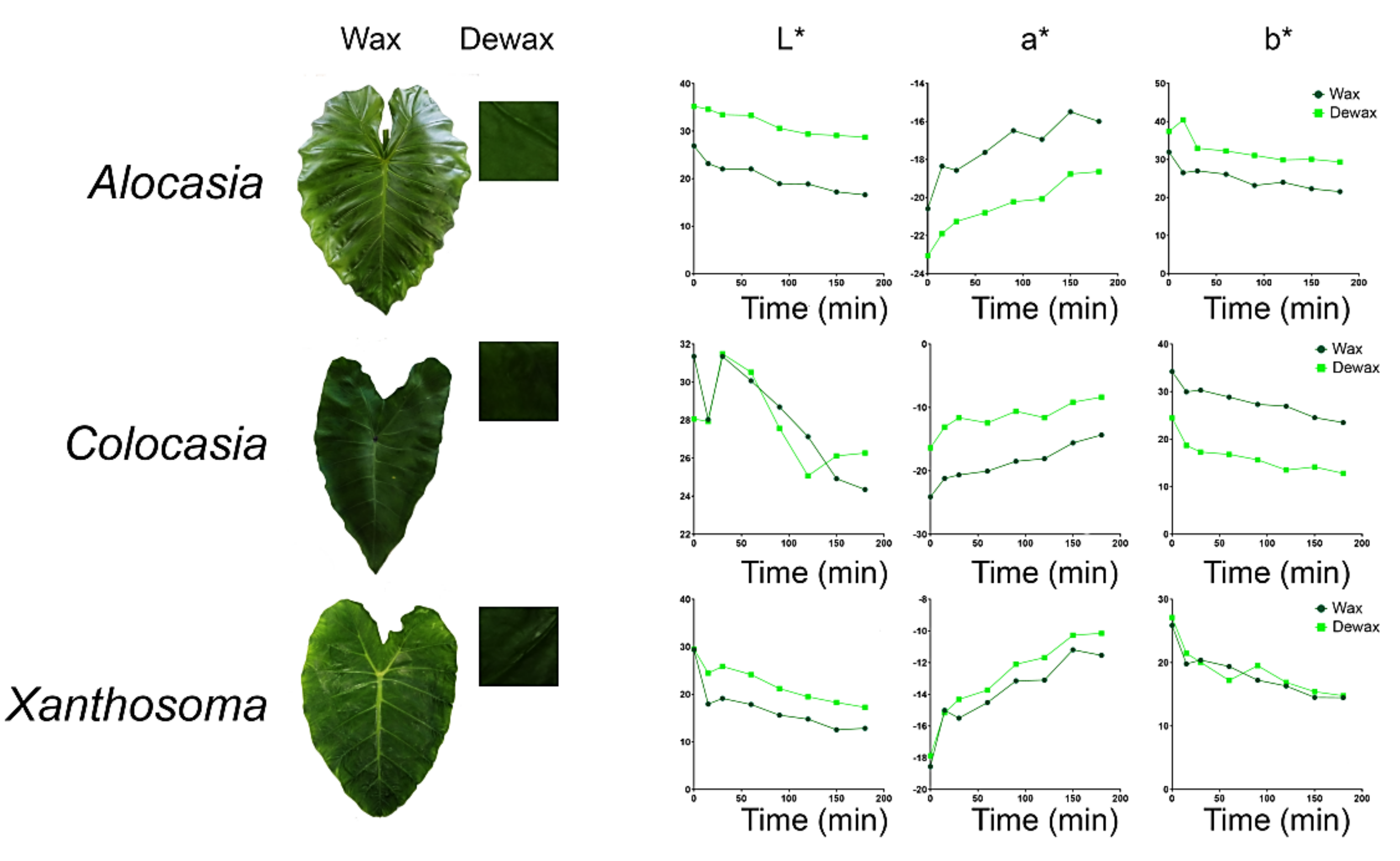
2.7. Color Parameters

Leaf samples of the three aroids with wax and after removal of wax were illuminated using a lamp (TL–D Deluxe, 169 Natural Daylight, 18W/965, Philips, NY, USA) with a color temperature of 6500 K 170 (D65, standard light source) and a color-rendering index (Ra) close to 90% [29].
Eighteen images from one side of each sample and eight regions of interest of each image were taken on the matte black background using the following camera settings: 174 manual modes with the lens aperture at the focus of 4.5 and speed 1/125, no zoom, no flash, 175, 3088 × 2056 pixel resolution, and stored in JPEG format.
The image segmentation and color quantification were processed by Adobe Photoshop CS6 (v18.0 Adobe Systems Incorporated, 2012, USA). L, a, and b values were transformed to CIE L*, a*, and b* using the algorithms [29].

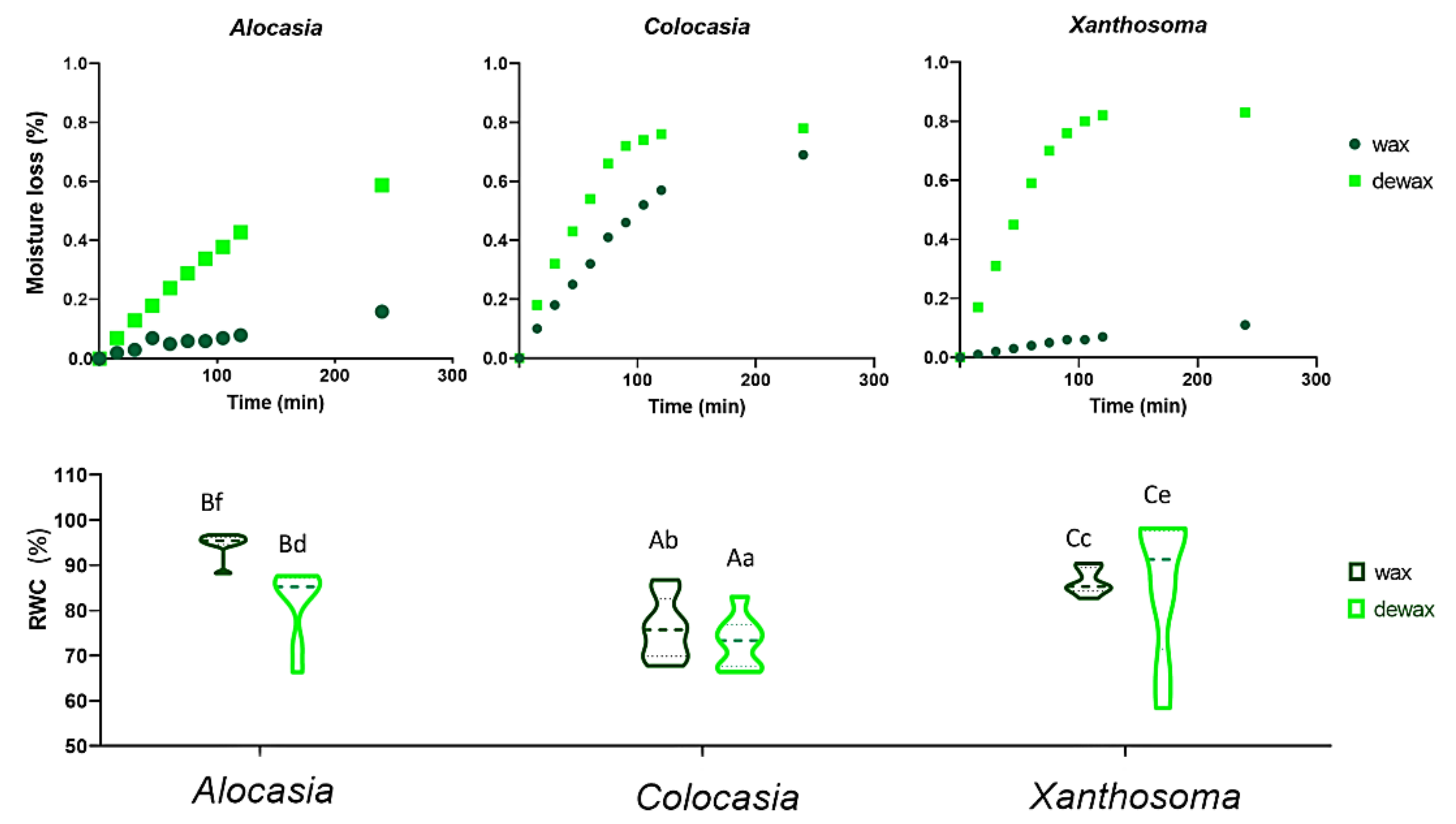
2.8. Relative Water Content (RWC) and Leaf Moisture Loss

RWC and leaf moisture loss were determined following the methods of Perez-Perez et al. [30] and Bueno et al. [31], respectively. Eight leaves of each aroid were cut into squares (5 × 5 cm2) using a scalpel and weighted to obtain the fresh weight (FW).
Leaves were submerged in Milli-Q water (Merck, Mumbai, India) at 25 °C for 4 h to obtain the turgid weight (TW), and the samples were dried in a hot air oven (REMI, India) at 70 °C for 96 h. RWC was calculated using the following formula:
RWC % = FW DW TW DW   ×   100 ,
where FW, TW, and DW are the fresh weight, turgid weight, and dry weight of the leaf samples, respectively. To measure leaf moisture loss, the waxed and dewaxed leaf samples were placed in a laminar airflow (Labtop Instruments Pvt. Ltd., Palghar, India). The declined leaf weight was measured at 15 min intervals for 240 min using an electronic balance (Shimadzu Analytical, New Delhi, India).

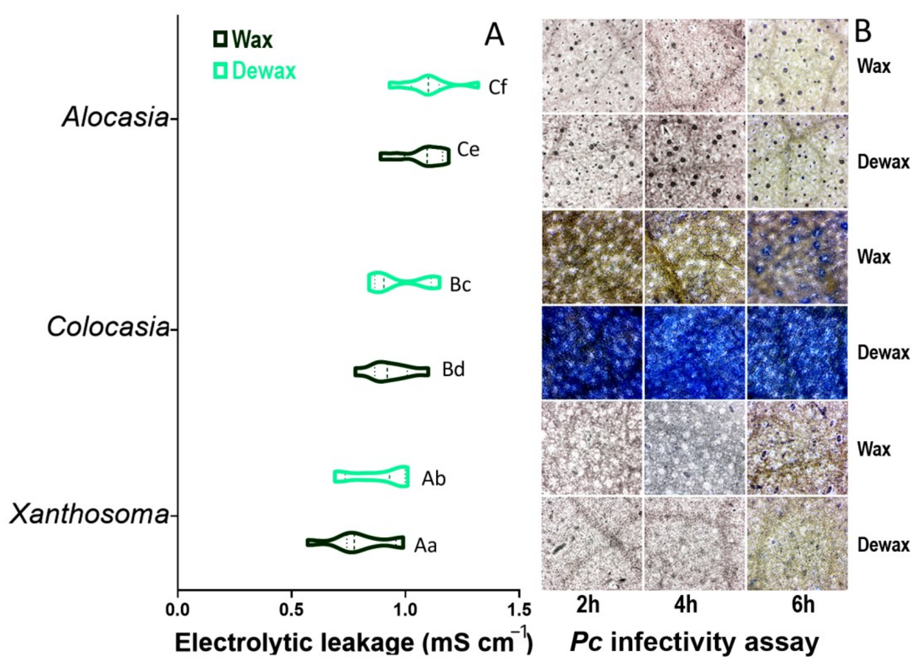
2.9. Cell Membrane Injury (CMI)

CMI was determined by comparing the electric conductivity (EC) of waxed and dewaxed leaves submerged in Milli-Q water (Merck, Mumbai, India) for 22 h followed by 2 h of hot water treatment at 70 °C. The electrolytic leakage related to the cell injuries was estimated with the variation of conductivity [32] as follows:
% Injury = 1 1 T 1 T 2 1 C 1 C 2 × 100 ,
where C1 and C2 are the EC of the water before and after submersion of control leaves (waxed) for 22 h in Milli-Q water at room temperature (25 ± 2 °C) followed by hot water treatment for 2 h, respectively. T1 and T2 are the EC of the water before and after submersion of the dewaxed leaves in Milli-Q water for 22 h followed by hot water treatment for 2 h, respectively.

2.10. In Vitro Phytophthora colocasiae (Pc) Infectivity Assay

Fungal infection in the leaf tissues was assessed by trypan blue staining, as demonstrated by Fernandez-Baustia et al. [33]. The fresh and dewaxed leaves from the Alocasia, Colocasia, and Xanthosoma plants were collected and placed in the Petri plate with moist filter paper (Whatman no.1) at room temperature (25 ± 2 °C). Phytophthora colocasiae (Pc) spores were collected from the infected Colocasia leaves using an artists’ paintbrush (size#1) in 10 mL of Milli-Q water. The leaves with and without wax were inoculated with 10 µL of Pc spore suspension (15,000 mL−1 spores) on the dorsal surface of the leaf. Pc infectivity was observed at different time points at 2, 4, and 6 h.
Infected leaves were immersed in 1.5 mL of trypan blue solution for 1 h. The leaves were then decolorized in 98–100% ethanol until green tissues became colorless, and the ethanol was discarded. Each leaf was mounted on a glass slide with the help of 50% glycerol and viewed under a light microscope (Magnus Opto Systems, New Delhi, India). The experiment was conducted thrice with three replications.

2.11. Statistical Analysis

All the data were analyzed by analysis of variance (ANOVA, Supplementary Table S1) in a complete randomized design (CRD) using XLSTAT statistical software (XLSTAT Premium 2020.2.1, Adinsoft, NY). The experiments were repeated thrice with eight determinations (n = 8). Differences among the mean values were compared using Tukey’s test [34] and were considered statistically significant when p ≤ 0.01. Correlation among the leaf physiological properties and wax content in three aroid species was performed following Pearson’s correlation coefficient (r-values) at p ≤ 0.05 and p ≤ 0.01.

3. Results and Discussion

3.1. Surface Properties, Extraction Process, and Estimation of Cuticular Wax

Leaf surface properties were visualized with SEM before wax extraction (Figure 1A). Electron micrographs showed the localization, distribution, and abundance of cuticular wax crystals embedded as a contour of cells with partially collapsed to prominent anticlinal cell walls. In the present study, we optimized the wax extraction process for the three aroid species by submerging the leaf pieces in chloroform for 1 min to obtain pure white wax crystals. The timepoint beyond 1 min resulted in green coloration of the solvent due to the removal of leaf chlorophyll. Upon extraction, CW concentration varied significantly among Alocasia (1.36 mg·dm−2), Colocasia (10.61 mg·dm−2), and Xanthosoma (11.36 mg·dm−2) leaf samples (Figure 1B). Colocasia and Xanthosoma leaves exhibited 10-fold higher CW than Alocasia, which was well visualized in the leaf ultrastructure (Figure 1A).
Wax microstructures were broad, aggregated, and observed in radiated cluster forms in Xanthosoma, unlike the wax crystals of Alocasia and Colocasia (Supplementary Figure S1). The wax crystals derived from Alocasia represented rough and irregular, but glossy surface properties. However, Colocasia leaf wax exhibited a smooth, compact, and opaque appearance. Xanthosoma leaf wax illuminated a bright and floral appearance with a friable texture (Supplementary Figure S1). Despite lower wax content, Alocasia featured a shinier leaf surface than Colocasia and Xanthosoma, probably due to glossy wax crystals. The quality and quantity of wax content in the leaf epidermis, its elemental composition, and its crystallization pattern represent the leaf surface properties and the protecting capacity [35].
The elemental composition of these three waxes indicated carbon and oxygen as the two major constituents as observed through the EDX system (Supplementary Figure S2). Alocasia wax comprised 52.89 wt.% of carbon and 39.86 wt.% of oxygen along with low levels of magnesium (0.51 wt.%), sulfur (0.32 wt.%), chlorine (2.1 wt.%), and potassium (4.33 wt.%). However, Colocasia wax comprised 79.94 wt.% of carbon and 20.06 wt.% of oxygen, while Xanthosoma wax constituted 86.63 wt.% of carbon and 13.37 wt.% of oxygen (Figure S2). The fresh aroid leaves also comprised similar carbon (49, 57.56, and 59.07 wt.%) and oxygen (45.96, 39.14, and 38.19 wt.%) levels in Alocasia, Colocasia, and Xanthosoma, respectively (Figure S2). Previous reports suggested that the composition of carbon in paraffin wax and beeswax varied from 10.33 to 12.99 wt.% and from 11.14 to 17.62 wt.%, respectively [36]. However, the oxygen composition ranged from 0 to 0.13 wt.% in paraffin wax and from 0.40 to 0.67 wt.% in beeswax. In our study, Colocasia wax showed better structural and elemental properties than Alocasia and Xanthosoma, which were comparatively higher than paraffin wax and beeswax available commercially [36]. The higher elemental composition in aroid waxes suggests the need for further comprehensive characterization of chemical compounds using GC–MS and/or nuclear magnetic resonance (NMR).

3.2. Sun Protection Factor (SPF)

The sun protecting factor (SPF) was increased significantly (p ≤ 0.01) with an increasing concentration of wax collected from the three aroid leaves (Figure 2). Alocasia registered a higher mean SPF (2.02) when compared to Xanthosoma (1.35) and Colocasia (0.24). Sun protection activity depends on the ability to prevent the plants from harmful UV radiation-led mutagenesis [37]. Higher SPF was positively correlated with the protective mechanisms and negatively correlated with the adverse effect of UV radiation [38].
Natural plant substances are considered potential resources for UV protection. In a pilot study, the SPF values were assessed to range from 0.4 to 23.5 in different plant species [39]. Eucalyptus showed a higher SPF of 23.5, which was positively correlated with the higher phenolic content [40]. Our results revealed that Alocasia leaves showed 10-fold higher SPF than Colocasia and twofold higher SPF than Xanthosoma; hence, they could be explored as a potential natural sun protector. Alocasia exhibited a shiny leaf surface with higher oxygen, sulfur, and potassium, resulting in higher SPF (Supplementary Figure S2). Colocasia leaf wax exhibited lower SPF compared to Alocasia and Xanthosoma, which might have been due to the compact and opaque appearance of the wax microstructure. In an aliphatic chain (sulfide) or with a double bond with oxygen (sulfoxide), sulfur induces a glossy leaf surface by accumulating bio-oil and bio-wax, which act as natural sun protectors [41].

3.3. Contact Angle (CA)

There were significant differences (p ≤ 0.01) in the CA of the three aroid leaves (Figure 3). The CA of the leaves decreased significantly upon wax removal while compared with the leaves with wax. Colocasia leaves exhibited superhydrophobicity with higher CA (153.1°), followed by Xanthosoma (128.7°) and Alocasia (105.7°). The static CA in dewaxed leaves of Colocasia, Xanthosoma, and Alocasia was observed to be 132.0°, 102.9°, and 99.7°, respectively.
Static CAs >90° and <150° are considered hydrophobic [42]. A surface with a static CA >150° is regarded as superhydrophobic [43], probably due to micro- and nanoscale hierarchical topography in the leaves. According to the classification [43], Colocasia leaves represented superhydrophobicity similar to the ‘Lotus’ hydrophobic state, a special state of Cassie’s superhydrophobic state [25]. Xanthosoma exhibited a transitional hydrophobic condition between Wenzel’s and Cassie’s states. However, Alocasia showed Wenzel’s form with the lowest static CA and inadequate hydrophobic capacity due to lower wax content and irregular distribution of CW. Results showed that the hydrophobic properties diminished once the wax was removed from the leaves, indicating the role of cuticular wax in static CA and hydrophobicity [44].

3.4. Wettability

The wettability test showed the capacity of CW to repel environmental water and protect the leaf surface. In our study, the filter paper pieces coated with aroid wax resisted the water droplets significantly (Supplementary Video S1: https://mfr.osf.io/render?url=https://osf.io/kby54/?direct%26mode=render%26action=download%26mode=render; accessed on 9 March 2022). Results showed that the filter paper without wax coating instantly absorbed the water droplet compared to the filter paper coated with aroid wax. The resistivity varied significantly (p ≤ 0.01) among the three types of aroid wax coating (Supplementary Video S1).
The wettability test witnessed a higher wax concentration correlated with higher water resistance and hydrophobicity. The above video showed that the Colocasia wax coating resisted the water droplet longer, which justified its superhydrophobicity. On the other hand, Alocasia and Xanthosoma wax showed poor hydrophobicity with low water droplet resistance. The uniform wax distribution and regular surface topography with inadequate pore space in the Colocasia wax coating were reasoned for better wettability. Oner and McCarthy [44] reported that wettability is correlated with synthetic compounds, hydrophobicity, and surface topography. Leaf cuticular wax film was also successfully examined as a model hydrophobic system [45], providing insights for the future potential of edible aroid leaf-based bio-wax films in food coating.

3.5. Chlorophyll Content and Chlorophyll Stability Index (CSI)

Significant differences (p ≤ 0.01) in chlorophyll content and stability index were observed among the Alocasia, Colocasia, and Xanthosoma leaves. Higher SPAD values for chlorophyll content (55.9) were obtained for Colocasia, followed by Xanthosoma (34.4) and Alocasia (12.6) (Figure 4A). In dewaxed leaves, SPAD values decreased significantly (p ≤ 0.01) in Xanthosoma (27.3), Colocasia (25.8), and Alocasia (9.2). Colocasia exhibited higher CSI, followed by Xanthosoma and Alocasia (Figure 4B), when exposed to hot water treatment. The CW layer in Colocasia protected the cell membrane and prevented the chlorophyll degradation from maintaining CSI. Rapid chlorophyll depletion occurred when the wax was removed from the cuticle, signifying the role of CW in maintaining the leaf chlorophyll content. The leaf chlorophyll content [46] and cuticular layer thickness [47] were decreased upon removal of leaf CW due to dismantling of the thylakoid membrane [48].

3.6. Color Parameters

Color parameters (L*, a*, and b* values) were significantly different (p ≤ 0.01) among the tested aroid leaves with and without wax (Figure 5). Leaf brightness (L*) decreased when the storage time after defoliation increased. The greenish leaf color related to a* values decreased when the time increased. The decrease in a* values was probably due to the chlorophyll degradation. During leaf pigment degradation, an increase in yellow color (b*) also played a vital role in manipulating leaf greenness.
In the present study, leaf discoloration in Colocasia under dewaxed conditions was higher when compared to leaves with wax. Dismantling of wax crystals from the leaf cuticle resulted in faster chlorophyll degradation, which ensured rapid leaf discoloration. Xanthosoma showed similar L*, a*, and b* values in waxed and dewaxed leaves, indicating low chlorophyll degradation upon wax removal. Cuticular wax exhibited a more predominant role in Colocasia leaf protection than in Alocasia and Xanthosoma. Similar results on leaf color pigmentation using a quantifiable RGB model were reported by Chen et al. [49]. The color variation was related to the chlorophyll degradation and other biological, chemical, and gas exchange processes occurring during photorespiration [50].

3.7. Relative Water Content (RWC) and Leaf Moisture Loss

As shown in Figure 6, RWC varied significantly (p ≤ 0.01) in the range of 76.1–94.7% in waxed leaves and 73.1–85.6% in dewaxed aroid leaves. Alocasia leaves with wax recorded higher RWC, followed by Xanthosoma and Colocasia. RWC in dewaxed leaves declined significantly in all three aroids. The rate of decrease in RWC in dewaxed Alocasia leaves was relatively higher compared to Colocasia and Xanthosoma. Lower wax content in Alocasia could be attributed to a higher reduction in RWC. In Colocasia and Xanthosoma, the samples showed a lower decrease in RWC due to the inherent higher wax content.
CW was shown to play an essential role in preventing leaf moisture loss by up to 95% through strengthening cuticle permeability [21]. In our study, Xanthosoma leaves exhibited the lowest moisture loss while embedded with CW, while it rapidly increased upon removing CW from the cuticle. Alocasia also showed a similar response in terms of leaf moisture loss to Xanthosoma. The rapid moisture loss occurred due to the lack of wax content or cuticular cracks upon wax removal [51]. On the other hand, Colocasia leaves showed a high dehydration rate in waxed and dewaxed conditions related to the leaf ultrastructure. The location and the structural basis of CW are responsible for the cuticular barrier, which restricts moisture loss. On the contrary, stomatal behavior, such as opening and closing of stomata, also cause reasonable moisture loss, which probably occurred in the case of Colocasia leaves. Rapid moisture loss is one of the major factors affecting leaf quality, and CW helped in leaf moisture retention in the tested aroids.

3.8. Cell Membrane Injury (CMI) and In Vitro Phytophthora colocasiae (Pc) Infectivity

CW maintains membrane integrity and acts as a protecting barrier against several environmental factors and invaders [51]. In our study, Alocasia leaves showed significantly higher CMI while submerged in hot water than Colocasia and Xanthosoma leaves under waxed and dewaxed conditions (Figure 7A). Higher CMI was attributed to the higher electrolytic leakage of subcellular components in the hot water treatment, which was distinctly related to the lower wax content in the leaf tissues of Alocasia. Xanthosoma and Colocasia exhibited lower CMI proportionate to their higher CW.
Aroid leaves, particularly Colocasia, usually experience leaf blight disease caused by the fungal pathogen Phytophthora colocasiae Racib (Pc). Figure 7B shows the intensity of in vitro Pc infestation assayed using trypan blue staining. The blue coloration indicated the cellular damage caused by Pc at different timepoints. Xanthosoma leaves showed less cellular disruption compared to Alocasia and Colocasia. CW outwardly acted as a physical barrier in the leaf epidermis against the invaders. However, Pc releases various cell wall-degrading enzymes (CWDEs) such as pectinase, cellulase, and hemicellulase to breach the cell-wall components other than CW. Consequently, the higher cellular damage observed in Colocasia leaves was probably due to several cell wall constituents such as pectin, cellulose, and hemicellulose. Previous reports also suggested that leaf resistance to Pc is associated with various reactive oxygen species and their scavengers [52]. Higher phenolics in Alocasia and Xanthosoma [53] could also be a major reason for the lower Pc infectivity than in Colocasia, as illustrated in the trypan blue staining studies.
On the other hand, wax solubility might be another reason for cellular depletion. Nonetheless, the dewaxed leaves showed higher incidence when compared to waxed leaves, which predicted the role of CW in Pc infestation. Several authors [54] reported evidence of natural leaf wax in preventing disease incidence. Our results indicated that the presence of CW in leaf tissues sustainably inhibited electrolytic leakage of the subcellular components to maintain the cellular integrity and defended the cellular damage caused by Pc, as evident by the lower acquisition of trypan blue coloration in the tested aroid leaves.

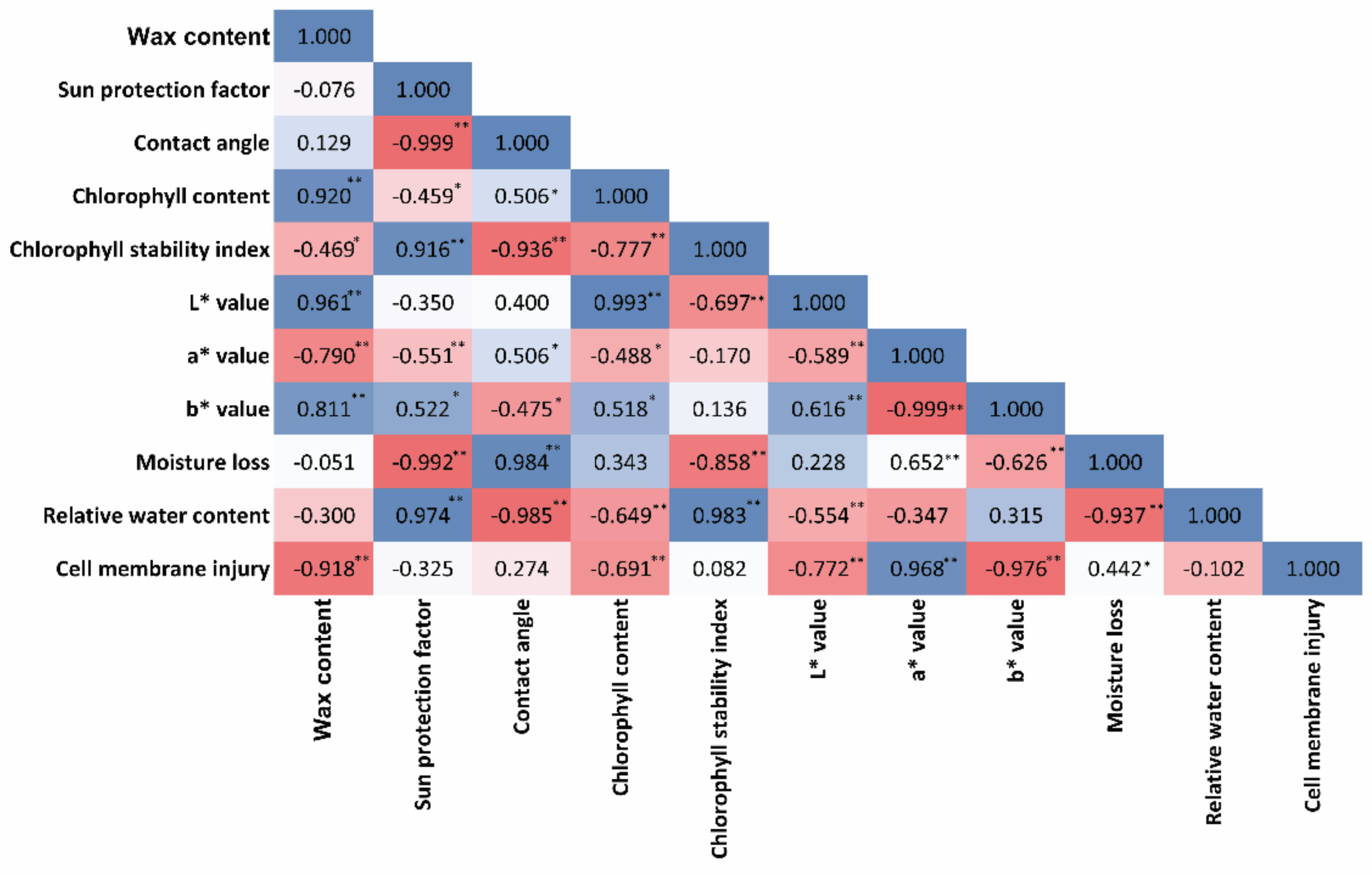
3.9. Correlation Studies

Significant and strong correlations among the wax content and leaf physiological properties were established (Figure 8). The wax content was positively correlated with chlorophyll content (r = 0.920), L* value (r = 0.961), and b* value (r = 0.811), and negatively correlated with CSI (r = −469), a* value (r = −790), and CMI (r = −0.918). SPF was positively correlated with CSI (r = 0.916), b* value (r = 0.522), and RWC (r = 0.974), and negatively correlated with CA (r = −0.999) and moisture loss (r = −0.992). CA had a positive influence on chlorophyll content (r = 506), a* value (r = 506), and moisture loss (r = 0.984), and negatively correlated with CSI (r = −936), b* value (r = −0.475), and RWC (r = −0.985). CMI was strongly and negatively correlated with wax content (r = −918), chlorophyll content (r = −0.691), L* value (r = −0.772), and b* value (r = −0.976). Strong correlation among the leaf physiological properties helps in plant defense against invaders [52]. The correlation studies on leaf physiological properties owing to wax accumulation would help in specific trait improvement in minor aroid species.

4. Conclusions

Significant differences in cuticular wax and its interaction with the qualitative and protective mechanisms in leaf tissues of three edible aroids, Alocasia, Colocasia, and Xanthosoma, were observed. Colocasia and Xanthosoma exhibited higher CW similar to lotus leaves, considered the most pronounced edible wax-rich terrestrial plants. Interestingly, Colocasia leaves showed superhydrophobic surfaces with higher contact angles and better wetting properties suitable for hydrophobic coatings. Higher CW occurrence in Colocasia and Xanthosoma showed a significant influence on all studied leaf properties except SPF. Alocasia exhibited higher SPF despite having lower CW content correlated with the thin and glossy appearance of wax crystals, which may be a potent source of natural sun protection. The study results revealed that the leaf cuticular wax coverage in aroids Colocasia and Xanthosoma strengthened the leaf epidermis and improved the physiological processes. The information on the role of cuticular wax in leaf physiological processes in these three lesser-known aroid species would be useful for augmenting future breeding strategies for biotic and abiotic stress tolerance through the selection of wax-rich plants. The evidence provides insight for further exploring the wax structure and composition of underutilized edible aroids to better understand their food, agricultural, and industrial applications.

Supplementary Materials

The following supporting information can be downloaded at https://www.mdpi.com/article/10.3390/agriculture12050724/s1: Figure S1. SEM images of cuticular wax structures of Alocasia (A), Colocasia (B), and Xanthosoma (C) at 500×, 1000×, and 2000×; Figure S2. Elemental composition analysis of Alocasia (A), Colocasia (B), and Xanthosoma (C) cuticular wax using SEM–EDX; Table S1. Analysis of variance (mean sum of squares) for the leaf physiological properties of three aroid species, Alocasia, Colocasia, and Xanthosoma, as influenced by leaf cuticular wax in a complete randomized design (CRD); Video S1. Wettability test.

Author Contributions

Conceptualization, M.R.S., F.P. and M.D.; data curation, F.P., M.D., M.P.D., Y.I.D., S.M. and S.S.; formal analysis, F.P., M.D., M.R.S. and V.M.; investigation, F.P. and M.D.; methodology, F.P., M.D. and M.R.S.; resources, M.R.S. and F.P.; software, F.P., M.R.S., B.B.S., G.C.A. and P.N.; writing—original draft, F.P., S.M. and M.D.; writing—review and editing, M.R.S., V.M., S.S., B.B.S., G.C.A. and P.N. All authors read and agreed to the published version of the manuscript.

Funding

The research training fellowship (RTF) for developing countries scientists (DCS) (DST–RTF–DCS/2019/000260) from the Department of Science and Technology (DST), Government of India, to Facundo Pieniazek is greatly acknowledged.

Institutional Review Board Statement

Not applicable.

Informed Consent Statement

Not applicable.

Data Availability Statement

Not applicable.

Acknowledgments

The financial support of the Department of Science and Technology (DST), Government of India, to Facundo Pieniazek as a research training fellowship (RTF) for developing countries scientists (DCS) (DST–RTF–DCS/2019/000260) is greatly acknowledged. The authors acknowledge the support of the Sophisticated Analytical Instrumentation Center (SAIC), Tezpur University, Assam, India, for expert assistance with the SEM–EDX analyses. We also sincerely acknowledge the Director and Joint Director, ICAR Research Complex for NEH Region, Manipur Center, for providing facilities.

Conflicts of Interest

The authors declare no conflict of interest.

References

  1. Irwin, S.V.; Kaufusi, P.; Banks, K.; De la Pena, R.; Cho, J.J. Molecular characterization of taro (Colocasia esculenta) using RAPD markers. Euphytica 1998, 99, 183. [Google Scholar] [CrossRef]
  2. Opara, L.U.; Mejía, D. Edible Aroids: Post–Harvest Operation. AGST/FAO: Danilo Mejía, PhD, FAO (Technical). 2003. Available online: https://www.fao.org/fileadmin/user_upload/inpho/docs/Post_Harvest_Compendium_-_Edible_aroids.pdf (accessed on 1 January 2020).
  3. Vieira, G.H.S.; Peterle, G.; Loss, J.B.; Peterle, G.; Poloni, C.M.M.; Colombo, J.N.; Monaco, P.A.V.L. Strategies for taro (Colocasia esculenta) irrigation. J. Exp. Agric. Int. 2018, 1–9. [Google Scholar] [CrossRef] [Green Version]
  4. FAOSTAT. Food and Agriculture Organization of the United Nations. Statistics Division; FAO: Rome, Italy, 2021; Available online: http://www.fao.org/statistics/en/ (accessed on 1 February 2021).
  5. Teron, R. Ethnobotanical study of dietary use and culinary knowledge of Aroids (family Araceae) in Karbi Anglong district, Assam. NeBIO 2019, 10, 80–84. [Google Scholar]
  6. Matthews, P.J.; Medhi, D. Feasibility study for field research: Ethnobotany and ecology of wild and cultivated aroids in Assam state, northeast India. AREIPGR 2014, 30, 159–183. [Google Scholar]
  7. Prajapati, R.; Kalariya, M.; Umbarkar, R.; Parmar, S.; Sheth, N. Colocasia esculenta: A potent indigenous plant. Int. J. Nutr. Pharmacol. Neurol. Dis. 2011, 1, 90. [Google Scholar] [CrossRef]
  8. Huang, W.; Li, C.; Wang, Y.; Yi, X.; He, X. Anti–inflammatory lignanamides and monoindoles from Alocasia macrorrhiza. Fitoterapia 2017, 117, 126132. [Google Scholar] [CrossRef]
  9. Mulla, W.A.; Kuchekar, S.B.; Thorat, V.S.; Chopade, A.R.; Kuchekar, B.S. Antioxidant, antinociceptive anti–inflammatory activities of ethanolic extract of leaves of Alocasia indica (Schott.). J. Young Pharm. 2010, 2, 137–143. [Google Scholar] [CrossRef] [Green Version]
  10. Roy, S.; Choudhury, M.D.; Paul, S.B. In vitro Antibacterial activity of Alocasia decipiens schott. Int. J. Pharm. Pharm. Sci. 2013, 5, 155–157. [Google Scholar]
  11. Adewumi, D.F.; Adewole, E.; Ogunmodede, O.T.; Ojo, A. Effect of Chemical Modifications on Pasting Properties of Cocoyam Starch (Xanthosoma Sagittifollium). J. Nat. Sci. Res. 2015, 5, 36–39. [Google Scholar]
  12. Zhu, F. Structure, properties, and applications of aroid starch. Food Hydrocoll. 2016, 52, 378–392. [Google Scholar] [CrossRef]
  13. Kalita, A.; Talukdar, N. Colocasia esculenta (L.) leaf bio–wax as a hydrophobic surface coating substance for paper for preparing hydrophobic paper bags. Int. J. Pharm. Biol. Sci. 2018, 8, 583–590. [Google Scholar]
  14. Acebuche, M.J.A.; Alvarez, M.L.C. Hydrophobic paper from the wax of Colocasia esculenta (taro) leaf and chitin from crab shell. Int. J. Res. Publ. 2019, 30, 1–7. Available online: https://www.ijrp.org (accessed on 1 January 2020).
  15. De Freitas, C.A.S.; de Sousa, P.H.M.; Soares, D.J.; da Silva, J.Y.G.; Benjamin, S.R.; Guedes, M.I.F. Carnauba wax uses in food—A review. Food Chem. 2019, 291, 38–48. [Google Scholar] [CrossRef] [PubMed]
  16. Hammam, A.R. Technological, applications, and characteristics of edible films and coatings: A review. SN Appl. Sci. 2019, 1, 632. [Google Scholar] [CrossRef] [Green Version]
  17. Boakye, A.A.; Wireko–Manu, F.D.; Oduro, I.; Ellis, W.O.; Gudjónsdóttir, M.; Chronakis, I.S. Utilizing cocoyam (Xanthosoma sagittifolium) for food and nutrition security: A review. Food Sci. Nutr. 2018, 6, 703–713. [Google Scholar] [CrossRef]
  18. Sharma, P.; Madhyastha, H.; Madhyastha, R.; Nakajima, Y.; Maruyama, M.; Verma, K.S.; Verma, S.; Prasad, J.; Kothari, S.L.; Gour, V.S. An appraisal of cuticular wax of Calotropis procera (Ait.) R. Br.: Extraction, chemical composition, biosafety and application. J. Hazard. Mater. 2019, 368, 397–403. [Google Scholar] [CrossRef]
  19. Sajeevan, R.S.; Parvathi, M.S.; Nataraja, K.N. Leaf wax trait in crops for drought and biotic stress tolerance: Regulators of epicuticular wax synthesis and role of small RNAs. Indian J. Plant Physiol. 2017, 22, 434–447. [Google Scholar] [CrossRef]
  20. Sharma, P.; Bala, N.; Saini, M.K.; Jain, A.; Kothari, S.L.; Gour, V.S. Assessment of role of cuticular wax in adaptive physiological responses of Calotropis procera and Calotropis gigantea. Plant Physiol. Rep. 2021, 26, 368–373. [Google Scholar] [CrossRef]
  21. Buschhaus, C.; Jetter, R. Composition and physiological function of the wax layers coating Arabidopsis Leaves: B–Amyrin negatively affects the intracuticular water barrier. Plant Physiol. 2012, 160, 1120–1129. [Google Scholar] [CrossRef] [Green Version]
  22. Zeisler–Diehl, V.V.; Barthlott, W.; Schreiber, L. Plant Cuticular Waxes: Composition, Function, and Interactions with Microorganisms. In Hydrocarbons, Oils and Lipids: Diversity, Origin, Chemistry and Fate; Wilkes, H., Ed.; Handbook of Hydrocarbon and Lipid Microbiology; Springer: Cham, Switzerland, 2018. [Google Scholar] [CrossRef]
  23. Sahoo, M.R.; Dasgupta, M.; Mukherjee, A.; Kole, P.C. In vitro and in vivo screening of taro [Colocasia esculenta (L.) Schott] for Phytophthora leaf blight disease. In Advances in Fungal Diversity and Host–Pathogen Interaction; Rodrigues, B.F., Gour, H.N., Bhat, D.J., Kamat, N., Eds.; Goa University: Goa, India, 2005; pp. 144–152. [Google Scholar]
  24. Pieniazek, F.; Messina, V. Texture and color analysis of freeze–dried potato (cv. Spunta) using instrumental and image analysis techniques. Int. J. Food Prop. 2017, 20, 1422–1431. [Google Scholar] [CrossRef] [Green Version]
  25. Kumar, M.; Bhardwaj, R. Wetting characteristics of Colocasia esculenta (Taro) leaf and a bioinspired surface thereof. Sci. Rep. 2020, 10, 935. [Google Scholar] [CrossRef] [PubMed]
  26. Mansur, J.d.S.; Breder, M.N.R.; Mansur, M.C.d.A. Determination of sun protection factor by spectrophotometry. An. Bras. De Dermatol. 1986, 61, 121–124. [Google Scholar]
  27. Sayre, R.M.; Agin, P.P.; LeVee, G.J.; Marlowe, E. A comparison of in vivo and in vitro testing of sunscreening formulas. Photochem. Photobiol. 1979, 29, 559–566. [Google Scholar] [CrossRef] [PubMed]
  28. Mohan, M.M.; Narayanan, S.L.; Ibrahim, S.M. Chlorophyll stability index (CSI): Its impact on salt tolerance in rice. Int. Rice Res. Notes 2000, 25, 38–39. [Google Scholar]
  29. Afshari–Jouybari, H.; Farahnaky, A. Evaluation of Photoshop software potential for food colorimetry. J. Food Eng. 2011, 106, 170–175. [Google Scholar] [CrossRef]
  30. Perez–Perez, J.G.; Syvertsen, J.P.; Botía, P.; García–Sánchez, F. Leaf water relations and net gas exchange responses of salinized carrizo citrange seedlings during drought stress and recovery. Ann. Bot. 2007, 100, 335–345. [Google Scholar] [CrossRef] [PubMed] [Green Version]
  31. Bueno, A.; Sancho-Knapik, D.; Gil-Pelegrín, E.; Leide, J.; Peguero-Pina, J.J.; Burghardt, M.; Riederer, M. Cuticular wax coverage and its transpiration barrier properties in Quercus coccifera L. leaves: Does the environment matter? Tree Physiol. 2020, 40, 827–840. [Google Scholar] [CrossRef]
  32. Liu, X.; Gao, S.; Liu, Y.; Cao, B.; Chen, Z.; Xu, K. Comparative analysis of the chemical composition and water permeability of the cuticular wax barrier in Welsh onion (Allium fistulosum L.). Protoplasma 2019, 257, 833–840. [Google Scholar] [CrossRef]
  33. Fernandez–Baustia, N.; Dominguez–Nunez, J.A.; Moreno, M.M.C.; Berrocal–Lobo, M. Plant tissue trypan blue staining during phytopathogen infection. Bio–Protoc. 2016, 6, 1–7. [Google Scholar] [CrossRef] [Green Version]
  34. Tukey, J.W. Comparing individual means in the analysis of variance. Biometrics 1949, 5, 99–114. [Google Scholar] [CrossRef]
  35. Jenks, M.A.; Ashworth, E.N. Plant epicuticular waxes: Function, production, and genetics. Hortic. Rev. 1999, 23, 1–68. [Google Scholar]
  36. Hossain, M.E.; Rahman, M.S.; Ketata, C.; Mann, H.; Islam, M.R. SEM–based structural and chemical analysis of paraffin wax and beeswax for petroleum applications. J. Charact. Dev. Nov. Mater. 2009, 1, 21–38. [Google Scholar]
  37. Mazumder, M.; Das, K.; Choudhury, A.D.; Khazeo, P. Determination of Sun Protection Factor (SPF) Number of Some Hydroalcoholic Vegetable Extracts. Pharma Tutor 2018, 6, 41–45. [Google Scholar]
  38. He, H.; Li, A.; Li, S.; Tang, J.; Li, L.; Xiong, L. Natural components in sunscreens: Topical formulations with sun protection factor (SPF). Biomed. Pharmacother. 2021, 134, 111161. [Google Scholar] [CrossRef]
  39. Srinivasan, P.; Inala, M.S.R.; Nandini, H.S.; Kutty, A.V.M.; Kiranmayee, P. A pilot study on sun protecting factor of plant extracts: An observational study. Asian J. Pharm. Clin. Res. 2016, 11, 67–71. [Google Scholar] [CrossRef]
  40. Yasmeen, S.; Gupta, P. In vitro demonstration of Dalbergia sissoo (Indian rosewood) methanolic extracts as potential agents for sun screening and Dan nick prevention. Int. J. Pharm. Sci. 2016, 8, 175–181. [Google Scholar]
  41. Chen, S.Y.; Mochizuki, T.; Nishi, M.; Takagi, H.; Yoshimura, Y.; Toba, M. Hydrotreating of Jatropha–derived Bio–oil over Mesoporous Sulfide Catalysts to Produce Drop–in Transportation Fuels. Catalysts 2019, 9, 392. [Google Scholar] [CrossRef] [Green Version]
  42. Gomes, D.J.C.; de Souza, N.C.; Silva, J.R. Using a monocular optical microscope to assemble a wetting contact angle analyser. Measurement 2013, 46, e3623–e3627. [Google Scholar] [CrossRef]
  43. Wang, S.; Jiang, L. Definition of Superhydrophobic States. Adv. Mater. 2007, 19, 3423–3424. [Google Scholar] [CrossRef]
  44. Oner, D.; McCarthy, T.J. Ultrahydrophobic Surfaces. Effects of topography length scales on wettability. Langmuir 2000, 16, 7777–7782. [Google Scholar] [CrossRef]
  45. De Carvalho Faria, M.A.; da Silva Sousa, M.; dos Santos, K.F.; de Souza, N.C.; Silva, J.R. Preparation and characterization of epicuticular wax films. Heliyon 2019, 5, e01319. [Google Scholar] [CrossRef] [PubMed] [Green Version]
  46. Medeiros, C.D.; Falcão, H.M.; Almeida–Cortez, J.; Santos, D.Y.A.C.; Oliveira, A.F.M.; Santos, M.G. Leaf epicuticular wax content changes under different rainfall regimes, and its removal affects the leaf chlorophyll content and gas exchanges of Aspidosperma pyrifolium in a seasonally dry tropical forest. S. Afr. J. Bot. 2017, 111, 267–274. [Google Scholar] [CrossRef]
  47. Ni, Y.; Guo, Y.J.; Han, L.; Tang, H.; Conyers, M. Leaf cuticular waxes and physiological parameters in alfalfa leaves as influenced by drought. Photosynthetica 2012, 50, 458–466. [Google Scholar] [CrossRef]
  48. Charuvi, D.; Nevo, R.; Shimoni, E.; Naveh, L.; Zia, A.; Farrant, J.M.; Kirchhoff, H.; Reich, Z. Photoprotection conferred by changes in photosynthetic protein levels and organization during dehydration of a homoiochlorophyllous resurrection plant. Plant Physiol. 2015, 167, 1554–1565. [Google Scholar] [CrossRef] [PubMed] [Green Version]
  49. Chen, Z.; Wang, F.; Zhang, P.; Ke, C.; Zhu, Y.; Cao, W.; Jiang, H. Skewed distribution of leaf color RGB model and application of Skewed parameters in leaf color description model. Plant Methods 2020, 16, 23. [Google Scholar] [CrossRef]
  50. Mohammadian, M.A.; Watling, J.R.; Hill, R.S. The impact of epicuticular wax on gas–exchange and photoinhibition in Leucadendron lanigerum (Proteaceae). Acta Oecologica 2007, 31, 93–101. [Google Scholar] [CrossRef]
  51. Koch, K.; Ensikat, H.-J. The hydrophobic coatings of plant surfaces: Epicuticular wax crystals and their morphologies, crystallinity and molecular self–assembly. Micron 2008, 39, 759–772. [Google Scholar] [CrossRef]
  52. Devi, Y.I.; Sahoo, M.R.; Mandal, J.; Dasgupta, M.; Prakash, N. Correlations between antioxidative enzyme activities and resistance to Phytophthora leaf blight in taro. J. Crop Improv. 2020, 35, 250–263. [Google Scholar] [CrossRef]
  53. Tresina, P.S.; Doss, A.; Mohan, V.R. Nutritional and antinutritional assessment of some underutilized corms, rhizomes and tubers. Trop. Subtrop. Agroecosystems 2020, 23, 1–11. [Google Scholar]
  54. Cajuste, J.F.; González–Candelas, L.; Veyrat, A.; García–Breijo, F.J.; Reig–Armiñana, J.; Lafuente, M.T. Epicuticular wax content and morphology as related to ethylene and storage performance of “Navelate” orange fruit. Postharvest Biol. Technol. 2010, 55, 29–35. [Google Scholar] [CrossRef] [Green Version]
Figure 1. (A) Leaf ultrastructure of the aroid leaves (Alocasia, Colocasia, and Xanthosoma) before extraction of cuticular wax; (B) cuticular wax content of the three aroids. Different letters in uppercase represent a significant difference in wax content among the three aroids according to Tukey’s test.
Agriculture 12 00724 g001
Figure 2. Sun protection factor (SPF) of Alocasia, Colocasia, and Xanthosoma at different wax concentrations. The bars in each column represent the standard error of mean (SEm). Different letters in uppercase represent a significant difference in SPF among the three aroids, while different letters in lowercase represent a significant difference at different wax concentrations according to Tukey’s test.
Agriculture 12 00724 g002
Figure 3. Contact angle (CA) of Alocasia, Colocasia, and Xanthosoma leaves under waxed and dewaxed conditions. Different letters in uppercase represent a significant difference in CA among the three aroids, while different letters in lowercase represent a significant difference in waxed and dewaxed conditions according to Tukey’s test.
Agriculture 12 00724 g003
Figure 4. (A) Chlorophyll content (SPAD value) and (B) chlorophyll stability index (CSI) of Alocasia, Colocasia, and Xanthosoma leaves under waxed and dewaxed conditions. The bars in each column represent the standard error of mean (SEm). Different letters in uppercase represent a significant difference in chlorophyll content and CSI among the three aroids, while different letters in lowercase represent a significant difference in waxed and dewaxed conditions according to Tukey’s test.
Agriculture 12 00724 g004
Figure 5. Color scheme (L*, a*, and b* values) of Alocasia, Colocasia, and Xanthosoma leaves under waxed and dewaxed conditions.
Agriculture 12 00724 g005
Figure 6. Relative water content (RWC) and moisture loss of Alocasia, Colocasia, and Xanthosoma leaves under waxed and dewaxed conditions. Different letters in uppercase represent a significant difference in RWC among the three aroids, while different letters in lowercase represent a significant difference in waxed and dewaxed conditions according to Tukey’s test.
Agriculture 12 00724 g006
Figure 7. (A) Cell membrane injury (CMI, electrolytic leakage) and (B) Phytophthora colocasiae infectivity assay of Alocasia, Colocasia, and Xanthosoma leaves under waxed and dewaxed conditions. Different letters in uppercase represent a significant difference in chlorophyll content and CSI among the three aroids, while different letters in lowercase represent a significant difference in waxed and dewaxed conditions according to Tukey’s test.
Agriculture 12 00724 g007
Figure 8. Pearson correlations among the leaf physiological properties in three aroids, Alocasia, Colocasia, and Xanthosoma. * p ≤ 0.05, ** p ≤ 0.01; the threshold correlation coefficients (r-values) for p ≤ 0.05 and p ≤ 0.01 were 0.413 and 0.526, respectively.
Agriculture 12 00724 g008
Publisher’s Note: MDPI stays neutral with regard to jurisdictional claims in published maps and institutional affiliations.

